autophagy regulates wolbachia populations across diverse
TRANSCRIPT
Autophagy regulates Wolbachia populations acrossdiverse symbiotic associationsDenis Voronin, Darren A. N. Cook, Andrew Steven, and Mark J. Taylor1
Molecular and Biochemical Parasitology, Liverpool School of Tropical Medicine, Liverpool L3 5QA, United Kingdom
Edited by Nancy A. Moran, Yale University, West Haven, CT, and approved May 7, 2012 (received for review February 29, 2012)
Wolbachia are widespread and abundant intracellular symbionts ofarthropods andfilarial nematodes. Their symbiotic relationships en-compass obligate mutualism, commensalism, parasitism, and path-ogenicity. A consequence of these diverse associations is thatWolbachia encounter a wide range of host cells and intracellularimmune defense mechanisms of invertebrates, which they mustevade to maintain their populations and spread to new hosts. Herewe show that autophagy, a conserved intracellular defense mech-anism and regulator of cell homeostasis, is a major immune recog-nition and regulatory process that determines the size ofWolbachiapopulations. The regulation of Wolbachia populations by autoph-agy occurs across all distinct symbiotic relationships and can bemanipulated either chemically or genetically to modulate the Wol-bachia population load. The recognition and activation of hostautophagy is particularly apparent in rapidly replicating strains ofWolbachia found in somatic tissues of Drosophila and filarial nem-atodes. In filarial nematodes, which host a mutualistic associationwith Wolbachia, the use of antibiotics such as doxycycline to elim-inate Wolbachia has emerged as a promising approach to theirtreatment and control. Here we show that the activation of hostnematode autophagy reduces bacterial loads to the same magni-tude as antibiotic therapy; thus we identify a bactericidal mode ofaction targeting Wolbachia that can be exploited for the develop-ment of chemotherapeutic agents against onchocerciasis, lymphaticfilariasis, and heartworm.
Brugia malayi | innate immunity | chemotherapy | helminth |endosymbiont
Wolbachia is a widespread and abundant endosymbioticbacterium of arthropods and filarial nematodes that resides
in vacuoles of host germline and somatic cells. Wolbachia showa diverse variety of symbiotic associations with their host, rangingfrom obligate mutualism in filarial nematodes to commensal,parasitic, or pathogenic associations in insects and other arthro-pod hosts (1–5).In filarial nematodes Wolbachia is obligatory for normal larval
growth and development, embryogenesis, and survival of adultworms (1). Although the molecular basis of this mutualistic re-lationship remains unknown, a comparison of host and bacterialgenomes suggests that intact biosynthetic pathways for haem,nucleotides, riboflavin, and FADmay be among the contributionsof the bacteria to the biology of the nematode host (6–8). Thebiological processesmost sensitive toWolbachia loss include larvalgrowth and development and embryogenesis in adult females.These processes have a high metabolic demand because of therapid growth, development, and organogenesis of the nematodeand are associated with the rapid expansion of Wolbachia pop-ulations following larval infection of mammalian hosts and in re-productively active adult females (9). Loss ofWolbachia results inextensive apoptosis of germline and somatic cells of embryos,microfilariae, and fourth-stage (L4) larvae, presumably because ofthe lack of provision of an essential nutrient or metabolite re-quired to prevent apoptosis of these cells and tissues (10); thusapoptosis due to loss of Wolbachia accounts for some of theantifilarial activities of antibiotic therapy.
Therefore we wished to investigate the mechanisms responsiblefor the regulation ofWolbachia population growth to determine ifactivation of host nematode defense could be turned against thehost’s symbiont, targeting Wolbachia for chemotherapeutic treat-ments as an alternative to antibiotics. Our studies revealed thatperiods of rapid population growth and expansion were accom-panied by activation of the autophagy pathway and that chemicaland genetic manipulation of this pathway could regulate bacterialpopulations directly at a level equivalent to that achieved withantibiotic treatment. We then extended our observation to otherWolbachia symbiotic relationships and showed that both parasiticand pathogenic strains of Wolbachia also could be regulated byinsect autophagy, demonstrating that this mechanism is a commonone for the control and regulation of Wolbachia populations.
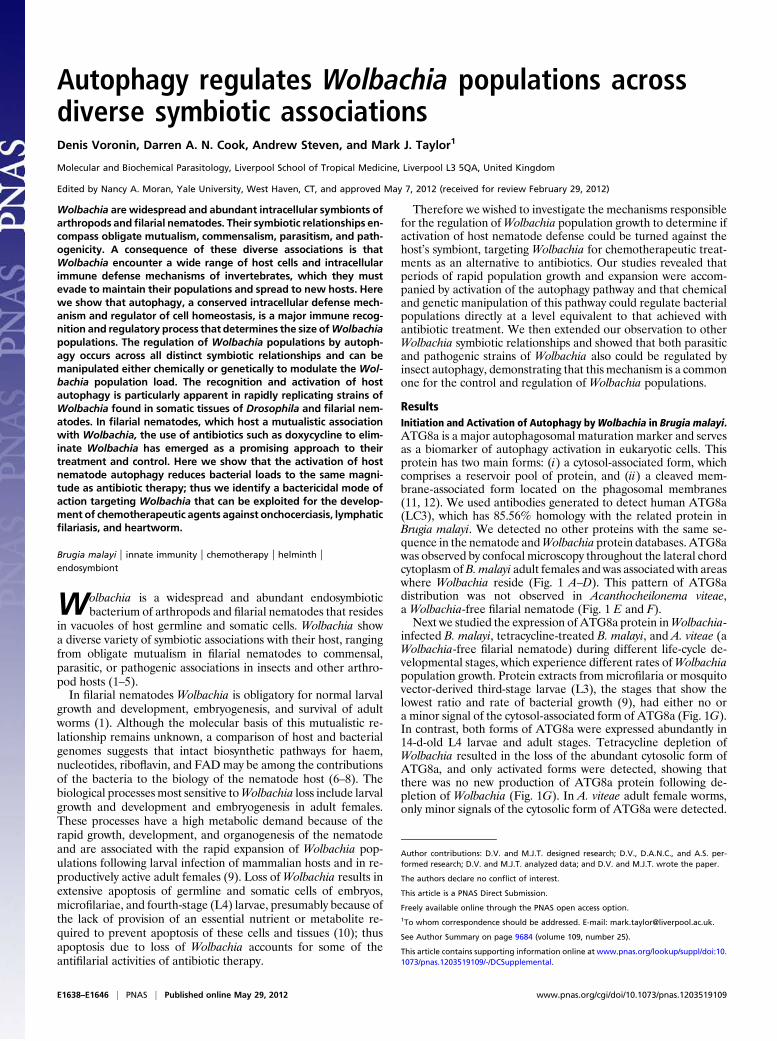
ResultsInitiation and Activation of Autophagy byWolbachia in Brugia malayi.ATG8a is a major autophagosomal maturation marker and servesas a biomarker of autophagy activation in eukaryotic cells. Thisprotein has two main forms: (i) a cytosol-associated form, whichcomprises a reservoir pool of protein, and (ii) a cleaved mem-brane-associated form located on the phagosomal membranes(11, 12). We used antibodies generated to detect human ATG8a(LC3), which has 85.56% homology with the related protein inBrugia malayi. We detected no other proteins with the same se-quence in the nematode andWolbachia protein databases. ATG8awas observed by confocal microscopy throughout the lateral chordcytoplasm ofB.malayi adult females and was associated with areaswhere Wolbachia reside (Fig. 1 A–D). This pattern of ATG8adistribution was not observed in Acanthocheilonema viteae,a Wolbachia-free filarial nematode (Fig. 1 E and F).Next we studied the expression of ATG8a protein inWolbachia-
infected B. malayi, tetracycline-treated B. malayi, and A. viteae (aWolbachia-free filarial nematode) during different life-cycle de-velopmental stages, which experience different rates ofWolbachiapopulation growth. Protein extracts from microfilaria or mosquitovector-derived third-stage larvae (L3), the stages that show thelowest ratio and rate of bacterial growth (9), had either no ora minor signal of the cytosol-associated form of ATG8a (Fig. 1G).In contrast, both forms of ATG8a were expressed abundantly in14-d-old L4 larvae and adult stages. Tetracycline depletion ofWolbachia resulted in the loss of the abundant cytosolic form ofATG8a, and only activated forms were detected, showing thatthere was no new production of ATG8a protein following de-pletion of Wolbachia (Fig. 1G). In A. viteae adult female worms,only minor signals of the cytosolic form of ATG8a were detected.
Author contributions: D.V. and M.J.T. designed research; D.V., D.A.N.C., and A.S. per-formed research; D.V. and M.J.T. analyzed data; and D.V. and M.J.T. wrote the paper.
The authors declare no conflict of interest.
This article is a PNAS Direct Submission.
Freely available online through the PNAS open access option.1To whom correspondence should be addressed. E-mail: [email protected].
See Author Summary on page 9684 (volume 109, number 25).
This article contains supporting information online at www.pnas.org/lookup/suppl/doi:10.1073/pnas.1203519109/-/DCSupplemental.
E1638–E1646 | PNAS | Published online May 29, 2012 www.pnas.org/cgi/doi/10.1073/pnas.1203519109
Next we investigated the gene expression of atg8a, a major markerof autophagy initiation, during the life-cycle stages [microfilariae, L3,L4 (14-d-old), and adults] of B. malayi. No expression of atg8a wasobserved in microfilaria, in which the number and ratio ofWolbachiais the lowest of all life-cycle stages (9). Expression of atg8a in L3larvae was detectable and was used as a basal level for comparisonwith the gene expression in other stages. An 11- to 14-fold increase inatg8a expression was observed in L4 (14-d-old) larvae and adultworms compared with L3 larvae (P < 0.003) (Fig. S1A).Together these results confirm that the activation of autoph-
agy in B. malayi is dependent on the presence of Wolbachia andis markedly elevated and activated during periods in which thebacterial population grows rapidly and in the developmentalstages with the highest bacterial density.
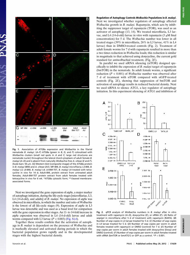
Regulation of Autophagy ControlsWolbachia Populations in B. malayi.Next we investigated whether regulators of autophagy affectedWolbachia growth in B. malayi. Rapamycin, which acts by inhib-iting the suppressor target of rapamycin (TOR), was used as anactivator of autophagy (13, 14). We treated microfilaria, L3 lar-vae, and L4 (14-d-old) larvae in vitro with rapamycin (5 μM finalconcentration) for 5 d. The Wolbachia number was lower in alltreated stages (39% in microfilaria, 26% in L3 larvae, 41% in L4larvae) than in DMSO-treated controls (Fig. 2). Treatment ofadult female worms for 7 d with rapamycin resulted in more thana two times reduction inWolbachia loads; this reduction is similarin magnitude to that achieved using doxycycline, the current goldstandard for antiwolbachial treatment, (Fig. 2E).In parallel we used siRNA silencing (siTOR) designed spe-
cifically to inhibit the expression of B. malayi target of rapamycin(bmTOR) in the nematode. In adult female worms, a significantreduction (P < 0.001) of Wolbachia number was observed after7 d of treatment with siTOR compared with siGFP-treatedcontrols (Fig. 2E), showing that suppression of bmTOR andactivation of autophagy results in reduced bacterial density. Nextwe used siRNA to silence ATG1, a key regulator of autophagyinitiation. In this experiment silencing of ATG1 and inhibition of
Fig. 1. Association of ATG8a expression and Wolbachia in the filarialnematode B. malayi. (A–F) ATG8a (green in B, D, and F) colocalized withWolbachia clusters (small red spots in A and C; large red structures arenematode nuclei) throughout the lateral chord cytoplasm of adult female B.malayi (A–D) and is absent from naturallyWolbachia-free A. viteae (E and F).(Scale bars: 50 μm. (G) Western blot (composite image) of the ATG8a proteinin B. malayi (BM) and A. viteae (AV). MF-BM, B. malayimicrofilaria; L3-BM, B.malayi L3; L4-BM, B. malayi L4; L4-BM-TET, B. malayi L4 treated with tetra-cycline in vivo for 14 d; Adult-BM, protein extract from untreated adultfemales; Adult-BM-TET protein extract from adult females treated withtetracycline in vivo for 6 wk. *ATG8a cytosolic form; **cleaved membrane-associated forms.
Fig. 2. qPCR analysis of Wolbachia numbers in B. malayi after in vitrotreatment with rapamycin (A–D), doxycycline (E), or siRNA (F). (A) Ratio ofwsp/gst in microfilaria after 5 d of treatment with rapamycin (RAPA). (B)Number of wsp copies in L3 larvae treated for 5 d. (C) Number of wsp copiesin L4 larvae treated for 5 d. (D) Number of wsp copies per worm in adultfemales treated with rapamycin or DMSO (control) for 7 d. (E) Number ofwsp copies per worm in adult females treated with doxycycline (Doxy) andDMSO (control). (F) Number ofwsp copies per worm in adult females treatedwith siRNA (bmTOR or bmATG1) or GFP as a control. *P < 0.001.
Voronin et al. PNAS | Published online May 29, 2012 | E1639
MICRO
BIOLO
GY
PNASPL
US
autophagy led to a significant increase in Wolbachia numbers inadult worms (Fig. 2F).Thus, the pharmacological or genetic activation and suppres-
sion of autophagy directly regulate Wolbachia populations inB. malayi.
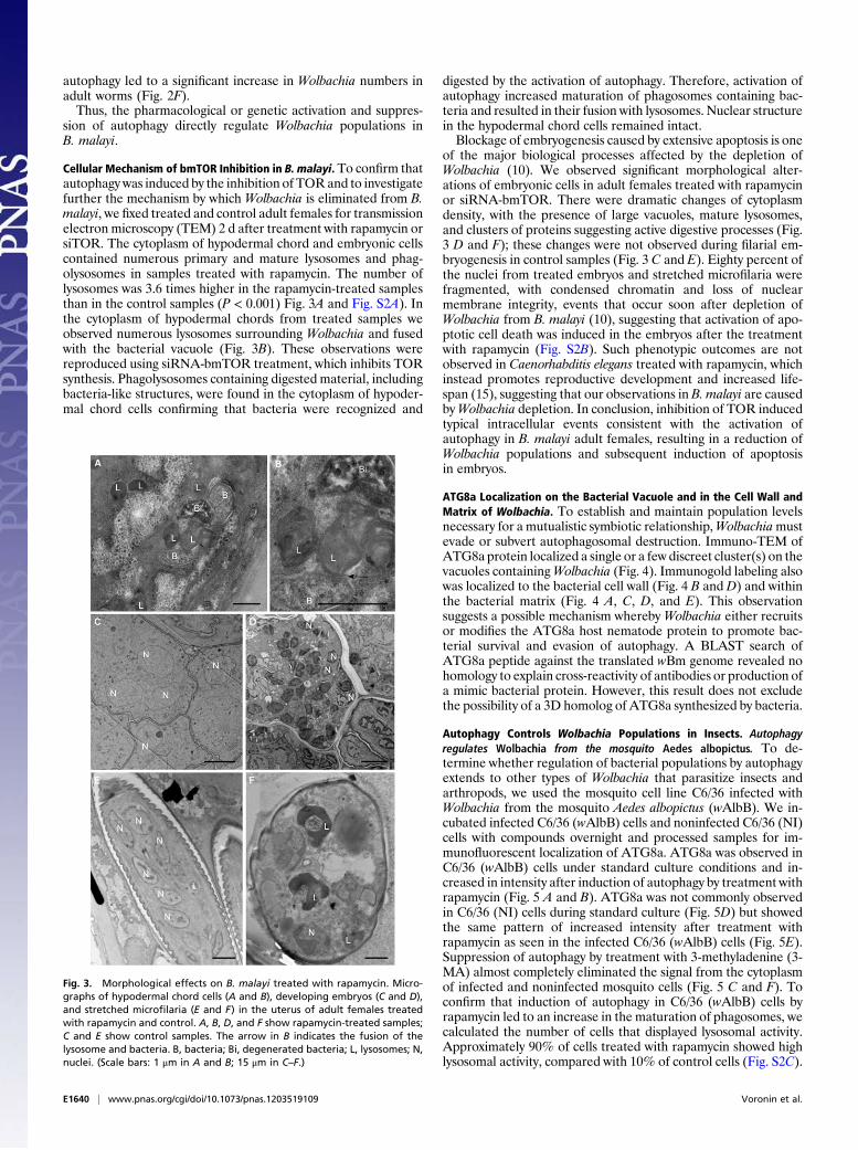
Cellular Mechanism of bmTOR Inhibition in B. malayi. To confirm thatautophagy was induced by the inhibition of TORand to investigatefurther the mechanism by which Wolbachia is eliminated from B.malayi, we fixed treated and control adult females for transmissionelectron microscopy (TEM) 2 d after treatment with rapamycin orsiTOR. The cytoplasm of hypodermal chord and embryonic cellscontained numerous primary and mature lysosomes and phag-olysosomes in samples treated with rapamycin. The number oflysosomes was 3.6 times higher in the rapamycin-treated samplesthan in the control samples (P < 0.001) Fig. 3A and Fig. S2A). Inthe cytoplasm of hypodermal chords from treated samples weobserved numerous lysosomes surrounding Wolbachia and fusedwith the bacterial vacuole (Fig. 3B). These observations werereproduced using siRNA-bmTOR treatment, which inhibits TORsynthesis. Phagolysosomes containing digested material, includingbacteria-like structures, were found in the cytoplasm of hypoder-mal chord cells confirming that bacteria were recognized and
digested by the activation of autophagy. Therefore, activation ofautophagy increased maturation of phagosomes containing bac-teria and resulted in their fusion with lysosomes. Nuclear structurein the hypodermal chord cells remained intact.Blockage of embryogenesis caused by extensive apoptosis is one
of the major biological processes affected by the depletion ofWolbachia (10). We observed significant morphological alter-ations of embryonic cells in adult females treated with rapamycinor siRNA-bmTOR. There were dramatic changes of cytoplasmdensity, with the presence of large vacuoles, mature lysosomes,and clusters of proteins suggesting active digestive processes (Fig.3 D and F); these changes were not observed during filarial em-bryogenesis in control samples (Fig. 3 C and E). Eighty percent ofthe nuclei from treated embryos and stretched microfilaria werefragmented, with condensed chromatin and loss of nuclearmembrane integrity, events that occur soon after depletion ofWolbachia from B. malayi (10), suggesting that activation of apo-ptotic cell death was induced in the embryos after the treatmentwith rapamycin (Fig. S2B). Such phenotypic outcomes are notobserved in Caenorhabditis elegans treated with rapamycin, whichinstead promotes reproductive development and increased life-span (15), suggesting that our observations in B. malayi are causedbyWolbachia depletion. In conclusion, inhibition of TOR inducedtypical intracellular events consistent with the activation ofautophagy in B. malayi adult females, resulting in a reduction ofWolbachia populations and subsequent induction of apoptosisin embryos.
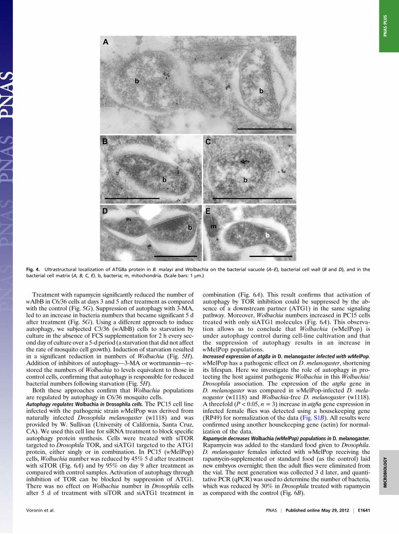
ATG8a Localization on the Bacterial Vacuole and in the Cell Wall andMatrix of Wolbachia. To establish and maintain population levelsnecessary for a mutualistic symbiotic relationship,Wolbachiamustevade or subvert autophagosomal destruction. Immuno-TEM ofATG8aprotein localized a single or a few discreet cluster(s) on thevacuoles containingWolbachia (Fig. 4). Immunogold labeling alsowas localized to the bacterial cell wall (Fig. 4 B and D) and withinthe bacterial matrix (Fig. 4 A, C, D, and E). This observationsuggests a possible mechanism whereby Wolbachia either recruitsor modifies the ATG8a host nematode protein to promote bac-terial survival and evasion of autophagy. A BLAST search ofATG8a peptide against the translated wBm genome revealed nohomology to explain cross-reactivity of antibodies or production ofa mimic bacterial protein. However, this result does not excludethe possibility of a 3D homolog of ATG8a synthesized by bacteria.
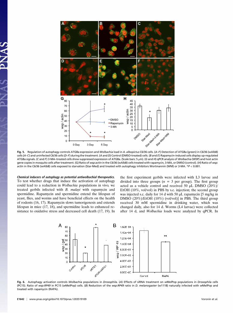
Autophagy Controls Wolbachia Populations in Insects. Autophagyregulates Wolbachia from the mosquito Aedes albopictus. To de-termine whether regulation of bacterial populations by autophagyextends to other types of Wolbachia that parasitize insects andarthropods, we used the mosquito cell line C6/36 infected withWolbachia from the mosquito Aedes albopictus (wAlbB). We in-cubated infected C6/36 (wAlbB) cells and noninfected C6/36 (NI)cells with compounds overnight and processed samples for im-munofluorescent localization of ATG8a. ATG8a was observed inC6/36 (wAlbB) cells under standard culture conditions and in-creased in intensity after induction of autophagy by treatment withrapamycin (Fig. 5 A and B). ATG8a was not commonly observedin C6/36 (NI) cells during standard culture (Fig. 5D) but showedthe same pattern of increased intensity after treatment withrapamycin as seen in the infected C6/36 (wAlbB) cells (Fig. 5E).Suppression of autophagy by treatment with 3-methyladenine (3-MA) almost completely eliminated the signal from the cytoplasmof infected and noninfected mosquito cells (Fig. 5 C and F). Toconfirm that induction of autophagy in C6/36 (wAlbB) cells byrapamycin led to an increase in the maturation of phagosomes, wecalculated the number of cells that displayed lysosomal activity.Approximately 90% of cells treated with rapamycin showed highlysosomal activity, compared with 10% of control cells (Fig. S2C).
Fig. 3. Morphological effects on B. malayi treated with rapamycin. Micro-graphs of hypodermal chord cells (A and B), developing embryos (C and D),and stretched microfilaria (E and F) in the uterus of adult females treatedwith rapamycin and control. A, B, D, and F show rapamycin-treated samples;C and E show control samples. The arrow in B indicates the fusion of thelysosome and bacteria. B, bacteria; Bi, degenerated bacteria; L, lysosomes; N,nuclei. (Scale bars: 1 μm in A and B; 15 μm in C–F.)
E1640 | www.pnas.org/cgi/doi/10.1073/pnas.1203519109 Voronin et al.
Treatment with rapamycin significantly reduced the number ofwAlbB in C6/36 cells at days 3 and 5 after treatment as comparedwith the control (Fig. 5G). Suppression of autophagy with 3-MA,led to an increase in bacteria numbers that became significant 5 dafter treatment (Fig. 5G). Using a different approach to induceautophagy, we subjected C3/36 (wAlbB) cells to starvation byculture in the absence of FCS supplementation for 2 h every sec-ond day of culture over a 5-d period (a starvation that did not affectthe rate of mosquito cell growth). Induction of starvation resultedin a significant reduction in numbers of Wolbachia (Fig. 5H).Addition of inhibitors of autophagy—3-MA or wortmannin—re-stored the numbers of Wolbachia to levels equivalent to those incontrol cells, confirming that autophagy is responsible for reducedbacterial numbers following starvation (Fig. 5H).Both these approaches confirm that Wolbachia populations
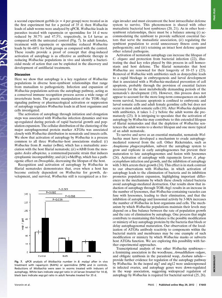
are regulated by autophagy in C6/36 mosquito cells.Autophagy regulates Wolbachia in Drosophila cells. The PC15 cell lineinfected with the pathogenic strain wMelPop was derived fromnaturally infected Drosophila melanogaster (w1118) and wasprovided by W. Sullivan (University of California, Santa Cruz,CA). We used this cell line for siRNA treatment to block specificautophagy protein synthesis. Cells were treated with siTORtargeted to Drosophila TOR, and siATG1 targeted to the ATG1protein, either singly or in combination. In PC15 (wMelPop)cells, Wolbachia number was reduced by 45% 5 d after treatmentwith siTOR (Fig. 6A) and by 95% on day 9 after treatment ascompared with control samples. Activation of autophagy throughinhibition of TOR can be blocked by suppression of ATG1.There was no effect on Wolbachia number in Drosophila cellsafter 5 d of treatment with siTOR and siATG1 treatment in
combination (Fig. 6A). This result confirms that activation ofautophagy by TOR inhibition could be suppressed by the ab-sence of a downstream partner (ATG1) in the same signalingpathway. Moreover, Wolbachia numbers increased in PC15 cellstreated with only siATG1 molecules (Fig. 6A). This observa-tion allows us to conclude that Wolbachia (wMelPop) isunder autophagy control during cell-line cultivation and thatthe suppression of autophagy results in an increase inwMelPop populations.Increased expression of atg8a in D. melanogaster infected with wMelPop.wMelPop has a pathogenic effect on D. melanogaster, shorteningits lifespan. Here we investigate the role of autophagy in pro-tecting the host against pathogenic Wolbachia in this Wolbachia/Drosophila association. The expression of the atg8a gene inD. melanogaster was compared in wMelPop-infected D. mela-nogaster (w1118) and Wolbachia-free D. melanogaster (w1118).A threefold (P < 0.05, n= 3) increase in atg8a gene expression ininfected female flies was detected using a housekeeping gene(RP49) for normalization of the data (Fig. S1B). All results wereconfirmed using another housekeeping gene (actin) for normal-ization of the data.Rapamycin decreasesWolbachia (wMelPop) populations in D. melanogaster.Rapamycin was added to the standard food given to Drosophila.D. melanogaster females infected with wMelPop receiving therapamycin-supplemented or standard food (as the control) laidnew embryos overnight; then the adult flies were eliminated fromthe vial. The next generation was collected 3 d later, and quanti-tative PCR (qPCR) was used to determine the number of bacteria,which was reduced by 30% in Drosophila treated with rapamycinas compared with the control (Fig. 6B).
Fig. 4. Ultrastructural localization of ATG8a protein in B. malayi and Wolbachia on the bacterial vacuole (A–E), bacterial cell wall (B and D), and in thebacterial cell matrix (A, B, C, E). b, bacteria; m, mitochondria. (Scale bars: 1 μm.)
Voronin et al. PNAS | Published online May 29, 2012 | E1641
MICRO
BIOLO
GY
PNASPL
US
Chemical inducers of autophagy as potential antiwolbachial therapeutics.To test whether drugs that induce the activation of autophagycould lead to a reduction in Wolbachia populations in vivo, wetreated gerbils infected with B. malayi with rapamycin andspermidine. Rapamycin and spermidine extend the lifespan ofyeast, flies, and worms and have beneficial effects on the healthof rodents (16, 17). Rapamycin slows tumorigenesis and extendslifespan in mice (17, 18), and spermidine leads to enhanced re-sistance to oxidative stress and decreased cell death (17, 19). In
the first experiment gerbils were infected with L3 larvae anddivided into three groups (n = 3 per group). The first groupacted as a vehicle control and received 50 μL DMSO (20%)/EtOH (10%, vol/vol) in PBS by s.c. injection; the second groupwas injected s.c. daily for 14 d with 50 μL rapamycin [5 mg/kg inDMSO (20%)/EtOH (10%) (vol/vol)] in PBS. The third groupreceived 30 mM spermidine in drinking water, which waschanged daily, also for 14 d. Worms (L4 larvae) were collectedafter 14 d, and Wolbachia loads were analyzed by qPCR. In
Fig. 5. Regulation of autophagy controls ATG8a expression andWolbachia load inA. albopictus C6/36 cells. (A–F) Detection of ATG8a (green) in C6/36 (wAlbB)cells (A–C) and uninfected C6/36 cells (D–F) during the treatment. (A andD) Control (DMSO-treated) cells. (B and E) Rapamycin-induced cells display up-regulatedATG8a signals. (C and F) 3-MA–treated cells show suppressed expression of ATG8a. (Scale bars: 5 μm). (G andH) qPCR analysis ofWolbachia (WSP) and host actingene copies inmosquito cells after treatment. (G) Ratio ofwsp:actin in the C6/36 (wAlbB) cells treatedwith rapamycin, 3-MA, or DMSO (control). (H) Ratio ofwsp:actin in the C6/36 (wAlbB) cells exposed to starvation (Star-Med) and treated with autophagy inhibitors Wortmannin (WM) or 3-MA. *P < 0.001.
Fig. 6. Autophagy activation controls Wolbachia populations in Drosophila. (A) Effects of siRNA treatment on wMelPop populations in Drosophila cells(PC15). Ratio of wsp:RP49 in PC15 (wMelPop) cells. (B) Reduction of the wsp:RP49 ratio in D. melanogaster (w1118) naturally infected with wMelPop andtreated with rapamycin (RAPA).
E1642 | www.pnas.org/cgi/doi/10.1073/pnas.1203519109 Voronin et al.
a second experiment gerbils (n = 4 per group) were treated as inthe first experiment but for a period of 35 d; then Wolbachialoads of adult worms were analyzed by qPCR.Wolbachia loads inparasites treated with rapamycin or spermidine for 14 d werereduced by 30.7% and 47.3%, respectively, in L4 larvae ascompared with the untreated control (Fig. 7). In adult females,treatment with rapamycin or spermidine reduced Wolbachialoads by 66–68% for both groups as compared with the control.These results provide a proof of concept that drug-inducedactivation of autophagy is as effective as antibiotic therapy inreducing Wolbachia populations in vivo and identify a bacteri-cidal mode of action that can be exploited in the discovery anddevelopment of antifilarial treatments.
DiscussionHere we show that autophagy is a key regulator of Wolbachiapopulations in diverse host–symbiont relationships that rangefrom mutualism to pathogenicity. Infection and expansion ofWolbachia populations activate the autophagy pathway, acting asa conserved immune recognition process across a wide range ofinvertebrate hosts. The genetic manipulation of the TOR–Atg1signaling pathway or pharmacological activation or suppressionof autophagy regulatesWolbachia loads in all host organisms andcells investigated.The activation of autophagy through initiation and elongation
steps was associated with Wolbachia infection dynamics and wasup-regulated during periods of rapid bacterial growth and pop-ulation expansion. The cellular distribution of the clustering of themajor autophagosomal protein marker ATG8a was associatedclosely with Wolbachia distribution in nematode and insects cells.We show that activation of autophagy by Wolbachia is a processcommon to all three Wolbachia–host associations studied: (i)Wolbachia from B. malayi (wBm), which has a mutualistic asso-ciation with the host filarial nematode; (ii) wAlbB from the mos-quito Aedes albopictus, a commensal/parasitic strain that inducescytoplasmic incompatibility; and (iii) wMelPop, which has a path-ogenic effect on Drosophila, decreasing the lifespan of the host.Recognition and activation of autophagy by Wolbachia in
filarial nematodes demonstrates that, even when a host hasbecome entirely dependent on Wolbachia for growth, de-velopment, and survival, Wolbachia still is recognized as a for-
eign invader and must circumvent the host intracellular defensesystem to survive. This phenomenon is shared with othermutualistic bacterial host symbioses (20, 21). As in other host–symbiont relationships, there must be a balance among (i) ac-commodating the symbiont to provide sufficient essential fac-tors that serve the mutualistic association, (ii) regulating thesymbiont population to avoid unnecessary fitness costs andpathogenicity; and (iii) retaining an intact host defense againstother related pathogens.Activation of nematode autophagy can increase the lifespan of
C. elegans and protection from bacterial infection (22), illus-trating the dual key roles played by this process in cell homeo-stasis and host defense. The filarial nematodes that hostWolbachia are renowned for their longevity of 10–15 y (23).Removal of Wolbachia with antibiotics such as doxycycline leadsto a rapid blockage in embryogenesis and larval developmentthat is associated with a Wolbachia-mediated prevention of cellapoptosis, probably through the provision of essential factorsnecessary for the most metabolically demanding periods of thenematode’s development (10). However, this process does notappear to account for the more long-term consequences on adultworm survival, because apoptosis is confined to embryonic andlarval somatic cells and adult female germline cells but does notoccur in most adult somatic cells (10). AfterWolbachia depletionwith antibiotics, it takes 1–2 y before the adult worms die pre-maturely (23). It is intriguing to speculate that the activation ofautophagy by Wolbachia may contribute to this extended lifespanof filarial nematodes and that the depletion of Wolbachia sen-tences the adult worms to a shorter lifespan and one more typicalof an adult nematode.To survive and serve as an essential mutualist, nematode Wol-
bachia must have developed a mechanism to evade autophagy-mediated removal from the cell. Other Ricketsiales, such asAnaplasma phagocytophilum, subvert the autophagy system togrow and replicate in early autophagosomes but prevent theirmaturation to late autophagosomes and fusion with lysosomes(24). Activation of autophagy with rapamycin favors A. phag-ocytophilum infection and growth, and the inhibition of autophagywith 3-MA arrests their growth (24). This effect is in stark contrastto our observations with Wolbachia, in which activation ofautophagy leads to the elimination of bacteria and its inhibitionpromotes population expansion, highlighting important differ-ences in the mechanisms by which these closely related bacteriaavoid autophagy-mediated destruction. Our results show that in-duction of autophagy through TOR-Atg1 results in an increase inthe number of lysosomes, thatWolbachia-containing vacuoles canfuse with lysosomes, leading to their elimination, and that theinhibition of autophagy and lysosomal activity by 3-MA increasesthe number of Wolbachia in host organisms and cells. The mech-anism by which Wolbachia populations maintain their levels maydepend on a fine balance between the rate of population growthand the rate of elimination by autophagy. One process that mightcontribute to maintaining this balance is the possible modificationor mimicry of key autophagy proteins by the bacteria that block ordelay autophagosomal maturation. Our observation of the local-ization of ATG8a antibody reactivity to components within thebacterial matrix and membranes may be one example of suchmodification or mimicry by which Wolbachia masks or subvertshost ATG8a function. We are exploring this possibility with fur-ther experimental approaches.Transcriptional analysis of two other Wolbachia symbioses—
a feminizing association in the woodlouse, Armadillidium vulgare,and obligate symbiosis in the parasitoid wasp, Asobara tabida—provide further evidence for regulation of the autophagy pathwayby Wolbachia. In the isopod atg7 and atg12 were underexpressedin infected ovaries, and autophagy genes were down-regulatedin the wasp association, suggesting widespread regulation ofautophagy by Wolbachia is required for bacterial survival (25, 26).
Fig. 7. qPCR analysis of Wolbachia number in B. malayi after in vivotreatment with rapamycin (RAPA) or spermidine (SPN) and in controls.Reductions of Wolbachia were seen in worms treated with inducers ofautophagy. White bars indicate wsp:gst ratio in L4 larvae (treated for 14 d),black bars indicate wsp:gst ratio in adult females treated for 35 d.
Voronin et al. PNAS | Published online May 29, 2012 | E1643
MICRO
BIOLO
GY
PNASPL
US
Autophagy is not the only host-defense mechanism that canbe activated by Wolbachia. Natural and experimental infectionsof Drosophila and mosquitoes with the overreplicating and life-shortening wMelPop strain can induce up-regulation of host im-mune responses and inhibit microbial infection with viruses, pro-tozoa, and helminth parasites (27–30). Nevertheless, not allWolbachia–host associations lead to activation of host immunity,and among the strains that do not activate host immunity arenatural strains infecting Drosophila and Aedes aegypti (31, 32). Theinduction of host defense and protection from microbial infectiontherefore is strain dependent and appears to be restricted tostrains that have a high replication rate and widespread tissuetropisms (29, 31, 33). Alternately, it has been suggested that themetabolic demands of such overreplicating bacteria may preventmicrobial infection and transmission through competition for hostcell resources (27).Although the mechanism by which Wolbachia protect host
insects from microbial infection remains to be fully resolved, ourresult suggests that autophagy activation and manipulation isa mechanism that might contribute to this phenomenon. The en-hanced activation of autophagy by rapidly replicating bacteria suchas wBm during larval development and in adult worm populationsand induced by wMelPop in Drosophila also may influence thesuccessful infection and transmission of viruses. For example, therequirement of arboviruses (Dengue and Chikungunya) for an in-tact host autophagy system and their use of autophagosomes forsuccessful replication and transmission (34, 35) may be blocked byWolbachia-mediated manipulation of autophagosomal maturation,a hypothesis we are testing currently.Finally, the use of antibiotics such as doxycycline to target
Wolbachia elimination from filarial nematodes has emerged asa promising approach to the treatment and control of onchocer-ciasis and lymphatic filariasis (23). Antiwolbachial therapy is moreeffective than existing standard antifilarial drugs because of thepermanent sterilization of adult worms and long-term macro-filaricidal effects. However, widespread mass administration ofdoxycycline is compromised by the relatively lengthy course oftreatment (4 wk) and the exclusion of pregnant women and chil-dren <9 y of age. These barriers stimulated the formation of theAnti-Wolbachia (A·WOL) consortium (http://www.a-wol.net) tosearch for drugs active against Wolbachia that overcome theserestrictions. Our observation that activation of nematode autoph-agy with drugs and small molecules leads to reductions of Wolba-chia populations similar in magnitude to those achieved with gold-standard antibiotics such as doxycycline, both in vitro and in vivo inanimal models, provides important proof of concept of a bacteri-cidal mode of action that could be exploited for the discovery anddevelopment of drugs against filarial diseases. This proof of con-cept can stimulate the search for drugs that preferentially activatehost nematode autophagy as an alternative approach to the elim-ination of this essential symbiont.In conclusion, we have described how the regulation of Wolba-
chia populations is under the control of host autophagy and showthat, to ensure their survival, the bacteria must manipulate ormodulate this process through mechanisms that are distinct fromthose adopted by closely related bacteria. All Wolbachia/hostassociations studied, ranging from mutualism through to patho-genicity, display similarities in the activation of autophagy, which isassociated particularly with overreplicating strains and periods ofrapid replication and population expansion. In filarial nematodes,which host a mutualistic association withWolbachia, the activationof host nematode autophagy provides a bactericidal mode of ac-tion to targetWolbachia for the development of chemotherapeuticagents against filarial diseases and in insects may represent an al-ternative host-defense mechanism to account for Wolbachia-me-diated protection against viruses and other microbial pathogens.
Materials and MethodsParasite Material. B. malayiwas maintained in the peritoneal cavity of gerbils(Meriones unguiculatus). Parasites originally were obtained from TRS Labs,and the life cycle was maintained in house at the Liverpool School ofTropical Medicine. L3 larvae were collected from Ae. aegypti mosquitoes.Microfilaria, L4 larvae, and adult worms were collected from the peritonealcavities using preheated (37 °C) standard culture medium (RPMI-1640 sup-plemented with 100 U/mL penicillin, 100 mg/mL streptomycin, 2 mM L-glu-tamine, 2.5 mg/mL amphotericin B, and 25 mM Hepes) (GIBCO). Individualworms were frozen at −80 °C for future extraction of protein/RNA/DNA. Theremaining worms were cultivated in 24-well plates with either rapamycin(Sigma) at a final concentration of 5 μM for 5 d or siRNA for 7 d (37 °C, 5%CO2). Adults were incubated individually with 10 worms per experimentalgroup; L3 and L4 larvae with 8–10 worms per group, and microfilaria with10,000 worms per experimental group. To treat worms with specific siRNA(45 mg/mL), freshly prepared siRNA molecules were dissolved in 0.5 mLnonsupplemented RPMI-1640 (without FBS), and worms were incubated at37 °C, 5% CO2 for 2 h (36). Then the medium was replaced with culturemedium (supplemented with heat-inactivated FBS) containing 45 mg/mLsiRNA. Worms were cultivated for 7 d before collection. After chemical orsiRNA treatment, all worms were washed individually with PBS and storedfor future analysis.
To obtain Wolbachia-depleted parasites, gerbils infected with L3 larvaewere treated with tetracycline administered in drinking water (2.5 mg/mLfinal concentration) for 14 d (for L4 larvae). Gerbils with adult worm infec-tions were treated for 6 wk. Worms were collected 2 wk following treat-ment, as described above, washed, and frozen for future analysis.
For in vivo treatment, gerbils (n = 3 or 4 animals per group) infected withL3 larvae were treated with (i) rapamycin injected s.c. in a concentration of5 mg/kg every day for 14 d for analysis of L4 larvae or for 35 d to collectadults. (ii) Spermidine was delivered in drinking water (30 mM final con-centration) daily for 14 for L4 larvae and 35 d adult worms. (iii) Controlanimals received vehicle solution (DMSO 20%/EtOH 10% in PBS) by s.c. fol-lowing the regime used in the rapamycin group. Worms were collected asdescribed above, washed, and frozen for future analysis.
All animal experiments were carried out in strict accordance with theAnimals Scientific (Procedures Act) 1986 (UK) under a license granted by theHome Office (London). Experimental procedures were reviewed and ap-proved by the Animal Welfare Committee, Liverpool School of TropicalMedicine and the Home Office (London).
Drosophila Maintenance. D. melanogaster (w1118) naturally infected withwMelPop andD.melanogaster (w1118NI)weremaintained at 25 °C and a 12-hdark/light regime. Agar-yeast standard food was changed every 20 d (37).
To study the effect of rapamycin on the Wolbachia loads in Drosophila,6-d-old females were placed overnight to lay new eggs in vials containingstandard food (as the control) or food supplemented with rapamycin (5 μM).Then adult flies were removed from the vials. The new generation wascollected from vials on day 5 and frozen for future analysis of Wolbachiapopulation by qPCR.
Mosquitoes and Drosophila Cell Lines. Mosquito cell line C6/36 (NI), originallyuninfected with Wolbachia, was established from Ae. albopictus. The cellline C6/36 (wAlbB) was infected with wAlbB derived from Aa23 (Ae. albo-pictus) at the Liverpool School of Tropical Medicine and was cultivatedsuccessfully for 4 y in the laboratory (38, 39). The cell lines were culturedroutinely in 25-cm2 plastic culture flasks at 26 °C in 5 mL of Leibovitz-15medium consisting 10% of heat-inactivated FBS, 50 U/mL penicillin, 50 mg/mL streptomycin, and 2 mM L-glutamine. Cells were transferred into a newflask every 4–5 d.
One day before the experiments, cells were transferred to a 96-well plateat 10,000 cells per well. On the next day the medium and nonattached cellswere removed, and fresh medium with compounds was added. Rapamycin(5 μM) and 3-MA (100 mM) were used to treat C6/36 cells for 3 and 5 d,respectively. For the starvation experiment, cells were cultivated in mediumwithout FBS for 2 h; then the medium was replaced with standard medium.This procedure was repeated every day during the 5-d experiment. To sup-press autophagy in the starved cells, medium was supplemented with 3-MA(100 mM) or Wortmannin (10 μM). At the end of the experiment cells werewashed twice with PBS, and DNA was extracted using a Promega DNA-ex-traction kit following the manufacturer’s instructions.
Drosophila PC15 (wMelPop) cells were derived from naturally infectedD. melanogaster (w1118) females. The cell line was cultured routinely in25-cm2 plastic culture flasks at 26 °C in 5 mL of Schneider’s insect mediumconsisting of 10% heat-inactivated FBS, 50 U/mL penicillin, and 50 mg/mL
E1644 | www.pnas.org/cgi/doi/10.1073/pnas.1203519109 Voronin et al.
streptomycin. Cells were transferred into a new flask routinely once every10 d. One day before the experiments, cells were transferred to a 96-wellplate at 10,000 cells per well. On the next day the medium and nonattachedcells were removed, medium without FBS and containing 20 mg siRNA wasadded in the wells, and cells were cultivated for 2 h. Then the modifiedmedium was replaced with standard medium containing 20 mg siRNA. Thisprocedure was repeated three times during the 7-d period of cell cultivation.At the end of the experiment (on day 7) cells were washed twice with PBS,and DNA was extracted as described above.
Production of RNA for dsRNA and siRNA. Total RNA was extracted fromB. malayi or D. melanogaster adult females by a TRIzol-based method (40).Purified RNA was treated with 1 U DNase I (Epicentre) at 37 °C for 30 minfollowed by inactivation by EDTA. Treated RNA (5 μg) was used as a tem-plate for cDNA synthesis performed by SuperScript III (Invitrogen). The cDNAtemplate was amplified by PCR using specific primers containing the T7promoter sequence (Table S1), and the product was used as a template forT7 RNA polymerase to synthesize the dsRNA by the HiScribe T7 in vitrotranscription kit (New England BioLabs). Quality and integrity of dsRNA waschecked by standard agarose gel electrophoresis. siRNA (18–25 bp) corre-sponding to the specific target was produced by digesting transcribeddsRNA with the ShortCut RNase III kit (New England BioLabs) following themanufacturer’s instructions. The siRNA was quantified by comparison onagarose gel to siRNA standard (New England BioLabs). RNA of the greenfluorescent protein (gfp) gene was used as a control for siRNA treatment.The RNA was extracted from Drosophila flies containing gfp-gene insertionusing the methods and procedures used for the production of experi-mental siRNA.
Gene Expression. Total RNA was extracted from 10,000 microfilaria, five L3larvae, five L4 (14-d old) larvae, five male or female adults, or individualfemale Drosophila. Then cDNA synthesis was performed as described above.Specific primers for detection of atg8a gene expression level were designedby tehePrimerPrimier 4.0 program using cDNA of atg8a B. malayi orD. melanogaster as templates (Table S1). All amplifications and fluorescencequantifications were performed by a Bio-Rad Chromo 4 real-time PCR De-tector (Bio-Rad). A ΔΔCt-based method was used to analyze atg8 levels andthe gst gene of B. malayi or the RP49 gene of Drosophila for normalization(10, 41, 42). All comparisons were replicated with at least three biologicalrepeats with three technical replicates for each repeat.
Western Blot. B. malayi worms (microfilaria, L4 larvae, and adults) werecollected from treated and untreated gerbils. L3 larvae were obtained bydissection of infected mosquitoes. All samples were washed three times inPBS and lysed with 50 μL of Tissue Extraction Reagent (Invitrogen). Theconcentration of the proteins was estimated by bicinchoninic acid assay(Invitrogen) following the manufacturer’s instructions. Lysates of wormswere mixed with LDS sample buffer (NuPAGE; Invitrogen), boiled, and run in12% PAGE. Protein was transferred to nitrocellulose membranes and used inthe Western blot as previously described (43). Western blot detection ofATG8a was performed using anti-ATG8a (LC3-II) antibody (Invitrogen andNew England BioLabs). Western blot was performed using three in-dependent protein samples analyzed in parallel.
DNA and qPCR. DNA was extracted from worms, mosquito or Drosophila cells,and Drosophila flies by using the QIAGenes Expression Kit (QIAGEN) fol-lowing the manufacturer’s instructions. Wolbachia numbers were quantifiedby qPCR using a single-copy gene: wsp (for Wolbachia) as previously de-scribed (9). To estimate the dynamics of the bacterial populations in
B. malayi microfilaria, we calculated the ratio of single-copy genes wsp andgst (B. malayi) (9, 10); to estimate Wolbachia loads in the cells, we calculatedthe ratio of wsp:actin for C6/36 cells and wsp:RP 49 for Drosophila to stan-dardize the data (39).
Microscopy. B. malayi adult females were fixed using 4% formaldehyde inPBS with 0.05% Triton-X100 (PBST) for 20 min for confocal microscopyanalysis of ATG8a protein localization. During fixation, worms were cut toimprove distribution of the fixative. Samples then were washed three timesin PBST and treated with RNase A (100 mg/mL) overnight at 4 °C (10). Thefollowing day, samples were washed in PBS and blocked with 5% BSA for15 min and incubated overnight at 4 °C with anti-Atg8a (LC3-II) antibody(Invitrogen) diluted 1:200. Secondary antibody labeled with FITC was used at1:500. After incubation with antibodies samples were costained with pro-pidium iodide for 20 min to visualize DNA (host nuclei and Wolbachia) andwere viewed with an LSM 5 Pascal confocal microscope (Zeiss).
For TEM, worms were fixed with 2.5% glutaraldehyde for 2 h. During thefixation worms were cut into ∼5-mm pieces. After fixation, samples werewashed three times in PBS and postfixed by 4% OsO4 for 1 h. Samples thenwere washed and dehydrated using a series of ethanol concentrations (50–100%) with a final wash of acetone. Samples were embedded in plastic(Agar 100) and prepared for sectioning. Ultrathin sections were contrastedwith uranyl acetate (1%) and lead citrate and then were analyzed under theTecnai G2 Spirit BioTWIN TEM by the TEM unit, University of Liverpool(Liverpool, UK).
For immuno-TEMworms were fixed by 4% paraformaldehyde dissolved inPBS for 4 h at 4 °C. During fixation, worms were cut. After fixation, sampleswere washed in PBS (three times on ice) and dehydrated in a series of eth-anol concentrations (50–100%) on ice. Dehydrated samples were embeddedin Lowicryl Gold plastic resin. Ultrathin sections were blocked by 5% BSA andincubated with primary anti-Atg8a (LC3-II) antibody (Invitrogen) diluted1:200 in 1% BSA overnight at 4 °C. On the next day, sections were washedthree times in PBS and incubated with secondary antibody labeled with10-nm gold particles. Sections then were washed with water and contrastedby uranyl acetate (1%) and lead citrate and then were analyzed under theTecnai G2 Spirit BioTWIN TEM by the TEM unit, University of Liverpool(Liverpool, UK).
C6/36 cell lines were grown on glass overnight and then were fixed by 4%formaldehyde in PBST for 20 min for confocal microscopy analysis of ATG8aprotein localization. After fixation, cells were washed three times in PBS andprocessed as described above. Stained cells were investigated under an LSM 5Pascal confocal microscope (Zeiss).
Statistical Analysis.Differences betweenmeans were analyzed using one-wayANOVA with Dunnett’s multiple comparison tests as appropriate. For in vitroexperiments, means were obtained from 8–10 biological replicates for qPCRand from three biological replicates for qRT-PCR. For in vivo experiments,means were obtained from three or four biological replicates. Analysis ofeach biological replicate was performed in triplicate. A Dunn–Šidák adjust-ment was made for multiple comparisons, using a normal P value of P =0.006 for individual tests to provide an overall significance (α) level of 0.05.All analyses were performed using the PASW Statistics 17 statistical com-puter program (IBM).
ACKNOWLEDGMENTS. We thank Prof. W. Sullivan, Dr. A. Debec, Dr. L. Serbus,and Dr. C. Casper Lindley (University of California, Santa Cruz) for providingPC15 Drosophila cell lines. This work was supported by a grant from the Billand Melinda Gates Foundation awarded to the Liverpool School of TropicalMedicine as part of the Anti-Wolbachia consortium.
1. Taylor MJ, Bandi C, Hoerauf A (2005) Wolbachia bacterial endosymbionts of filarialnematodes. Adv Parasitol 60:245–284.
2. Dedeine F, Boulétreau M, Vavre F (2005) Wolbachia requirement for oogenesis:Occurrence within the genus Asobara (Hymenoptera, Braconidae) and evidence forintraspecific variation in A. tabida. Heredity (Edinb) 95:394–400.
3. Werren JH, Baldo L, Clark ME (2008) Wolbachia: Master manipulators of invertebratebiology. Nat Rev Microbiol 6:741–751.
4. Ferree PM, Avery A, Azpurua J, Wilkes T, Werren JH (2008) A bacterium targetsmaternally inherited centrosomes to kill males in Nasonia. Curr Biol 18:1409–1414.
5. McMeniman CJ, et al. (2009) Stable introduction of a life-shortening Wolbachiainfection into the mosquito Aedes aegypti. Science 323:141–144.
6. Foster J, et al. (2005) The Wolbachia genome of Brugia malayi: Endosymbiontevolution within a human pathogenic nematode. PLoS Biol 3:e121.
7. Wu B, et al. (2009) The heme biosynthetic pathway of the obligate Wolbachiaendosymbiont of Brugia malayi as a potential anti-filarial drug target. PLoS Negl TropDis 3:e475.
8. Slatko BE, Taylor MJ, Foster JM (2010) The Wolbachia endosymbiont as an anti-filarialnematode target. Symbiosis 51:55–65.
9. McGarry HF, Egerton GL, Taylor MJ (2004) Population dynamics of Wolbachiabacterial endosymbionts in Brugia malayi. Mol Biochem Parasitol 135:57–67.
10. Landmann F, Voronin D, Sullivan W, Taylor MJ (2011) Anti-filarial activity of antibiotictherapy is due to extensive apoptosis after Wolbachia depletion from filarialnematodes. PLoS Pathog 7:e1002351.
11. Klionsky DJ, et al. (2008) Guidelines for the use and interpretation of assays formonitoring autophagy in higher eukaryotes. Autophagy 4:151–175.
12. Deretic V, Levine B (2009) Autophagy, immunity, and microbial adaptations. Cell HostMicrobe 5:527–549.
13. Hansen IA, Attardo GM, Roy SG, Raikhel AS (2005) Target of rapamycin-dependentactivation of S6 kinase is a central step in the transduction of nutritional signalsduring egg development in a mosquito. J Biol Chem 280:20565–20572.
14. Wullschleger S, Loewith R, Hall MN (2006) TOR signaling in growth and metabolism.Cell 124:471–484.
Voronin et al. PNAS | Published online May 29, 2012 | E1645
MICRO
BIOLO
GY
PNASPL
US
15. Meléndez A, Levine B Autophagy in C. elegans. WormBook, ed The C. elegansResearch Community, 10.1895/wormbook.1.7.1. Available at http://www.wormbook.org.
16. Harrison DE, et al. (2009) Rapamycin fed late in life extends lifespan in geneticallyheterogeneous mice. Nature 460:392–395.
17. Eisenberg T, et al. (2009) Induction of autophagy by spermidine promotes longevity.Nat Cell Biol 11:1305–1314.
18. Liu M, et al. (2005) Antitumor activity of rapamycin in a transgenic mouse model ofErbB2-dependent human breast cancer. Cancer Res 65:5325–5336.
19. Madeo F, Eisenberg T, Büttner S, Ruckenstuhl C, Kroemer G (2010) Spermidine: Anovel autophagy inducer and longevity elixir. Autophagy 6:160–162.
20. Anselme C, et al. (2008) Identification of the weevil immune genes and theirexpression in the bacteriome tissue. BMC Biol 6:43.
21. Taylor M, Mediannikov O, Raoult D, Greub G (2012) Endosymbiotic bacteriaassociated with nematodes, ticks and amoebae. FEMS Immunol Med Microbiol 64:21–31.
22. Jia K, Chen D, Riddle DL (2004) The TOR pathway interacts with the insulin signalingpathway to regulate C. elegans larval development, metabolism and life span.Development 131:3897–3906.
23. Taylor MJ, Hoerauf A, Bockarie M (2010) Lymphatic filariasis and onchocerciasis.Lancet 376:1175–1185.
24. Niu H, Yamaguchi M, Rikihisa Y (2008) Subversion of cellular autophagy byAnaplasma phagocytophilum. Cell Microbiol 10:593–605.
25. Kremer N, et al. (2012) Influence of Wolbachia on host gene expression in anobligatory symbiosis. BMC Microbiol 12(Suppl 1):S7.
26. Chevalier F, et al. (2012) Feminizing Wolbachia: A transcriptomics approach withinsights on the immune response genes in Armadillidium vulgare. BMC Microbiol 12(Suppl 1):S1.
27. Moreira LA, et al. (2009) A Wolbachia symbiont in Aedes aegypti limits infection withdengue, Chikungunya, and Plasmodium. Cell 139:1268–1278.
28. Kambris Z, Cook PE, Phuc HK, Sinkins SP (2009) Immune activation by life-shorteningWolbachia and reduced filarial competence in mosquitoes. Science 326:134–136.
29. Frentiu FD, Robinson J, Young PR, McGraw EA, O’Neill SL (2010) Wolbachia-mediatedresistance to dengue virus infection and death at the cellular level. PLoS ONE 5:e13398.
30. Kambris Z, et al. (2010) Wolbachia stimulates immune gene expression and inhibitsplasmodium development in Anopheles gambiae. PLoS Pathog 6:e1001143.
31. Wong ZS, Hedges LM, Brownlie JC, Johnson KN (2011) Wolbachia-mediatedantibacterial protection and immune gene regulation in Drosophila. PLoS ONE 6:e25430.
32. Pan X, et al. (2012) Wolbachia induces reactive oxygen species (ROS)-dependentactivation of the Toll pathway to control dengue virus in the mosquito Aedes aegypti.Proc Natl Acad Sci USA 109:E23–E31.
33. Popovici J, et al. (2010) Assessing key safety concerns of a Wolbachia-based strategyto control dengue transmission by Aedes mosquitoes. Mem Inst Oswaldo Cruz 105:957–964.
34. Lee YR, et al. (2008) Autophagic machinery activated by dengue virus enhances virusreplication. Virology 374:240–248.
35. Krejbich-Trotot P, et al. (2011) Chikungunya triggers an autophagic process whichpromotes viral replication. Virol J 8:432.
36. Ford L, et al. (2009) Functional analysis of the cathepsin-like cysteine protease genesin adult Brugia malayi using RNA interference. PLoS Negl Trop Dis 3:e377.
37. Voronin DA, Bochernikov AM, Baricheva EM, Zakharov IK, Kiseleva EV (2009)[Influence of Drosophila melanogaster genotype on biological effects of endosym-biont Wolbachia (stamm wMelPop)]. Tsitologiia 51:335–345. Russian.
38. Turner JD, et al. (2006) Wolbachia endosymbiotic bacteria of Brugia malayi mediatemacrophage tolerance to TLR- and CD40-specific stimuli in a MyD88/TLR2-dependentmanner. J Immunol 177:1240–1249.
39. Voronin D, Tran-Van V, Potier P, Mavingui P (2010) Transinfection and growthdiscrepancy of Drosophila Wolbachia strain wMel in cell lines of the mosquito Aedesalbopictus. J Appl Microbiol 108:2133–2141.
40. Knox DP, Geldhof P, Visser A, Britton C (2007) RNA interference in parasiticnematodes of animals: A reality check? Trends Parasitol 23:105–107.
41. LaLonde M, et al. (2006) A role for Phospholipase D in Drosophila embryoniccellularization. BMC Dev Biol 6:60.
42. Matta BP, Bitner-Mathé BC, Alves-Ferreira M (2011) Getting real with real-time qPCR:A case study of reference gene selection for morphological variation in Drosophilamelanogaster wings. Dev Genes Evol 221:49–57.
43. Johnston KL, et al. (2010) Lipoprotein biosynthesis as a target for anti-Wolbachiatreatment of filarial nematodes. Parasit Vectors 3:99.
E1646 | www.pnas.org/cgi/doi/10.1073/pnas.1203519109 Voronin et al.