autophosphorylationactivates dictyostelium ... sites of the pet-28a vector ... cells were cultured...
TRANSCRIPT

Autophosphorylation Activates Dictyostelium Myosin II HeavyChain Kinase A by Providing a Ligand for an AllostericBinding Site in the �-Kinase Domain*□S
Received for publication, August 19, 2010, and in revised form, October 24, 2010 Published, JBC Papers in Press, November 11, 2010, DOI 10.1074/jbc.M110.177014
Scott W. Crawley‡, Mojdeh Samimi Gharaei‡, Qilu Ye‡, Yidai Yang‡, Barak Raveh§, Nir London§, Ora Schueler-Furman§,Zongchao Jia‡1, and Graham P. Cote‡2
From the ‡Department of Biochemistry, Queen’s University, Kingston, Ontario K7L 3N6, Canada and the §Department ofMicrobiology and Molecular Genetics, Institute for Medical Research Israel-Canada, Hadassah Medical School, The HebrewUniversity, Jerusalem, 91120 Israel
Dictyostelium discoideummyosin II heavy chain kinase A(MHCKA), amember of the atypical �-kinase family, phosphor-ylates sites in themyosin II tail that block filament assembly.Here we show that the catalytic activity of A-CAT, the �-kinasedomain ofMHCKA (residues 552–841), is severely inhibited bythe removal of a disordered C-terminal tail sequence (C-tail; resi-dues 806–841). The key residue in the C-tail was identified asThr825, which was found to be constitutively autophosphorylated.Dephosphorylation of Thr825 using shrimp alkaline phosphatasedecreased A-CAT activity. The activity of a truncated A-CATlacking Thr825 could be rescued by Pi, phosphothreonine, and aphosphorylated peptide, but not by threonine, glutamic acid, as-partic acid, or an unphosphorylated peptide. These results fo-cused attention on a Pi-binding pocket located in the C-terminallobe of A-CAT.Mutational analysis demonstrated that the Pi-pocket was essential for A-CAT activity. Based on these results, itis proposed that autophosphorylation of Thr825 activates ACATby providing a covalently tethered ligand for the Pi-pocket.Abinitiomodeling studies using the Rosetta FloppyTail and Flex-PepDock protocols showed that it is feasible for the phosphory-lated Thr825 to dock intramolecularly into the Pi-pocket. Allo-steric activation is predicted to involve a conformational changein Arg734, which bridges the bound Pi to Asp762 in a key active siteloop. Sequence alignments indicate that a comparable regulatorymechanism is likely to be conserved inDictyosteliumMHCKB-Dandmetazoan eukaryotic elongation factor-2 kinases.
Dictyostelium discoideumMHCK A3 is a highly specializedprotein kinase that targets three threonine residues located in
the �-helical coiled-coil tail of myosin II (1–4). Phosphoryla-tion of these sites results in the disassembly of myosin II bipo-lar filaments and inhibits processes, such as cytokinesis, thatdepend on myosin II contractile activity (5, 6). MHCK A is130-kDa in size and consists of an N-terminal �-helicalcoiled-coil domain, a central kinase domain, and a C-terminalWD-repeat domain (7). The coiled-coil domain assemblesinto trimers or tetramers, binds, and cross-links actin fila-ments and is responsible for targeting MHCK A to actin-richcellular protrusions (8–10). The WD-repeat domain interactswith filamentous myosin II and is required for MHCK A toefficiently phosphorylate myosin II (10, 11). The kinase do-main of MHCK A bears no sequence similarity to the super-family of “conventional” eukaryotic protein kinases but in-stead belongs to a small but widespread family of atypicalprotein kinases termed the �-kinases (12).
In addition to MHCK A, D. discoideum expresses five pro-teins with �-kinase domains. Three of the proteins, termedMHCK B, C, and D, are closely related to MHCK A and atleast two to them, MHCK B and C, function cooperatively toregulate myosin II filament assembly in vivo (13–15). Theother two �-kinases, AK1 and VwkA, have domain structuresunrelated to MHCK A. AK1 contains an Arf GTPase-activat-ing protein domain and VwkA contains an N-terminal vonWillebrand factor A-like domain. The function of AK1 is notknown, whereas VwkA is involved in the regulation of con-tractile vacuole function (16, 17). Mammals express six mul-tidomain proteins with �-kinase domains (12). These includeTRPM6 and TRPM7, which function as divalent cation chan-nels and phosphorylate the tail of non-muscle myosin II (18,19) and eEF2K, which regulates protein synthesis (20).The isolated �-kinase domain of MHCK A, termed A-CAT,
displays a high level of protein kinase activity and stronglyprefers to phosphorylate threonine residues (21, 22). The x-ray crystal structure of A-CAT reveals that it is composed ofan N-terminal and C-terminal lobe with the active site situ-ated in a cleft at the interface between the two lobes (23). Aninvariant catalytic residue in the active site (Asp766) can bephosphorylated, suggesting that the �-kinase catalytic mecha-
* This work was supported in part by Canadian Institutes of Health ResearchGrant MOP8603 and Heart and Stroke Foundation of Ontario Grant T6054(to G. P. C.), Natural Sciences and Engineering Research Council of CanadaGrant 203705 (to Z. J.), the Israel Science Foundation funded by Israel Acad-emy of Science and Humanities ISF Grant 306/6, and USA-Israel Bi-nationalScience Foundation Grant 2009418 (to O. S.-F.).
□S The on-line version of this article (available at http://www.jbc.org) con-tains supplemental Table S1 and Figs. S1–S5.
The atomic coordinates and structure factors (code 3PDT) have been depositedin the Protein Data Bank, Research Collaboratory for Structural Bioinformat-ics, Rutgers University, New Brunswick, NJ (http://www.rcsb.org/).
1 Canada Research Chair in Structural Biology.2 To whom correspondence should be addressed. Tel.: 613-533-2998; Fax:
613-533-2497; E-mail: [email protected] The abbreviations used are: MHCK, D. discoideum myosin II heavy chain
kinase; A-CAT, the �-kinase domain of MHCK A; AK1, D. discoideum
�-kinase 1; eEF2K, eukaryotic elongation factor-2 kinase; MBP, myelinbasic protein; TRPM, transient receptor potential melastatin; Pi, inorganicphosphate; SAP, shrimp alkaline phosphatase; TES, 2-{[2-hydroxy-1,1-bis(hydroxymethyl)ethyl]amino}ethanesulfonic acid.
THE JOURNAL OF BIOLOGICAL CHEMISTRY VOL. 286, NO. 4, pp. 2607–2616, January 28, 2011© 2011 by The American Society for Biochemistry and Molecular Biology, Inc. Printed in the U.S.A.
JANUARY 28, 2011 • VOLUME 286 • NUMBER 4 JOURNAL OF BIOLOGICAL CHEMISTRY 2607
by guest on May 23, 2018
http://ww
w.jbc.org/
Dow
nloaded from
nism differs from that of conventional eukaryotic protein ki-nases. A-CAT contains a tightly bound zinc atom that is re-quired for stability and binds two Mg2� ions, one in an activesite pocket and one at the center of the glycine-rich N/D-loop. The Mg2� binding sites are regulatory, because millimo-lar concentrations of Mg2�, in excess of that required to bindto ATP, are required for A-CAT to exhibit maximal catalyticactivity (22). A-CAT also binds a Pi molecule at a highly basicsite in the C-terminal lobe that we term the Pi-pocket (23).
The catalytic activity of MHCK A is enhanced at least 50-fold by autophosphorylation (24). The cellular signalingmechanisms that activate MHCK A remain to be elucidated,but in vitro the rate of autophosphorylation is stimulated byactin filaments, myosin II, and negatively charged com-pounds, such as DNA and acidic phospholipids (25, 26).MHCK A incorporates up to 10 mol of phosphate/mol, but 3mol of phosphate are sufficient for maximal activation (24).Here we identify Thr825 as a key autophosphorylation site thatis required for the activity of A-CAT and MHCK A. Thr825 islocated within a disordered sequence that links the �-kinasedomain to the WD-repeat domain. We further demonstratethat the Pi-pocket functions as a positive allosteric bindingsite, and propose a model in which the phosphorylated Thr825(Thr(P)825) activates A-CAT by providing a covalently teth-ered ligand for the Pi-pocket. Sequence alignments indicatethat the proposed regulatory mechanism is conserved in theother MHCKs and the metazoan eEF2Ks.
EXPERIMENTAL PROCEDURES
Materials—Peptides used in this study were synthesized bythe Sheldon Biotechnology Facility, McGill University. ATP,MBP, aspartic acid, glutamic acid, threonine, and phospho-threonine were purchased from Sigma and [�-32P]ATP wasobtained from PerkinElmer Life Sciences.Plasmid Constructs—DNAmanipulations were carried out
using standard methods (27). Truncation and deletion con-structs of the MHCK A �-kinase domain (A-CAT; residues552–841) were created using PCR (22). DNA constructs en-coding site-directed mutants of A-CAT were generated usingthe QuikChange XL site-directed mutagenesis system (Strat-agene). Constructs were cloned in-frame into the NcoI andXhoI sites of the pET-28a vector (Novagen) to add an N-ter-minal His tag and a TEV protease site as described (23). Full-length MHCK A constructs (NCBI accession XP_635600)were cloned into the plasmid pTX-FLAG vector (28) that hadbeen adapted to GATEWAY technology (Invitrogen). Briefly,the parent plasmid pTX-FLAG was digested by BamHI andXhoI and blunted with T4 DNA polymerase. The Gatewayrecombination cassette (reading frame cassette B) was theninserted to create destination vector pTX-FLAG-GATE. Ex-pression constructs encoding MHCK A were then createdusing pTX-FLAG-GATE according to the manufacturer’sinstructions.Protein Expression and Purification—Wild-type and mutant
forms of A-CAT were expressed in Escherichia coliBL21(DE3) and purified by chromatography over a His-Bindcolumn (Novagen) as described (supplemental Fig. S1) (23,29). FLAG-tagged MHCK A was expressed in the D. discoi-
deum AX3 cell line (30). Cells were cultured on 9-cm plasticPetri dishes in HL5 medium (31) supplemented with 10,000units/ml of penicillin and 10 mg/ml of streptomycin (Sigma).Plasmids were introduced into AX3 cells by electroporation at0.85 KV and 25 microfarads using a Bio-Rad Gene Pulser IIElectroporation System (32). Clonal cell lines expressingFLAG-tagged MHCK A were produced by dilution platingand selection in 20 �g/ml of geneticin (G418) (Invitrogen).Cells were lysed by homogenization in 500 mM NaCl, 1 mM
EDTA, 0.3% Triton X-100, and 50 mM Tris-HCl, pH 8.0, con-taining one complete mini protease inhibitor tablet (RocheApplied Diagnostics) per 50 ml of buffer at 4 °C. The superna-tant obtained following centrifugation at 12,000 � g for 1 hwas passed over a column of anti-FLAGM2 Affinity gel(Sigma) (33). FLAG-MHCK A was eluted with TBS buffer(150 mM NaCl, 50 mM Tris-HCl, pH 7.4) containing 200�g/ml of FLAG peptide and dialyzed against 20 mM NaCl and50 mM Tris-HCl, pH 7.4.Kinase, ATPase, and Autophosphorylation Assays—ATPase
and kinase activity was assayed at 22 °C in kinase buffer (2 mM
MgCl2, 1 mM dithiothreitol, 0.25 mM ATP, and 20 mM TES,pH 7.0) containing [�-32P]ATP at a specific activity of 5–500cpm/pmol of ATP. Kinase assays included MBP or the MH-3synthetic peptide (RKKFGEAEKTKAKEFL) at a concentra-tion of 30 or 300 �M, respectively. Other additions to the ki-nase and ATPase assays are described in the figure legends.Assays were initiated by the addition of 1.45 �M A-CAT or38.8 nM MHCK A. Aliquots were removed at 1, 2, 3, 4, and 5min for kinase assays and 5, 10, 20, and 40 min for ATPaseassays. In some cases kinase assays were performed at 4 °Cand aliquots were removed at 20, 40, 60, and 90 s. ATP hy-drolysis was measured by following the release of 32P (34).Kinase activity was measured by spotting 20-�l aliquots ontosquares of Whatman P81 phosphocellulose paper (35). Thesquares were washed in 1% phosphoric acid, immersed inScintiVerse Universal LS Mixture (Fisher Scientific), andcounted using a Beckman LS 9000 scintillation counter. Overthe assay time courses linear rates of ATPase activity andphosphate incorporation were obtained. Under the assay con-ditions used here A-CAT exhibited an ATPase activity of0.022 � 0.002 s�1 and a kinase activity of 0.047 � 0.004 s�1
and 0.101 � 0.0029 s�1 toward MBP and MH-3, respectively.MHCK A exhibited a kinase activity of 0.19 � 0.033 s�1 to-ward MBP. Autophosphorylation assays contained 14.5 �M
A-CAT-5xA and were carried out in kinase buffer containing[�-32P]ATP at a specific activity of 500 cpm/pmol of ATP.Aliquots of 20 �l were taken at 1, 5, 10, 20, 40, and 60 min andsubjected to SDS-PAGE. After staining with Coomassie Bluethe A-CAT-5xA band was excised and counted in scintillationfluid to measure incorporation of 32P. For some assaysA-CAT-5xA was dephosphorylated by treatment at roomtemperature for 1 h with 10 units of calf intestinal alkalinephosphatase-agarose beads (Sigma) or for 5 h with 10 units ofSAP (Fermentas). A total of 200 �g of A-CAT-RRGT or A-CAT-RRGS were incubated with 0.5 �g of recombinant PKAcatalytic subunit (Active Motif) for 30 min at 22 °C in kinasebuffer. Following dialysis against 20 mM Tris-HCl, pH 7.4,kinase assays were carried out using the MH-3 peptide as de-
A Phosphate Binding Site Activates MHCK A
2608 JOURNAL OF BIOLOGICAL CHEMISTRY VOLUME 286 • NUMBER 4 • JANUARY 28, 2011
by guest on May 23, 2018
http://ww
w.jbc.org/
Dow
nloaded from
scribed above. Data were fit to a hyperbolic equation by non-linear regression analysis using the program SigmaPlot (SystatSoftware Inc.). Where shown, mean � S.D. are derived from 3to 6 determinations.Crystallization Procedures and Data Collection and
Analysis—A-CAT truncated at Leu809 (A-CAT-�809) wasconcentrated to 230 �M using a 10,000 molecular weight cut-off Ultrafree-4 Centrifugal Filtration Unit (Millipore). The Histag was cleaved off by incubation overnight at 4 °C withAcTEV protease (Invitrogen), and the protease was removedby chromatography over a His-Bind column. A 1-�l aliquot ofA-CAT-�809 was mixed with 1 �l of reservoir solution con-sisting of 0.1 M Tris-HCl, pH 8.5, 0.2 M NaH2PO4, and 18%(w/v) PEG 8000. Crystallization was performed by the hang-ing drop vapor diffusion method at 4 °C. Crystals of A-CAT-�809 appeared after a few days and grew to their maximumsize in 10 days. Crystals were flash frozen in a stream of liquidnitrogen after dipping them in a cryoprotectant solution thatconsisted of the mother liquor component and 25% (w/v) eth-ylene glycol. A 1.9-Å dataset for the A-CAT-�809 crystal wascollected at the F1 beamline at the Cornell High Energy Syn-chrotron Source (CHESS). The dataset was processed andscaled with DENZO and SCALEPACK or with the HKL2000suite program (36) and solved by molecular replacement withthe initially solved A-CAT structure (23) as the search modelwith PHASER software (37). CNS and REFMAC5 were usedto build and refine the final model (38, 39).Modeling of the C-tail—Ab initio structure prediction
methods were used to generate a three-dimensional model ofthe C-terminal tail sequence (C-tail) with Thr825 at the Pi-pocket or the active site. Residues 806 to 830 were first addedin an extended conformation (�135° �/� angles) to the crys-tal structure of A-CAT (Protein Data Bank code 3LLA, chainB). The side chains of the starting model were prepacked us-ing the Rosetta-fixed backbone design/packing application(using the parameters -ex1, -ex2, use_input_sc). A three-stagemodeling protocol was then applied. In the first step theFloppyTail protocol was used to generate 5500 structuralmodels of the C-tail using a Rosetta fragment library for3-mer (40). In the second step, the positions of residues 823–830 from the two top scoring FloppyTail models were refinedby applying the FlexPepDock protocol, which accurately mod-els the conformation of peptides that fold upon binding totheir receptors (41). In the last step, the Rosetta loop refine-ment protocol over residues 806–824 was used to connectthe FlexPepDock model with the rest of the FloppyTail model(42). To model the C-tail at the Pi-pocket, the protocol wasconstrained to force the phosphate group of Thr825 to createsimilar hydrogen bonds and salt bridges as the original Pimolecule solved in the crystal structure. To model the C-tailat the active site a soft constraint was imposed in the initiallow resolution step that required that the C� atom of Thr825be within 3–6 Å of the C� atom of Asp756, which is presumedto act as the catalytic base in the phosphotransferase reaction(23). This constraint was removed in the subsequent high-resolution modeling steps of all three stages. An ATP mole-cule at the active site was included, starting from its orienta-tion in the A-CAT-D766A structure (PDB code 3LMI).
RESULTS
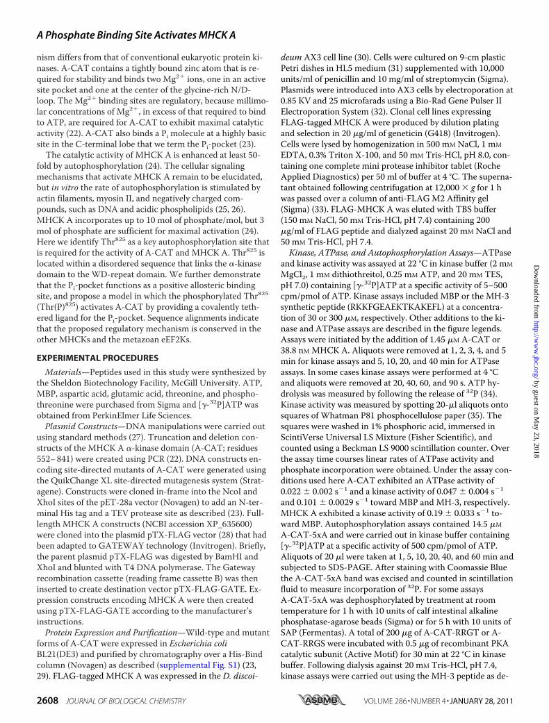
The Disordered C-terminal Sequence Is Required for A-CATActivity—A-CAT is a catalytically active fragment of MHCKA that encompasses the �-kinase domain (residues 552–805)as well as part of the flexible linker that connects the �-kinasedomain to the WD-repeat domain (residues 806–841) (Fig.1A) (22, 23). In the x-ray crystal structure of A-CAT, residues806–841 lack defined electron density and thus form a disor-dered C-terminal tail (C-tail) (23). Truncation of the C-tail atresidue 831 did not alter the kinase activity of A-CAT, buttruncation at residues 823 or 809 resulted in the loss of 90–95% of kinase activity (Fig. 1B, left panel). The severe loss ofkinase activity was observed with both myelin basic proteinand the MH-3 peptide, which corresponds to the Thr2029 sitein the myosin II tail, as substrate (24). In the absence of a pro-tein or peptide substrate A-CAT exhibits a basal rate ofATPase activity (23). Truncation of the C-tail at residues 823or 809 reduced the ATPase activity of A-CAT by �95% (Fig.1B, right panel). A section of the C-tail between residues 823and 831 is therefore required for both the kinase and ATPaseactivities of A-CAT.An Essential Threonine Residue Is Conserved in the C-tail—A
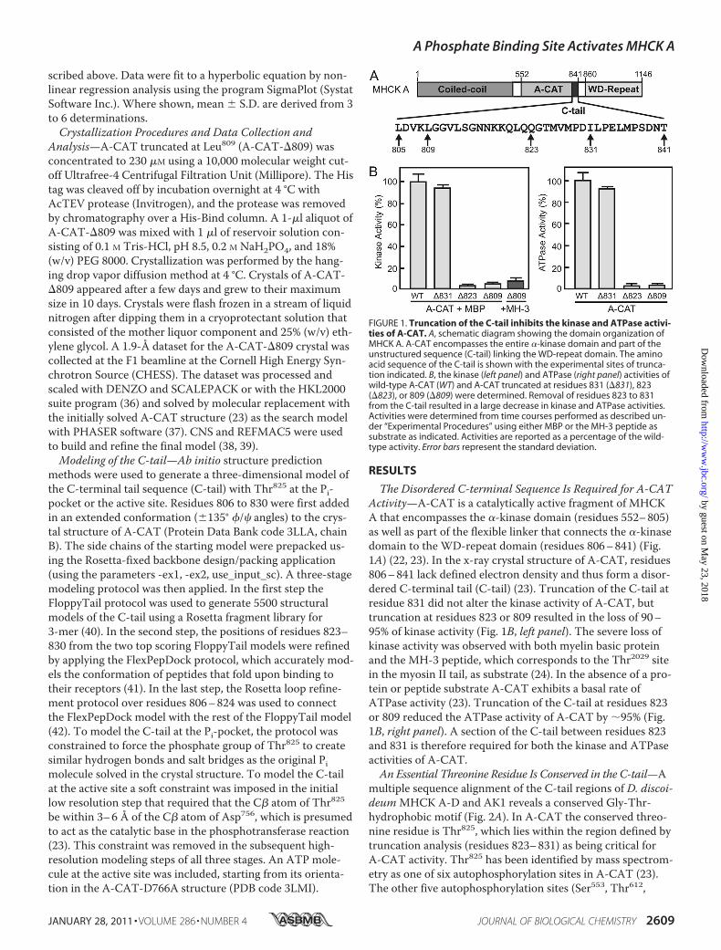
multiple sequence alignment of the C-tail regions of D. discoi-deumMHCK A-D and AK1 reveals a conserved Gly-Thr-hydrophobic motif (Fig. 2A). In A-CAT the conserved threo-nine residue is Thr825, which lies within the region defined bytruncation analysis (residues 823–831) as being critical forA-CAT activity. Thr825 has been identified by mass spectrom-etry as one of six autophosphorylation sites in A-CAT (23).The other five autophosphorylation sites (Ser553, Thr612,
FIGURE 1. Truncation of the C-tail inhibits the kinase and ATPase activi-ties of A-CAT. A, schematic diagram showing the domain organization ofMHCK A. A-CAT encompasses the entire �-kinase domain and part of theunstructured sequence (C-tail) linking the WD-repeat domain. The aminoacid sequence of the C-tail is shown with the experimental sites of trunca-tion indicated. B, the kinase (left panel) and ATPase (right panel) activities ofwild-type A-CAT (WT) and A-CAT truncated at residues 831 (�831), 823(�823), or 809 (�809) were determined. Removal of residues 823 to 831from the C-tail resulted in a large decrease in kinase and ATPase activities.Activities were determined from time courses performed as described un-der “Experimental Procedures” using either MBP or the MH-3 peptide assubstrate as indicated. Activities are reported as a percentage of the wild-type activity. Error bars represent the standard deviation.
A Phosphate Binding Site Activates MHCK A
JANUARY 28, 2011 • VOLUME 286 • NUMBER 4 JOURNAL OF BIOLOGICAL CHEMISTRY 2609
by guest on May 23, 2018
http://ww
w.jbc.org/
Dow
nloaded from
Thr613, Thr614, and Thr634) are located within the N-terminallobe. Mutation of Ser553, Thr612, Thr613, Thr614, and Thr634 toalanine (A-CAT-5xA) had little effect on the kinase orATPase activities of A-CAT (Fig. 2B). In contrast, mutation ofThr825 to alanine decreased kinase and ATPase activities by95% (Fig. 2B). This result suggests that autophosphorylationof Thr825 is critical for the activity of A-CAT. Mutation ofThr825 to glutamic acid or aspartic acid resulted in the loss of�90 and 75% of kinase activity, respectively, indicating that anegatively charged residue only weakly compensates for theloss of phosphothreonine (Fig. 2B). A mutant with serine inplace of Thr825 also exhibited low kinase and ATPase activity(Fig. 2B). Serine residues are poor substrates for A-CAT, sug-gesting that the low activity may reflect the incomplete auto-phosphorylation of Ser825 (21, 29).Phosphorylation of the C-tail Activates A-CAT—Incubation
of A-CAT-5xA with [�-32P]ATP resulted in the incorporationof less than 0.1 mol of phosphate/mol (Fig. 3A). The amount ofphosphate incorporated into A-CAT-5xA increased to 0.2mol/mol after a 1-h treatment with calf intestinal alkalinephosphatase and to 0.33 mol/mol after a 5-h treatment withSAP (Fig. 3A). We interpret these results to indicate thatThr825 is nearly fully phosphorylated in bacterially expressedA-CAT-5xA and that it is only partially dephosphorylatedeven after extensive phosphatase treatment. The phosphory-lated state of Thr825 was confirmed by mass spectrometry(data not shown). Kinase assays showed that SAP treatmentproduced only a small decrease in the kinase activity ofA-CAT-5xA (Fig. 3B). It was reasoned that this may reflectthe ability of A-CAT-5xA to rapidly autophosphorylateThr825 in the kinase assay (Fig. 3A). When kinase assays werecarried out at 4 °C to reduce the rate of Thr825 autophosphor-
ylation, a significant decrease in the activity of SAP-treatedA-CAT-5xA could be detected (Fig. 3B). These results areconsistent with the conclusion that phosphorylation of Thr825is required for A-CAT activity.The ability of Thr825 phosphorylation to activate A-CAT-
5xA was further examined by mutating Gln822 and Gln823 toarginine to convert Thr825 into a PKA phosphorylation site(i.e.QQGT to RRGT). A mutant with serine in place of Thr825(RRGS) was also created. A-CAT-RRGS exhibited a lowerinitial activity than A-CAT-RRGT, which is consistent withserine being a poorer autophosphorylation site than threo-nine. Incubation with PKA and MgATP increased the activityof A-CAT-RRGT by 60% and A-CAT-RRGS by more than3-fold (Fig. 3C). The increase in activity was accompanied bythe incorporation of 0.25 mol of Pi/mol into A-CAT-RRGS byPKA. Control experiments showed that the activity ofA-CAT-5xA was not altered by incubation with PKA and thatthe MH-3 peptide was not a substrate for PKA (Fig. 3C). This
FIGURE 2. The conserved Thr825 residue in the C-tail is required for A-CAT activity. A, a multiple sequence alignment of the C-tail regions of D.discoideum MHCK A, MHCK B, MHCK C, MHCK D, and AK1. Conserved resi-dues are shaded in gray. A Gly-Thr motif (residues 824 and 825 in MHCK A)is present in all of the C-tail sequences. The NCBI accession numbers are:MHCK A, XP_635119; MHCK B, XP_636368; MHCK C, XP_635600; MHCK D,XP_640080; and AK1, XP_629868. B, the kinase (left panel) and ATPase (rightpanel) activities of wild-type A-CAT (WT), A-CAT-5xA, and the indicatedThr825 mutants were determined. Mutation of Thr825 severely inhibited boththe kinase and ATPase activities of A-CAT. Activities were determined fromtime courses performed as described under “Experimental Procedures”and the activities are reported as a percentage of A-CAT activity. Error barsrepresent the standard deviation.
FIGURE 3. Phosphorylation of the C-tail activates A-CAT. A, time courseof the autophosphorylation of A-CAT-5xA prior to treatment (F) and follow-ing treatment for 1 h with calf intestinal alkaline phosphatase (E) or for 5 hwith SAP (�). The low level of 32P incorporation indicates that Thr825 ishighly phosphorylated and is resistant to dephosphorylation by calf intesti-nal alkaline phosphatase and SAP. Autophosphorylation assays were per-formed by incubating A-CAT-5xA with [�-32P]ATP as described under “Ex-perimental Procedures.” B, the kinase activity of A-CAT-5xA incubated for5 h in the presence or absence of SAP was assayed at 22 and 4 °C. The as-says at 4 °C, which limits the ability of A-CAT-5xA to autophosphorylateThr825, showed that dephosphorylation of Thr825 by SAP decreased the ki-nase activity of A-CAT-5xA. Activities were determined from time coursesperformed as described under “Experimental Procedures” and are reportedas a percentage of the untreated A-CAT-5xA activity. Error bars representthe standard deviation. C, the kinase activities of A-CAT-5xA (5xA) or A-CAT-5xA with the QQGT sequence in the C-tail mutated to RRGT or RRGS weredetermined before and after incubation with PKA as indicated. Activitieswere determined from time courses performed as described under “Experi-mental Procedures” with the MH-3 peptide as substrate. The MH-3 peptidewas not a substrate for PKA. Incubation with PKA activated the RRGT andRRGS mutants, but not A-CAT-5xA, showing that phosphorylation of theThr825 site enhances kinase activity. Activities are reported as a percentageof A-CAT-5xA activity. Error bars represent the standard deviation.
A Phosphate Binding Site Activates MHCK A
2610 JOURNAL OF BIOLOGICAL CHEMISTRY VOLUME 286 • NUMBER 4 • JANUARY 28, 2011
by guest on May 23, 2018
http://ww
w.jbc.org/
Dow
nloaded from

result provides additional evidence that A-CAT activity de-pends on the phosphorylation of the Thr825 (or Ser825) site inthe C-tail.To examine whether autophosphorylation of Thr825 is re-
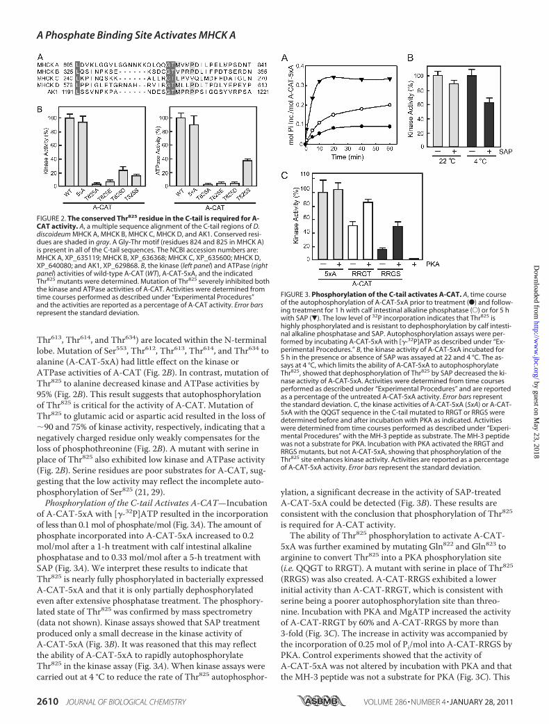
quired for the activation of full-length MHCK A, wild-typeMHCK A and T825A and T825E mutants were expressed inD. discoideum as FLAG-tagged proteins (Fig. 4A). The kinaseactivities of the MHCK A T825A and T825E mutants wereless than 10% that of wild-type MHCK A (Fig. 4B). This showsthat autophosphorylation of Thr825 is a necessary step in theactivation of MHCK A.The effect of moving Thr825 closer to the �-kinase domain
was examined by deleting three (A-CAT-�3) or six (A-CAT-�6) residues from the intervening linker sequence (Fig. 4C).A-CAT-�3 and A-CAT-�6 were considerably less active thanA-CAT, indicating that the function of Thr825 depends tosome extent on its position within the C-tail (Fig. 4C). Theconserved nature of the Gly-Thr-hydrophobic sequence sug-
gested that the C-tails of AK1 and MHCK B-D may havefunctions comparable with that of MHCK A (Fig. 2A). To testthis possibility the C-tail of MHCK B (residues 326–354) wasfused to the C terminus of A-CAT truncated at Leu805 (Fig.4C). The C-tail of MHCK B fully rescued both the kinase andATPase activity of the truncated A-CAT (Fig. 4C). This resultsupports the view that the MHCK C-tail regions performanalogous regulatory functions.X-ray Crystal Structure of A-CAT-�809—A-CAT truncated
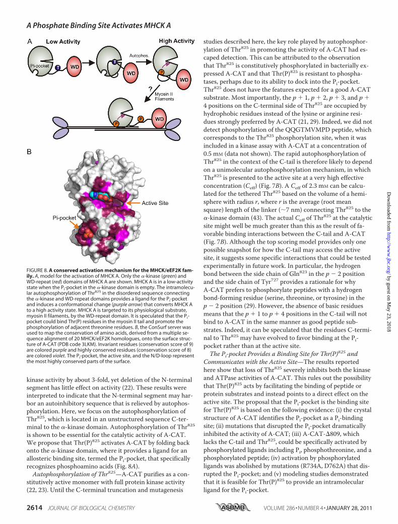
at residue 809 (A-CAT-�809) was purified in yields compara-ble with that of A-CAT, showing that the loss of the C-taildoes not compromise the stability of the core �-kinase do-main. Indeed, A-CAT-�809 formed diffraction quality crys-tals much more readily than A-CAT, indicating that the C-tail
FIGURE 4. Regulation of A-CAT and MHCK A by Thr825. A, the CoomassieBlue-stained SDS gel shows wild-type MHCK A (WT) and the MHCK A T825Aand T825E mutants purified from D. discoideum as described under “Experi-mental Procedures.” B, the kinase activities of MHCK A (WT) and the MHCK AT825A and T825E mutants were assayed as described under “ExperimentalProcedures.” Mutation of Thr825 severely inhibited the kinase activity ofMHCK A. Activities are reported as a percentage of MHCK A activity. Errorbars represent the standard deviation. C, the top line shows the sequence ofthe A-CAT C-tail and indicates the residues deleted to generate the �3 and�6 constructs. The second line shows the sequence of the MHCK B C-tail(residues 326 –354) that was fused to Leu805 of A-CAT to generate the A-CAT-BT chimera. The conserved Gly-Thr sequence is underlined. The kinase(left panel) and ATPase (right panel) activities of wild-type A-CAT (WT), the�3 and �6 constructs, and the A-CAT-BT chimera (BT) were determinedfrom time courses performed as described under “Experimental Proce-dures.” The C-tail of MHCK B rescues the activity of the truncated A-CAT.Activities are reported as a percentage of wild-type A-CAT activity. Error barsrepresent the standard deviation.
FIGURE 5. X-ray crystal structure of A-CAT-�809. A, the structure of A-CAT-�809 is shown with �-strands in blue and �-helices in strawberry. Mg2�
ions are rendered as dark blue spheres and the zinc atom as a yellow sphere.Arg734, Asp762, ADP, and Pi are shown as sticks with the C, O, N, and P atomscolored green, red, blue, and orange, respectively. N, N terminus; C, C termi-nus. B, the distribution of electrostatic potential is shown on the molecularsurface of A-CAT-�809, with blue indicating areas of net positive charge andred areas of net negative charge. ADP and Pi are rendered as spheres. Theview is rotated 40° to the right from the view in panel A. C, detailed view ofthe Pi-pocket. The crystal structure of A-CAT (left panel) shows that the Pimolecule interacts with the side chains of Arg734, Lys684, and Thr736 andthe main chain carbonyl of Ser735 (dotted lines). The bent Arg734 sidechain bridges the Pi molecule to Asp762 in an active site loop. Ab initiomodeling of the Pi-pocket in the absence of Pi (right panel) shows thatthe side chain of Arg734 can switch from the bent to a straight configura-tion that does not interact with Asp762. Although the straight conforma-tion of Arg734 can occur when modeling in the presence of a ligand, it isfavored by an empty Pi-pocket. The conformational change in Arg734 ispostulated to act as an allosteric switch that controls the activity of A-CAT in response to Pi binding.
A Phosphate Binding Site Activates MHCK A
JANUARY 28, 2011 • VOLUME 286 • NUMBER 4 JOURNAL OF BIOLOGICAL CHEMISTRY 2611
by guest on May 23, 2018
http://ww
w.jbc.org/
Dow
nloaded from

impairs crystal formation. The x-ray crystal structure of A-CAT-�809 was solved to a resolution of 1.8 Å (Fig. 5A andsupplemental Table S1). When the C� atoms of A-CAT-�809and A-CAT were superimposed, a root mean square differ-ence of 0.37 Å was obtained, which indicates that the twostructures are virtually identical. The structure of A-CAT-�809 further shows that the absence of the C-tail did not alterthe binding of ligands. A-CAT-�809, like A-CAT, contained azinc atom bound to the C-terminal lobe, a Pi molecule boundto the Pi-pocket and Mg2� ions bound at the active site and atthe center of the N/D-loop (23). The active site of A-CAT-�809 contained ADP, even though crystallization was carriedout in a buffer containing ATP (Fig. 5A). This result is sur-prising given the low ATPase activity exhibited by A-CAT-�809 (Fig. 1B) and raises the possibility that the crystallizationconditions induce A-CAT-�809 to adopt a catalytically activeconformation.The Pi-pocket Regulates A-CAT Activity—It was reasoned
that Thr(P)825 must bind to a site in the core �-kinase domainto stimulate catalytic activity. The Pi-pocket, which is locatednear to the bottom of the C-terminal lobe, provides an obvi-ous candidate for a phosphate-dependent binding site (Fig.5B). The Pi molecule makes electrostatic interactions with theside chains of Lys684, located in the loop following �-helix C,and Arg734, located at the start of �-helix D. The Pi moleculealso forms hydrogen bonds with the main chain carbonyl ofSer735 and the main chain carbonyl and side chain hydroxylgroup of Thr736 (Fig. 5C and supplemental Fig. S2). Mutationof Lys684, Arg734, and Thr736 to alanine reduced the kinaseactivity of A-CAT by 90–95% showing that an intact Pi-pocket is essential for activity (Fig. 6A). A mechanism bywhich Pi binding may influence the conformation of the ac-tive site is apparent from the structure of A-CAT, whichshows that Arg734 forms a salt bridge with Asp762 in one ofthe principal catalytic loops (Fig. 5C). Mutation of Asp762 toalanine drastically reduced catalytic activity, demonstratingthat the salt bridge with Arg734 is of functional importance(Fig. 6A).The possibility that Pi might be able to compensate for the
loss of Thr825 by providing a ligand for the Pi-pocket wastested by adding NaH2PO4 to the kinase assays. The additionof Pi stimulated the kinase activity of A-CAT-�809 by �4-fold, to a level �20% that of A-CAT (Fig. 6B). Half-maximalactivation was achieved at a Pi concentration of 440 � 150�M. At higher concentrations of Pi a slight inhibition of A-CAT activity was observed, likely because of the increasedionic strength (1). Phosphothreonine proved to be a betteractivator than Pi, stimulating the kinase activity of A-CAT-�809 to a level approaching 65% that of A-CAT (Fig. 6C).Half-maximal activation occurred at a phosphothreonineconcentration of 250 � 80 �M. No activation of A-CAT-�809occurred when threonine was added to the kinase assays (Fig.6C). Glutamic acid and aspartic acid also did not activate (notshown). A peptide with a sequence corresponding to residues823–830 of the C-tail (QQGTMVMPD) had no effect on theactivity of A-CAT-�809, whereas the phosphorylated versionof the peptide (QQG(p)TMVMPD) was as effective an activa-tor as phosphothreonine (Fig. 6C). Half-maximal activation
by the QQG(p)TMVMPD peptide was achieved at a concen-tration of 40 � 15 �M. Mutation of Arg734 to alanine abol-ished the ability of phosphothreonine to activate A-CAT-�809 (Fig. 6C) as did mutation of Asp762 to alanine (notshown). These results confirm that activation of A-CAT-�809is the result of ligand binding to the Pi-pocket.Phosphothreonine and the QQG(p)TMVMPD peptide also
restored the ATPase activity of A-CAT-�809 (Fig. 6D). Bothactivators increased the ATPase activity of A-CAT-�809 to alevel approaching 95% that of A-CAT. Half-maximal activa-tion was achieved at QQG(p)TMVMPD and phosphothreo-nine concentrations of 120 � 40 and 360 � 40 �M, respec-tively. Taken together, the data show that phosphorylatedligands dramatically and specifically rescue the catalytic activ-ity of A-CAT-�809.Modeling of the C-tail at the Pi-pocket—Analytical ultracen-
trifugation studies show that A-CAT is a monomer in solu-
FIGURE 6. Ligand binding to the Pi-pocket activates A-CAT. A, the kinaseactivity of wild-type A-CAT (WT) and the indicated Pi-pocket mutants weredetermined as described under “Experimental Procedures.” Error bars repre-sent the standard deviation. Mutation of residues that bind Pi resulted in asevere loss in kinase activity. B, the kinase activity of A-CAT (closed symbols)and A-CAT-�809 (open symbols) were assayed in the presence of NaH2PO4.The addition of Pi enhanced the kinase activity of A-CAT-�809. A hyperboliccurve fit to the A-CAT-�809 data yielded a Ka � 440 � 150 �M and a Vmax �22 � 3%. Activities are reported as a percentage of the A-CAT activity in theabsence of Pi. C, the kinase activity of A-CAT (closed symbols) and A-CAT-�809 (open symbols) were assayed in the presence of phosphothreonine(squares), the QQG(p)TMVMPD peptide (circles), threonine (triangles), andthe unphosphorylated QQGTMVMPD peptide (inverted triangles). TheA-CAT-�809-R734A mutant was assayed in the presence of phosphothreo-nine (diamonds). Hyperbolic curves fit to the A-CAT-�809 data yielded aKa � 260 � 80 �M and a Vmax � 65 � 6% for phosphothreonine and a Ka �35 � 12 �M and a Vmax � 64 � 3% for the QQG(p)TMVMPD peptide. Activi-ties are reported as a percentage of the A-CAT activity in the absence of anyadditions. D, the ATPase activity of A-CAT-�809 was assayed in the pres-ence of phosphothreonine (squares), QQG(p)TMVMPD peptide (circles), orthreonine (triangles). Hyperbolic curves fit to the data yielded a Ka � 360 �40 �M and a Vmax � 93 � 2% for phosphothreonine and a Ka � 120 � 40�M and a Vmax � 97 � 8% for the QQG(p)TMVMPD peptide. The kinase orATPase activity shown for each ligand concentration in panels B–D was de-termined from a time course performed as described under “ExperimentalProcedures.”
A Phosphate Binding Site Activates MHCK A
2612 JOURNAL OF BIOLOGICAL CHEMISTRY VOLUME 286 • NUMBER 4 • JANUARY 28, 2011
by guest on May 23, 2018
http://ww
w.jbc.org/
Dow
nloaded from

tion (23). To determine whether it is feasible for Thr(P)825 toact as an intramolecular ligand for the Pi-pocket, residues806–830 of the C-tail were modeled using the Rosetta Flop-pyTail and FlexPepDock protocols (40, 41). The only con-straint on the model was that the Thr825 phosphoryl groupwas forced to adopt the same binding interactions as the Pimolecule in the crystal structure. The modeling process re-sulted in 5,500 independent models. The two top-scoringmodels converged on a single binding mode that allowsThr(P)825 to dock into the Pi-pocket (Fig. 7A and supplemen-tal Fig. S3). Reassuringly, the backbone and side chain confor-mations of residues 823–830 are nearly identical in both inde-pendent models. The model predicts that residues 806–817form a random coil and make little or no contact withA-CAT, which is consistent with multiple sequence align-ments that show this section of the C-tail to be poorly con-served (Fig. 2A). Residues 818–823 in the model form a short�-helix that, again, makes no contact with A-CAT. The con-served Gly824 allows the polypeptide chain to make a sharpturn that places the Thr825 phosphoryl group into the Pi-
pocket in the same orientation as the Pi in the crystal struc-ture. Residues on the C-terminal side of Thr825 take up anextended conformation and are involved in interactions withA-CAT. This stretch of sequence corresponds to the C-tailpeptide (QQGTMVMPD) used in the A-CAT-�809 activa-tion experiments described above. Most notably, Met826makes hydrophobic contacts with Pro683 and Val799.Interestingly, the modeling studies predict that the side
chain of Arg734 in the Pi-pocket can switch from the bent con-formation present in the A-CAT crystal structure to a straightconformation (Fig. 5C). The conformational change movesthe Arg734 guanidino group 3 to 6 Å away from the Pi pocketand thus disrupts the electrostatic interactions with the Pimolecule and Asp762. The tendency of Arg734 to adopt thetwo orientations was evaluated by repacking the side chainconformations in the region of the Pi-pocket 200 times, bothin the absence and presence of the C-tail. In the free state,99.5% of the models had the side chain of Arg734 in thestraight conformation, whereas when Thr(P)825 is bound asignificant fraction of the models (42.3%) had the Arg734 sidechain in the bent conformation. These studies suggest thatbinding of a phosphorylated ligand induces a conformationalchange in Arg734 that promotes formation of a salt bridgewith Asp762.Modeling of the C-tail in the Active Site—The same three-
stage modeling protocol was carried out to examine whether itis feasible for Thr825 to interact in an intramolecular mannerwith the A-CAT active site. A soft constraint that requiredthe C� atom of Thr825 to be within 3–6 Å of the C� atom ofAsp756 (the presumed catalytic base) was applied in the initiallow-resolution modeling using the FloppyTail protocol butwas then removed in the subsequent high-resolution model-ing steps. The modeling studies clearly indicate that the flexi-ble C-tail allows Thr825 to access the active site (Fig. 7B). Al-though there was some convergence near the active site, thetop-scoring models were not identical, indicating that theflexible C-tail is likely to be able to reach the catalytic site viaan ensemble of conformations (supplemental Fig. S4). The topscoring model provides one possible snapshot of this ensem-ble and is stabilized by several favorable interactions, includ-ing a salt bridge between Lys808 and Glu746, hydrophobic in-teractions between Leu813 and Thr793, and a hydrogen bondbetween Gln822 and the main chain amide of Lys722 (Fig. 7B).In addition, the model predicts that the side chain of Tyr727rotates from its position in the crystal structure toward theC-tail so that it can form a hydrogen bond with the side chainof Gln823 and participate in hydrophobic interactions withMet826.
DISCUSSION
MHCK A is potently activated by autophosphorylation butthe identity of the autophosphorylation sites and the mecha-nisms involved in the activation process have remained ob-scure. Previous studies have localized several MHCK-A auto-phosphorylation sites to a segment (amino acids 499–550)immediately N-terminal to the �-kinase domain (22). Studieson A-CAT, the isolated �-kinase domain of MHCK A, showthat autophosphorylation of the N-terminal sites stimulates
FIGURE 7. Models of the C-tail bound to the Pi-pocket and active site.The Rosetta FloppyTail and FlexPepDock protocols were used to model theC-tail (residues 806 – 830) using the procedure described under “Experimen-tal Procedures.” A, model of the C-tail with the phosphorylated Thr825
bound into the Pi-pocket. The C-tail is colored blue, key residues in the C-tailthat interact with the �-kinase domain are shown in stick representation,and the original Pi in the crystal structure is represented as transparentspheres. The surface of A-CAT (PBD code 3LLA, chain B) is shown in gray. Themodel shows that the Thr825 phosphoryl group can act as an intramolecularligand for the Pi-pocket. B, model of the C-tail with the unphosphorylatedThr825 at the active site. The C-tail is colored blue, residues in the C-tail thatinteract with the �-kinase domain are shown in stick representation, andATP (from PBD code 3LMI) is shown as spheres. The surface of A-CAT isshown in gray and is transparent to illustrate the position of Tyr727 pre-dicted by the model (cyan sticks) and in the crystal structure (green sticks).The model illustrates that it is feasible for Thr825 to be autophosphorylatedvia an intramolecular mechanism.
A Phosphate Binding Site Activates MHCK A
JANUARY 28, 2011 • VOLUME 286 • NUMBER 4 JOURNAL OF BIOLOGICAL CHEMISTRY 2613
by guest on May 23, 2018
http://ww
w.jbc.org/
Dow
nloaded from
kinase activity by about 3-fold, yet deletion of the N-terminalsegment has little effect on activity (22). These results wereinterpreted to indicate that the N-terminal segment may har-bor an autoinhibitory sequence that is relieved by autophos-phorylation. Here, we focus on the autophosphorylation ofThr825, which is located in an unstructured sequence C-ter-minal to the �-kinase domain. Autophosphorylation of Thr825is shown to be essential for the catalytic activity of A-CAT.We propose that Thr(P)825 activates A-CAT by folding backonto the �-kinase domain, where it provides a ligand for anallosteric binding site, termed the Pi-pocket, that specificallyrecognizes phosphoamino acids (Fig. 8A).Autophosphorylation of Thr825—A-CAT purifies as a con-
stitutively active monomer with full protein kinase activity(22, 23). Until the C-terminal truncation and mutagenesis
studies described here, the key role played by autophosphor-ylation of Thr825 in promoting the activity of A-CAT had es-caped detection. This can be attributed to the observationthat Thr825 is constitutively phosphorylated in bacterially ex-pressed A-CAT and that Thr(P)825 is resistant to phospha-tases, perhaps due to its ability to dock into the Pi-pocket.Thr825 does not have the features expected for a good A-CATsubstrate. Most importantly, the p � 1, p � 2, p � 3, and p �4 positions on the C-terminal side of Thr825 are occupied byhydrophobic residues instead of the lysine or arginine resi-dues strongly preferred by A-CAT (21, 29). Indeed, we did notdetect phosphorylation of the QQGTMVMPD peptide, whichcorresponds to the Thr825 phosphorylation site, when it wasincluded in a kinase assay with A-CAT at a concentration of0.5 mM (data not shown). The rapid autophosphorylation ofThr825 in the context of the C-tail is therefore likely to dependon a unimolecular autophosphorylation mechanism, in whichThr825 is presented to the active site at a very high effectiveconcentration (Ceff) (Fig. 7B). A Ceff of 2.3 mM can be calcu-lated for the tethered Thr825 based on the volume of a hemi-sphere with radius r, where r is the average (root meansquare) length of the linker (�7 nm) connecting Thr825 to the�-kinase domain (43). The actual Ceff of Thr825 at the catalyticsite might well be much greater than this as the result of fa-vorable binding interactions between the C-tail and A-CAT(Fig. 7B). Although the top scoring model provides only onepossible snapshot for how the C-tail may access the activesite, it suggests some specific interactions that could be testedexperimentally in future work. In particular, the hydrogenbond between the side chain of Gln823 in the p � 2 positionand the side chain of Tyr727 provides a rationale for whyA-CAT prefers to phosphorylate peptides with a hydrogenbond-forming residue (serine, threonine, or tyrosine) in thep � 2 position (29). However, the absence of basic residuesmeans that the p � 1 to p � 4 positions in the C-tail will notbind to A-CAT in the same manner as good peptide sub-strates. Indeed, it can be speculated that the residues C-termi-nal to Thr825 may have evolved to favor binding at the Pi-pocket rather than at the active site.The Pi-pocket Provides a Binding Site for Thr(P)825 and
Communicates with the Active Site—The results reportedhere show that loss of Thr825 severely inhibits both the kinaseand ATPase activities of A-CAT. This rules out the possibilitythat Thr(P)825 acts by facilitating the binding of peptide orprotein substrates and instead points to a direct effect on theactive site. The proposal that the Pi-pocket is the binding sitefor Thr(P)825 is based on the following evidence: (i) the crystalstructure of A-CAT identifies the Pi-pocket as a Pi-bindingsite; (ii) mutations that disrupted the Pi-pocket dramaticallyinhibited the activity of A-CAT; (iii) A-CAT-�809, whichlacks the C-tail and Thr825, could be specifically activated byphosphorylated ligands including Pi, phosphothreonine, and aphosphorylated peptide; (iv) activation by phosphorylatedligands was abolished by mutations (R734A, D762A) that dis-rupted the Pi-pocket; and (v) modeling studies demonstratedthat it is feasible for Thr(P)825 to provide an intramolecularligand for the Pi-pocket.
FIGURE 8. A conserved activation mechanism for the MHCK/eEF2K fam-ily. A, model for the activation of MHCK A. Only the �-kinase (green) andWD-repeat (red) domains of MHCK A are shown. MHCK A is in a low-activitystate when the Pi-pocket in the �-kinase domain is empty. The intramolecu-lar autophosphorylation of Thr825 in the disordered sequence connectingthe �-kinase and WD-repeat domains provides a ligand for the Pi-pocketand induces a conformational change (purple arrow) that converts MHCK Ato a high activity state. MHCK A is targeted to its physiological substrate,myosin II filaments, by the WD-repeat domain. It is speculated that the Pi-pocket could bind Thr(P) residues in the myosin II tail and promote thephosphorylation of adjacent threonine residues. B, the ConSurf server wasused to map the conservation of amino acids, derived from a multiple se-quence alignment of 20 MHCK/eEF2K homologues, onto the surface struc-ture of A-CAT (PDB code 3LKM). Invariant residues (conservation score of 9)are colored purple and highly conserved residues (conservation score of 8)are colored violet. The Pi-pocket, the active site, and the N/D-loop representthe most highly conserved parts of the surface.
A Phosphate Binding Site Activates MHCK A
2614 JOURNAL OF BIOLOGICAL CHEMISTRY VOLUME 286 • NUMBER 4 • JANUARY 28, 2011
by guest on May 23, 2018
http://ww
w.jbc.org/
Dow
nloaded from

Importantly, a molecular mechanism that allows the Pi-pocket to communicate directly with the active site is appar-ent from the crystal structure of A-CAT and gains supportfrom the modeling studies (Fig. 5C). The mechanism hingeson a salt bridge that is formed between Arg734 in the Pi-pocket and Asp762 in the active site loop that harbors catalyticresidues Asp756 and Asp766. Asp766 accepts the �-phosphatefrom ATP to form an aspartylphosphate intermediate andAsp756 is presumed to be the catalytic base in the phospho-transferase reaction (23, 44). By anchoring the catalytic loop,the salt bridge between Asp762 and Arg734 helps to fix the po-sitions of Asp756 and Asp766 within the active site. The model-ing studies indicate that the bent conformation of the Arg734side chain that bridges the Pi molecule to Asp762 is not ener-getically favorable, especially when the Pi-pocket is empty.Thus, a plausible allosteric model can be proposed in whichthe binding of a phosphorylated ligand to the Pi-pocket stabi-lizes the bent conformation of the Arg734 side chain, which inturn promotes the electrostatic interaction with Asp762 andhelps to properly align the Asp756–Asp766 catalytic loopwithin the active site. It is interesting that interactions involv-ing the Asp756–Asp766 loop have also been implicated in theactivation of TRPM7 by dimer assembly (45). The results sug-gest that regulatory mechanisms that stabilize what may be aninherently mobile Asp756–Asp766 catalytic loop may provide acommon pathway by which to activate the �-kinases.In several experiments the kinase and ATPase activities of
A-CAT were found to diverge. Phosphothreonine and theQQG(p)TMVMPD peptide restored about 60–65% ofthe kinase activity of A-CAT-�809 but close to 95% of theATPase activity. Differential effects on the kinase and ATPaseactivities of A-CAT were also noted with the T825D, T825S,and �3 mutants (Figs. 2B and 4C). It can be speculated thatthe balance between the two enzymatic activities may dependon the active site conformation, which is influenced by theligand for the Pi-pocket. Whether or not the ATPase (andADPase) activities exhibited by A-CAT are physiologicallyrelevant is currently not clear (23).The identification of the Pi-pocket as a positive allosteric
binding site provides a rationale for why A-CAT-�809 andA-CAT have virtually identical crystal structures. Both A-CAT and A-CAT-�809 were crystallized from buffers con-taining 0.2 M NaH2PO4 and in both crystal structures Pi occu-pies the Pi-pocket (23). It can be concluded that in the case ofA-CAT, Pi in the crystallization buffer displaced Thr(P)825from the Pi-pocket, causing the C-tail to take up a disorderedconformation that is not visible in the crystal structure. In thecase of A-CAT-�809, Pi provided a ligand for the Pi-pocket inthe absence of the C-tail and induced A-CAT-�809 to adoptthe active conformation. This analysis highlights the need toidentify crystallization conditions that lack Pi. In the absenceof Pi, it may be possible to obtain a structure of the inactivestate of A-CAT-�809 with an empty Pi-pocket and a structureof A-CAT in the autoactivated state with Thr(P)825 dockedinto the Pi-pocket.Substrate Specificity of the Pi-pocket—Glutamic acid and
aspartic acid did not activate A-CAT-�809, although glutamicacid, and especially aspartic acid, supported a low level of cat-
alytic activity when substituted for Thr835 in the C-tail. Thissuggests that the Pi-pocket exhibits a weak affinity for car-boxyl groups. The primary interaction with the Pi-pocket isthrough the phosphoryl group. However, Pi was less effectivethan phosphothreonine in stimulating the activity of A-CAT-�809, indicating that the amino acid side chain plays somerole in the binding interaction. The QQG(p)TMVMPD pep-tide activated A-CAT-�809 at a 3–5-fold lower concentrationthan phosphothreonine, which is consistent with the predic-tion from modeling studies that Met826 can act as a secondaryrecognition site to promote binding of the C-tail at the Pi-pocket (Fig. 7A). The importance of the potential secondaryinteractions in mediating the binding of Thr825 to the Pi-pocket can be tested experimentally in future work. The esti-mated binding affinity of the phosphorylated QQG(p)TM-VMPD peptide for the Pi-pocket is in the range of 35–120 �M.This is well below the Ceff for the tethered Thr825 of 2.3 mM
calculated above, and implies that autophosphorylation ofThr825 would be sufficient to saturate the Pi-pocket and fullyswitch on kinase activity.A Pi-binding site has been identified in casein kinase I and
glycogen synthase kinase 3� (46–48). In both cases, the Pi-binding site is located within the active site cleft and is re-sponsible for the recognition of substrates that contain aphosphoamino acid. It is interesting to speculate that the Pi-pocket might act, along with the WD-repeat domain, to targetMHCK A to bipolar myosin II filaments (Fig. 8A). Multiplethreonine residues within each myosin II monomer need tobe phosphorylated before the filament falls apart. Once onethreonine residue in the filament is phosphorylated, it couldact as a ligand for the Pi-pocket and enhance the rate of phos-phorylation of nearby threonine residues (Fig. 8A). Such amechanism could result in a more efficient disassembly ofbipolar filaments and may provide an explanation for the abil-ity of myosin II filaments to activate MHCK A (24). Nega-tively charged compounds, including DNA and vesicles com-posed of phosphatidylinositol, also activate MHCK A in vitro(26). The mechanism of activation is not known, but couldinvolve the Pi-pocket. It will be important to establishwhether the Pi-pocket is able to recognize phosphorylatedlipids, nucleotides, and other phosphorylated molecules, orwhether it is specific for phosphopeptides.Conservation of the Pi-pocket in theMHCKs and eEF2Ks—
Phylogenetic analysis demonstrates that the �-kinase domainsof the DictyosteliumMHCKs and metazoan eEF2Ks grouptogether to form one branch of the �-kinase family (12, 23). Amultiple sequence alignment of the MHCKs and eEF2Ksshows that the residues that bind Pi (Lys684, Arg734, andThr736) and the aspartic acid that forms a salt bridge withArg734 (Asp762) are invariant (supplemental Fig. S5). Thehighly conserved nature of the Pi-pocket can be visualized bymapping residues conserved in the multiple sequence align-ment onto the surface of A-CAT (Fig. 8B) (49). This resultimplies that an allosteric regulatory mechanism dependent onthe binding of phosphorylated ligands may underlie the regu-lation of the entire MHCK/eEF2K family. Although the C-tailsequences of the MHCK/eEF2K family members are diver-gent, a threonine residue followed by a hydrophobic residue is
A Phosphate Binding Site Activates MHCK A
JANUARY 28, 2011 • VOLUME 286 • NUMBER 4 JOURNAL OF BIOLOGICAL CHEMISTRY 2615
by guest on May 23, 2018
http://ww
w.jbc.org/
Dow
nloaded from

always present (supplemental Fig. S5). The conserved natureof the hydrophobic residue in the p � 1 position is consistentwith the observation that its predicted binding partner,Pro683, is an invariant residue in the MHCK/eEF2K family(supplemental Fig. S5). eEF2K rapidly autophosphorylatesmultiple serine and threonine residues via an intramolecularmechanism but the sites have not been mapped (50). Severalresidues in the C-tail of eEF2K are targets for exogenous pro-tein kinases and their phosphorylation can either inhibit oractivate kinase activity (20). Further studies will be required todetermine whether any of the C-tail phosphorylation sitesregulate eEF2K by acting as a ligand for the Pi-pocket.In summary, we have demonstrated that the �-kinase do-
main of MHCK A contains a previously undetected allostericbinding site for phosphorylated peptides. The allosteric bind-ing site provides a mechanism to switch on the activity ofMHCK A in response to autophosphorylation and is likely toplay a central role in regulating the cellular activities ofMHCK A and its close homologues, including eEF2K.
Acknowledgments—We thank David McLeod of the Protein Func-tion Discovery Facility, Queen’s University for carrying out massspectrometry experiments. Portions of this research were carried outat the Cornell High Energy Synchrotron Source.
REFERENCES1. Cote, G. P., and Bukiejko, U. (1987) J. Biol. Chem. 262, 1065–10722. De La Roche, M. A., Smith, J. L., Betapudi, V., Egelhoff, T. T., and Cote,
G. P. (2002) J. Muscle Res. Cell Motil. 23, 703–7183. Vaillancourt, J. P., Lyons, C., and Cote, G. P. (1988) J. Biol. Chem. 263,
10082–100874. Luck-Vielmetter, D., Schleicher, M., Grabatin, B., Wippler, J., and
Gerisch, G. (1990) FEBS Lett. 269, 239–2435. Kolman, M. F., Futey, L. M., and Egelhoff, T. T. (1996) J. Cell Biol. 132,
101–1096. Egelhoff, T. T., Lee, R. J., and Spudich, J. A. (1993) Cell 75, 363–3717. Futey, L. M., Medley, Q. G., Cote, G. P., and Egelhoff, T. T. (1995) J. Biol.
Chem. 270, 523–5298. Steimle, P. A., Licate, L., Cote, G. P., and Egelhoff, T. T. (2002) FEBS
Lett. 516, 58–629. Russ, M., Croft, D., Ali, O., Martinez, R., and Steimle, P. A. (2006) Bio-
chem. J. 395, 373–38310. Kolman, M. F., and Egelhoff, T. T. (1997) J. Biol. Chem. 272,
16904–1691011. Steimle, P. A., Naismith, T., Licate, L., and Egelhoff, T. T. (2001) J. Biol.
Chem. 276, 6853–686012. Middelbeek, J., Clark, K., Venselaar, H., Huynen, M. A., and Van Leeu-
wen, F. N. (2010) Cell Mol. Life Sci. 67, 875–89013. Yumura, S., Yoshida, M., Betapudi, V., Licate, L. S., Iwadate, Y., Naga-
saki, A., Uyeda, T. Q., and Egelhoff, T. T. (2005)Mol. Biol. Cell 16,4256–4266
14. Nagasaki, A., Itoh, G., Yumura, S., and Uyeda, T. Q. (2002)Mol. Biol.Cell 13, 4333–4342
15. Liang, W., Licate, L., Warrick, H., Spudich, J., and Egelhoff, T. (2002)BMC Cell Biol. 3, 19
16. Betapudi, V., Mason, C., Licate, L., and Egelhoff, T. T. (2005)Mol. Biol.Cell 16, 2248–2262
17. Betapudi, V., and Egelhoff, T. T. (2009) Traffic 10, 1773–178418. Clark, K., Langeslag, M., van Leeuwen, B., Ran, L., Ryazanov, A. G., Fig-
dor, C. G., Moolenaar, W. H., Jalink, K., and van Leeuwen, F. N. (2006)EMBO J. 25, 290–301
19. Clark, K., Middelbeek, J., Lasonder, E., Dulyaninova, N. G., Morrice,N. A., Ryazanov, A. G., Bresnick, A. R., Figdor, C. G., and Van Leeuwen,F. N. (2008) J. Mol. Biol. 378, 790–803
20. Proud, C. G. (2007) Biochem. J. 403, 217–23421. Luo, X., Crawley, S. W., Steimle, P. A., Egelhoff, T. T., and Cote, G. P.
(2001) J. Biol. Chem. 276, 17836–1784322. Cote, G. P., Luo, X., Murphy, M. B., and Egelhoff, T. T. (1997) J. Biol.
Chem. 272, 6846–684923. Ye, Q., Crawley, S. W., Yang, Y., Cote, G. P., and Jia, Z. (2010) Sci. Signal.
3, ra1724. Medley, Q. G., Gariepy, J., and Cote, G. P. (1990) Biochemistry 29,
8992–899725. Egelhoff, T. T., Croft, D., and Steimle, P. A. (2005) J. Biol. Chem. 280,
2879–288726. Medley, Q. G., Bagshaw, W. L., Truong, T., and Cote, G. P. (1992) Bio-
chim. Biophys. Acta 1175, 7–1227. Sambrook, J., Fritsch, E. F., and Maniatis, T. (1989)Molecular Cloning: A
Laboratory Manual, 2nd Ed., Cold Spring Harbor Laboratory, ColdSpring Harbor, NY
28. Levi, S., Polyakov, M., and Egelhoff, T. T. (2000) Plasmid 44, 231–23829. Crawley, S. W., and Cote, G. P. (2008) Biochim. Biophys. Acta 1784,
908–91530. Hadwiger, J. A., and Firtel, R. A. (1992) Genes Dev. 6, 38–4931. Sussman, M. (1987)Methods Cell Biol. 28, 9–2932. Knecht, D. A., and Shelden, E. (1995) Dev. Biol. 170, 434–44433. Crawley, S. W., de la Roche, M. A., Lee, S. F., Li, Z., Chitayat, S., Smith,
S. P., and Cote, G. P. (2006) J. Biol. Chem. 281, 6307–631534. Pollard, T. D., and Korn, E. D. (1973) J. Biol. Chem. 248, 4682–469035. Casnellie, J. E. (1991)Methods Enzymol. 200, 115–12036. Otwinowski, Z., and Minor, W. (1997) inMethods in Enzymology
(Carter, C., and Sweet, R., eds) Vol. 276, pp. 307–326, Academic Press,New York
37. McCoy, A. J., Grosse-Kunstleve, R. W., Storoni, L. C., and Read, R. J.(2005) Acta Crystallogr. D Biol. Crystallogr. 61, 458–464
38. Brunger, A. T., Adams, P. D., Clore, G. M., DeLano, W. L., Gros, P.,Grosse-Kunstleve, R. W., Jiang, J. S., Kuszewski, J., Nilges, M., Pannu,N. S., Read, R. J., Rice, L. M., Simonson, T., and Warren, G. L. (1998)Acta Crystallogr. D Biol. Crystallogr. 54, 905–921
39. Murshudov, G. N., Vagin, A. A., Lebedev, A., Wilson, K. S., and Dodson,E. J. (1999) Acta Crystallogr. D Biol. Crystallogr. 55, 247–255
40. Kleiger, G., Saha, A., Lewis, S., Kuhlman, B., and Deshaies, R. J. (2009)Cell 139, 957–968
41. Raveh, B., London, N., and Schueler-Furman, O. (2010) Proteins 78,2029–2040
42. Wang, C., Bradley, P., and Baker, D. (2007) J. Mol. Biol. 373, 503–51943. Kramer, R. H., and Karpen, J. W. (1998) Nature 395, 710–71344. Yamaguchi, H., Matsushita, M., Nairn, A. C., and Kuriyan, J. (2001)Mol.
Cell 7, 1047–105745. Crawley, S. W., and Cote, G. P. (2009) Biochem. J. 420, 115–12246. Xu, R. M., Carmel, G., Sweet, R. M., Kuret, J., and Cheng, X. (1995)
EMBO J. 14, 1015–102347. Dajani, R., Fraser, E., Roe, S. M., Young, N., Good, V., Dale, T. C., and
Pearl, L. H. (2001) Cell 105, 721–73248. Longenecker, K. L., Roach, P. J., and Hurley, T. D. (1996) J. Mol. Biol.
257, 618–63149. Glaser, F., Pupko, T., Paz, I., Bell, R. E., Bechor-Shental, D., Martz, E.,
and Ben-Tal, N. (2003) Bioinformatics 19, 163–16450. Redpath, N. T., and Proud, C. G. (1993) Eur. J. Biochem. 212, 511–520
A Phosphate Binding Site Activates MHCK A
2616 JOURNAL OF BIOLOGICAL CHEMISTRY VOLUME 286 • NUMBER 4 • JANUARY 28, 2011
by guest on May 23, 2018
http://ww
w.jbc.org/
Dow
nloaded from
London, Ora Schueler-Furman, Zongchao Jia and Graham P. CôtéScott W. Crawley, Mojdeh Samimi Gharaei, Qilu Ye, Yidai Yang, Barak Raveh, Nir
-Kinase DomainαProviding a Ligand for an Allosteric Binding Site in the Myosin II Heavy Chain Kinase A byDictyosteliumAutophosphorylation Activates
doi: 10.1074/jbc.M110.177014 originally published online November 11, 20102011, 286:2607-2616.J. Biol. Chem.
10.1074/jbc.M110.177014Access the most updated version of this article at doi:
Alerts:
When a correction for this article is posted•
When this article is cited•
to choose from all of JBC's e-mail alertsClick here
http://www.jbc.org/content/286/4/2607.full.html#ref-list-1
This article cites 49 references, 19 of which can be accessed free at
by guest on May 23, 2018
http://ww
w.jbc.org/
Dow
nloaded from