autosomal recessive chondrodysplasia with severe short
TRANSCRIPT

1
Autosomal recessive chondrodysplasia with severe short stature caused by a biallelic
COL10A1 variant
Noor ul Ain1, Outi Makitie
2,3,4, Sadaf Naz
1*
1. School of Biological Sciences, University of the Punjab, Lahore, Pakistan
2. Children’s Hospital, University of Helsinki and Helsinki University Hospital, Helsinki,
Finland
3. Folkhälsan Institute of Genetics, Helsinki, Finland
4. Department of Molecular Medicine and Surgery and Center for Molecular Medicine,
Karolinska Institutet, Stockholm, Sweden
*Correspondence to
Dr. Sadaf Naz, School of Biological Sciences, University of the Punjab
Quaid-i-Azam Campus, Lahore, 54590, Pakistan

2
ABSTRACT
Background Heterozygous mutations in COL10A1 underlie metaphyseal chondrodysplasia,
Schmid type (MCDS), an autosomal dominant skeletal dysplasia.
Objective To identify the causative variant in a large consanguineous Pakistani family with
severe skeletal dysplasia and marked lower-limb deformity.
Methods Whole-Exome sequencing was completed followed by Sanger sequencing to verify
segregation of the identified variants. In silico variant pathogenicity predictions and amino acid
conservation analyses were performed.
Results A homozygous c.133 C>T (p.Pro45Ser) variant was identified in COL10A1 in all six
severely affected individuals (adult heights 119 cm-130 cm, mean~-6.33 SD). The individuals
heterozygous for the variant had mild phenotype of short stature (adult heights 140 cm-162 cm,
mean~-2.15 SD) but no apparent skeletal deformities. The variant was predicted to be pathogenic
by in silico prediction tools and was absent from public databases and hundred control
chromosomes. Pro45 is conserved in orthologues and is located in the non-collagenous 2 domain
of COL10A1, variants of which have never been associated with skeletal dysplasia.
Conclusions This first report of individuals with a homozygous variant in COL10A1 defines a
new type of autosomal recessive skeletal dysplasia. The observations in COL10A1 variant
carriers suggest a phenotypic overlap between the mildest forms of MCDS and idiopathic short
stature.
Keywords: Metaphyseal chondrodysplasia, Schmid type, MCDS, SMCD, COL10A1, Collagen,
Pakistan, Skeletal Dysplasia, Short stature

3
INTRODUCTION
Metaphyseal chondrodysplasia, Schmid type (MCDS; OMIM 156500) is an autosomal dominant
disorder. It is characterized by short stature, long bone deformities such as genua vara and coxa
vara, and radiographic signs of methaphyseal dysplasia. Fifty heterozygous mutations in
COL10A1 encoding type X collagen have been reported to cause MCDS (Human Gene Mutation
Database; http://www.hgmd.cf.ac.uk/ac, accessed in June 2017). Type X collagen is a
homotrimer non-fibrillar collagen, composed of three alpha-1(X) chains of 680 amino acids. It is
present in the extracellular matrix and is exclusively produced by the hypertrophic chondrocytes
during the endochondral ossification.1 The protein plays a role in distribution of proteoglycans
and vesicles in bone matrix. COL10A1 is comprised of a signal peptide (amino acids 1-18) at the
N-terminal followed by a non-collagenous 2 (NC2) region (19-56), a triple-helical region (57-
519) and a non-collagenous 1 (NC1) region (520-680) at the C-terminal.1
All except three mutations2 3
known to cause MCDS affect the NC1 region. Haploinsufficiency is
one of the proposed mechanisms for the disorder since a 50% reduction of COL10A1 is observed
in bone cartilage of MCDS patients who are heterozygous for some nonsense and frameshift
mutations.4 5
Dominant-negative effects of COL10A1 nonsense and missense variants have also
been shown to be responsible for causing the disorder.6 The NC1 region contains motifs required
for trimerization of COL10A1 and a loss of NC1 leads to improper trimerization of collagen,7
resulting in accumulation of these misfolded proteins in the cells. This leads to endoplasmic
stress and apoptosis in hypertrophic chondrocytes during endochondral ossification.8 9
However,
mutations that result in a change of amino acid at the exterior of NC1 region (p.Asp617Lys and
p.Gly618Val) allow trimerization but hinder supramolecular assembly and interaction of

4
COL10A1 with endoplasmic reticulum and proteins in extracellular matrix.10 11
In addition,
mutations of residues which introduce a charge deep inside the hydrophobic NC1 domain affect
the trimerization process and produce unstable COL10A1 which is degraded in the endoplasmic
reticulum8 resulting in haploinsufficiency. Some NC1 missense variants, for example
(p.Tyr598Asp) have been demonstrated to give rise to formation of abnormal disulfide bonds by
exposing sulfhydryl groups of cysteine which are normally buried inside protein in the normal
configuration.8
To date, all patients reported with pathogenic variants in COL10A1 have been heterozygous. In
the present study, we describe a large consanguineous family with a novel missense variant of
COL10A1 leading to a pronounced phenotype with severe short stature and lower-limb
deformities in homozygous individuals while family members harbouring a heterozygous variant
were only mildly affected. This is the first disease-causing variant in COL10A1 located in the
NC2 domain of the protein. Further, to the best of our knowledge, this is the first report of
individuals with a homozygous variant in COL10A1.
MATERIALS AND METHODS
Patients
The study was approved by the Institutional Review Board at the School of Biological Sciences,
University of the Punjab, Lahore, Pakistan. Family NAD-02 (figure 1A) was recruited to the
study after obtaining written informed consents from the participants or their parents. A detailed
history about the onset and progression of growth failure and deformities was obtained. Family
history was recorded and photographs were taken. Ages and heights of all family members were

5
recorded and standard deviation scores for heights were calculated using WHO population-based
reference values. Medical radiographs could not be obtained as all family members refused the
testing. Peripheral blood samples were drawn from the participants. DNA was isolated using a
standard protocol.12
Molecular Analysis
Whole-exome sequencing (WES) was performed for one severely affected individual IV:15,
using Agilent V4 enrichment kit (Agilent Technologies, Santa Clara, CA). Paired-end reads were
obtained at 50× coverage on an Illumina Hi-Seq 2000 sequencer (Otogenetics, Norcross, GA).
Reads were mapped to UCSC hg19 reference human genome (http://genome.ucsc.edu/). The web
based program wANNOVAR was used for further annotating variants. All heterozygous variants
and variants in dbSNP database, Exome Aggregation Consortium (ExAC), 1000 Genomes and in
6500 exon sequence project with minor allele frequency (MAF) greater than 0.0099 were
removed. Exonic and splice site variants were considered. Homozygous chromosomal intervals
in the exome data were identified using Agilevariant mapper and intronic and synonymous
variants located in these regions were also scrutinized. The disease causing potential of sorted
variants was checked by using online prediction programs which included Polyphen2, Mutation
taster, Mutation Assessor and fathmm. ClustalO was used for generating protein alignment of
COL10A1 orthologous sequences retrieved from UCSC genome browser. Primers for the
amplification of genomic regions containing the selected variants were designed using Primer3.
The segregation of each variant with the phenotype was checked after PCR amplification of the
specific product followed by Sanger sequencing with Big Dye Terminator V3.1 cycle
Sequencing kit (Applied Biosystems).

6
RESULTS
Clinical Features
Family NAD-02 had eight severely affected individuals born in four consanguineous marriages
(figure 1A). Six affected individuals participated in the study. All severely affected individuals
had severe disproportionate short stature with short limbs and significant lower limb varus or
valgus deformity but normal facial features (Table 1). Their growth was reported to be normal
until the age of 6 years. However, no longitudinal height data were available. The adult heights
ranged from 114 cm to 130 cm (Table 1). Lower limb deformities progressed with age in all,
with older individuals complaining of joint pain in knees. None of the patients had significant
scoliosis. Bowing of legs was visible but hands and feet were normal. One individual (IV:2)
homozygous for the variant had genu valgum while all other severely affected individuals had
genu varus. Apart from the skeletal findings, no other clinical signs, including intellectual
disability, infections, respiratory problems or hearing deficits were present in any of the affected
individuals. Family members who were obligate carriers and those who were subsequently found
to be heterozygous for the disease-causing variant were apparently normal but had mild short
statures (adult heights from 140 cm to 162 cm) (Table 1). They did not complain of joint pain or
other heath related issues. None of these individuals considered themselves as affected.
Molecular Findings
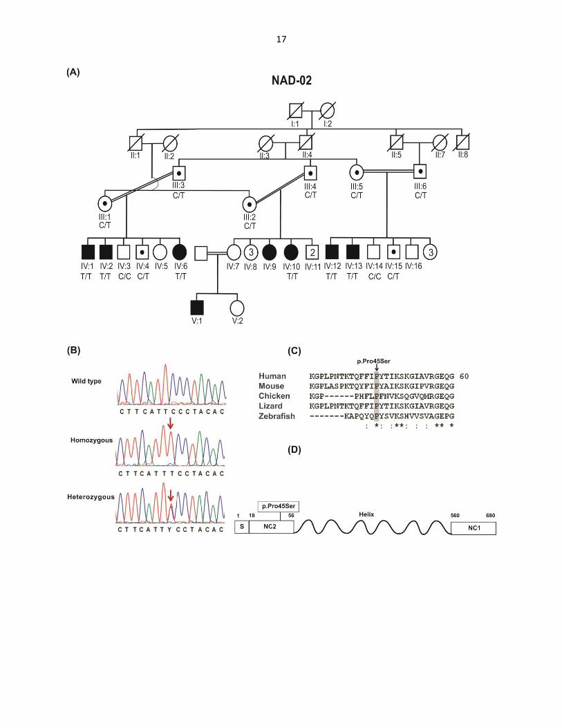
Analysis of the whole exome sequencing data revealed a novel homozygous missense mutation
in exon 2 of COL10A1, c.133 C>T (p.Pro45Ser) which fully co-segregated with the disease
phenotype in the family, with the six severely affected individuals being homozygous for the
variant (figures 1A, 1B, Supplementary Table 1). Analysis of data for individual 1V:15 indicated

7
that all coding exons located in the homozygous chromosome 6 interval (114-130 MB, according
to GRCh37/hg19, data not shown), in which COL10A1 is located, were fully covered by exome
sequencing. No other potential pathogenic variant was identified in the exome data within this
region of homozygosity on chromosome 6 and none of the other variants on different
chromosomal intervals segregated with the phenotype (Supplementary Table 1).
The COL10A1, c.133 C>T (p.Pro45Ser) mutation was absent from public databases including
gnomAD (http://gnomad.broadinstitute.org/) as well as in ethnically matched 100 control
chromosomes. The variant was predicted to be pathogenic or damaging by all four prediction
programs and also had a high Combined Annotation Dependent Depletion (CADD) score of
14.55 (Supplementary Table 1). The Pro45 amino acid is absolutely conserved in COL10A1
orthologues (figure 1C).
DISCUSSION
Variants of COL10A1 have been shown to cause metaphyseal chondrodysplasia, Schmid type in
humans and chondrodysplasia in mice. Both haploinsufficiency and dominant-negative gain of
function are proposed mechanisms for MCDS. Here we describe a new form of skeletal
dysplasia caused by biallelic mutations in COL10A1 resulting in a skeletal phenotype which is
reminiscent of MCDS caused by heterozygous COL10A1 variants but with more severe
symptoms. Interestingly, the carriers of (p.Pro45Ser) variant had mild short statures but no other
phenotypic manifestations. One explanation for this variant causing the severe manifestations of
the disorder only in the homozygous form could be due to the unusual site of the mutation
(figure 1D). The variant identified in the present family is located in the NC2 region of
COL10A1, in which no pathogenic variants have been identified previously. All reported

8
dominantly inherited mutations in MCDS patients are located in the NC1 region of COL10A1
except for three variants.2 3
These COL10A1 heterozygous variants affect the signal peptide
(p.Gly18Arg), (p.Gly18Glu) or the triple helical domain (p.Gly288Arg) of COL10A1 (figure 1D,
Supplementary Table 2). The three variants are associated with late onset and mild forms of
MCDS.2 3
All except one (p.Val677Glu) of the pathogenic variants of NC1 region of COL10A1 (figure 1D
and Supplementary Table 2) result in the development of a severe form of the disease. The NC1
domain has been shown to play a vital role in the trimerization of collagen X.7 Moreover, it has
been proposed that the NC1 region interacts with other proteins and any alteration in the NC1
region may also lead to accumulation of its interacting proteins inside cells, adding to pathology
of disease.13
Homozygous Col10a1-/-
null mutant mice exhibit late onset extremely mild abnormalities.14
These mutant mice develop unilateral coxa vara on ageing. The observed difference in the
severity of phenotype between mice and humans was proposed to be due to the difference in the
weight borne by growth plates in both species.14
However, transgenic wild-type mice transfected
with mutant COL10A1 develop a severe phenotype with genu valgum, coxa vara and short
stature similar to that found in human MCDS patients heterozygous for these mutations.15 16
The
severity of the disease in these mice is gene dosage dependent with homozygous mutant mice
being more severely affected than their heterozygous littermates.15 16
We speculate that the
(p.Pro45Ser) variant causes the disease in a dosage dependent manner similar to that seen for
transgenic mice with COL10A1 mutations.
9
The lack of radiographs for the participants in our study hinders an unequivocal phenotypic
diagnosis for both individuals heterozygous and homozygous for the variant. Nevertheless, the
variant (p.Pro45Ser) in the present family results in a later onset (at around 6 years of age) of an
extremely severe disorder only in individuals carrying the homozygous variant. The usual age of
onset in dominantly inherited MCDS is between 1 to 4 years.17
It has been speculated that late
onset of the disease associates with mutations leading to complete nonsense-mediated RNA
decay of the mutated transcripts, while the more common early onset forms are caused by
mutations with incomplete nonsense-mediated decay and dominant-negative impairment of
protein folding, endoplasmic reticulum stress response and altered hypertrophic chondrocyte
differentiation.18
The heterozygous carriers of the (p.Pro45Ser) variant had short heights, but did not exhibit
apparent skeletal anomalies or other associated clinical features observed in those with
dominantly inherited MCDS or in the homozygous affected individuals in their own family.
Although we cannot exclude the possibility of a skeletal phenotype detectable by radiography in
heterozygous individuals with (p.Pro45Ser) variant, the absence of visible skeletal defects in
these individuals suggest variants in COL10A1 are attractive candidates for idiopathic short
stature. The phenotypic variability among patients harbouring different COL10A1 mutations also
indicates complexity in the disease mechanism.
The pathogenic mechanism through which the missense variant (p.Pro45Ser) leads to severe
short stature in our patients remains to be elucidated. The crystal structure for NC-2 domain of
COL10A1 is not available, thus precluding prediction of the potential effects of the variant on

10
the protein. However, proline has unusual properties since it is a cyclic -imino acid rather than
an -amino acid. Unlike other amino acids, the -carbon atom is part of a cyclic pyrrolidine ring,
which is thus introduced into the polypeptide backbone on incorporation of proline.19
Moreover,
there is no hydrogen bond donor in the amide bond. Incorporation of proline produces bends
and its inclusion can serve as a helix breaker in the polypeptides. Additionally, though usually
found in the cis state, it has been proposed that proline residues may act as molecular switches in
mature proteins by transitioning between the cis and trans states.20
The substitution of serine
residue at position 45 may affect some of these unique properties conferred by proline. In
addition, serine is an amino acid with a hydrogen bond donor. This may affect the bonding of
COL10A1 or lead to its aberrant interactions with other proteins. Determination of the crystal
structure of NC-2 domain is required to elucidate the precise mechanism of (p.Pro45Ser)
variant’s effect. The knowledge gained from in vitro and in vivo analyses of COL10A1 variants
will lead to better understanding of its function in health and disease.
Web resources
1000 Genomes Project (http://www.1000genomes.org)
Agilevariant mapper (http://dna.leeds.ac.uk/agile/AgileGenotyper/)
ClustalO (http://www.ebi.ac.uk/Tools/msa/clustalo/)
dbSNP (https://www.ncbi.nlm.nih.gov/projects/SNP/)
Exome Aggregation Consortium (http://exac.broadinstitute.org/)
Exome Variant Server (http://evs.gs.washington.edu/EVS/)
Fathmm (http://fathmm.biocompute.org.uk/inherited.html)
gnomAD (http://gnomad.broadinstitute.org/)

11
Human gene mutation database (http://www.hgmd.cf.ac.uk/)
Mutation taster (http://www.mutationtaster.org/)
Mutation Assessor (http://mutationassessor.org/r3/)
Online Mendelian Inheritance in Man, OMIM, (http://www.omim.org)
Polyphen2, (http:// http://genetics.bwh.harvard.edu/pph2/)
Primer3 (http://bioinfo.ut.ee/primer3-0.4.0/primer3/input.htm)
UCSC Genome Browser Feb. 2009 (GRCh37/hg19), https://genome.ucsc.edu/cgi-bin/hgGateway
wANNOVAR (http://wannovar.usc.edu/)
ACKNOWLEDGMENTS
We thank the family NAD-2 for participating in the study.
CONTRIBUTORS
SN designed and supervised the study; NA and SN Conducted experiments and analysed data;
OM Reviewed clinical data and offered diagnosis; NA, OM and SN wrote the manuscript.
Funding This research was funded by Koshish Foundation, USA (SN).
COMPETING INTERESTS
None
ETHICS REVIEW
University of the Punjab, Lahore, Pakistan (IRB00005281)
REFERENCES
12
1. Bateman JF, Wilson R, Freddi S, Lamandé SR, Savarirayan R. Mutations of COL10A1 in
Schmid metaphyseal chondrodysplasia. Hum Mut 2005;25:525-34.
2. Ikegawa S, Nakamura K, Nagano A, Haga N, Nakamura Y. Mutations in the N-terminal
globular domain of the type X collagen gene (COL10A1) in patients with Schmid
metaphyseal chondrodysplasia. Hum Mut 1997;9:131.
3. Park H, Hong S, Im Cho S, Cho T-J, Choi IH, Jin D-K, Sohn YB, Park SW, Cho H-H, Cheon
J-E. Case of mild Schmid-type metaphyseal chondrodysplasia with novel sequence
variation involving an unusual mutational site of the COL10A1 gene. Eur J Med Genet
2015;58:175-9.
4. Bateman JF, Freddi S, Nattrass G, Savarirayan R. Tissue-specific RNA surveillance?
Nonsense-mediated mRNA decay causes collagen X haploinsufficiency in Schmid
metaphyseal chondrodysplasia cartilage. Hum Mol Genet 2003;12:217-25.
5. Chan D, Weng YM, Graham HK, Sillence DO, Bateman JF. A nonsense mutation in the
carboxyl-terminal domain of type X collagen causes haploinsufficiency in schmid
metaphyseal chondrodysplasia. JClin Invest 1998;101:1490.
6. Ho MS, Tsang KY, Lo RL, Susic M, Makitie O, Chan TW, Ng VC, Sillence DO, Boot-
Handford RP, Gibson G, Cheung KM, Cole WG, Cheah KS, Chan D. COL10A1
nonsense and frame-shift mutations have a gain-of-function effect on the growth plate in
human and mouse metaphyseal chondrodysplasia type Schmid. Hum Mol Genet
2007;16:1201-15.
7. Brass A, Kadler KE, Thomas JT, Grant ME, Boot-Handford RP. The fibrillar collagens,
collagen VIII, collagen X and the C1q complement proteins share a similar domain in
their C-terminal non-collagenous regions. FEBS Lett 1992;303:126-8.

13
8. Wilson R, Freddi S, Chan D, Cheah KS, Bateman JF. Misfolding of collagen X chains
harboring Schmid metaphyseal chondrodysplasia mutations results in aberrant disulfide
bond formation, intracellular retention, and activation of the unfolded protein response. J
Biol Chem 2005;280:15544-52.
9. Tsang KY, Chan D, Cheslett D, Chan WC, So CL, Melhado IG, Chan TW, Kwan KM,
Hunziker EB, Yamada Y. Surviving endoplasmic reticulum stress is coupled to altered
chondrocyte differentiation and function. PLoS Biol 2007;5:e44.
10. Bonaventure J, Chaminade F, Maroteaux P. Mutations in three subdomains of the carboxy-
terminal region of collagen type X account for most of the Schmid metaphyseal
dysplasias. Hum Genet 1995;96:58-64.
11. Chan D, Cole WG, Rogers JG, Bateman JF. Type X collagen multimer assembly in vitro is
prevented by a Gly618 to Val mutation in the α1 (X) NC1 domain resulting in Schmid
metaphyseal chondrodysplasia. J Biol Chem 1995;270:4558-62.
12. Grimberg J, Maguire S, Belluscio L. A simple method for the preparation of plasmid and
chromosomal E. coli DNA. Nucleic Acids Res 1989;17:8893-.
13. Bogin O, Kvansakul M, Rom E, Singer J, Yayon A, Hohenester E. Insight into Schmid
metaphyseal chondrodysplasia from the crystal structure of the collagen X NC1 domain
trimer. Structure 2002;10:165-73.
14. Kwan KM, Pang MK, Zhou S, Cowan SK, Kong RY, Pfordte T, Olsen BR, Sillence DO,
Tam PP, Cheah KS. Abnormal compartmentalization of cartilage matrix components in
mice lacking collagen X: implications for function. J Cell Biol 1997;136:459-71.

14
15. Jacenko O, LuValle P, Solum K, Olsen B. A dominant negative mutation in the alpha 1 (X)
collagen gene produces spondylometaphyseal defects in mice. Prog Clin Biol Res
1992;383:427-36.
16. Jacenko O, LuValle PA, Olsen BR. Spondylometaphyseal dysplasia in mice carrying a
dominant negative mutation in a matrix protein specific for cartilage-to-bone transition.
Nature 1993;365:56-61.
17. Mäkitie O, Susic M, Ward L, Barclay C, Glorieux FH, Cole WG. Schmid type of
metaphyseal chondrodysplasia and COL10A1 mutations—findings in 10 patients. Am J
Med Genet A 2005;137:241-8.
18. Makitie O, Susic M, Cole WG. Early-onset metaphyseal chondrodysplasia type Schmid
associated with a COL10A1 frame-shift mutation and impaired trimerization of wild-type
alpha1(X) protein chains. J Orthop Res 2010;28:1497-501.
19. Berg JM, Tymoczko JL, Stryer L. Biochemistry. 5th
ed. New York: W. H. Freeman and
Company., 2002.
20. Lu KP, Finn G, Lee TH, Nicholson LK. Prolyl cis-trans isomerization as a molecular timer.
Nat Chem Biol 2007;3:619-29.
Legends
Figure 1: Family NAD-2 with the c.133 C>T (p.Pro45Ser) variant, sequence conservation
and schematic representation of COL10A1.
(A) Pedigree of family NAD-2. Double lines indicate consanguineous marriages, filled symbols
represent homozygous affected individuals and symbols with dots denote the heterozygous

15
carriers of the variant. Genotypes for COL10A1 variant c.133 C>T are provided for all
participants below their symbols.
(B) Partial chromatograms of DNA sequence of COL10A1. Arrows indicate the point of
mutation.
(C) Clustal Omega sequence alignment of COL10A1 from diverse vertebrate species showing
conservation of Proline in all orthologues. The conserved amino acids are highlighted in grey.
The asterisk signs below the alignment indicate evolutionary conserved amino acids, a colon
indicates highly conserved amino acids, and the periods symbolize less conserved amino acid
changes.
(D) Schematic representation of COL10A1. The amino acid numbers corresponding to each
domain are shown by integers. Vertical lines with integers show all variants of COL10A1 in the
signal peptide, NC2 and the triple helical region as well as selected changes from 47 variants
affecting NC1. The newly identified variant is boxed. S, signal peptide; NC2, non-collagenous
domain 2; Helix, triple helical domain and NC1, non-collagenous domain 1. The amino acids for
start and ends of domains are: 1-18 S, 19-56 NC2, 57-519 Helix and 520-680 NC1.

16
Table 1: Genotype and phenotype of individuals in family NAD-02
Name Sex Age (years) Height (cm) SD Deformity Zygosity Gait
III:1 F 58 145 -2.8 No Heterozygous Normal
III:2 F 56 140 -3.5 No Heterozygous Normal
III:3 M 60 160 -2.3 No Heterozygous Normal
III:4 M 58 162 -2.0 No Heterozygous Normal
III:5 F 58 147 -2.5 No Heterozygous Normal
III:6 M 62 162 -2.0 No Heterozygous Normal
IV:1 M 23 130 -6.4 Genu varum Homozygous Waddling
IV:2 M 18 130 -6.1 Genu valgum Homozygous Waddling
IV:3 M 29 168 -1.2 No Wild type Normal
IV:4 M 18 160 -2.2 No Heterozygous Normal
IV:6 F 21 114 -7.5 Genu varum Homozygous Waddling
IV:11 F 16 119 -6.8 Genu varum Homozygous Waddling
IV:15 M 21 130 -6.4 Genu varum Homozygous Waddling
IV:16 M 14 122 -4.8 Genu varum Homozygous Waddling
IV:17 M 28 168 -1.2 No Wild type Normal
IV:4 M 18 150 -3.7 No Heterozygous Normal
SD: standard deviation, cm: centimeter
17