axial skeleton honors anatomy & physiology. i. skull a.22 bones b.cranium - houses and protects...
TRANSCRIPT
Axial SkeletonHonors Anatomy & Physiology
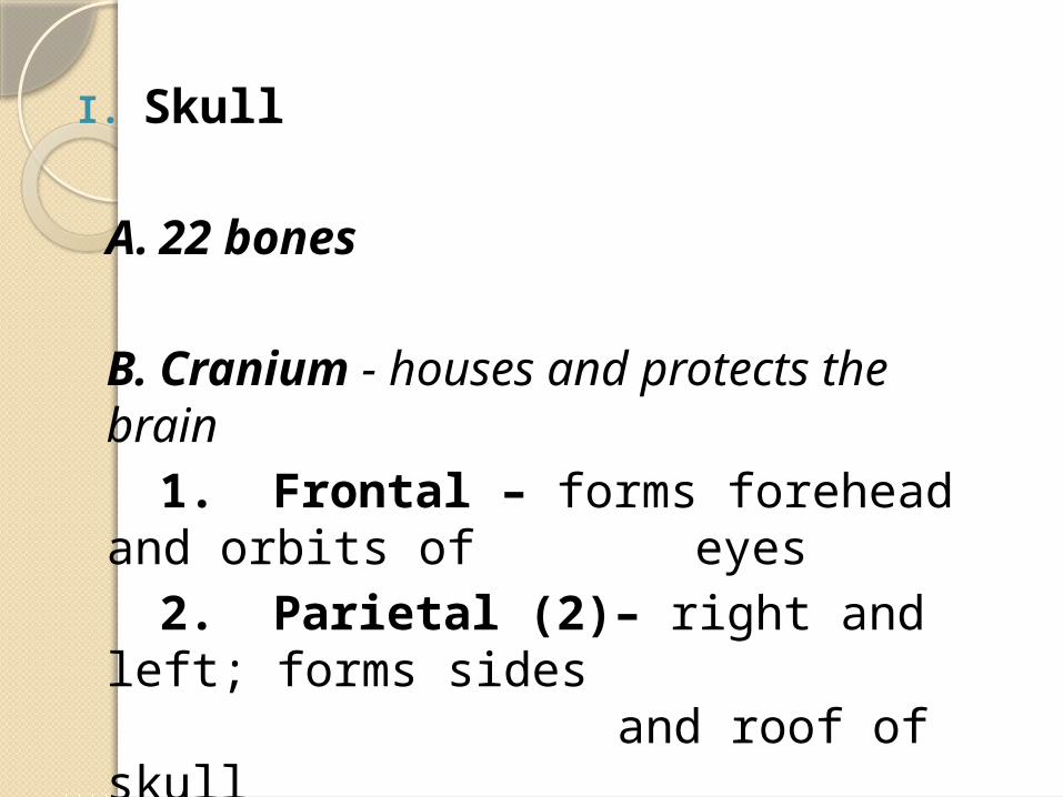
I. Skull
A. 22 bones
B. Cranium - houses and protects the brain
1. Frontal – forms forehead and orbits of eyes 2. Parietal (2)– right and left; forms sides and roof of skull
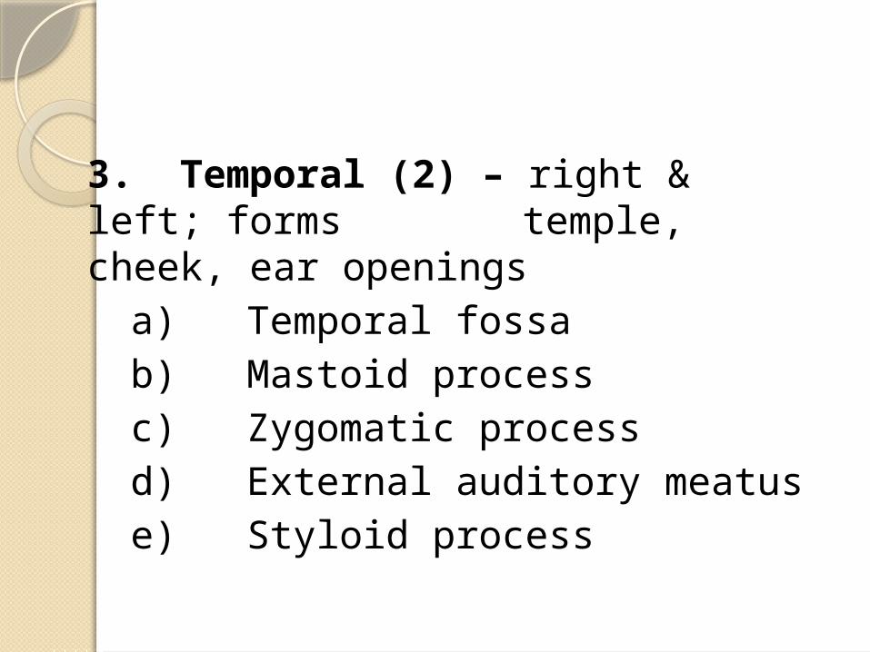
3. Temporal (2) – right & left; forms temple, cheek, ear openings
a) Temporal fossab) Mastoid processc) Zygomatic processd) External auditory meatuse) Styloid process
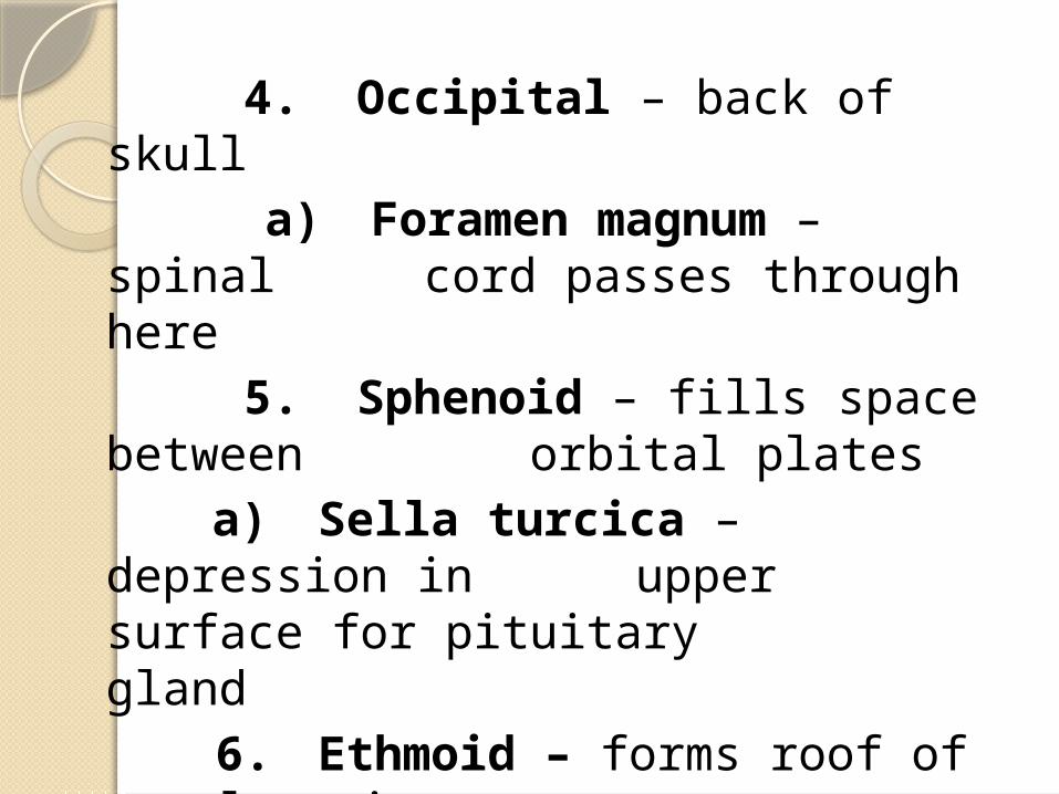
4. Occipital – back of skull a) Foramen magnum –
spinal cord passes through here
5. Sphenoid – fills space between orbital plates
a) Sella turcica – depression in upper surface for pituitary gland
6. Ethmoid – forms roof of nasal cavity
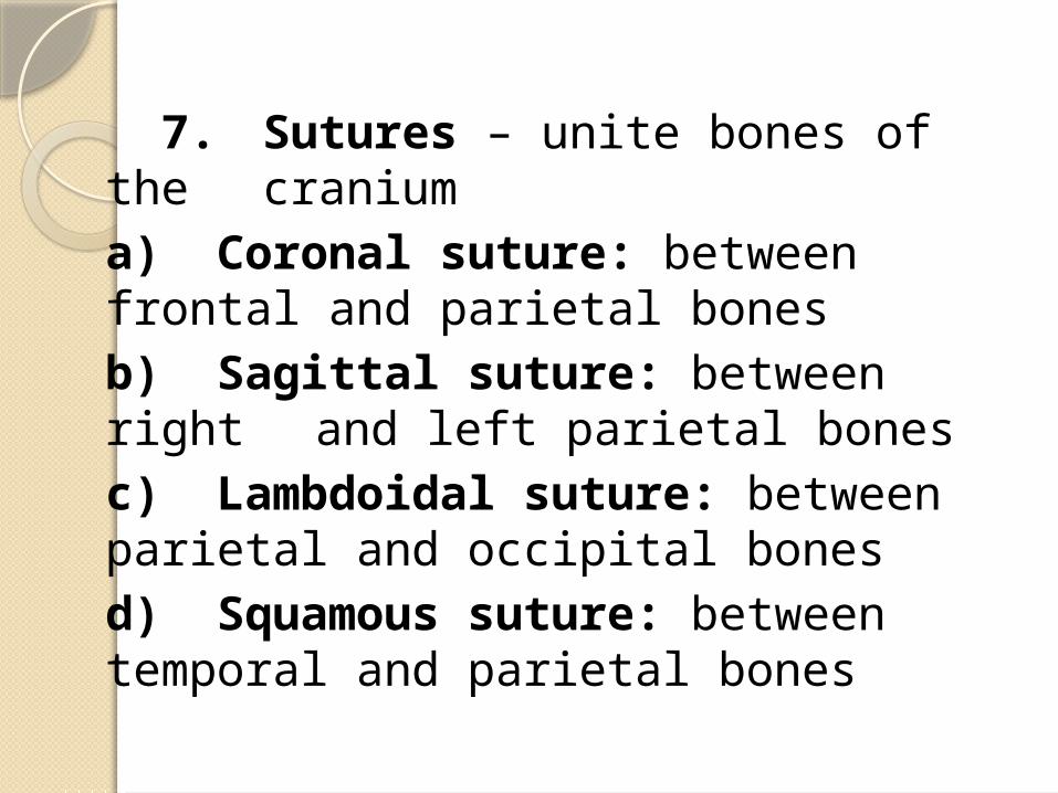
7. Sutures – unite bones of the cranium a) Coronal suture: between frontal and parietal bonesb) Sagittal suture: between right and left parietal bonesc) Lambdoidal suture: between parietal and occipital bonesd) Squamous suture: between temporal and parietal bones
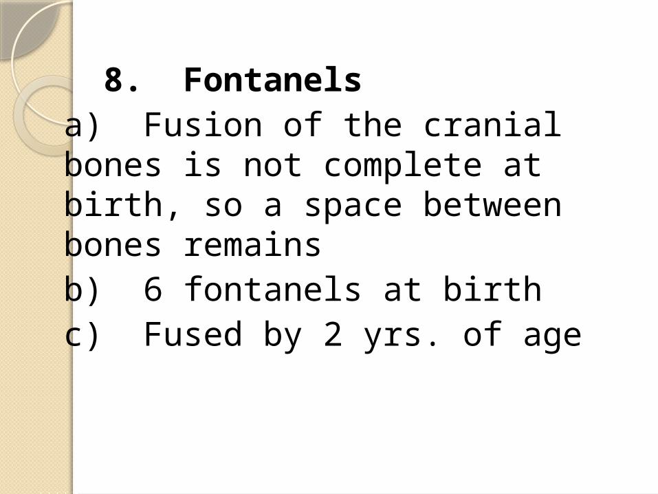
8. Fontanels a) Fusion of the cranial
bones is not complete at birth, so a space between bones remains
b) 6 fontanels at birthc) Fused by 2 yrs. of
age
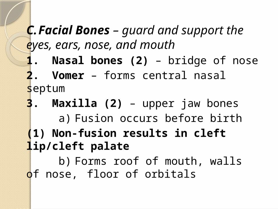
C. Facial Bones – guard and support the eyes, ears, nose, and mouth1. Nasal bones (2) – bridge of nose2. Vomer – forms central nasal septum3. Maxilla (2) – upper jaw bones a) Fusion occurs before birth(1) Non-fusion results in cleft lip/cleft palate b) Forms roof of mouth, walls of nose, floor of orbitals
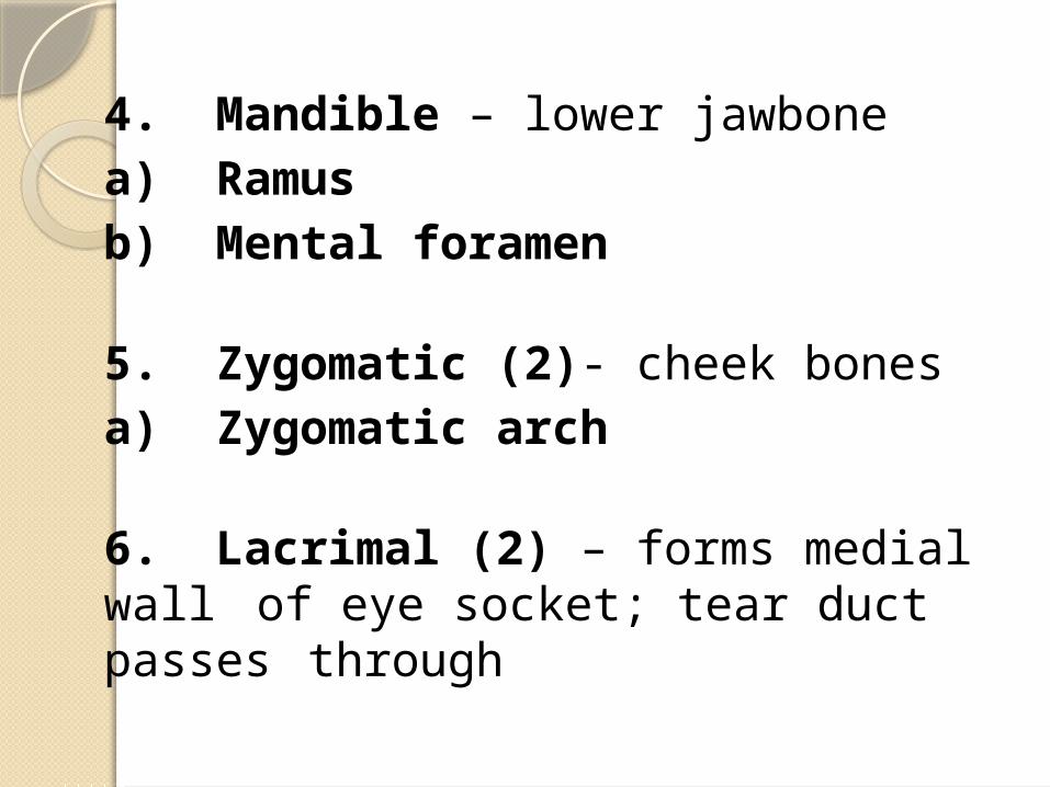
4. Mandible – lower jawbonea) Ramusb) Mental foramen
5. Zygomatic (2)- cheek bonesa) Zygomatic arch
6. Lacrimal (2) – forms medial wall of eye socket; tear duct passes through
7. Palatine (2) – forms back roof of mouth and floor of nose; L-shaped
8. Inferior turbinate (2) – forms curved ledge inside side wall of nose
D. Ear Bones1. Malleus - hammer2. Incus - anvil3. Stapes - stirrups
E. Hyoid Bone – U-shaped bone in the neck at the base of the tongue; only bone that does not touch another boneF. Cranial Sinuses
1. Cavities within the cranium2. Functions as resonance
chambers in voice production3. Decreases weight of skull4. Lined with mucous membrane
II.Vertebral ColumnA.Spinal Curvature
1.Cervical, thoracic, lumbar, and sacral curvatures
2.Primary Curves – appears late in fetal development
a)Thoracic and sacral curvesb)Accommodation curves - Accommodate the thoracic and abdominopelvic organs
c)Present in the newborn – the only curves present
3.Secondary Curvesa)Lumbar and cervical curvesb)Compensation curves - Shifts the trunk weight over the lower limbs
c)Appear several months after birth(1)Cervical curve – develops when infant can hold head upright
(2)Lumbar curve – develops with ability to stand
(3)Both curves become accentuated with walking and running
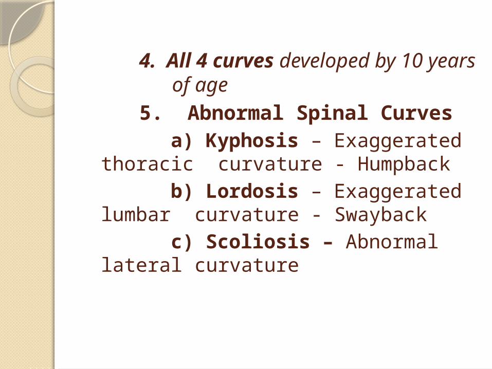
4. All 4 curves developed by 10 years of age
5. Abnormal Spinal Curves a) Kyphosis –
Exaggerated thoracic curvature - Humpback
b) Lordosis – Exaggerated lumbar curvature - Swayback
c) Scoliosis – Abnormal lateral curvature
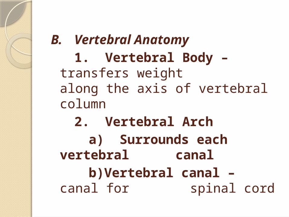
B. Vertebral Anatomy1. Vertebral Body –
transfers weight along the axis of vertebral column
2. Vertebral Arch a) Surrounds each
vertebral canalb)Vertebral canal –
canal for spinal cord
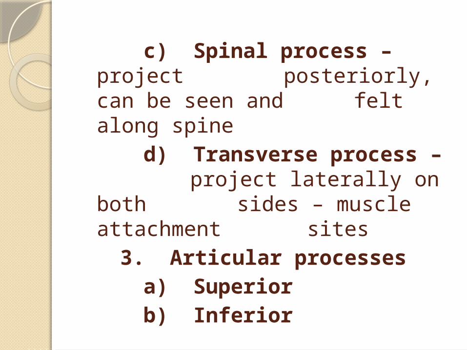
c) Spinal process – project posteriorly, can be seen and felt along spine
d) Transverse process – project laterally on both sides – muscle attachment
sites3. Articular processes
a) Superiorb) Inferior
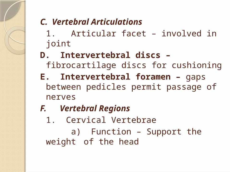
C. Vertebral Articulations1. Articular facet – involved in joint
D. Intervertebral discs – fibrocartilage discs for
cushioningE. Intervertebral foramen – gaps
between pedicles permit passage of nerves
F. Vertebral Regions1. Cervical Vertebrae a) Function – Support the weight
of the head
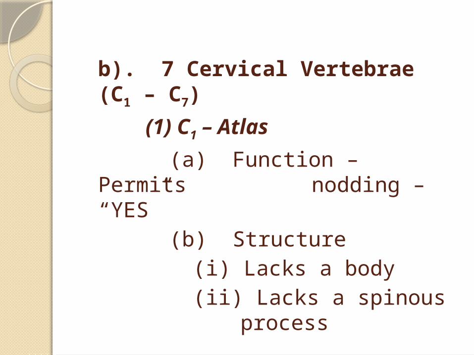
b). 7 Cervical Vertebrae (C1 – C7)
(1) C1 – Atlas
(a) Function – Permits nodding – “YES”
(b) Structure(i) Lacks a body(ii) Lacks a
spinous process
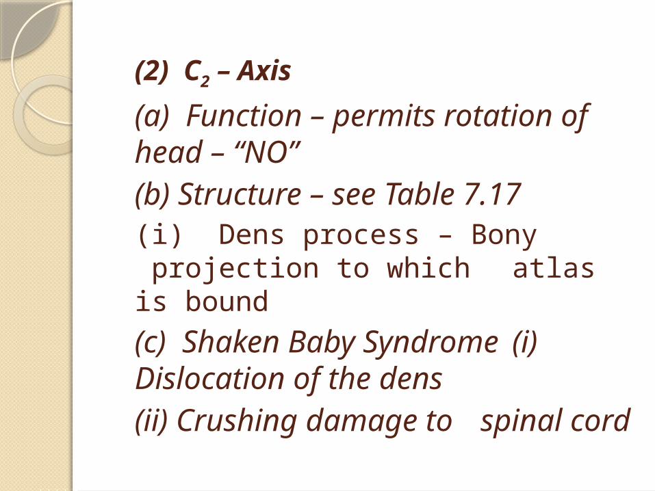
(2) C2 – Axis
(a) Function – permits rotation of head – “NO”(b) Structure – see Table 7.17(i) Dens process – Bony projection to which atlas is bound
(c) Shaken Baby Syndrome (i) Dislocation of the dens (ii) Crushing damage to spinal cord
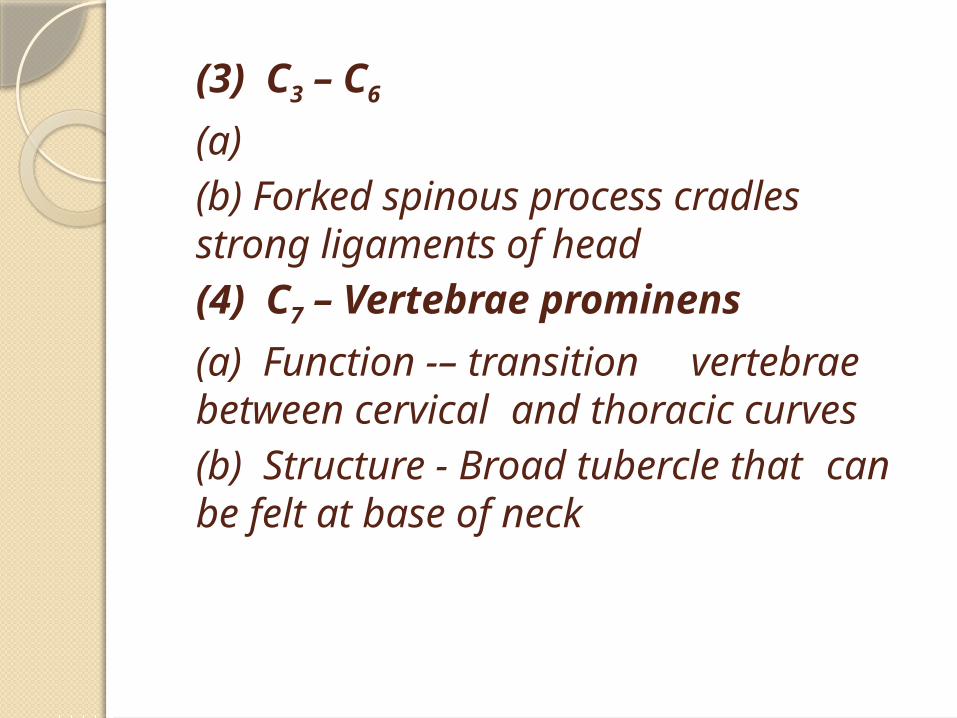
(3) C3 – C6
(a) (b) Forked spinous process
cradles strong ligaments of head
(4) C7 – Vertebrae prominens
(a) Function -– transition vertebrae between
cervical and thoracic curves
(b) Structure - Broad tubercle that can be felt at base of neck
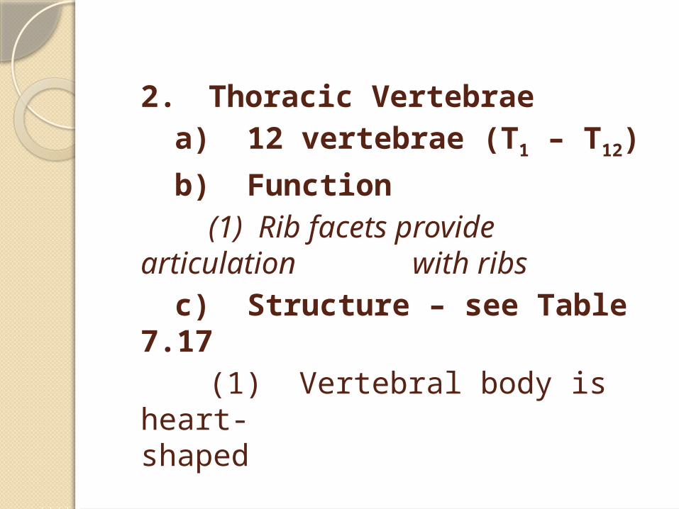
2. Thoracic Vertebraea) 12 vertebrae (T1 – T12)
b) Function (1) Rib facets provide
articulation with ribs
c) Structure – see Table 7.17
(1) Vertebral body is heart-
shaped
3. Lumbar Vertebraea) 5 vertebrae – L1 – L5
b) Function(1) Bears most of the weight
of the bodyc) Structure – see Table 7.17
(1) Large, oval vertebral body(2) Massive spinous
processes provide increased surface area for large muscle attachment
4. Sacruma) Function
(1) Protection of reproductive, digestive, and excretory organs
(2) Attaches axial skeleton to appendicular skeleton
b) Structure (1) Fusion of 5 sacral
vertebrae (2) Triangular, curved bone
5. Coccyxa) Function
(1) Attachment site for ligaments
(2) Attachment site for muscle that constricts the anal opening
b) Structure (1) Fusion of 3-5
coccygeal vertebrae
(2) Slightly movable to assist in childbirth
III. ThoraxA. 25 bones and cartilageB. Functions 1. Protects and supports
heart and lungs 2. Serves as attachment
point for muscles involved ina) Respirationb) Position of vertebral
columnc) Movements of the pectoral girdle and upper extremity
C. Sternum - breastbone1. Manubrium a) Articulates with clavicle
and 1st rib b) Jugular notch – shallow
indentation2. Body a) Individual costal cartilages
form rib pairs 2 – 7 are attached
3. Xiphoid process a) Diaphragm and rectus
abdominis muscles attach here
D. Costal Cartilages1. Hyaline cartilage2. Connects ribs to sternum in 1 – 7 and to anterior rib (#7) in 8 - 10
E. Ribs (12 pairs)1. Attached posteriorly with vertebrae and anteriorly with costal cartilage2. True ribs: a) 1st seven pairs b) Direct connection-each costal bone connects directly to sternum with its own piece of cartilage
3. False ribs: a) Pairs 8 – 10 b) Indirect connection –
cartilage from each of these three costal bones fuses together before
fusing with cartilage on rib 74. Floating ribs:
a) Pairs 11 – 12 b) No costal cartilage
c) NO connection to sternum