axilla & upper limb - wordpress.com · 07.03.2015 · ... fhs122.org axilla & upper limb 1...
TRANSCRIPT
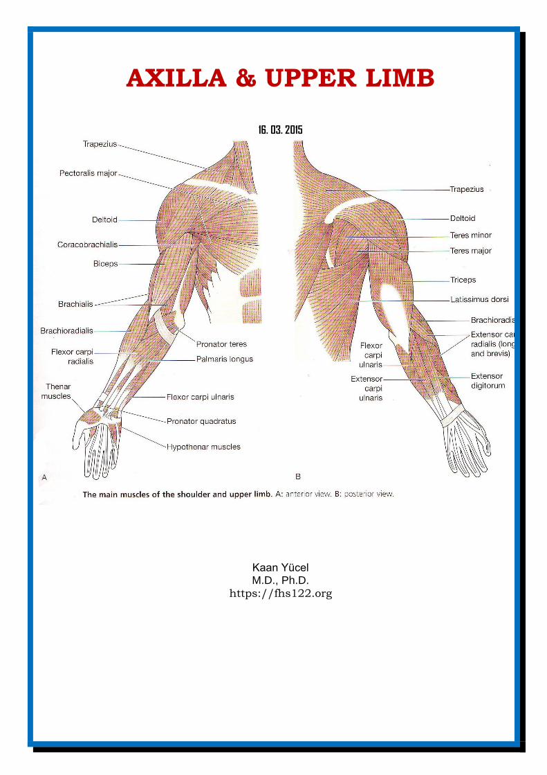
AXILLA & UPPER LIMB
16. 03. 2015
Kaan Yücel M.D., Ph.D.
https://fhs122.org
Dr.Kaan Yücel fhs122.org Axilla & Upper limb
1
1. AXILLA
AARRMMPPIITT,, KKOOLLTTUUKK66
gateway to the upper limb from the trunk An area of transition between the neck and the arm Irregular pyramidal space inferior to the shoulder joint @ the junction of the arm & thorax. Formed by clavicle,scapula,upper thoracic wall, humerus, & related muscles. Provides a passageway, or “distribution center,” usually protected by the adducted upper limb, for the
neurovascular structures that serve the upper limb. Has 4 walls, an inlet (apex) and a floor (base)
Contains 1) Axillary artery and its branches supply blood to the upper limb (Subclavian artery: considered as artery of the upper limb) 2) Axillary vein and its tributaries drain blood from the upper limb; 3) Lymph vessels and lymph nodes drain lymph from a) upper limb b) breast c) skin of the trunk, down as far as the level of the umbilicus. Axillary inlet (Apex)= Cervico-axillary canal
passageway between the neck and the axilla bounded by the 1st rib, clavicle, and superior edge of the scapula.
The arteries, veins, lymphatics, & nerves traverse axillary inlet to pass to or from the arm. Subclavian artery major blood vessel supplying the upper limb becomes the axillary artery as it crosses the lateral margin of rib I and enters the axilla. Similarly, the axillary vein becomes the subclavian vein as it passes over the lateral margin of rib I and leaves the axilla to enter the neck. @ Axillary inlet, Axillary vein anterior to axillary artery anterior to trunks of brachial plexus. (VAP) Anterior wall pectoralis major muscle, underlying pectoralis minor & subclavius muscles Medial wall thoracic wall (1st-4th ribs and intercostal muscles) and the overlying serratus anterior. Lateral wall narrow bony wall formed only by the intertubercular groove in the humerus. Posterior wall costal surface of the scapula. The posterior wall of axilla is formed chiefly by the scapula and subscapularis on its anterior surface and inferiorly by the teres major and latissimus dorsi.
AAxxiillllaarryy aarrtteerryy Supplies the walls of the axilla and related regions. Begins @ lateral border of 1st rib = continuation of the subclavian artery Ends at the inferior border of the teres major Continues as brachial artery.
Gateways in the posterior wall
Quadrangular space Contents Axillary nerve + Posterior circumflex humeral artery and vein
Triangular interval Radial nerve passes out of the axilla to reach the posterior compartment of the arm. The profunda brachii artery (deep artery of arm) and associated veins also pass through the triangular interval.
2. BRACHIAL PLEXUS
A major somatic nerve network Most nerves @ upper limb originate from brachial plexus Supplies the upper limb Begins @ neck ==== Ends @ axilla. Formed by anterior rami (branches) of C5-C8 +T1 spinal nerves Anterior rami of last 4 cervical spinal nerves + of T1 = ROOTS OF THE BRACHIAL PLEXUS Roots originate @ neck, pass laterally & inferiorly over rib I = enter axilla.
From roots SUPERIOR, MIDDLE & INFERIOR TRUNKS are formed. • Superior trunk is formed by the union of C5 and C6 roots; • Middle trunk is a continuation of the C7 root; • Inferior trunk is formed by the union of the C8 and T1 roots.
Each trunk is then divided into two divisions. Anterior division. Posterior division. The three cords of the brachial plexus originate from the divisions. Lateral cord (C5-C7), Medial cord (C8-T1), Posterior cord (C5 to T1).
Dr.Kaan Yücel fhs122.org Axilla & Upper limb
2
All major nerves that innervate the upper limb originate mostly from the cords.
Important branch (peripheral nerves) of LATERAL CORD
Musculocutaneous nerve “Anterior compartment of arm” muscles
Important branches (peripheral nerves) of MEDIAL CORD
Medial cutaneous nerve of arm/forearm
Ulnar nerve Flexor carpi ulnaris+ Medial half of flexor digitorum profundus, some intrinsic musles of
hand
Median nerve All the muscles of the anterior compartment forearm except the muscles innervated by
the ulnar nerve + Some muscles of the intrinsic muscles of hand
Important branches (peripheral nerves) of POSTERIOR CORD
Thoracodorsal nerve Latissimus dorsi
Axillary nerve Deltoid and teres major
Radial nerve Posterior compartment of arm/forearm muscles
3. SHOULDER
region of upper limb attachment to the trunk the proximal segment of the limb overlaps parts of the trunk (thorax and back) and lower lateral neck includes the pectoral, scapular, and deltoid regions of the upper limb, and the lateral part of the lateral
cervical region. The bone framework of the shoulder consists of: • clavicle and scapula, which form the pectoral girdle (shoulder girdle); and • proximal end of the humerus.
The three joints in the shoulder complex are the sternoclavicular, acromioclavicular, and glenohumeral joints. SUPERFICIAL EXTRINSIC SHOULDER MUSCLES
TRAPEZIUS attaches the scapula and clavicle to the trunk. LATISSIMUS DORSI
DEEP EXTRINSIC SHOULDER MUSCLES levator scapulae and rhomboids:provide direct attachment of the appendicular skeleton to the axial skeleton.
attaches the scapula and clavicle to the humerus INTRINSIC SHOULDER MUSCLES Deltoid Main abductor of the arm (Nerve: Axillary nerve) Teres major Supraspinatus Infraspinatus Teres minor Subscapularis
Deltoid and subscapularis are not @ posterior scapular region. Others are @ posterior scapular region. DELTOID MUSCLE
large and triangular in shape attaches the scapula and clavicle to the humerus.
ABDUCTION OF THE ARM Supraspinatus starts the abduction of the arm. It does the initial 15-20° of abduction of the
shoulder/arm. Beyond this, after this: Deltoid is the abductor. If you want to abduct your arm beyond the 90°, you need trapezius and serratus anterior muscles.
Subscapularis coming from anterior part of the shoulder and medial part Medial rotation+adduction of arm. Teres minor+Infraspinatus Lateral rotation of arm
Teres major Medial rotation of arm (just like subscapularis) The two major nerves of the posterior scapular region are the suprascapular and axillary nerves.
4. ARM
region of the upper limb between shoulder & elbow Superiorly => communicates medially with the axilla
The famous 4 “Rotator cuff muscles” . Just like a cuff (kelepçe) hold the head of humerus in
glenoid cavity of scapula @ shoulder joint. stabilize this joint during movements of the elbow, wrist, and hand
3 of them also Rotation of the shoulder. Supraspinatus Starts the abduction of the
shoulder!
Dr.Kaan Yücel fhs122.org Axilla & Upper limb
3
Inferiorly => a number of important structures pass between the arm & forearm through cubital fossa positioned anterior to the elbow joint
Divided into two compartments: Anterior & Posterior compartmens 2 types of movement occur between the arm and the forearm at the elbow joint: flexion-extension and pronation-supination. Muscles @ anterior compartment of arm flex the elbow joint. Muscles @ posterior compartment of arm extend the elbow joint. Major nerves and vessels supply and pass through each compartment. The chief action of both groups is at the elbow joint, but some muscles also act at the glenohumeral joint.
The skeletal support for the arm is the humerus. Anterior compartment of the arm contains three muscles.
Coracobrachialis Flexes & adducts the arm (@ medial side of arm) Brachialis Chief flexor of forearm (under biceps brachii) Biceps brachii 3 joint muscle: Shoulder, elbow, radio-ulnar joints Powerful flexor of forearm & main
supinator muscle innervated predominantly by musculocutaneous nerve. Posterior compartment of the arm contains 1 muscle Triceps brachii (Nerve: Radial nerve) The long head also aids in extension and adduction of the arm. The medial head is the workhorse of forearm extension.
BRACHIAL ARTERY
The major artery of the arm Brachial artery @ anterior compartment. Starts as a continuation of the axillary artery @ lower border of teres major muscle Ends @ just distal to the elbow joint, opposite to the neck of the radius. Divides into into radial and ulnar arteries.
Deep artery of the arm (L. arteria profunda brachii) is the largest branch of the brachial artery. Superficial veins @ subcutaneous tissue Deep veins accompany arteries. Two main superficial veins of the upper limb 1) Cephalic vein 2) Basilic vein. Cephalic vein ascends @ superficial fascia on the lateral side of the biceps and drains into axillary vein. Basilic vein (on medial side) joins with brachial veins forms axillary vein.
Four main nerves pass through the arm: median, ulnar, musculocutaneous, and radial nerves. The musculocutaneous nerve leaves the axilla and enters the arm by passing through the
coracobrachialis muscle. It passes diagonally down the arm in the plane between the biceps brachii and brachialis muscles. The musculocutaneous nerve provides: motor innervation to all muscles in the anterior compartment of the arm; and sensory innervation to skin on the lateral surface of the forearm.
The median nerve passes vertically down the medial side of the arm in the anterior compartment. The median and ulnar nerves have no major branches @ axilla & arm. The radial nerve in the arm supplies all the muscles in the posterior compartment of the arm (and
forearm). The radial nerve originates from the posterior cord of the brachial plexus. Accompanied by the profunda brachii artery, the radial nerve enters the posterior compartment of the arm by passing through the triangular interval.
5. FOREARM
The forearm is the part of the upper limb between the elbow wrist joints. Proximally, most major structures pass between the arm and forearm through, or in relation to, the cubital fossa, which is anterior to the elbow joint. The exception is the ulnar nerve, which passes posterior to the medial epicondyle of the humerus.
Muscles in the anterior compartment of the forearm flex the wrist and digits and pronate the hand. Muscles in the posterior compartment extend the wrist and digits and supinate the hand. Major nerves and vessels supply or pass through each compartment. The flexors and pronators of the forearm in the anterior compartment are served mainly by the median nerve; the one and a half exceptions are innervated by the ulnar nerve. The pronator quadratus (a muscle like a bracelet @ distal part of ant.forearm) is the main
Dr.Kaan Yücel fhs122.org Axilla & Upper limb
4
pronator. Pronator teres is close to the elbow joint and median nerve passes between the two heads of pronator teres.
The extensors and supinators of the forearm are in the posterior compartment and are all served by the radial nerve (directly or by its deep branch).
The main arteries of the forearm are the ulnar and radial arteries usually arise opposite the neck of the radius in the inferior part of the cubital fossa as terminal
branches of the brachial artery. The superficial veins of the forearm lie in the superficial fascia. The cephalic vein arises from the
lateral side of the dorsal venous arch on the back of the hand and winds around the lateral border of the forearm; it then ascends into the cubital fossa and up the front of the arm on the lateral side of the biceps. It terminates in the axillary vein. The basilic vein arises from the medial side of the dorsal venous arch on the back of the hand and winds around the medial border of the forearm; it then ascends into the cubital fossa and up the front of the arm on the medial side of the biceps. Its terminates, by joining the brachial veins to form the axillary vein.
Nerves in the anterior compartment of the forearm are the median and ulnar nerves, and the superficial branch of the radial nerve.
The median nerve is the principal nerve of the anterior compartment of the forearm. It supplies muscular branches directly to the muscles of the superficial layerof forearm flexors (except flexor carpi ulnaris), and deep muscles (except for the medial [ulnar] half of the flexor digitorum profundus; ring and little fingers).
Radial nerve serves motor and sensory functions in both the arm and the forearm (but only sensory functions in the hand). However, its sensory and motor fibers are distributed in the forearm by two separate branches, the superficial (sensory or cutaneous) and deep radial/posterior interosseous nerve (motor).
Muscles in the posterior compartment of the forearm occur in two layers: a superficial and a deep layer. The muscles are associated with: 1) Movement of the wrist 2) Extension of fingers 3) Supination
All muscles in the posterior compartment of the forearm are innervated by the radial nerve. Common origin of most of the muscles of the anterior compartment Medial epicondyle of humerus Common origin of most of the muscles of the posterior compartment Lateral epicondyle of humerus
Brachioradialis @ posterolateral side of forearm. A muscles of the posterior compartment of forearm, innervated by radial nerve Flexion of wrist!
Except for the supinator muscle, all these deep layer muscles originate from the posterior surfaces of the radius, ulna, and interosseous membrane and pass into the thumb and fingers.
The blood supply to the posterior compartment of the forearm occurs predominantly through branches of the radial, posterior interosseous, and anterior interosseous arteries (branches of ulnar artery).
The nerve of the posterior compartment of the forearm is the radial nerve.
6. CUBITAL FOSSA
The pronator teres makes the medial border, whereas the brachioradialis makes the lateral border of the cubital fossa.
The contents of the cubital fossa are the: • Terminal part of the brachial artery and the commencement of its terminal branches, the radial and
ulnar arteries. The brachial artery lies between the biceps tendon and the median nerve. • (Deep) accompanying veins of the arteries • Biceps brachii tendon • Median nerve • Radial nerve Ulnar nerve passes posterior to medial epicondyle of humerus & IS NOT @ Cubital Fossa.
7.HAND
a mechanical and sensory tool Many of the features of the upper limb are designed to facilitate positioning the hand in space. The hand is the region of the upper limb distal to the wrist joint.
Dr.Kaan Yücel fhs122.org Axilla & Upper limb
5
Subdivided into three parts: 1) Wrist (carpus) 2) Metacarpus 3) Digits (five fingers including the thumb). The carpal tunnel is formed anteriorly at the wrist by a deep arch formed by the carpal bones and the
flexor retinaculum. The four tendons of the flexor digitorum profundus, the four tendons of the flexor digitorum superficialis, and the tendon of the flexor pollicis longus pass through the carpal tunnel, as does the median nerve.
The extensor tendons pass into the hand on the medial, lateral, and posterior surfaces of the wrist in six compartments defined by an extensor retinaculum (dorsal carpal ligament) and lined by synovial sheaths.
The palmar aponeurosis is a triangular condensation of deep fascia that covers the palm and is anchored to the skin in distal regions.
INTRINSIC MUSCLES OF THE HAND
Ulnar nerve Hypothenar muscles, adductor pollicis palmar and dorsal interossei, two medial lumbrical muscles Median nerve Three thenar muscles, two lateral lumbrical muscles Palmar interossei adduct, dorsal interossei abduct the fingers. Lumbrical muscles flex metacarpophalangeal joints + extend interphalangeal joints.
Ulnar artery & ulnar nerve enter the hand on the medial side of the wrist. Distally, the ulnar artery swings laterally across the palm forms superficial palmar arch.
Radial artery curves around the lateral side of the wrist Passes into the deep plane of the palm by penetrating anteriorly through the back of the hand. Accesses the deep plane of the palm forms the deep palmar arch.
Superficial palmar arch Palmar digitial arteries (fingers) Deep palmar arch Palmar metacarpal arteries (metacarpals) a palmar digital artery and three large, common palmar digital arteries
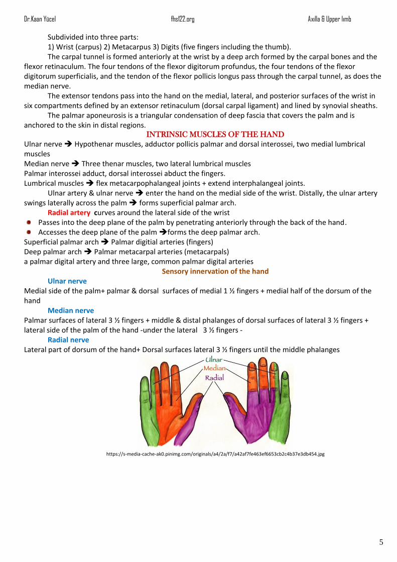
Sensory innervation of the hand Ulnar nerve
Medial side of the palm+ palmar & dorsal surfaces of medial 1 ½ fingers + medial half of the dorsum of the hand
Median nerve Palmar surfaces of lateral 3 ½ fingers + middle & distal phalanges of dorsal surfaces of lateral 3 ½ fingers + lateral side of the palm of the hand -under the lateral 3 ½ fingers -
Radial nerve Lateral part of dorsum of the hand+ Dorsal surfaces lateral 3 ½ fingers until the middle phalanges
https://s-media-cache-ak0.pinimg.com/originals/a4/2a/f7/a42af7fe463ef6653cb2c4b37e3db454.jpg