bach proteins belong to a novel family of btb-basic leucine zipper transcription factors that
TRANSCRIPT

MOLECULAR AND CELLULAR BIOLOGY, Nov. 1996, p. 6083–6095 Vol. 16, No. 110270-7306/96/$04.0010Copyright q 1996, American Society for Microbiology
Bach Proteins Belong to a Novel Family of BTB-Basic LeucineZipper Transcription Factors That Interact with MafK and
Regulate Transcription through the NF-E2 SiteTATSUYA OYAKE,1 KEN ITOH,1 HOZUMI MOTOHASHI,1† NORIO HAYASHI,1 HIDETO HOSHINO,2
MAKOTO NISHIZAWA,3 MASAYUKI YAMAMOTO,4,5 AND KAZUHIKO IGARASHI4,5*
Department of Biochemistry, Tohoku University School of Medicine, Seiryo-machi 2-1, Aoba-ku, Sendai 980-77,1
Graduate School of Medicine,2 Center for Tsukuba Advanced Research Alliance,4 and Institute of BasicMedical Sciences,5 University of Tsukuba, Tenno-dai 1-1-1, Tsukuba 305, and Department of Molecular
Oncology, Kyoto University Faculty of Medicine, Sakyo-ku, Kyoto 606,3 Japan
Received 5 July 1996/Returned for modification 26 July 1996/Accepted 9 August 1996
Members of the small Maf family (MafK, MafF, and MafG) are basic region leucine zipper (bZip) proteinsthat can function as transcriptional activators or repressors. The dimer compositions of their DNA bindingforms determine whether the small Maf family proteins activate or repress transcription. Using a yeasttwo-hybrid screen with a GAL4-MafK fusion protein, we have identified two novel bZip transcription factors,Bach1 and Bach2, as heterodimerization partners of MafK. In addition to a Cap’n’collar-type bZip domain,these Bach proteins possess a BTB domain which is a protein interaction motif; Bach1 and Bach2 showsignificant similarity to each other in these regions but are otherwise divergent. Whereas expression of Bach1appears ubiquitous, that of Bach2 is restricted to monocytes and neuronal cells. Bach proteins bind in vitro toNF-E2 binding sites, recognition elements for the hematopoietic transcription factor NF-E2, by formingheterodimers with MafK. Furthermore, a DNA binding complex that contained MafK as well as Bach2 or aprotein related closely to Bach2 was found to be present in mouse brain cells. Bach1 and Bach2 function astranscription repressors in transfection assays using fibroblast cells, but they function as a transcriptionalactivator and repressor, respectively, in cultured erythroid cells. The results suggest that members of the Bachfamily play important roles in coordinating transcription activation and repression by MafK.
Protein interactions within and between various families oftranscription factors play important roles in gene regulationduring the development of multicellular organisms. Het-erodimer formation by basic leucine zipper (bZip) proteins istypical of such interactions, conferring novel functions thatdiffer from those of the individual proteins. Hence, to under-stand the roles of bZip proteins in gene regulation, it is crucialthat the physiological partners for each factor be identified andthat dimer formation as regulated spatially and temporallyduring embryogenesis be analyzed.Transcription factor NF-E2, which was originally identified
as an erythrocyte-specific DNA binding activity (45), is a het-erodimeric factor that consists of a large and a small bZipsubunit (1, 3, 33, 52). The large subunit of NF-E2 is p45, whichbelongs to the Cap’n’collar (CNC)-type bZip protein family (1,8, 52). The mammalian CNC family includes p45 NF-E2, Nrf1/LCR-F1/TCF11 (7, 9, 43), and Nrf2 (47), the last being closelyrelated to chicken ECH (34). These proteins and the Drosoph-ila transcription factor CNC (46) show high structural similar-ity in their bZip domains as well as conservation of the pre-ceding regions, referred to as the CNC domain (7). The otherhalf of NF-E2 is contributed by the so-called small Maf family,which consists of MafK (also known as p18), MafG, and MafF(3, 21, 31, 33, 37). These small Maf family proteins possess a
conserved basic region that is characteristic of the Maf family(21) but lack any canonical transactivation domain. Variousdimeric combinations of the small Maf family proteins bind invitro to a DNA sequence motif called T-MARE [TGCTGA(G/C)TCAGCA] containing a 12-O-tetradecanoylphorbol-13-acetate-responsive element [TRE; TGA(G/C)TCA] (37, 38).Furthermore, the small Maf family proteins form heterodimerswith p45 and ECH and the resultant heterodimers can bind tothe NF-E2 consensus site [TGCTGA(G/C)TCA(T/C)] that isrelated to the T-MARE, whereas p45 and ECH show very lowlevels of DNA-binding activity by themselves (1, 31, 33, 34). Incontrast, Nrf1/LCR-F1/TCF11 appears to bind to the NF-E2site as a homodimer (7).NF-E2 sites have attracted particular attention because of
their presence in the locus control region (LCR) of the b-glo-bin gene cluster. The b-globin LCR was initially recognized asa series of DNase I-hypersensitive sites and is composed of fiveDNase-hypersensitive sites (HS-1 to HS-5) (19, 68). In trans-genic mice, the LCR was found to direct copy-number-depen-dent, position-independent gene expression (25). This activityis a result of redundant combinations of cis regulatory sites andis not attributable to a single regulatory site (6). In contrast, thevery strong erythroid enhancer activity of the LCR has beenmapped within HS-2 and established to be dependent on thetandem NF-E2 sites (6, 48, 53, 54, 69). However, mysteriesregarding effector molecules of NF-E2 sites still abound, since(i) p45 NF-E2 is dispensable for functions of the b-globin LCRin erythroid cells (61), (ii) p45-related proteins that can bind tothe NF-E2 sites, as summarized above, also exist and (iii)NF-E2 sites in HS-2 are occupied by proteins in hematopoieticcells even before commitment to erythroid lineage, suggesting
* Corresponding author. Mailing address: Institute of Basic MedicalSciences, University of Tsukuba, Tenno-dai 1-1-1, Tsukuba 305, Japan.Phone: 81-298-53-6963. Fax: 81-298-53-6965. Electronic mail address:[email protected].† Present address: Center for Tsukuba Advanced Research Alliance
and Institute of Basic Medical Sciences, University of Tsukuba, Tenno-dai 1-1-1, Tsukuba 305, Japan.
6083
Dow
nloa
ded
from
http
s://j
ourn
als.
asm
.org
/jour
nal/m
cb o
n 09
Dec
embe
r 20
21 b
y 91
.236
.133
.10.
that the program of b-globin activation is set in motion at anearlier stage (35).Another feature of NF-E2 sites is that similar sequences are
also frequently found in regulatory regions of various noner-ythroid genes (38), suggesting that Maf- and CNC-related fac-tors regulate gene expression through NF-E2-like sites notonly in hematopoietic cells but also in a variety of systems. Forexample, antioxidant response elements contain NF-E2 site-like sequences (reference 71 and references therein). Our pre-vious observation that the mafK gene is expressed specificallyin neuronal, mesenchymal, and hematopoietic cells duringmouse embryogenesis also suggests importance for the smallMaf network in various developmental programs (50).Previous studies showed that the small Maf family proteins
are unique transcription factors which can switch from tran-scriptional repressors to activators, depending on the dimercompositions (31, 33, 37). For example, the small Maf-p45heterodimers activate transcription whereas small Maf-Fosheterodimers exert a repressive effect. To understand the mo-lecular mechanisms and roles of the small Maf interactor net-work, we set out to define constituent components. In thisreport, we describe the cloning and characterization of mem-bers of a novel bZip transcription factor family, Bach, whichform heterodimers with MafK and bind to NF-E2 sites. Inaddition to a leucine zipper, the Bach family contains a BTBdomain (Broad complex–Tramtrack–Bric-a-brac domain, alsoknown as the POZ [poxvirus and zinc finger] domain), anemerging protein interaction motif (4, 10, 75). The Bach familyappears to connect the MafK network with different regulatorynetworks or to different levels of transcriptional regulationthrough unique structural and functional features.
MATERIALS AND METHODS
Two-hybrid system. Saccharomyces cerevisiae two-hybrid screening was per-formed by the Matchmaker two-hybrid system (Clontech). To construct a baitplasmid, the entire open reading frame (ORF) of mouse MafK (31) was fused tothe DNA binding domain of GAL4 on the pGBT9 plasmid (Clontech). A mouseday 17 post coitus (pc) embryonic Matchmaker cDNA library (Clontech) wastransformed into the HF7c yeast strain along with the MafK bait plasmid.Approximately 23 107 double transformants were tested for their ability to growon His2 medium. Of 950 His1 transformants, 67 clones were scored positive forLacZ expression. To obtain larger cDNA clones, a mouse brain cDNA library inLambda ZAPII (Stratagene) was screened by hybridization with cDNA frag-ments. Inserts of the positive phage clones were rescued into plasmids by in vivoexcision, resulting in pBSA1-3 and pBSF69-J. Sequences of the cDNA cloneswere determined on both strands with an ABI Prism dye terminator cycle-sequencing ready reaction kit (Perkin-Elmer) and an automated DNA sequenceanalyzer (ABI, model 373A). Additional screening of the brain cDNA librarywith a bach2 cDNA probe yielded another cDNA clone termed pBSF69-F.To examine protein interactions within yeast cells, HF7c cells were trans-
formed with various combinations of plasmids that express DNA binding domain(DBD)-tagged and activation domain (AD)-tagged molecules. Transformantswere diluted in water and spotted onto His2 and His1 media to test for activa-tion of the GAL4-dependent HIS3 reporter gene.Plasmids. Expression plasmids of Bach1 and Bach2 were constructed as fol-
lows. Eukaryotic expression plasmid pGFP-c2 (Clontech) was digested with NheIand XhoI to remove the green fluorescent protein-coding region and circularizedagain, resulting in pCMVKM. The entire ORF of Bach1 was isolated as a SalIfragment from pA1 and inserted into the SmaI site of pCMVKM, resulting inpCMV/Bach1. To construct pCMV/Bach2, the entire ORF of Bach2 was isolatedas an XbaI-XhoI fragment from pBSF69-J and inserted into the SmaI site ofpCMVKM. The F69-J and F69-F cDNAs were recombined at the internal ApaIsite to construct a cDNA encoding a presumptive full-size F69-F-type ORF. Theresulting composite cDNA was transferred into pCMVKM, resulting in pCMV/Bach2(F).To construct prokaryotic expression plasmids, cDNA fragments of Bach1 and
Bach2 that included the bZip-coding domains were inserted into pMal-c2 (NewEngland Biolabs). The resulting plasmids encoded the carboxy-terminal 453 and329 amino acids of Bach1 and Bach2, respectively, fused to the maltose-bindingprotein (MBP). The prokaryotic expression plasmids of chicken ECH and mouseMafK, the eukaryotic expression plasmid of mouse MafK, and the luciferasereporter plasmids pRGBP2 and pRGBP4 were described previously (30, 34).Expression plasmids for GAL4 DBD-tagged Bach1 were constructed as follows.
The SalI fragment of pA1 was inserted into the BamHI site of pGBT9 after fillingin the relevant DNA ends. The resulting plasmid, pGBT/Bach1, encoded theentire Bach1 ORF fused to the DBD of GAL4. pGBT/Bach1 was digested withBglII and SalI and recircularized after being filled in with T4 DNA polymerase.This treatment resulted in pGBT/Bach1DBS, with removal of the entire carboxy-terminal region of Bach1, leaving only the amino-terminal 163 residues of thepredicted Bach1 polypeptide. To remove the bZip-coding domain from pA1,pA1 was digested with NcoI and recircularized, generating pA1DNc. This plas-mid encoded the amino-terminal 287 residues of the predicted Bach1 polypep-tide.RNA blot hybridization. Poly(A)1 RNAs were isolated from various cell lines
or tissues of adult mice and embryos by the acid guanidinium thiocyanate-phenol-chloroform extraction method (12) and with oligo(dT)-latex beads(Takara). Approximately 2 to 3 mg of poly(A)1 RNA was separated on 1%agarose–formaldehyde gels and transferred onto Zetaprobe membranes (Bio-Rad). Hybridization with 32P-labeled DNA or RNA probes and subsequentwashing was carried out as described previously (20). Probe DNA fragmentswere either the NcoI 1.4-kb DNA of bach1 cDNA or the ApaI-HindIII 1.4-kbDNA of bach2 cDNA.Recombinant transcription factors and antisera. Expression and purification
of MBP fusion proteins were carried out as previously described (38) withEscherichia coli SG 12036. The purified MBP-Bach fusion proteins were used toimmunize Japanese White rabbits by the RIBI ImmunoChem adjuvant systemafter collection of preimmune sera. Three different antisera were obtained, andtheir specificities were examined by immunoblotting analysis with overproducedBach1, Bach2, p45 NF-E2, and ECH proteins obtained by transfection of fibro-blast cells. The first, raised against Bach1, was specific for Bach1. The second wasraised against Bach2 and found to react with both Bach1 and Bach2. The thirdwas raised against Bach2 and was specific for Bach2. These three antisera arereferred to as anti-Bach1, anti-Bach panspecific, and anti-Bach2, respectively.Since none of these three reacted with p45 NF-E2 or ECH in an immunoblottinganalysis or in an electrophoretic gel mobility shift analysis (EGMSA), we as-sumed that they do not react with known CNC family proteins. In EGMSA, eachof the three antisera reacted with both Bach1 and Bach2.Transient-transfection assay. The quail fibroblast cell line QT6 (49) was main-
tained in Dulbecco’s modified Eagle’s medium supplemented with 10% fetalbovine serum and seeded in 12-well dishes 24 h before transfection. QT6 cellswere transfected with the reporter and effector plasmids by the calcium phos-phate precipitation method as previously described (60). The chicken erythroidcell line HD3 (5) was maintained and transfected by the DEAE-dextran methodas previously described (41). Luciferase assays were performed by the luciferaseassay system (Promega) by following the supplier’s protocol and with a Biolumatluminometer (Berthold). Transfection efficiencies were normalized with a co-transfected b-galactosidase expression plasmid, pENL. To analyze DNA bindingactivities generated by the effector plasmids in transfected QT6 cells, transfec-tions were carried out with 2 mg of effector plasmids, 1.5 mg of pENL, and carrierplasmid DNA (a total of 8 mg of plasmids per 100-mm-diameter dish).EGMSA. Nuclear extracts were prepared from transfected QT6 cells by the
previously described protocol (2). Nuclear extracts of mouse embryo brain cellswere as described previously (50). An oligonucleotide containing the chickenb-globin enhancer NF-E2 site (59-TCGACCCGAAAGGAGCTGACTCATGCTAGCCC-39) was labeled with [g-32P]ATP with T4 polynucleotide kinase. Oli-gonucleotides containing either the mutated b-globin enhancer NF-E2 site, theNF-E2 sites of the b-globin LCR, or the NF-E2 site-like sequence of the ery-throid d-aminolevulinate synthase gene promoter were as described previously(33). Binding reactions and electrophoresis were carried out as previously de-scribed (38) with bacterially synthesized proteins or nuclear extracts. Whereindicated in the figures and figure legends, rabbit preimmune serum or antisera
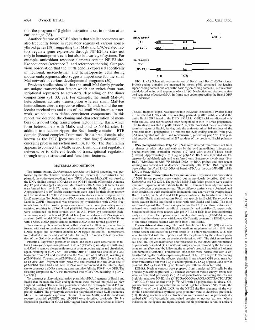
FIG. 1. (A) Schematic representation of Bach1 and Bach2 cDNA clones.Protein-coding domains are indicated by boxes. pF69 contained the leucinezipper-coding domain but lacked the basic region-coding domain. (B) Nucleotideand deduced amino acid sequences of bach1. (C) Nucleotide and deduced aminoacid sequences of bach2 cDNA. In-frame stop codons preceding the Bach2 ORFare underlined.
6084 OYAKE ET AL. MOL. CELL. BIOL.
Dow
nloa
ded
from
http
s://j
ourn
als.
asm
.org
/jour
nal/m
cb o
n 09
Dec
embe
r 20
21 b
y 91
.236
.133
.10.
VOL. 16, 1996 BTB-bZip TRANSCRIPTION FACTOR FAMILY 6085
Dow
nloa
ded
from
http
s://j
ourn
als.
asm
.org
/jour
nal/m
cb o
n 09
Dec
embe
r 20
21 b
y 91
.236
.133
.10.

were included in the binding reactions at 1/10 to 1/100 dilutions, and the reac-tions were incubated for 10 min on ice before addition of the probe.Selection and amplification of binding sites. PCR-assisted DNA binding site
selection was performed essentially as described previously (38). Briefly, a 55-bpdegenerate oligonucleotide (59-CATAGATGGATCCTCTGTN18GGCTCAGAATTCTCGAACC-39) was converted to double-stranded DNA with KlenowDNA polymerase by being primed with the 16-bp reverse primer (59-GGTTCGAGAATTCTGA-39). The double-stranded degenerate DNA was purified on apolyacrylamide gel, recovered, and labeled at the 59 end with 32P. About 2 mg ofthe DNA was incubated with 2 mg of MBP-Bach2 in the binding reaction buffer,in a total volume of 100 ml for 10 min at 258C. The reaction mixture wasseparated as in the EGMSA described above and processed for autoradiography.The upper half of the gel, which included a faint retarded band, was then excised.DNA was eluted from the gel, recovered, and subjected to PCR with the reverseprimer and a forward primer (59-CATAGATGGATCCTCT-39). The resultantPCR product was purified on a polyacrylamide gel and used for the secondselection. From the second selection onwards, we used 100 ng of labeled DNAfor binding reactions. After four rounds of selection, selected DNA was clonedin pGEM-T (Promega) and sequences were determined. Sequences from ran-dom portions of the selected DNA were compiled to make a tally.Immunoblot analysis. Nuclear extracts from transfected QT6 cells and brain
cells of mouse embryos were separated with sodium dodecyl sulfate (SDS)-polyacrylamide gels; proteins were then transferred onto polyvinylidene difluo-ride membranes (Waters) and processed for reaction with the primary antiseraagainst Bach proteins (1,000-fold dilution), and secondary antibodies were con-jugated with horseradish peroxidase as described previously (26). Where indi-cated in the figures and figure legends, the antisera (5 ml) were preabsorbed withMBP (100 mg) or MBP-Bach2 (30 mg) for 2 h on ice. Detection of peroxidaseactivity was carried out by the enhanced chemiluminescence system (Amer-sham).Nucleotide sequence accession number. The nucleotide sequence data de-
scribed in Fig. 1 will appear in the DDBJ, EMBL, and GenBank nucleotidesequence databases with accession numbers D86603 (Bach1) and D86604(Bach2).
RESULTS
Molecular cloning of new bZip factors. Yeast two-hybridscreening (18) was used to identify proteins that bind to MafKprotein. The bait plasmid expressed the full-length mouseMafK protein fused to the DBD of GAL4. From 2 3 107 yeastcells transformed with the bait plasmid and mouse day 17 pcembryo cDNA library plasmids, we isolated plasmids that ac-tivated reporter genes in conjunction with the bait plasmid.The majority of such plasmids were found to encode eitherNF-E2 p45 or Nrf2, verifying that the bait plasmid can detectinteractions between MafK and its known heterodimerizingpartners within yeast cells. Among the other plasmids, two(pA1 and pF69) were found to encode novel leucine zipperproteins, which we named Bach1 and Bach2 (see below), re-spectively. pA1 and pF69 plasmids failed to activate reportergene expression when transformed into the reporter yeaststrain in conjunction with a bait plasmid carrying mutations inthe leucine zipper motif of MafK, indicating that the leucinezipper of MafK mediated the interactions (data not shown).The fusion proteins encoded by plasmids pA1 and pF69 alsointeracted with MafG in the two-hybrid system (data notshown).In order to obtain larger cDNAs covering entire ORFs, we
screened a newborn mouse brain cDNA library with the pA1and pF69 cDNA fragments recovered from the two-hybridscreening. The resulting two cDNA clones, one for each probe,completely overlapped the cDNA fragments obtained from thetwo-hybrid screening (Fig. 1A). Their cDNA sequences andconceptual translations are shown in Fig. 1B and C. The bach2cDNA clone was found to contain an ORF which starts with aKozak consensus site for initiation of translation (39), is pre-ceded by an in-frame stop codon, and was predicted to encodea protein of 716 amino acid residues with a calculated molec-ular mass of 79 kDa. No preceding in-frame stop codon wasnoted for the ORF for bach1 cDNA. However, on the basis ofits structural similarity with Bach2, we tentatively assigned aninitiation methionine to it, as shown in Fig. 1B, so that it would
conform to the Kozak consensus. Bach1 was predicted to con-sist of 739 amino acid residues with a calculated molecularmass of 81 kDa.A comparison of the deduced amino acid sequences of
Bach1 and Bach2 is shown in Fig. 2A, with the two proteinsshowing 38% identity. Bach1 and Bach2 contain well-con-served bZip domains most closely related to the CNC-typebZip domains among the known bZip factors (Fig. 2A and B).CNC family proteins are characterized by the presence of anadditional conserved region preceding the bZip domain. How-ever, the corresponding regions of Bach proteins were lessconserved than those of murine CNC family members.As shown in Fig. 2C, another hallmark of Bach proteins is
the presence of a BTB domain (4, 75). As with previouslydescribed examples, the BTB domains of the Bach proteinswere found to be located close to the amino termini. Most BTBdomains characterized thus far are associated with zinc fingermotifs (4, 75), and hence Bach proteins are the first transcrip-tion factors in which the BTB domains are associated withbZip domains. This characteristic defines them as belonging toa new family of bZip factors, and thus we adopted the nameBach for “BTB and CNC homology.” Since BTB domains are
FIG. 2. (A) Comparison of amino acid sequences of Bach1 and Bach2. Iden-tical amino acids are indicated by asterisks. The BTB and CNC-bZip domainsare indicated by boxes. (B) Comparison of the bZip and surrounding regions ofBach1, Bach2, murine CNC family proteins, and Drosophila CNC. Amino acidsconserved among at least three proteins are shaded. The CNC domain and basicregion are indicated with broken and thick lines, respectively, and the leucinezippers are indicated by filled rectangles. (C) Comparison of the BTB domainsof mod(mdg4), Tramtrack, the GAGA factor, Bach1, Bach2, ZF5, and BCL6.Amino acids conserved among at least four proteins are shaded.
6086 OYAKE ET AL. MOL. CELL. BIOL.
Dow
nloa
ded
from
http
s://j
ourn
als.
asm
.org
/jour
nal/m
cb o
n 09
Dec
embe
r 20
21 b
y 91
.236
.133
.10.

known to mediate protein-protein interactions, we speculatethat they confer upon Bach proteins a specific function that isunique to them among known bZip transcription factors.Other than the similarity between the bZip and BTB domains,only limited similarity was observed between Bach1 and Bach2.Expression pattern of bach genes. To begin to address the
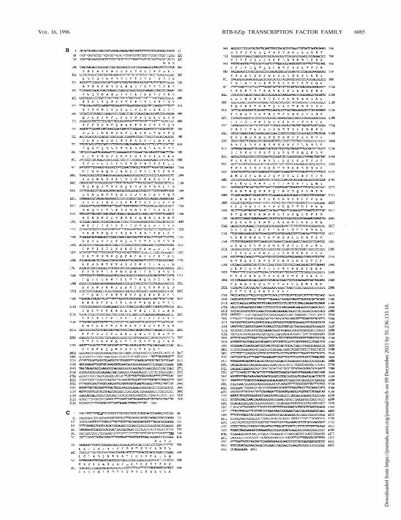
role of the Bach family in vivo, we examined the expressionprofiles of the two genes by RNA blotting (Fig. 3). bach1 andbach2 transcripts were detected as relatively long mRNAswhose sizes were estimated to be 8 and 11 kb, respectively.Because the cDNA sequences of bach1 and bach2 that weredetermined here lack an authentic polyadenylation signal atthe 39 ends, we assume that each mRNA possesses a relativelylong 39 untranslated region which might account for the lengthof the mRNA. bach1 mRNA was detected in all of the tissuesexamined, including the spleen, with the small intestine ex-pressing the highest level (Fig. 3A). In contrast, expression ofbach2mRNA was essentially restricted to the brain and spleen.In the brain, expression of bach2 but not bach1 was found to belower in the adult than in the neonate. Despite their expres-sions in adult spleen cells, bach1 and bach2 mRNAs werebarely detectable in the livers of day 17 pc embryos, at whichstage of development the liver is the major hematopoieticorgan. During mouse embryogenesis, both of the bach RNAswere detectable in day 10, 13, and 17 pc whole embryos (Fig.3A and B and data not shown).Because the expression sites of MafK are in neuronal, mes-
enchymal, and hematopoietic tissues (50), we examined ex-pression of bach genes in several cell lines representing thesecell lineages. Among the hematopoietic cell lines tested, bach1mRNA was expressed abundantly in P815 (mastocytoma [16]),FDCP-1 (bipotential myeloid progenitor), M1 (myelomono-
cytic leukemic [29]), murine erythroleukemia (MEL) DS-19(erythroleukemic [62]), and BW5147 (T-lymphoid) cells (Fig.3B). The level of bach1 mRNA was low in B-lymphoid BaF/3cells (58). MEL cells are known to undergo terminal erythroiddifferentiation upon treatment with dimethyl sulfoxide (44).However, the level of bach1 mRNA did not change signifi-cantly during this induced differentiation in vitro (data notshown). In contrast to the expression pattern of bach1, thelevel of bach2 expression was high only in the M1 cells amongthe hematopoietic cell lines. In addition to the major mRNA,M1 cells expressed a less abundant and smaller mRNA species.This mRNA was also found to be expressed in the neuroblas-toma cells (see below). The level of expression of bach2 did notincrease during the induced differentiation of MEL cells (datanot shown). The specific expression of bach2 in hematopoieticcell lines suggests a role of bach2 in lineage-related gene reg-ulation. Its expression in M1 but not in FDCP-1 suggests thatbach2 plays an important role during development of themonocyte/macrophage lineage.Consistent with its widespread expression in mouse tissues,
bach1 mRNA was also evident in the C1300 neuroblastomacells and NIH 3T3 cells (Fig. 3B). In contrast to bach1, bach2mRNA was significantly more abundant in C1300 cells than inNIH 3T3 cells. Comparison of the expression patterns of thebach genes in these cell lines with those of the p45 and nrf2genes indicated that these MafK interactor genes showed over-lapping but distinct expression profiles.Formation of heterodimers with MafK. The primary struc-
tures of the bZip domains of Bach1 and Bach2 and theirinteractions with the leucine zipper of MafK suggested thatBach1 and Bach2, like NF-E2 p45, bind to a NF-E2 site byforming heterodimers with MafK. To test this possibility and to
VOL. 16, 1996 BTB-bZip TRANSCRIPTION FACTOR FAMILY 6087
Dow
nloa
ded
from
http
s://j
ourn
als.
asm
.org
/jour
nal/m
cb o
n 09
Dec
embe
r 20
21 b
y 91
.236
.133
.10.
confirm interactions between Bach proteins and MafK, eachprotein was expressed in E. coli as a MBP fusion and purified,and their DNA binding activities were then examined byEGMSA. Binding of MBP-MafK, MBP-ECH, which we em-ployed as a control, MBP-Bach1, and MBP-Bach2 fusion pro-teins to the NF-E2 site of the chicken b-globin enhancer wasbarely detectable with 5 ng of protein (Fig. 4A, lanes 2, 4, 6,and 8). Addition of MafK to Bach resulted in strong DNAbinding (lanes 5 and 7), as was the case with ECH (lane 3).Because formation of the complexes required both MafK andBach fusion proteins, these complexes should be heterodimersof MafK and Bach proteins (see below). Formation of thesecomplexes was efficiently inhibited by competition with theunlabeled chicken b-globin enhancer NF-E2 site probe (Fig.4B, lanes 3 and 8). The NF-E2 site on the probe was essentialfor binding, since mutations within the NF-E2 site abolishedbinding (lanes 4 and 9). A DNA fragment containing theNF-E2 site from HS-2 of the b-globin LCR competed effi-ciently with the b-globin enhancer probe (lanes 5 and 10). Incontrast, the NF-E2 site-related sequence from the erythroidd-aminolevulinate synthase gene promoter, which cannot bindNF-E2 efficiently (1), barely competed with the probe (lanes 6and 11). These results showed that the nucleoprotein com-plexes reflect specific interactions between the NF-E2 site andMafK-Bach heterodimers. The observed DNA recognitionspecificities of the Bach-MafK heterodimers are very similar tothat of NF-E2 (1).To compare the relative affinities, various amounts of Bach
fusion proteins were incubated with the probe in the presenceor absence of an excess amount of MafK fusion protein. Asshown in Fig. 4C, Bach1 showed much stronger NF-E2 sitebinding activity in the presence than in the absence of MafK.On the other hand, Bach2 showed significant DNA bindingactivity by itself, especially when high doses of proteins wereused in the binding reaction mixtures. However, in the pres-
ence of MafK, Bach2 bound to the probe preferentially as aheterodimer with MafK. These results suggested that in cells,the availability of MafK or other small Maf family proteins maygovern the DNA binding forms and activities of Bach proteins.The results also suggested that one of the differences betweenBach1 and Bach2 is their efficacy of DNA binding as ho-modimers.Bach2 binds to a TRE sequence. Because homodimers and
heterodimers of bZip proteins often bind to different se-quences, we determined optimal binding sites for Bach2 byPCR-assisted selection of binding sites. We used the MBP-Bach2 fusion protein for this experiment, because the EGMSAexperiments described above showed that Bach2 bound moreactively to DNA than did Bach1 in the absence of MafK. Afterfour rounds of selection, Bach2 exhibited clear binding pref-erences over a 7-bp region whose consensus sequence wasidentical to the TRE consensus sequence (Fig. 5A). In additionto the TRE sequence, Bach2 showed preference for purine atthe 24 position and pyrimidine at the 14 position. The in-verted repeat structure of the consensus sequence suggestedthat Bach2 bound to the selected sites as a homodimer, justlike other bZip proteins. Comparison of the Bach2-selectedsequences with the sequence of the NF-E2 site revealed thatthe Bach2 consensus site is identical to the last 8 of the 11nucleotides in the NF-E2 site (Fig. 5A and B). On the otherhand, the half site of the T-MARE is identical to the longerhalf of the NF-E2 site (38). These features of the Maf andBach binding sites lead to the conclusion that, upon het-erodimeric DNA binding, MafK and Bach proteins recognizethe longer and shorter halves of the NF-E2 site, respectively.Andrews and colleagues reached a similar conclusion on thetopology of NF-E2 subunits using a different experimentalapproach (3). The results suggest that the DNA recognitionspecificity of Bach2 is regulated by differential dimer formationwith other bZip proteins like MafK.
FIG. 3. Expression of bachmRNAs. Poly(A)1 RNAs (2 to 3 mg), isolated from various sources, were hybridized with bach1- or bach2-specific RNA or DNA probesafter separation on agarose-formaldehyde gels and transfer onto nylon membranes. RNA loadings were verified by hybridizing with glyceraldehyde-3-phosphatedehydrogenase (GAPDH)- or b-actin-specific probes. (A) RNAs were isolated from tissues of adult mice (lanes 1 and 3 to 8), day 17 pc fetal liver cells (lane 2), newbornmouse brain cells (lane 9), or day 13 pc whole embryos (lane 10), as indicated above the lanes. Positions of 28S and 18S RNAs are indicated on the right side with bars.(B) RNAs were isolated from the indicated murine cultured cells (lanes 1 to 8) or from day 17 pc embryos (lane 9). Expressions of p45 NF-E2 and nrf2 mRNAs werealso analyzed by subsequent hybridizations. Positions of 28S RNAs are indicated on the right.
6088 OYAKE ET AL. MOL. CELL. BIOL.
Dow
nloa
ded
from
http
s://j
ourn
als.
asm
.org
/jour
nal/m
cb o
n 09
Dec
embe
r 20
21 b
y 91
.236
.133
.10.

Bach proteins participate in unique DNA binding complexeswithin cells. To determine if Bach and MafK form het-erodimers in vivo, we expressed these proteins in fibroblastcells by transient transfection of expression plasmids, prepared
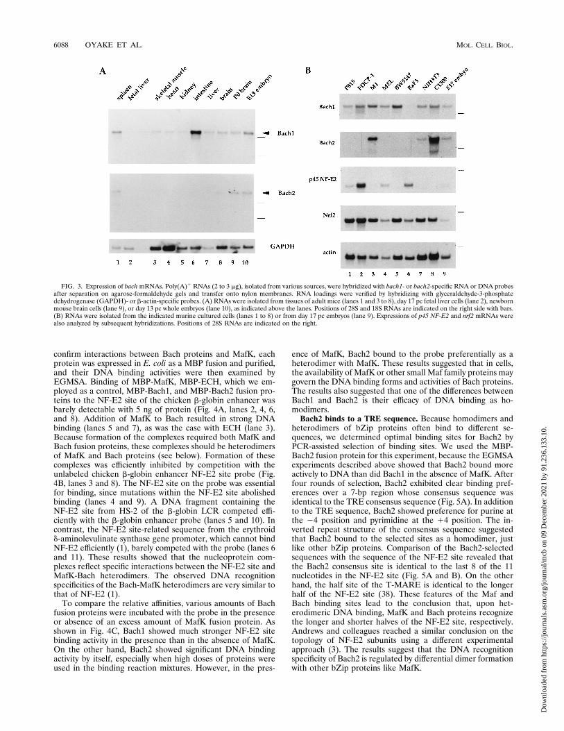
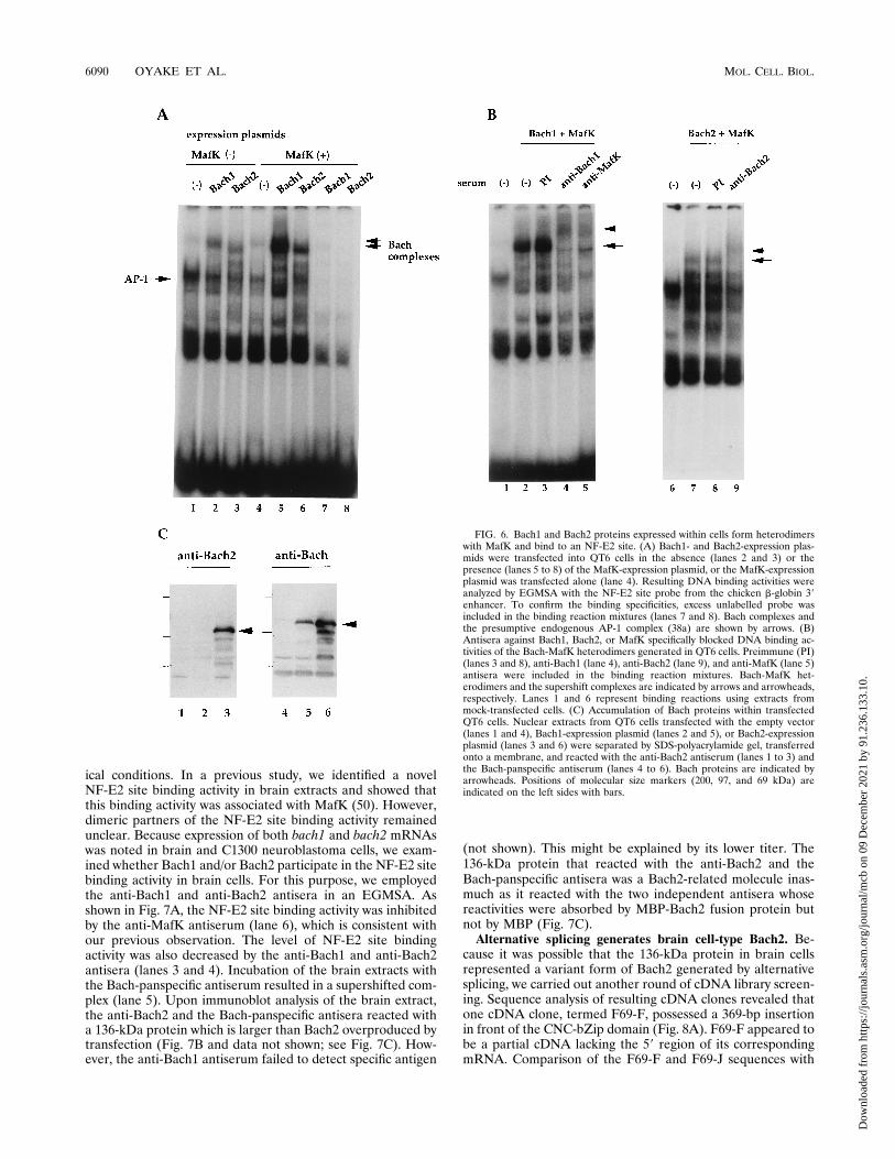
nuclear extracts, and tested resultant binding activities to theNF-E2 site by EGMSA experiments. Expression of Bach1 orBach2 alone resulted in nucleoprotein complexes with distinctmobilities (Fig. 6A, lanes 2 and 3). The amounts of thesecomplexes increased significantly without a change in theirmobilities when MafK was simultaneously expressed (lanes 5and 6). Because formation of the Bach1 complex was inhibitedby anti-Bach1 or anti-MafK antisera, it contained Bach1 andMafK (Fig. 6B). Formation of the Bach2 complex was inhib-ited by the anti-Bach2 antiserum (Fig. 6B). The complexes thatwere formed by expression of Bach proteins alone may containthe exogenous Bach and endogenous MafK or MafK-relatedproteins. Expression of Bach1 and Bach2 proteins within trans-fected cells was checked by immunoblot analysis utilizing theanti-Bach1 and anti-Bach2 antisera and the Bach-panspecificantiserum recognizing both Bach1 and Bach2 (Fig. 6C anddata not shown). The estimated sizes of the overproducedBach proteins were roughly 110 kDa and thus larger than theircalculated molecular masses predicted from the ORFs. Suchaberrant mobility of Bach proteins in denaturing protein gelsmight result from posttranslational modifications or the pres-ence of clustered basic amino acid residues on Bach proteins.Taken together, these results indicate that Bach proteins formDNA binding complexes with MafK when they are overex-pressed in cells.The next question we asked was whether Bach proteins
function by forming heterodimers with MafK under physiolog-
FIG. 4. Binding of Bach and MafK heterodimers to NF-E2 sites. (A) Fivenanograms of MBP (lane 1), MBP-ECH (lanes 3 and 4), MBP-Bach1 (lanes 5and 6), or MBP-Bach2 fusion proteins (lanes 7 and 8) was incubated with a probecontaining the NF-E2 site from the chicken b-globin 39 enhancer in the presence(lanes 2, 3, 5, and 7) or the absence of 5 ng of MBP-MafK (lanes 1, 4, 6, and 8).Formation of nucleoprotein complexes was examined by EGMSA. (B) DNArecognition specificities of Bach-MafK heterodimers were examined by EGMSA.Oligonucleotides containing various sequences were tested for their affinities forBach1-MafK (lanes 2 to 6) and Bach2-MafK (lanes 7 to 11) heterodimers bycompetition with the b-globin 39 enhancer probe. A 100-fold excess of cold DNAwas used for the competition. Competitors were cold probe DNA (bE) (lanes 3and 8), b-globin 39 enhancer DNA with mutations in the NF-E2 site (bE/M)(lanes 4 and 9), the sequence of the mouse HS-2 NF-E2 site (lanes 5 and 10), andan erythroid d-aminolevulinate synthase (ALAS) NF-E2 site-like sequence(lanes 6 and 11). (C) Twofold increments (0 to 40 ng) of Bach1 and Bach2 fusionproteins (upper and lower panels, respectively) were incubated with the b-globin39 enhancer probe in the absence (lanes 1 to 5) or presence of 50 ng of MBP-MafK (lanes 6 to 10). homo, homodimer; hetero, heterodimer.
FIG. 5. Selection of TRE-like sequences by Bach2. (A) The tally was com-piled by aligning sequences of 26 independent clones such that the centers ofpalindromic sequences were occupied by a purine. The number of samples foreach position varies, because whenever nonrandom portions of the DNAs over-lapped with the aligned sequence, these sequences were excluded from theanalysis. The consensus site shown below the tally was derived by selectingnucleotides at each position that were present in more than 70% (uppercaseletters) or 50% (lowercase letters) of clones. (B) Comparison of the half sites ofthe Bach2 consensus site and the TRE-type Maf recognition element with theNF-E2 consensus site (2).
VOL. 16, 1996 BTB-bZip TRANSCRIPTION FACTOR FAMILY 6089
Dow
nloa
ded
from
http
s://j
ourn
als.
asm
.org
/jour
nal/m
cb o
n 09
Dec
embe
r 20
21 b
y 91
.236
.133
.10.
ical conditions. In a previous study, we identified a novelNF-E2 site binding activity in brain extracts and showed thatthis binding activity was associated with MafK (50). However,dimeric partners of the NF-E2 site binding activity remainedunclear. Because expression of both bach1 and bach2 mRNAswas noted in brain and C1300 neuroblastoma cells, we exam-ined whether Bach1 and/or Bach2 participate in the NF-E2 sitebinding activity in brain cells. For this purpose, we employedthe anti-Bach1 and anti-Bach2 antisera in an EGMSA. Asshown in Fig. 7A, the NF-E2 site binding activity was inhibitedby the anti-MafK antiserum (lane 6), which is consistent withour previous observation. The level of NF-E2 site bindingactivity was also decreased by the anti-Bach1 and anti-Bach2antisera (lanes 3 and 4). Incubation of the brain extracts withthe Bach-panspecific antiserum resulted in a supershifted com-plex (lane 5). Upon immunoblot analysis of the brain extract,the anti-Bach2 and the Bach-panspecific antisera reacted witha 136-kDa protein which is larger than Bach2 overproduced bytransfection (Fig. 7B and data not shown; see Fig. 7C). How-ever, the anti-Bach1 antiserum failed to detect specific antigen
(not shown). This might be explained by its lower titer. The136-kDa protein that reacted with the anti-Bach2 and theBach-panspecific antisera was a Bach2-related molecule inas-much as it reacted with the two independent antisera whosereactivities were absorbed by MBP-Bach2 fusion protein butnot by MBP (Fig. 7C).Alternative splicing generates brain cell-type Bach2. Be-
cause it was possible that the 136-kDa protein in brain cellsrepresented a variant form of Bach2 generated by alternativesplicing, we carried out another round of cDNA library screen-ing. Sequence analysis of resulting cDNA clones revealed thatone cDNA clone, termed F69-F, possessed a 369-bp insertionin front of the CNC-bZip domain (Fig. 8A). F69-F appeared tobe a partial cDNA lacking the 59 region of its correspondingmRNA. Comparison of the F69-F and F69-J sequences with
FIG. 6. Bach1 and Bach2 proteins expressed within cells form heterodimerswith MafK and bind to an NF-E2 site. (A) Bach1- and Bach2-expression plas-mids were transfected into QT6 cells in the absence (lanes 2 and 3) or thepresence (lanes 5 to 8) of the MafK-expression plasmid, or the MafK-expressionplasmid was transfected alone (lane 4). Resulting DNA binding activities wereanalyzed by EGMSA with the NF-E2 site probe from the chicken b-globin 39enhancer. To confirm the binding specificities, excess unlabelled probe wasincluded in the binding reaction mixtures (lanes 7 and 8). Bach complexes andthe presumptive endogenous AP-1 complex (38a) are shown by arrows. (B)Antisera against Bach1, Bach2, or MafK specifically blocked DNA binding ac-tivities of the Bach-MafK heterodimers generated in QT6 cells. Preimmune (PI)(lanes 3 and 8), anti-Bach1 (lane 4), anti-Bach2 (lane 9), and anti-MafK (lane 5)antisera were included in the binding reaction mixtures. Bach-MafK het-erodimers and the supershift complexes are indicated by arrows and arrowheads,respectively. Lanes 1 and 6 represent binding reactions using extracts frommock-transfected cells. (C) Accumulation of Bach proteins within transfectedQT6 cells. Nuclear extracts from QT6 cells transfected with the empty vector(lanes 1 and 4), Bach1-expression plasmid (lanes 2 and 5), or Bach2-expressionplasmid (lanes 3 and 6) were separated by SDS-polyacrylamide gel, transferredonto a membrane, and reacted with the anti-Bach2 antiserum (lanes 1 to 3) andthe Bach-panspecific antiserum (lanes 4 to 6). Bach proteins are indicated byarrowheads. Positions of molecular size markers (200, 97, and 69 kDa) areindicated on the left sides with bars.
6090 OYAKE ET AL. MOL. CELL. BIOL.
Dow
nloa
ded
from
http
s://j
ourn
als.
asm
.org
/jour
nal/m
cb o
n 09
Dec
embe
r 20
21 b
y 91
.236
.133
.10.

the genomic DNA sequence revealed that the F69-F-specificinsertion was generated by omitting splicing of an intron (27a).The results of RNA-PCR analysis showed that Bach2 mRNAwith the F69-F-type insertion was prevalent in brain cells (datanot shown). F69-F-specific insertion encodes a region that isrich in serine (Fig. 8B). To express Bach2 with the serine-richregion, a presumptive full-length cDNA was constructed byfusing the F69-F and F69-J cDNAs. Upon transfection intofibroblast cells, the composite cDNA expressed a protein witha mobility identical to that of the Bach2-related 136-kDa an-
tigen in brain cells in a denaturing protein gel (Fig. 8C). Theseresults suggested strongly that the Bach2-related antigen in thebrain cells was actually a variant form of Bach2 generated byalternative splicing of mRNA. Taken together, the resultspoint to an importance for the Bach-MafK network in generegulation in the brain.The BTB domain mediates protein interactions. Previous
studies showed that BTB domains mediate protein interactionsamong various zinc finger proteins that contain this domain (4,10). To examine whether the BTB domain of Bach1 was ca-pable of mediating protein interactions, we used the yeasttwo-hybrid system. The entire Bach1 protein was fused toeither the DBD or the AD of GAL4. None of the fusionproteins activated individually the GAL4-dependent HIS3 re-porter gene (Fig. 9). However, when the fusion proteins were
FIG. 7. Bach2 participates in an NF-E2 site binding complex within braincells. (A) Nuclear extracts were prepared from the brains of day 17 pc mouseembryos, and NF-E2 site binding activity was analyzed by EGMSA with thechicken b-globin 39 enhancer probe (lane 1). Preimmune (PI) (lane 2), anti-Bach1 (lane 3), anti-Bach2 (lane 4), and Bach-panspecific (lane 5) antisera wereincluded in the binding reaction mixtures. The effect of the anti-MafK antiserumwas also examined (lane 6). The positions of the specific binding activity and thesupershift complex generated by the Bach-panspecific antiserum are indicated byan arrow and arrowhead, respectively. (B) Nuclear extracts from QT6 cellstransfected with the empty vector (lane 1), Bach1-expression plasmid (lane 2),Bach2-expression plasmid (lane 3), and the day 17 pc fetal brain extract (lane 4)were analyzed for the presence of Bach proteins by immunoblotting with theBach-panspecific antiserum as described for Fig. 6C. A reactive protein in thebrain extract is indicated by an arrow. Size markers used were as indicated in thelegend to Fig. 6C. (C) Specificities of the anti-Bach2 and Bach-panspecificantisera were examined in a preabsorption experiment. Each antiserum waspreincubated with either buffer alone (lanes 1 and 4), excess MBP (lanes 2 and5), or MBP-Bach2 (lanes 3 and 6) before performing immunoblot reactions withday 17 pc fetal brain extract. The position of the specific antigen is indicated byan arrow. Preabsorption of the antisera with MBP (lanes 2 and 5) resulted in arelatively high background for unknown reasons. Size markers noted by bars atthe left are as indicated in the legend to Fig. 6C.
FIG. 8. (A) Schematic representation of two distinct Bach2 cDNA clones.Protein-coding domains are indicated by boxes. Correlation between the twocDNAs is indicated by the dotted lines. (B) Nucleotide and deduced amino acidsequences surrounding the F69-F-specific insertion. 59 and 39 junctions of theinsertion are indicated by vertical arrows above the lines. (C) Nuclear extractsfrom QT6 cells transfected with the empty vector (lane 1), Bach2-expressionplasmid (lane 2), Bach2(F)-expression plasmid (lane 3), and day 17 pc fetal brainextract (lane 4) were analyzed for the presence of Bach proteins by immuno-blotting with the Bach-panspecific antiserum as described in the legend to Fig.6C. A reactive protein in the brain extract is indicated by an arrow. Size markersused were as indicated in the legend to Fig. 6C.
VOL. 16, 1996 BTB-bZip TRANSCRIPTION FACTOR FAMILY 6091
Dow
nloa
ded
from
http
s://j
ourn
als.
asm
.org
/jour
nal/m
cb o
n 09
Dec
embe
r 20
21 b
y 91
.236
.133
.10.

coexpressed, reporter gene expression was induced, resultingin histidine autotrophy. To determine whether the BTB do-main was involved in the homophilic interaction, the amino-terminal region of Bach1, including the BTB domain, wasfused to the DBD or AD and expressed in the reporter cells.The results indicated the bZip region in AD- or DBD-taggedBach1 to be dispensable for the interaction. Furthermore, theamino-terminal region showed self-interaction, albeit to aweaker extent than those of the other combinations tested.These results indicated that the BTB domain of Bach1 func-tions as a protein interaction motif within cells.Transcription regulation by Bach. Because one of the target
sites of Bach proteins appears to be a NF-E2 site, we examinedthe regulatory activities of Bach proteins with a promoter con-taining NF-E2 sites. Cotransfection of either the Bach1- orBach2-expression plasmid with the reporter plasmid in QT6fibroblasts repressed promoter activity in a dose-dependentmanner (Fig. 10A). Similar effects of the Bach-expression plas-mids were observed in the presence of the MafK-expressionplasmid (data not shown). Neither Bach1 nor Bach2 showedany significant effect on a reporter gene carrying mutatedNF-E2 sites that could not bind NF-E2 or AP-1. Thus, theeffects of Bach1 and Bach2 were specific for promoters withfunctional NF-E2 sites. Since functions of transcription factorsmay be dependent on the presence of specific cofactors, wecarried out a similar transfection experiment using the HD3chicken erythroid cell line. Unexpectedly, Bach1 and Bach2showed distinct activities. Bach2 functioned as a transcrip-tional repressor, whereas Bach1 acted as a transcriptional ac-tivator (Fig. 10B). They showed no effect on the reporter genecarrying the mutated NF-E2 sites (data not shown), verifyingthat Bach proteins acted through the NF-E2 sites. These re-
sults clearly indicated that Bach1 can function as both anactivator and a repressor of transcription, depending on thecellular context. Even though Bach2 functioned as a transcrip-tion repressor in these experiments, it might also function as atranscription activator in particular types of cells.
DISCUSSION
Previous studies implicated MafK in gene regulation duringvarious developmental events, such as erythroid and neuronalcell differentiation (1, 32, 50). The present report describes thecloning and characterization of members of a novel family ofbZip transcription factors, Bach1 and Bach2, that form het-erodimers with MafK. In addition to their contribution tonovel small Maf heterodimers, Bach1 and Bach2 may functionto connect the small Maf protein network with different regu-latory networks or different levels of transcriptional regulationthrough their unique structures and activities.The intriguing and definitive feature of Bach proteins is the
presence of both bZip and BTB domains. In Drosophila mela-nogaster, BTB domain proteins are involved in a variety ofprocesses, including chromatin modeling, regional specifica-tion in the early embryo, metamorphosis, oogenesis, photore-ceptor development, formation of neural connections, andlimb development (14, 22–24, 27, 28, 63, 66, 70, 72, 73). Thus,BTB domain proteins play important roles in various develop-mental programs. However, little is known about BTB domainproteins in vertebrates, known examples of which include zincfinger proteins like ZFPJS (65), ZF5 (56), ZID (4), PLZF (11),and BCL6 (74). Among the BTB domain proteins identifiedthus far, those of the Bach family are the first demonstratingassociation with a bZip domain. This novel combination ofBTB and bZip domains is presumably a reflection of uniquefunctions not shared by other zinc finger and bZip proteinfamilies.Some BTB domain proteins in Drosophila melanogaster reg-
ulate transcription by modulating chromatin structure. Themod(mdg4) gene imparts directionality on a chromatin insula-tor (22). The same gene is also known as the enhancer ofposition effect variegation gene, E(Var)3-93D (15). Anotherenhancer of position effect variegation gene, Trithorax-like, en-codes a GAGA factor whose biochemical function is to disruptnucleosomes in concert with the ATP-dependent nucleosome-remodeling factor (17, 66, 67). Because of its conservationamong modifier genes of position effect variegation, the BTBdomain has been suggested to mediate protein interactionsresponsible for establishing certain chromatin structures (15,17). In this regard, it is noteworthy that the b-globin LCRcontains multiple NF-E2 sites (reference 64 and referencestherein). Hence, an interesting question that stems from thepresent study is whether the Bach proteins contribute to thefunctions of the b-globin LCR to activate or repress expressionof the locus.The known biochemical function of BTB domains is to me-
diate homodimerization and/or heterodimerization with otherproteins containing this domain (4, 10). Our findings for Bach1are consistent with this. Bach1, and presumably Bach2, con-tains two independent motifs for oligomer formation, a leucinezipper and a BTB domain. While we do not know at presentany specific heterotypic interactions or physiological targets,they would be expected by analogy with GAGA and Tramtrack(4). From the observations described in this report, we mayinfer that Bach proteins form heterodimers with MafK orother bZip proteins through the leucine zipper, bind to TRE-or NF-E2 site-related DNA sequences, and interact with an-other Bach protein or other proteins through the BTB do-
FIG. 9. The BTB domain of Bach1 mediates homophilic interaction withincells. (A) Schematic representation of Bach1 proteins that were fused to theDBD and AD of GAL4. (B) Various combinations of plasmids encoding DBDand AD fusions were introduced into the reporter yeast strain, and the resultingtransformants were tested for the His1 phenotype by spotting onto His2 or His1
plates. Duplicate clones were tested for each transformation.
6092 OYAKE ET AL. MOL. CELL. BIOL.
Dow
nloa
ded
from
http
s://j
ourn
als.
asm
.org
/jour
nal/m
cb o
n 09
Dec
embe
r 20
21 b
y 91
.236
.133
.10.

mains. Our hypothesis that multiple interactions, with forma-tion of multiprotein complexes on the NF-E2 sites, in turnexecute unique regulatory roles is now testable.In the above-described transient-transfection assays, both
Bach1 and Bach2 acted as transcription regulators that werespecific for a promoter with functional NF-E2 sites (Fig. 10).At present, there are two possibilities with regard to the DNAbinding forms of Bach proteins in these experiments. They maybind to the target sites as homodimers or as heterodimers withendogenous small Maf or related proteins. The results of theEGMSA experiment (Fig. 6) support the latter alternative. Infibroblasts, both Bach1 and Bach2 repressed transcription. Be-cause NF-E2 sites contain TRE and can bind AP-1 and relatedfactors, repression of NF-E2 site-driven promoters can beachieved by competitive binding of proteins which do not pos-sess intrinsic transactivation activity. Bach1 fused to the DNAbinding domain of GAL4 did not activate expression of GAL4-dependent reporter genes in yeast cells (Fig. 9) or in fibroblastcells (30), indicating that Bach1 lacks general transactivationactivity. Thus, competition for binding sites is likely to be oneof the mechanisms by which Bach proteins repress transcrip-tion. However, it remains possible that Bach proteins exerttheir effects by interacting with other transcription factors. Therepressor activities of Bach proteins are intriguing, sinceknown CNC family proteins activate, rather than repress, tran-scription (7–9, 33, 34, 47). Thus, the Bach family may playimportant roles in coordinating transcriptional activation andrepression by the small Maf family proteins.In our transient-transfection assays using an erythroid cell
line, Bach1 functioned as an activator of transcription. In con-
trast, Bach2 acted as a repressor, irrespective of the experi-mental system (Fig. 10). The difference between Bach1 andBach2 in terms of transregulation in the erythroid cells may beexplained by their divergence in primary structures other thanthose in the BTB and bZip domains. These structural differ-ences would be expected to cause differential interactions ofthe proteins with other proteins, like coactivators, resulting indifferent regulatory properties. Our observations suggest thatfunctions of the small Maf interactor network depend not onlyon deployment of different combinations of bZip proteins butalso on the presence of other factors interacting with specificportions of the molecules.During mouse embryogenesis, the mafK gene is expressed
predominantly in hematopoietic, mesenchymal, and neuronalcells (50). The results of the two-hybrid screening indicatedthat, in the day 17 pc mouse embryo, at least four MafKinteractors, namely, p45 NF-E2, Nrf2, Bach1, and Bach2, werepresent. Furthermore, their expression profiles suggested thatall of them can participate in the MafK interactor network inhematopoietic cells. Another place where the Bach family maycontribute to MafK function is in neuronal cells, as evidencedby the above-described demonstration that brain nuclear ex-tracts contain a Bach2-related molecule which binds to theNF-E2 site as a complex with MafK (Fig. 7 and 8). BecauseC1300 neuroblastoma cells expressed high levels of bach2mRNA (Fig. 3), the presumptive Bach2-MafK complex waslikely to have been derived from neuronal cells. Consistentwith this inference, an in situ hybridization experiment showedthat Bach2 mRNA was actually expressed in neural tubes inthe day 11 pc mouse embryo (51). On the basis of these ob-servations, we propose that Bach2 functions as a partner ofMafK in neuronal cells. Whereas the NF-E2 site binding ac-tivity in brain cells was also reactive to the anti-Bach1 anti-serum in the EGMSA experiment, the antiserum did not reactwith specific protein upon immunoblotting. Even though it isspecific for Bach1 in an immunoblot analysis, the anti-Bach1antiserum cross-reacts with Bach2 under the conditions of the
FIG. 10. Regulation of transcription by Bach proteins. (A) Increasingamounts of the Bach1-expression plasmid (filled bars) and Bach2-expressionplasmid (open bars) were transfected into QT6 cells together with the pRGBP2or pRGBP4 reporter plasmid. pRBGP2 carries NF-E2 sites from the chickenb-globin 39 enhancer. pRBGP4 carries mutated NF-E2 sites that do not bindNF-E2 or AP-1. Luciferase activities were normalized with b-galactosidase ac-tivity, and the value of pRGBP2 without the effector plasmid was set at 100%.(B) Increasing amounts of the Bach1-expression plasmid (filled bars) and Bach2-expression plasmid (open bars) were transfected into HD3 cells together with thereporter plasmid pRGBP2.
VOL. 16, 1996 BTB-bZip TRANSCRIPTION FACTOR FAMILY 6093
Dow
nloa
ded
from
http
s://j
ourn
als.
asm
.org
/jour
nal/m
cb o
n 09
Dec
embe
r 20
21 b
y 91
.236
.133
.10.
EGMSA experiment (not shown; see Materials and Methods),which could explain the effect of the anti-Bach1 antiserumapparent in Fig. 7A. Alternatively, the complex might actuallycontain Bach1 in addition to Bach2 and MafK. Interactionsthrough the BTB domains would be expected to allow forma-tion of such a ternary protein complex.Several reports have pointed to important roles of the Maf
family in the development of the brain. The Kreisler gene,which is a mouse homolog of mafB, is essential for hindbraindevelopment (13, 36). Nrl, another maf family gene, is ex-pressed in postmitotic neurons and is supposed to regulateretina-specific gene expression (42, 59). c-Maf was suggested tofunction in Purkinje cell-specific gene regulation (40). It isquite possible that Bach proteins regulate the functions ofthese Maf family proteins by competing for binding sites or byinteracting directly with Maf proteins. Indeed, we could detectan interaction between c-Maf and Bach1 in the yeast two-hybrid system (55).In conclusion, the results described in this report suggest
multiple roles for Bach family proteins in coordinating tran-scription activation and repression by MafK and other Maf-related factors and underscore the importance of decipheringcross-talk among various bZip regulatory networks during cellproliferation and differentiation.
ACKNOWLEDGMENTS
We thank M. Yamagishi for advice regarding the yeast experiments,K. Kataoka for various suggestions, and J. Akasaka for constructions ofplasmids. We also thank K. Shirato and M. Ichinose for their supportand J. D. Engel, B. Roizman, and K. Lim for comments on the manu-script.This work was supported in part by grants-in-aid from the Ministry
of Education, Science, and Culture of Japan and a grant from theJapanese Society for the Promotion of Science.
REFERENCES1. Andrews, N. C., H. Erdjument-Bromage, M. B. Davidson, P. Tempst, andS. H. Orkin. 1993. Erythroid transcription factor NF-E2 is a haematopoietic-specific basic-leucine zipper protein. Nature (London) 362:722–728.
2. Andrews, N. C., and D. V. Faller. 1991. A rapid micropreparation techniquefor extraction of DNA-binding proteins from limiting numbers of mamma-lian cells. Nucleic Acids Res. 19:2499.
3. Andrews, N. C., K. J. Kotkow, P. A. Ney, H. Erdjument-Bromage, P. Tempst,and S. H. Orkin. 1993. The ubiquitous subunit of erythroid transcriptionfactor NF-E2 is a small basic-leucine zipper protein related to the v-mafoncogene. Proc. Natl. Acad. Sci. USA 90:11488–11492.
4. Bardwell, V. J., and R. Treisman. 1994. The POZ domain: a conservedprotein-protein interaction motif. Genes Dev. 8:1664–1677.
5. Beug, H., S. Palmieri, C. Freudenstein, H. Zentgraf, and T. Graf. 1982.Hormone-dependent terminal differentiation in vitro of chicken erythroleu-kemia cells transformed by ts mutants of avian erythroblastosis virus. Cell28:907–919.
6. Caterina, J., D. J. Ciavatta, D. Donze, R. R. Behringer, and T. M. Townes.1994. Multiple elements in human b-globin locus control region 59 HS 2 areinvolved in enhancer activity and position-independent, transgene expres-sion. Nucleic Acids Res. 22:1006–1011.
7. Caterina, J. J., D. Donze, C. W. Sun, D. J. Ciavatta, and T. M. Towns. 1994.Cloning and functional characterization of LCR-F1: a bZip transcriptionfactor that activates erythroid-specific, human globin gene expression. Nu-cleic Acids Res. 12:2383–2391.
8. Chan, J. Y., X.-L. Han, and Y. W. Kan. 1993. Isolation of cDNA encoding thehuman NF-E2 protein. Proc. Natl. Acad. Sci. USA 90:11366–11370.
9. Chan, J. Y., X.-L. Han, and Y. W. Kan. 1993. Cloning of Nrf1, an NF-E2-related transcription factor, by genetic selection in yeast. Proc. Natl. Acad.Sci. USA 90:11371–11375.
10. Chen, W., S. Zollman, J.-L. Couderc, and F. A. Laski. 1995. The BTBdomain of bric a brac mediates dimerization in vitro. Mol. Cell. Biol. 15:3424–3429.
11. Chen, Z., N. J. Brand, A. Chen, S. Chen, J.-H. Tong, Z.-Y. Wang, S. Waxman,and A. Zelent. 1993. Fusion between a novel Kruppel-like zinc finger geneand the retinoic acid receptor-a locus due to a variant t(11;17) translocationassociated with acute promyelocytic leukemia. EMBO J. 12:1161–1167.
12. Chomczynski, I., and N. Sacchi. 1987. Single-step method of RNA isolationby acid guanidium thionate-phenol-chloroform extraction. Anal. Biochem.162:156–159.
13. Cordes, S. P., and G. S. Barsh. 1994. The mouse segmentation gene krencodes a novel basic domain-leucine zipper transcription factor. Cell 79:1025–1034.
14. DiBello, P., D. Withers, C. A. Bayer, J. W. Fristrom, and G. M. Guild. 1991.The Drosophila broad-complex encodes a family of related, zinc finger-containing proteins. Genetics 129:385–397.
15. Dorn, R., V. Krauss, G. Reuter, and H. Saumweber. 1993. The enhancer ofposition-effect variegation of Drosophila, E(var)3-93D, codes for a chromatinprotein containing a conserved domain common to several transcriptionalregulators. Proc. Natl. Acad. Sci. USA 90:11376–11380.
16. Dunn, T. B., and M. Potter. 1957. A transplantable Mast-cell neoplasm in themouse. J. Natl. Cancer Inst. 18:587–601.
17. Farkas, G., J. Gausz, M. Galloni, G. Reuter, H. Gyurkovics, and F. Karch.1994. The Trithorax-like gene encodes the Drosophila GAGA factor. Nature(London) 371:806–808.
18. Fields, S., and O. Song. 1989. A novel genetic system to detect protein-protein interactions. Nature (London) 340:245–247.
19. Forrester, W. C., C. Thompson, J. T. Elder, and M. Groudine. 1986. Adevelopmentally stable chromatin structure in the human b-globin genecluster. Proc. Natl. Acad. Sci. USA 83:1359–1363.
20. Fujita, H., M. Yamamoto, T. Yamagami, N. Hayashi, and S. Sassa. 1991.Erythroleukemia differentiation. Distinctive responses of the erythroid-spe-cific and the nonspecific d-aminolevulinate synthase mRNA. J. Biol. Chem.266:17494–17502.
21. Fujiwara, K. T., K. Kataoka, and M. Nishizawa. 1993. Two new members ofthe maf oncogene family, mafK and mafF, encode nuclear b-Zip proteinslacking putative trans-activator domain. Oncogene 8:2371–2380.
22. Gerasimova, T. I., D. A. Gdula, D. V. Gerasimov, O. Simonova, and V. G.Corces. 1995. A Drosophila protein that imparts directionality on a chroma-tin insulator is an enhancer of position-effect variegation. Cell 82:587–597.
23. Giniger, E., K. Tietje, L. Y. Jan, and Y. N. Jan. 1994. lola encodes a putativetranscription factor required for axon growth and guidance in Drosophila.Development 120:1385–1398.
24. Godt, D., J.-L. Couderc, S. E. Cramton, and F. A. Laski. 1993. Patternformation in the limbs of Drosophila: bric a brac is expressed in both agradient and a wave-like pattern and is required for specification and propersegmentation of the tarsus. Development 119:799–812.
25. Grosveld, F., G. Blom van Assendelft, D. R. Greaves, and G. Kollias. 1987.Position-independent, high-level expression of the b-globin gene in trans-genic mice. Cell 51:975–985.
26. Harlow, E., and D. Lane. 1988. Antibodies: a laboratory manual. Cold SpringHarbor Laboratory Press, Plainview, N.Y.
27. Harrison, S. D., and A. A. Travers. 1990. The tramtrack gene encodes aDrosophila finger protein that interacts with the ftz transcriptional regulatoryregion and shows a novel embryonic expression pattern. EMBO J. 9:207–216.
27a.Hoshino, H., and K. Igarashi. Unpublished observation.28. Hu, S., D. Fambrough, J. R. Atashi, C. S. Goodman, and S. T. Crews. 1995.
The Drosophila abrupt gene encodes a BTB-zinc finger regulatory proteinthat controls the specificity of neuromuscular connections. Genes Dev.9:2936–2948.
29. Ichikawa, Y. 1969. Differentiation of a cell line of myeloid leukemia. J. Cell.Physiol. 74:223.
30. Igarashi, K. Unpublished data.31. Igarashi, K., K. Itoh, H. Motohashi, N. Hayashi, Y. Matuzaki, H. Nakauchi,
M. Nishizawa, and M. Yamamoto. 1995. Activity and expression of murinesmall Maf family protein MafK. J. Biol. Chem. 270:7615–7624.
32. Igarashi, K., K. Itoh, M. Nishizawa, N. Hayashi, and M. Yamamoto. 1995.Conditional expression of the ubiquitous transcription factor MafK induceserythroleukemia cell differentiation. Proc. Natl. Acad. Sci. USA 92:7445–7449.
33. Igarashi, K., K. Kataoka, K. Itoh, N. Hayashi, M. Nishizawa, and M.Yamamoto. 1994. Regulation of transcription by dimerization of erythroidfactor NF-E2 p45 with small Maf proteins. Nature (London) 367:568–572.
34. Itoh, K., K. Igarashi, N. Hayashi, M. Nishizawa, and M. Yamamoto. 1995.Cloning and characterization of a novel erythroid-derived CNC family tran-scription factor heterodimerizing with the small Maf family proteins. Mol.Cell. Biol. 15:4184–4193.
35. Jimenez, G., S. D. Griffiths, A. M. Ford, M. Greaves, and T. Enver. 1992.Activation of the b-globin locus control region precedes commitment to theerythroid lineage. Proc. Natl. Acad. Sci. USA 89:10618–10622.
36. Kataoka, K., K. T. Fujiwara, M. Noda, and M. Nishizawa. 1994. MafB, a newMaf family transcription activator that can associate with Maf and Fos butnot with Jun. Mol. Cell. Biol. 14:7581–7591.
37. Kataoka, K., K. Igarashi, K. Itoh, K. T. Fujiwara, M. Noda, M. Yamamoto,and M. Nishizawa. 1995. Small Maf proteins heterodimerize with Fos andpotentially act as competitive repressors of NF-E2 transcription factor. Mol.Cell. Biol. 15:2180–2190.
38. Kataoka, K., M. Noda, and M. Nishizawa. 1994. Maf nuclear oncoproteinrecognizes sequences related to an AP-1 site and forms heterodimers withboth Fos and Jun. Mol. Cell. Biol. 14:700–712.
38a.Kataoka, K., M. Noda, and M. Nishizawa. 1996. Transcription activity of
6094 OYAKE ET AL. MOL. CELL. BIOL.
Dow
nloa
ded
from
http
s://j
ourn
als.
asm
.org
/jour
nal/m
cb o
n 09
Dec
embe
r 20
21 b
y 91
.236
.133
.10.
Maf nuclear oncoprotein is modulated by Jun, Fos and small Maf proteins.Oncogene 12:53–62.
39. Kozak, M. 1989. The scanning model for translation: an update. J. Cell Biol.108:229.
40. Kurschner, C., and J. I. Morgan. 1995. The maf proto-oncogene stimulatestranscription from multiple sites in a promoter that directs Purkinje neuron-specific gene expression. Mol. Cell. Biol. 15:246–254.
41. Lim, K. C., H. Ishihara, R. D. Riddle, Z. Yang, N. Andrews, M. Yamamoto,and J. D. Engel. 1994. Structure and reglation of the chicken erythroidd-aminolevulinate synthase gene. Nucleic Acids Res. 22:1226–1233.
42. Liu, Q., M. L. Breitman, P. F. Hitchcock, and A. Swaroop. 1996. Expressionof the bZip transcription factor gene Nrl in the developing nervous system.Oncogene 12:207–211.
43. Luna, L., O. Johnsen, A. H. Skartlien, F. Pedeutour, C. Turc-Carel, H.Prydz, and A. Kolsto. 1994. Molecular cloning of a putative novel humanbZIP transcription factor on chromosome 17q22. Genomics 22:553–562.
44. Marks, P. A., and R. A. Rifkind. 1978. Erythroleukemic differentiation.Annu. Rev. Biochem. 47:419–448.
45. Mignotte, V., L. Wall, E. deBohr, F. Grosveld, and P.-H. Romeo. 1989. Twotissue-specific factors bind the erythroid promoter of human porphobilino-gen deaminase gene. Nucleic Acids Res. 17:37–54.
46. Mohler, J., K. Vani, S. Leung, and A. Epstein. 1991. Segmentally restricted,cephalic expression of a leucine zipper gene during Drosophila embryogen-esis. Mech. Dev. 34:3–10.
47. Moi, P., K. Chan, I. Asunis, A. Cao, and Y. W. Kan. 1994. Isolation ofNF-E2-related factor 2 (Nrf2), a NF-E2-like basic leucine zipper transcrip-tional activator that binds to the tandem NF-E2/AP1 repeat of the b-globinlocus control region. Proc. Natl. Acad. Sci. USA 91:9926–9930.
48. Moi, P., and Y. W. Kan. 1990. Synergistic enhancement of globin geneexpression by activator protein-1-like proteins. Proc. Natl. Acad. Sci. USA87:9000–9004.
49. Moscovici, C., M. G. Moscovici, H. Jimenez, M. M. C. Lai, M. J. Hayman,and P. K. Vogt. 1980. Continuous tissue culture cell lines derived fromchemically induced tumors of Japanese quail. Cell 11:95–103.
50. Motohashi, H., K. Igarashi, K. Onodera, S. Takahashi, H. Ohtani, M.Nakafuku, M. Nishizawa, J. D. Engel, and M. Yamamoto. 1996. Mesoder-mal- vs neuronal-specific expression of MafK is elicited by different promot-ers. Genes Cells 1:223–238.
51. Motohashi, H., M. Yamamoto, and K. Igarashi. Unpublished data.52. Ney, P. A., N. C. Andrews, S. M. Jane, B. Safer, M. E. Purucker, S. Wer-
emowicz, C. C. Morton, S. A. Goef, S. H. Orkin, and A. W. Nienhuis. 1993.Purification of the human NF-E2 complex: cDNA cloning of the hemato-poietic cell-specific subunit and evidence for an associated partner. Mol.Cell. Biol. 13:5604–5612.
53. Ney, P. A., B. P. Sorrentino, K. T. McDonagh, and A. W. Nienhuis. 1990.Tandem AP-1-binding sites within the human b-globin dominant controlregion function as an inducible enhancer in erythroid cells. Genes Dev.4:993–1006.
54. Ney, P. A., B. P. Sorrentino, C. H. Lowrey, and A. W. Nienhuis. 1990.Inducibility of the HS II enhancer depends on binding of an erythroidspecific nuclear protein. Nucleic Acids Res. 18:6011–6017.
55. Nisizawa, M. Unpublished data.56. Numoto, M., O. Niwa, J. Kaplan, K. Wong, K. Merrell, K. Kamiya, Y.
Yanagihara, and K. Calame. 1993. Transcriptional repressor ZF5 identifiesa new conserved domain in zinc finger proteins. Nucleic Acids Res. 21:3767–3775.
57. Oyake, T., and K. Igarashi. Unpublished data.58. Palacios, R., and M. Steinmetz. 1985. IL3-dependent mouse clones that
express B-220 surface antigen, contain Ig genes in germ-line configuration,and generate B lymphocytes in vivo. Cell 41:727–734.
59. Rehemtulla, A., R. Warwar, R. Kumar, X. Ji, D. J. Zack, and A. Swaroop.1996. The basic motif-leucine zipper transcription factor Nrl can positivelyregulate rhodopsin gene expression. Proc. Natl. Acad. Sci. USA 93:191–195.
60. Sambrook, J., E. F. Fritsch, and T. Maniatis. 1989. Molecular cloning: alaboratory manual, 2nd ed. Cold Spring Harbor Laboratory Press, ColdSpring Harbor, N.Y.
61. Shivdasani, R. A., M. F. Rosenblatt, D. Zucker-Franklin, C. W. Jackson, P.Hunt, C. J. M. Saris, and S. H. Orkin. 1995. Transcription factor NF-E2 isrequired for platelet formation independent of the actions of thrombopoi-etin/MGDF in megakaryocyte development. Cell 81:695–704.
62. Singer, D., M. Cooper, G. Maniatis, P. A. Marks, and R. A. Rifkind. 1974.Erythropoietic differentiation in colonies of cells transformed by Friendvirus. Proc. Natl. Acad. Sci. USA 71:2668–2670.
63. Soeller, W. C., C. E. Oh, and T. B. Kornberg. 1993. Isolation of cDNAsencoding the Drosophila GAGA transcription factor. Mol. Cell. Biol. 13:7961–7970.
64. Stamatoyannopoulos, J. A., A. Goodwin, T. Joyce, and C. H. Lowrey. 1995.NF-E2 and GATA binding motifs are required for the formation of DNaseI hypersensitive site 4 of the human b-globin locus control region. EMBO J.14:106–116.
65. Sugawara, M., T. Scholl, P. D. Ponath, and J. L. Strominger. 1994. A factorthat regulates the class II major histocompatibility complex gene DPA is amember of a subfamily of zinc finger proteins that includes a Drosophiladevelopmental control protein. Mol. Cell. Biol. 14:8438–8450.
66. Tsukiyama, T., P. B. Becker, and C. Wu. 1994. ATP-dependent nucleosomedisruption at a heat-shock promoter mediated by binding of GAGA tran-scription factor. Nature (London) 367:525–532.
67. Tsukiyama, T., and C. Wu. 1995. Purification and properties of an ATP-dependent nucleosome remodeling factor. Cell 83:1011–1020.
68. Tuan, D., W. Solomon, Q. Li, and I. M. London. 1985. The b-like-globin genedomain in human erythroid cells. Proc. Natl. Acad. Sci. USA 82:6384–6388.
69. Tuan, D. Y. H., W. B. Solomon, I. M. London, and D. P. Lee. 1989. Anerythroid-specific, developmental-stage-independent enhancer far upstreamof the human b-like globin genes. Proc. Natl. Acad. Sci. USA 86:2554–2558.
70. Weber, U., V. Siegel, and M. Mlodizk. 1995. pipsqueak encodes a novelnuclear protein required downstream of seven-up for the development ofphotoreceptor R3 and R4. EMBO J. 14:6247–6257.
71. Xie, T., M. Belinsky, Y. Xu, and A. K. Jaiswal. 1995. ARE- and TRE-mediated regulation of gene expression. J. Biol. Chem. 270:6894–6900.
72. Xiong, W.-C., and C. Montell. 1993. tramtrack is a transcriptional repressorrequired for cell fate determination in the Drosophila eye. Genes Dev.7:1085–1096.
73. Xue, F., and L. Cooley. 1993. Kelch encodes a component of intracellularbridges in Drosophila egg chambers. Cell 72:681–693.
74. Ye, B. H., F. Lista, F. L. Coco, D. M. Knowles, K. Offit, R. S. K. Chaganti,and R. Dalla-Favera. 1993. Alterations of a zinc finger-encoding gene,BCL-6, in diffuse large-cell lymphoma. Science 262:747–750.
75. Zollman, S., D. Godt, G. G. Prive, J.-L. Couderc, and F. A. Laski. 1994. TheBTB domain, found primarily in zinc finger proteins, defines an evolution-arily conserved family that includes several developmentally regulated genesin Drosophila. Proc. Natl. Acad. Sci. USA 91:10717–10721.
VOL. 16, 1996 BTB-bZip TRANSCRIPTION FACTOR FAMILY 6095
Dow
nloa
ded
from
http
s://j
ourn
als.
asm
.org
/jour
nal/m
cb o
n 09
Dec
embe
r 20
21 b
y 91
.236
.133
.10.