back of leg ii - kgmu.org
TRANSCRIPT

Back of Leg
II
Dr Garima Sehgal
Associate Professor
King George’s Medical University
UP, Lucknow

DISCLAIMER
Presentation has been made only for educational purpose
Images and data used in the presentation have been taken from various textbooks and other online resources
Author of the presentation claims no ownership for this material

Learning Objectives
By the end of this teaching session on Back of leg – II all the MBBS 1st year students must be able to:
• Enumerate the deep muscles of back of leg
• Describe the origin, insertion, nerve supply & actions of deep muscles of back of leg
• Describe locking and unlocking of knee joint
• Name the nerve and arteries of posterior compartment of leg
• Describe the origin, course relations & branches of tibial nerve
• Discuss the applied anatomy of structures in posterior compartment of leg

Deep Muscles of Back of Leg
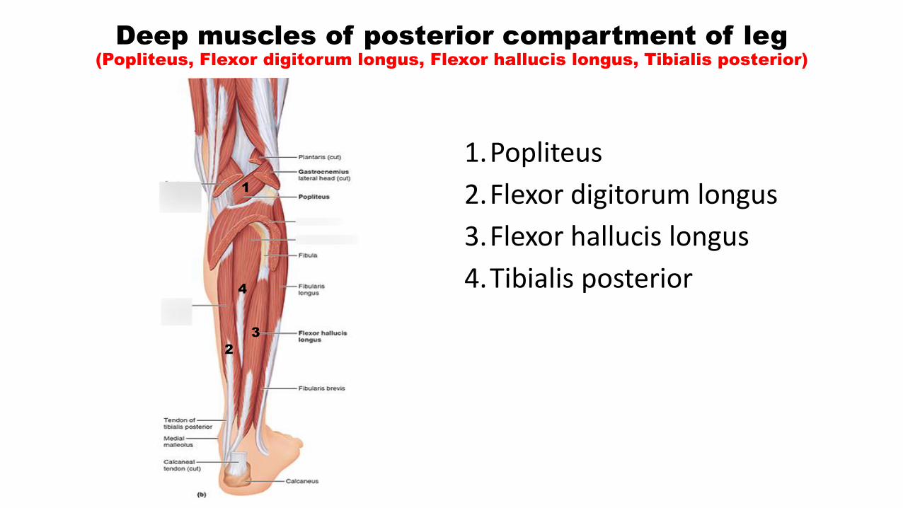
Deep muscles of posterior compartment of leg
(Popliteus, Flexor digitorum longus, Flexor hallucis longus, Tibialis posterior)
1.Popliteus
2.Flexor digitorum longus
3.Flexor hallucis longus
4.Tibialis posterior
1
4
2
3

Deep muscles contd…..
Origin:
• Lateral surface of lateral condyle of femur
• Outer margin of lateral meniscus
Insertion:
Posterior surface of tibial shaft
Above soleal line
Actions:
Unlocking of knee joint prior to flexion
Popliteus

Locking and Unlocking of knee- foot on the ground
• At the end of extension – knee joint locked
MECHANISM- when foot is on the ground:
LOCKING: Femur rotates medially on the tibia for LOCKING the knee joint at the end of extension
UNLOCKING: Femur rotates Laterally on the tibia for UNLOCKING the knee joint before flexion can be initiated/ prior to flexion
What locks the knee joint?
Articular surface geometry
NOT
MusclesMedial rotation of femur (MRF)
What Unlocks the knee joint?
POPLITEUS

Locking and Unlocking of knee- foot off the ground
MECHANISM- when foot is off the ground:
LOCKING: Tibia rotates laterally on the femur for LOCKING the knee joint at the end of extension
UNLOCKING: Tibia rotates Medially on the femur for UNLOCKING the knee joint before flexion can be initiated
What locks the knee joint?
Articular surface geometry
NOT
Muscles
What Unlocks the knee joint?
POPLITEUS

Deep muscles contd…..
Origin:
• Posterior surface of tibia
• Upper 2/3rd of medial part below soleal line
Ends in tendon that divides into 4 slips 1 for each of lateral 4 toes
Insertion:
Plantar surface of base of distal phalanges of lateral 4 toes
Flexor Digitorum Longus
Upper
2/3rd
of
medial
part

Flexor Digitorum Longus contd……
• FDL tendon crosses Tibialis posterior in lower leg
• FDL tendon crosses tendon of FHL in sole
• Tendon of FDL receives insertion of flexor digitorum accessories (quadratusplantae)
• Digital slips of tendons give origin to 4 lumbrical muscles

Deep muscles contd…..
Origin:
1. Lower 3/4th of posterior surface of fibula
Insertion:
Plantar surface of base of distal phalanx of great toe
Flexor Hallucis Longus
1

1. Tendon related to posterior surface of lower end of tibia
3. Then between the two tubercles on the posterior surface of the body of the talus 4. Runs forward below sustentaculum tali
5. Passes deep to flexor retinaculum
6. In sole crossed by tendon of FDL
7. then distally on the plantar surface of the foot in the second layer of muscles (5) where it gives rise to the Knot of Henry, then into a synovial sheath within the flexor sheath of the great toe.
Flexor Hallucis Longus contd……

Tibialis posterior
Origin:
1. Posterior surface of tibia below soleal line, upper 2/3rd of lateral part
2. Posterior surface of fibula in front of medial crest
3. Posterior surface of interosseous membrane
Insertion:
• Tuberosity of navicular bone
• Other tarsal bones (except talus)
• 2nd, 3rd , 4th metatarsal bones
13
2

Tibialis posterior contd….
• Tendon passes behind medial malleolus-in a groove
• Beneath flexor retinaculum
• Terminal part supports the spring ligament

Muscles Nerve
Supply
Actions
Popliteus Tibial nerve Unlocking of locked knee prior to flexion
Flexor Digitorum
Longus
Tibial nerve Flexes distal phalanges of lateral 4 toes , plantarflexion of ankle, supports longitudinal arches
Flexor Hallucis
Longus
Tibial nerve Flexes distal phalanx of great 4 toe , plantar flexion of ankle, supports medial longitudinal arch
Tibialis Posterior Tibial nerve Plantar flexion of ankle, inversion at subtalar joint, supports medial longitudinal arch
Deep muscles- nerve supply & actions

Posterior tibial artery
Beginning-
Lower border of popliteus, between tibia & fibula
Enters leg – deep to soleal arch
Descends medially – reaches medial ankle midway between medial malleolus and medial tubercle on calcaneum
Termination-
Deep to flexor retinaculum
Divides into medial & lateral plantar arteries

SUPERFICIAL
In upper 2/3rd
Gastrocnemius, Soleus & superficial septum
In lower 1/3rd
Skin & fascia (2.5cm in front and parallel to tendoachilles)
At ankle
Flexor retinaculum
Posterior tibial artery- relations
DEEP
In upper 2/3rd
Tibialis posterior
In lower 1/3rd
Flexor Digitorum longus
At ankle
Capsule of ankle joint (between FDL & FHL)

Branches
• Peroneal artery (largest branch)
• Muscular branches (to muscles of back)
• Nutrient artery to tibia
• Anastomotic branches• Circumflex fibular (around knee joint)
• Malleolar (medial malleolus)
• Communicating (5cm above ankle)
• Calcanean (around heel)
• Terminal branches• Medial plantar
• Lateral plantar

Peroneal artery
(largest branch of posterior tibial artery)
Beginning-
2.5 cm below lower border of popliteus
Descends laterally – along medial crest of fibula
Passes behind inferior tibiofibular& ankle joints
Termination-
Divides into many calcaneanbranches

Branches of peroneal artery
• Muscular branches (to muscles of posterior & lateral comparment)
• Nutrient artery to fibula
• Anastomotic branches• Communicating (5cm above ankle)
• Perforating branch (pierces interosseus membrane 4 cm above ankle)
• Lateral malleolar
• Calcanean (around heel)

Enters leg – deep to soleal arch
Descends medially – reaches medial ankle midway between medial malleolus and medial tubercle on calcaneum
Termination-
Deep to flexor retinaculum
Divides into medial & lateral plantar arteries
Tibial nerve
(course in leg)

SUPERFICIAL
In upper 2/3rd
Gastrocnemius, Soleus & superficial septum
In lower 1/3rd
Skin & fascia (2.5cm in front and parallel to tendoachilles)
At ankle
Flexor retinaculum
Tibial nerve- relations in leg
(similar to posterior tibial artery)
DEEP
In upper 2/3rd
Tibialis posterior
In lower 1/3rd
Flexor Digitorum longus
At ankle
Capsule of ankle joint (between FDL & FHL)

Tibial nerve – Branches in leg
MUSCULAR:
Muscles of posterior compartment superficial & deep (to popliteus & gastrocnemius in popliteal fossa)
CUTANEOUS:
Medial calcanean branches
ARTICULAR:
To the ankle joint
TERMINAL:
Medial & Lateral plantar nerves

Tibial nerve injury in the leg
DUE TO:
1. Fracture of tibia
2. Tight plasters
3. Compression under flexor retinaculum
SENSORY LOSS:
MOTOR LOSS:
• Superficial and deep muscles of calf
• Muscles of sole

Ankle jerk reflex

Venous Thrombosis
• Sitting immobile for long periods like-Long distance air travel
• Thrombosis of solealvenous sinuses
• May cause thromoembolism

Posterior tibial Pulse
LOCATION:
2 cm below and behind the medial malleolus
Felt against Calcaneum

Achilles Tendon Rupture
• most common initial symptom of Achilles tendon rupture is a sudden snap at the lower calf, intense pain, and inability to point the foot downward.
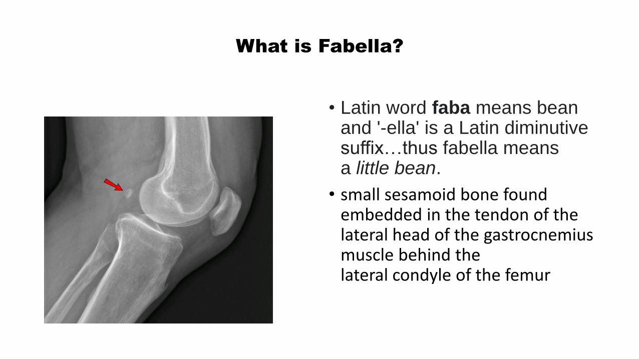
What is Fabella?
• Latin word faba means bean and '-ella' is a Latin diminutive suffix…thus fabella means a little bean.
• small sesamoid bone found embedded in the tendon of the lateral head of the gastrocnemius muscle behind the lateral condyle of the femur

Tarsal Tunnel syndrome
Tarsal tunnel syndrome causes burning pain with pins and needles or numbness in the heel and arch of the foot.

Sural nerve & Plantaris Tendon Graft
Nerve grafting
Replacement of an area of defective nerve with a segment from a sound one
The plantaris tendon is an extremely tensile structure used for flexor tendon replacement in hand surgery,Removal has no effect on normal limb function.Absent in 9% of the population

• Muscles that help to plantar flex the foot at ankle joint
• Muscles that flex the toes• Muscle that brings about unlocking of knee
joint• Posterior tibial artery……..• Tibial nerve……..• Structures deep to flexor retinaculum…..TDANH
