background radiation in radiography departments and who
TRANSCRIPT
Background Radiation in Radiography Departments and Who Wears Thermoluminescent Dosimeter (TLD)
Badges
Tehun Terfasa Dube
Diploma in Radiography University of Iceland
Medical Faculty Radiography
School of Health Sciences

Bakgrunnsgeislun á röntgendeildum og hverjir eru með TLD geislamæla
Tehun Terfasa Dube
Dissertation for Diploma degree
Supervisor: Díana Óskarsdóttir, Guðlaug Björnsdóttir, Jónína Guðjónsdóttir,
University of Iceland and and Guðlaugur Einarsson, the Icelandic Radiation Safety Authority.
Medical Faculty
Radiography
School of Health Sciences, University of Iceland June 2015


Dissertation for Diploma Degree in MS Radiography and may not be copied in any form without the permission of the copyright holder.
© Tehun Terfasa Dube, 2015
Printed: Háskólaprent
Reykjavík, Iceland, 2015

3
Abstract
The purpose of this study was to monitor background radiation leakage; using TLD badges. The study was performed by placing the TLD in the radiography control rooms’ windows; as well as the opposite sides of those same windows; where higher radiation exposure is likely. The secondary purpose of performing this study was to also observe how frequently employees wear their TLD monitor.
Background radiation measurement is utilized to determine whether or not there are any radiation leaks escaping from the radiography departments rooms. The secondary purpose of performing this study was to also observe how frequently employees wear their TLD monitor appropriately; which is of course at all times when working within the radiation area. In this study, this was accomplished by placing thermoluminescent dosimeters (TLD) badges in different medical facilities. They include; Hospital A, B, B-RT, B-NM and the clinic. These facilities were observed for a period of three-months, and provide evidence of the presence of excess background radiation. In hospital-A CT the measurement showed 63,39 mSv inside exposure room and Angiography 47,5 mSv inside exposure room and controlling room 0,39 mSv. In the hospital-B CT the scan measured 93,76 mSv inside exposure room and in the employee area inside controlling room measured 0,61 mSv. Average background radiation measurements for hospital A were 27,25 mSv over the three months inside exposure rooms. In hospital B-RT 28,4 mSv was measured, inside the radiography room and 0,122 mSv in the control rooms. In hospital B-NM scan room, waiting area, hot lab, injection room and I-131 rooms measured 1,91 mSv. In the clinic the scan measured mSv 6,5; which is inside where radiations exposure.
The observation of those wearing the TLD badge on an appropriate basis was performed simultaneously at these facilities. Those monitored for this purpose include all of the employees who operate equipment or otherwise work within the radiography departments; this is done with allowances from those health care facilities. Overall averages of all employees from Hospitals A, B, B-NM and the clinic who are wearing their TLD appropriately or not, showed as following: 10% unsure who are with TLDs, 33% with TLDs and 57% without TLDs. Inside of the exposure room in hospital-B CT the scan measured levels that were very high in comparison to the other departments where exposure. It exceeds the limit dose put in place by safety standards of 1 mSv/year; if you add up the data presented over one year. The amount of radiation in the workplace can be kept to a minimum by using radiation detectors; which can measure the amount of radiation that is present in the workplace. For those employees that it was possible to observe; the data showed that there are very few employees who wear their TLDs at all times.
Attending assistant doctors; and other staff; could potentially be exposed to inappropriately high doses of radiation over the course of one year; based upon the doses recommended by regulation officials. The information presented in these figures demonstrates the importance of employees wearing protective shielding; and staying away from these high radiation areas as much as possible. This information is particular pertinent for staff that regularly stand/work inside of the highly radiated areas within the CT scan, fluoroscopy and angiography rooms.

4

5
Thanks
I want to thank all of the people at the University of Iceland who helped me during my study. Especially thank to supervisor: Díana Óskarsdóttir, Guðlaug Björnsdóttir, Jónína Guðjónsdóttir university of Iceland and Guðlaugur Einarsson, the Icelandic Radiation Safety Authority, for their assistance provided on monitoring the TLD. The research was undertaken in five different departments within a hospital and also at a clinic; for comparisons. The radiography technicians and the supervisors who allowed to putting up the TLD to measure background radiations all deserve many thanks.
Finally, thanks to those individuals at the health care faculty of Medicine in Iceland, Reykjavik City; and assistants with the set-up of the TLD and all the others who have participated in any way within with study period.
6
Contents
Abstract..................................................................................................................................................... 3 Thanks ...................................................................................................................................................... 5 Contents ................................................................................................................................................... 6 List of figures ............................................................................................................................................ 8 List of tables ............................................................................................................................................. 8 List of Abbreviations ................................................................................................................................. 9 1 Introduction .....................................................................................................................................10
1.1 The Discovery ........................................................................................................................ 10 1.1.1 Ionization detectors ................................................................................................... 11 1.1.2 Radiation detection .................................................................................................... 11 1.1.3 The measurement conversion ................................................................................... 11
1.2 Radiation Safety Authority’s .................................................................................................. 12
1.3 Natural background radiation ................................................................................................ 12
1.4 Manmade background radiation ............................................................................................ 12 1.4.1 X-ray diagnostic machines ........................................................................................ 14 1.4.2 Radioactive material and contamination ................................................................... 14 1.4.3 Radiation therapy....................................................................................................... 15
1.5 Risks of radiation exposure ................................................................................................... 15 1.5.1 Dose Limits for employees ........................................................................................ 15 1.5.2 Placement of Radiation dose measurement ............................................................. 17 1.5.3 Radiation measuring instruments different types of TLD´s ....................................... 18
1.6 Background Radiation Measurement from the Radiology X-Ray Rooms of 13 different Hospitals ............................................................................................................................................ 19
2 Goals ..............................................................................................................................................22
2.1 Measuring background radiation ........................................................................................... 22
2.2 Observing how many employees wear TLD .......................................................................... 22 3 Materials and methods ...................................................................................................................23
3.1 Health care centers Hospital ................................................................................................. 23
3.2 How many employees wear TLD on a regular basis? ........................................................... 23 4 Results ............................................................................................................................................25
4.1 Background radiation ............................................................................................................. 25 4.1.1 Background radiation in hospital A ............................................................................ 26 4.1.2 Background radiation in Hospital B-RT ..................................................................... 26 4.1.3 Background radiation in Hospital B-NM .................................................................... 27 4.1.4 Background radiation in the clinic .............................................................................. 28
4.2 Who Wear Their TLDs ........................................................................................................... 30 4.2.1 How many employees wear TLD badges in Hospital A ............................................ 30 4.2.2 How many employees wear TLD badges in Hospital B ............................................ 31 4.2.3 How many employees wear TLD badges in Hospital B-NM ..................................... 31 4.2.4 How many employees wear TLD badges in the clinic ............................................... 31

7
5 Discussion ......................................................................................................................................33
5.1 Background radiation ............................................................................................................. 33
5.2 How many employees wear TLD badges .............................................................................. 37 6 Conclusions ....................................................................................................................................39 References .............................................................................................................................................40 Appendix 1 ..............................................................................................................................................42 Appendix 2 ..............................................................................................................................................43

8
List of figures
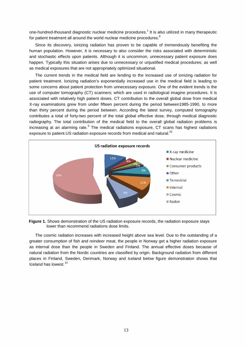
Figure 1. Shows demonstration of the US radiation exposure records, the radiation exposure stays lower than recommend radiations dose limits. ...................................................................................... 13
Figure 2. Show the estimated average effective dose to the public from natural radiation. ............ 14 Figure 3. Point out in median mSv/yr ranges that are measured at for four times for all 13 hospitals.
............................................................................................................................................................... 20 Figure 4. Above indicates range of the radiation the amount of scatter from Hospital-F1-F2. ........ 20 Figure 5. Shows background radiation in Hospital A ....................................................................... 25 Figure 6. Shows background radiation in Hospital B ....................................................................... 26 Figure 7. Shows background radiation in Hospital B-NM ................................................................ 27 Figure 8. Shows background radiation in Clinic ............................................................................... 28 Figure 9. Shows total background radiation of all hospitals as one average. .................................. 29 Figure 10. Shows total background radiation between the hospitals and Clinic in average. ........... 29 Figure 11. Shows how many employees are wearing TLD badges in Hospital A. In some instances
TLD was unable to be counted, because it was covered by clothing, or otherwise hidden from view. 30 Figure 12. Shows how many employees are wearing TLD badge in Hospital B. In some instances
TLD was unable to be counted, because it was covered by clothing, or otherwise hidden from view. 30 Figure 13. Shows how many employees are wearing TLD badge in Hospital B-NM. In some
instances TLD was unable to be counted, because it was covered by clothing, or otherwise hidden from view. .............................................................................................................................................. 31
Figure 14. Shows how many employees are wearing TLD badge in the clinic. In some instances TLD was unable to be counted, because it was covered by clothing, or otherwise hidden from view. 31
Figure 15. Shows how many employees on average are wearing TLD badge and how many are not in Hospital A, B and B-NM and at clinic. ................................................................................................ 32
Figure 16. Shows a pie chart representation of the overall averages of all how many Hospitals A, B, B-NM and clinic employees are wearing their TLD appropriately ......................................................... 32
List of tables
Table 1. Radiation and conversion units. ......................................................................................... 12 Table 2. The radiation doses limits recommendation by ICRP. ....................................................... 17 Table 3. Shows how many TLD´s were used to measure the background radiation ....................... 25 Table 4. Shows number of employees for each medical institution ................................................. 30 Table 5. Show Background radiation from the Nordic countries 10 .................................................. 36

9
List of Abbreviations
According to International Atomic Energy Agency (IAEA)
Becquerel (Bq)
Computed Tomography (CT)
Curie (Ci)
Geiger-Muller counters (GM)
Gray (Gy)
Icelandic Radiation Safety Authority’s (IRSA)
International Commission on Radiological Protection (ICRP)
National Council on Radiation Protection (NCRP)
Nuclear Medicine (NM)
millibecquerel (mBq)
millicurie (mCi)
Peripheral Dual energy X-ray absorptiometry (pDEXA)
Optically Stimulated Light-emitting Dosimeters (OSL)
Radiation therapy (RT)
Sievert (Sv)
Thermo luminescent dosimeters (TLD)
10
1 Introduction
Radiation is defined as the emission of energy as electromagnetic waves, or as moving subatomic particles, especially high-energy particles that cause ionization. Ionizing radiation has multiple uses. The two most common uses in the medical field are medical treatment of patients and sterilization of medical equipment used during the procedure and diagnostic imaging.1
Radiation comes from both natural and artificial sources. Radiation is found all around us in nature; from the Earth’s radon gas, the sun, as well as the natural radioactive materials in our bodies. Man-made sources include medical exposure and sterilization within medical facilities. They can produce what is known as background radiation; which can lead to dangerous radiation exposure of patients and employees.11
Background radiation is defined as the radiant energy that remains after the source of the radiation exposure; and is found everywhere. This remaining radiation can cause serious biological side effects if patients or employees are exposed to high doses. Therefore device most commonly used in the medical field for the measurement of background radiation is known as the thermo luminescent dosimeters (TLD). This device is used to observe background radiation in the radiography department; as well as the surrounding departments to ensure there are no background radiation leaks. It is the duty of the radiation safety department to analyze background radiation measurements within medical exposure rooms.1-2
When dose unit are introduced, background radiation caused by natural/man-made components levels are analyzed. The equivalent and effective doses; both relevant; are absorbed. Potential biological abnormalities caused by irradiation are considered; including results of low and abnormally high doses. Information is also provided on the levels of background radiation in different countries; as well as the specified annual effective background radiation doses for both sources. Background radiation can also cause other disorders; therefore it is very important to measure it in order to keep the doses as low as possible; and decrease long term health risk. When someone is exposed to radiation there is always a health risk; however it is possible to reduce the radiation when equipment is managed correctly; and by following the regulations as is required. It’s important that individuals receive proper training before managing devices within the radiography department. Radioactive materials have high radiations effective in small doses. Radiation monitors must be used to check the surroundings each and every time that radioactive materials are handled.1-2
Evaluating radiation leakage within a radiography department is very important. One must measure sources of background radiation emitted from the entire radiography department, where the X-rays are taken. Such as using Computed Tomography (CT) is world widely in use consistently for the managing health care in needs of such as emergency circumstance for patients. This technology’s fast, accurate diagnostics have saved several of lives. The world widespread use of CT for medical x-rays has unfortunately led to a global increase in average radiation doses. The results of this extreme increase in the use of imaging technology have raised additional awareness of the issue of radiation protecting in health care professionals and patients; especially children; from the dangers of high dose radiation exposure. Diagnostic imaging is steadily growing in the health care field; there is a consistently rising risk of exposure to radiation. High doses of radiation can cause serious tissue damage and raise individual’s risk of cancer.3
1.1 The Discovery The history of the discovery and utilization of radiation is rather interesting. Many individuals contributed their ideas and hard work to make radiation; particularly background radiation; less dangerous to the public. On the evening of November 8th in 1895; while working in a darkened room; Wilhelm Conrad Rontgen saw a flickering of a greenish light, such as fluorescent light with electrical discharges. Rontgen had discovered a new kind of radiation. On December 22nd of that same year, Rontgen demonstrated his newfound wonder for the first time; in the form of X-ray images of his wife’s hand. Photographic
11
plates were used in these initial X-ray studies. Wilhelm Conrad Rontgen was awarded the first Nobel Prize in Physics for his work in 1901. X-rays were soon widely used in the medical field. People were amazed by this invisible ray and its ability to pass through solid material; and with the help of a photographic plate, deliver pictures of the bones and the inside of the human body.4
William Herbert Rollins was a Boston dentist from 1896 to 1904; as well as another contributor to the field of radiation technology. He frequently supported the developing science of radiography in reference to radiation safety; such as leaded tube housings, collimators, and other techniques containing the development of high voltage tubes to reduce patient dose. Rollins also performed an experiment which presented evidence that X-rays might possibly kill guinea pigs. His experiments involved exposing pregnant guinea pigs to the device and recording the deaths of fetuses. This caused some distress for Rollins; concerning the use of X-rays in pelvic exams for pregnant women. For a few years Rollins worked on forcing radiation reduction systems. He made a real contribution in the area of X-ray safety. It was additionally discovered in April of 1896 that the X-ray equipment was harmful to living tissue, and can cause severe tissue damages following a continuous exposure to X-rays. Years later, P. Curie deliberately exposed part of his arm to radiation for roughly 10 hours; which began like sunburn wound; and took four months to completely heal.4
1.1.1 Ionization detectors In 1908 Rutherford and Geiger made the first cylindrical electrical counter for alpha particles, and then improved it in 1912 by familiarizing a spherical counter. Additional development was made in 1913 when a detector for the counting of beta particles was established. In 1928 Geiger and Muller announced kinds of gas-filled counter that answers to individual radiation-induced with the powerful output signal. It is called the Geiger-Muller counter; or GM counter; which is used to monitor radiation. The counter was even more improved in the 1930s to better facilitate monitoring, as well as make it more affordable. Soon it was in worldwide use. The GM counters were not beneficial for direct monitoring of the energy of radiation; and it was initially somewhat limited, having low counting rates. On the other hand it is still used worldwide in various laboratories for low energy X-rays in nuclear laboratories and for alpha or beta radiation, pulse it’s cost-effective.4
1.1.2 Radiation detection By 1942 the radiation detection film badge was introduced; and was used for a period of time. During dental surgeries, the device was kept in the pocket for regular monitoring of potential employee radiation exposure. The first thermo luminescent dosimeters (TLD), which were used to measure radiation exposure; were solid state detectors that were introduced by Robert Boyle in 1663. In 1927-1928 another scientist named Wick made some modifications and improved the thermo luminescent dosimeter (TLD); making it possible to use even now.4
1.1.3 The measurement conversion The development of the measuring device that was meant to standardize units for the radiation was piloted by an internal committee for weights and measures in 1984.Conversion of Radiation Protection Rules of SI unit’s measure radiation exposure from background radiation in Roentgen’s experiments are in the following forms: R coulomb/kg Absorbed Dose rad gray, Gy, Dose Equivalent rem, Sievert, Sv. Radioactivity curie, Ci, Becquerel, Bq. Ionizing radiation can change the chemical bonds in molecules of cells and therefore cause damage and cause biological effects.19
12
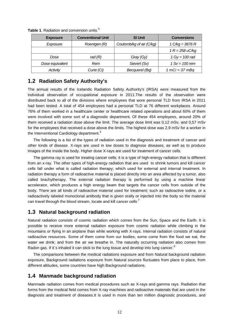
Table 1. Radiation and conversion units.5
Exposure Conventional Unit SI Unit Conversions Exposure Roentgen (R) Coulomb/kg of air (C/kg) 1 C/kg = 3876 R
1 R = 258 uC/kg Dose rad (R) Gray (Gy) 1 Gy = 100 rad
Dose equivalent Rem Sievert (Sv) 1 Sv = 100 rem Activity Curie (Ci) Becquerel (Bq) 1 mCi = 37 mBq
1.2 Radiation Safety Authority’s The annual results of the Icelandic Radiation Safety Authority’s (IRSA) were measured from the individual observation of occupational exposure in 2011.The results of the observation were distributed back to all of the divisions where employees that wore personal TLD from IRSA in 2011 had been tested. A total of 454 employees had a personal TLD at 76 different workplaces. Around 76% of them worked in a healthcare center or healthcare related operations and about 60% of them were involved with some sort of a diagnostic department. Of these 454 employees, around 20% of them received a radiation dose above the limit. The average dose limit was 0,12 mSv, and 0,57 mSv for the employees that received a dose above the limits. The highest dose was 2,9 mSv for a worker in the Interventional Cardiology department.6
The following is a list of the types of radiation used in the diagnosis and treatment of cancer and other kinds of disease. X-rays are used in low doses to diagnose diseases, as well as to produce images of the inside the body. Higher dose X-rays are used for treatment of cancer cells.
The gamma ray is used for treating cancer cells; it is a type of high-energy radiation that is different from an x-ray. The other types of high-energy radiation that are used to shrink tumors and kill cancer cells fall under what is called radiation therapy; which used for external and internal treatment. In radiation therapy a form of radioactive material is placed directly into an area affected by a tumor, also called brachytherapy. The external radiation therapy is performed by using a machine linear accelerator, which produces a high energy beam that targets the cancer cells from outside of the body. There are all kinds of radioactive material used for treatment; such as radioactive iodine, or a radioactively labeled monoclonal antibody that is given orally or injected into the body so the material can travel through the blood stream, locate and kill cancer cells.7
1.3 Natural background radiation Natural radiation consists of cosmic radiation which comes from the Sun, Space and the Earth. It is possible to receive more external radiation exposure from cosmic radiation while climbing in the mountains or flying in an airplane than while working with X-rays. Internal radiation consists of natural radioactive resources. Some of them come from our bodies; some come from the food we eat, the water we drink; and from the air we breathe in. The naturally occurring radiation also comes from Radon gas. If it´s inhaled it can stick to the lung tissue and develop into lung cancer.8
The comparisons between the medical radiations exposure and from Natural background radiation exposure, Background radiations exposure from Natural sources fluctuates from place to place, from different altitudes, some countries have high Background radiations.
1.4 Manmade background radiation Manmade radiation comes from medical procedures such as X-rays and gamma rays. Radiation that forms from the medical field comes from X-ray machines and radioactive materials that are used in the diagnosis and treatment of diseases.It is used in more than ten million diagnostic procedures, and
13
one-hundred-thousand diagnostic nuclear medicine procedures.1 It is also utilized in many therapeutic for patient treatment all around the world nuclear medicine procedures.8
Since its discovery, ionizing radiation has proven to be capable of tremendously benefiting the human population. However, it is necessary to also consider the risks associated with deterministic and stochastic effects upon patients. Although it is uncommon, unnecessary patient exposure does happen. Typically this situation arises due to unnecessary or unjustified medical procedures; as well as medical exposures that are not appropriately optimized situational.
The current trends in the medical field are lending to the increased use of ionizing radiation for patient treatment. Ionizing radiation’s exponentially increased use in the medical field is leading to some concerns about patient protection from unnecessary exposure. One of the evident trends is the use of computer tomography (CT) scanners; which are used in radiological imagine procedures. It is associated with relatively high patient doses. CT contribution to the overall global dose from medical X-ray examinations grew from under fifteen percent during the period between1985-1990, to more than thirty percent during the period between. According the latest survey, computed tomography contributes a total of forty-two percent of the total global effective dose, through medical diagnostic radiography. The total contribution of the medical field to the overall global radiation problems is increasing at an alarming rate.9 The medical radiations exposure, CT scans has highest radiations exposure to patient.US radiation exposure records from medical and natural.20
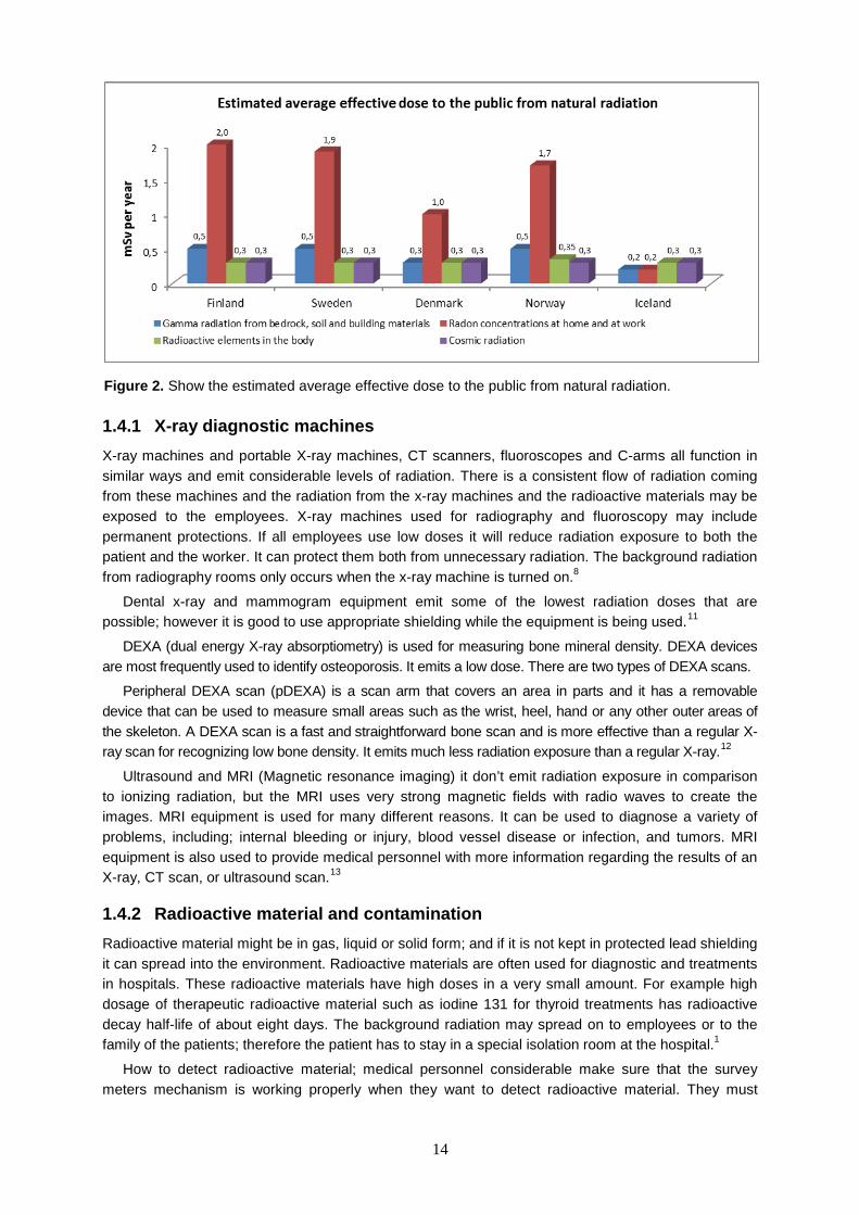
The cosmic radiation increases with increased height above sea level. Due to the outstanding of a greater consumption of fish and reindeer meat, the people in Norway get a higher radiation exposure as internal dose than the people in Sweden and Finland. The annual effective doses because of natural radiation from the Nordic countries are classified by origin. Background radiation from different places in Finland, Sweden, Denmark, Norway and Iceland below figure demonstration shows that Iceland has lowest.10
Figure 1. Shows demonstration of the US radiation exposure records, the radiation exposure stays lower than recommend radiations dose limits.
14
1.4.1 X-ray diagnostic machines X-ray machines and portable X-ray machines, CT scanners, fluoroscopes and C-arms all function in similar ways and emit considerable levels of radiation. There is a consistent flow of radiation coming from these machines and the radiation from the x-ray machines and the radioactive materials may be exposed to the employees. X-ray machines used for radiography and fluoroscopy may include permanent protections. If all employees use low doses it will reduce radiation exposure to both the patient and the worker. It can protect them both from unnecessary radiation. The background radiation from radiography rooms only occurs when the x-ray machine is turned on.8
Dental x-ray and mammogram equipment emit some of the lowest radiation doses that are possible; however it is good to use appropriate shielding while the equipment is being used.11
DEXA (dual energy X-ray absorptiometry) is used for measuring bone mineral density. DEXA devices are most frequently used to identify osteoporosis. It emits a low dose. There are two types of DEXA scans.
Peripheral DEXA scan (pDEXA) is a scan arm that covers an area in parts and it has a removable device that can be used to measure small areas such as the wrist, heel, hand or any other outer areas of the skeleton. A DEXA scan is a fast and straightforward bone scan and is more effective than a regular X-ray scan for recognizing low bone density. It emits much less radiation exposure than a regular X-ray.12
Ultrasound and MRI (Magnetic resonance imaging) it don’t emit radiation exposure in comparison to ionizing radiation, but the MRI uses very strong magnetic fields with radio waves to create the images. MRI equipment is used for many different reasons. It can be used to diagnose a variety of problems, including; internal bleeding or injury, blood vessel disease or infection, and tumors. MRI equipment is also used to provide medical personnel with more information regarding the results of an X-ray, CT scan, or ultrasound scan.13
1.4.2 Radioactive material and contamination Radioactive material might be in gas, liquid or solid form; and if it is not kept in protected lead shielding it can spread into the environment. Radioactive materials are often used for diagnostic and treatments in hospitals. These radioactive materials have high doses in a very small amount. For example high dosage of therapeutic radioactive material such as iodine 131 for thyroid treatments has radioactive decay half-life of about eight days. The background radiation may spread on to employees or to the family of the patients; therefore the patient has to stay in a special isolation room at the hospital.1
How to detect radioactive material; medical personnel considerable make sure that the survey meters mechanism is working properly when they want to detect radioactive material. They must
Figure 2. Show the estimated average effective dose to the public from natural radiation.

15
check the battery charge and select the radiation survey style, so that record background radiation reads the interpretation radiation search. The whole body is scanned for any external contamination and trace for radioactive material with Montero the whole body for radiation.
External contamination; the consequences can be very serious when someone is exposed to external contamination from radioactive material. If the material gets onto the skin, hair, eyes or any other body part, it sticks just like a splash of liquid and stays there if it´s not washed off immediately. Therefore external contamination can be stopped if the material is washed appropriately right away.
Internal contamination; exposure can occur when a radioactive material is swallowed, inhaled or absorbed through the skin, and then transferred into the blood stream. These types of exposure can stay in the body until the decay time of radioactive material is complete. Drinking a lot of fluids can accelerate the clearance from the body, somewhat. Even so, it may not be completely out of the body until half-life clearance time. To measure internal contamination, collect 70 mL spot urine sample for measurement of radioactive levels.14
1.4.3 Radiation therapy Radiation therapy is a form of high-energy. It includes x-rays, gamma rays and electron beams, which are often used for treating different kinds of cancers. After removing cancer by surgery, radiation is often used to kill the left over cancer cells of the affected part, or to slow down the growth speed of cancer cells. Radiation treatment doses are much higher than doses that are used in medical imaging for the form of radiation therapy which is known as a Linear accelerators machine; used for the treatment of cancer and for direct targeting of cancer.8-15
1.5 Risks of radiation exposure When ionizing radiation passes through a body part it can cause radiation damage to the DNA; this is called a biological effect from ionization radiation. When higher doses are used, ionization radiation can cause severe tissue damage and increase the risk of developing cancer later in life. If the radiation hit an organ directly with high doses, cells can die. Often cells can repair themselves, but not always. Some of the cells reproduce to make abnormal cells, which may cause leukemia cancer later in life.
For patients who undergo diagnostic imaging, the doses range from 15 mSv to (in an adult) 30 mSv per dose; an average of two to three CT scans for each examinations.20 CT scan machines are well-known for their contribution of higher radiation doses to the scanned body part. Usage of CT scan highly benefits results in diagnostics however, and it is still in very high demand for every day uses. Even though it increases people’s exposure to radioactive material, it continues to increase in usage as a main diagnostic in the radiography department.16
Radiography employees are specially trained to operate radiation equipment such as; x-rays, CT scans, etc. These employees may be exposed to radiation indirectly when performing their usual duties. Medical employees can also be exposed indirectly by patients that are being treated with radioactive material. If the employees follow all of the guidelines of safety regulations however, health risk is generally low.8
1.5.1 Dose Limits for employees The International Atomic Energy Agency (IAEA) and the International Commission on Radiological Protection (ICRP) publication of approved radiation dose limits for professional health care employees, dose amounts must be kept as low as reasonably achievable.
ICRP recommended annual average dose limit for the occupational employees is 20 mSv for 5 years which is allowed as flexibility, dose limit should never exceed 50 mSv within that time frame. The dose limit for the public for the same number of years is 1 mSv. The ICRP recommendation of dose limit shall be

16
controlled as shown in Table 2. Based on the data, the NCRP or ICRP estimates that doses rising above 1 mSv per year will justify the introduction of protection actions for members of the public. Deterministic effects were also considered. This became the dose limit from all sources for members of the public.17
The ICRP also recommends that controls be placed on each source, for example, limits on emission of radionuclides from installations including the emission of naturally occurring radionuclides from installations such as waste disposal sites.18
The following documentation from 2003 was recorded by the Ministry of Health and Social Security Icelandic. In their research, radiation dose limits are given for both employees and the public who encounter the ionizing radiation area. It is focused upon the employees who are working in the radiography department’s areas; as well as the monitoring of employee radiation doses.
These dose limits do not include the individuals who are exposed to radiation for medical purposes, those non-radiographer individuals who freely and voluntarily help and support individuals that are exposed to radiation for medical purposes, or the people who are exposed to ionizing radiation for voluntary participation in scientific research. It also excludes natural radiation that people are exposed to in their work environment; except in the case of increased natural radiation in their work environment; such as flight. The following definitions were for supervision; an employee with appropriate training and experience, who is responsible for activities in terms of radiation protection. Rules must be followed through an effort of action to protect employees against radiation exposure. The individuals were introduced to indicators. The intention to continue wearing radiation reading meters for these individuals with radiation reading meters was to assess the radiation that their body; and specific body parts; receive.
Radiation reading meters were utilized to assess the effective dose of ionizing radiation received by employees. Given certain assumptions, these radiation meters readings can also assist in the estimation of the amount of radioactivity within certain areas; such as a contamination zone. A partial dose can determine the biological effects of ionizing radiation in certain tissues or body parts. Internal radiation is from radioactive materials consumed; or within the body; such as X-rays or other radiation that has similar biological effects.
Ministry of Health and Social Security in Iceland stated Radiation Protection dose, in August 15, 2003. For medical Staff; Radiation Dosage to the individuals should not be exposed to more than the radiation maximum specified in this regulation. The limit for annual effective dose and effective dose object are given as follows; effective dose share of employees, student’s apprentices age 18 and older, and the public are classified as maximum annual radiation dose of 20 mSv/year, 150 mSv for the lens of the eye, 500 mSv for both limbs and skin. For those age16-18 who is in training in ionizing radiation the effective dose/year is 6 mSv, 50mSv for the lenses of the eye, and 150 mSv for the limbs and skin. For the public and other employee workers in the ionizing radiation areas the effective dose/ year is 1mSv, skin 50 mSv and the lens of the eye is 15 mSv effective doses.
The areas affected by ionizing radiation within the workplace should be divided into separate areas. The work areas where employees are exposed to annual radiation doses higher than the 30 percent annual dose limit can leak and spread to contaminate other areas. It is necessary to take measures in order to prevent these areas from becoming contaminated.
The area is defined as a closed area where employees are exposed to more radiation than the 30 percent limit; where radioactive contamination may pass from the area. Secluded individuals should only work in enclosed areas due to the possibility of radioactive contamination, or other high radiation exposure areas. Those areas shall be provided indications that the area is a source of risk. The occupants shall be required to follow appropriate procedures, and instructions shall be provided. They must take into account the risks related to their source and activity.
The Radiation Safety Authority is responsible for monitoring radiation doses for protection against exposure. That all radiation workers and the public from the activities which use ionizing radiation is always as low as possible, and taking into account at if higher radiations exposure place.

17
If radiation is higher at a single point during measurement readings, this may indicate an effective dose greater than 5 mSv; under which circumstance, the Radiation Safety Authority shall notify the sponsor immediately to request clarification on the outcome and the taking of appropriate measures. Therefore radiation workers need to wear their measurement devices. If they do not the supervisor may also ask for individual monitoring of ionizing radiation of other employees.
The reference should be made to equalize the distribution of 1 cm2 regardless of the size of the area that really will be the radiation, for the Ionizing radiation effective dose shall be according to the regulation documentations number 627 from 2003.19 For the annual effective dose and effective dose share of employees, students are the classification by groups as Maximum annual radiation dose that are similar to each other.
Table 2. The radiation doses limits recommendation by ICRP.
The limit of specified annual dose limits in this Regulation shall not apply to: 1) Situations in which the individual is exposed to radiation for medical purposes.2) Situations in which individuals who voluntarily; and not as part of their work; are exposed to ionizing radiation from nurturing and supporting individuals exposed to radiation for medical purposes. 3) Situations in which individuals are exposed to ionizing radiation due to voluntarily participation; in research which uses ionizing radiation. 4) Situations in which people are exposed to natural radiation in the course of their work; except in the case of increased natural radiation because of their work.19
1.5.2 Placement of Radiation dose measurement During a CT scan, the interventional fluoroscopy or C-arm specifies the need for two or more TLDs for operation. The placement of the TLD should be; one TLD adjacent to the chest area, inside of the protective apron, one outside of the protective apron, and finally one at eye or neck level. It is also crucial to use a thyroid shield. Ring TLD should be used for a primary radiation beam; although it is very important to keep hands out of the primary beam if possible; and best to use real time TLD in those type of circumstances. Radiation can be maximized by focusing the beam on the region which needs to be viewed. Portable screen shields should be used to protect the employees. There are various methods of reducing radiation exposure in healthcare professions for the occupational individuals. The most effective manner of operative approach for radiation safety is to spend as little time as possible in radiation areas; and remembers to keep their distance, and use protective shielding. Working longer hours in a radiation area can increase the doses received .For C-arm and others fluoroscopy machines, the X-ray tube should be kept under the patients table not angling it over the patient.20
Annual Dose limits for the worker
Employees Exposures Dose limits
Whole body 50 mSv
Lens of eye 150 mSv
Skin/extremities 500 mSv
Whole body over life time 10 mSv
Fetus for nine-month 5 mSv
Fetus one-month 0.5 mSv
Public Exposure
At most time exposure 1 mSv
At most time exposure shall not exceed 5 mSv
18
1.5.3 Radiation measuring instruments different types of TLD´s The TLD measures the radiation dose received and is used to find out if the radiation dose received by an employee is within the permitted limit. The TLD´s are used for individual radiation measuring, so that if workers are overexposed to radiation they can take action to prevent such incidents from happening again. There are many different types of TLD´s that are used for measuring the levels of radiation within the instruments in radiography departments. This is to insure radiation safety for the workers. The different types of TLDs are divided into categories; Immediate Read, Pocket Ionization Chambers and Handheld Ionization detectors with dose accumulation function, and delayed read Personnel monitors, Film Badges, TLD (Thermo Luminescent Dosimeters), OSL (Optically Stimulated Light-emitting Dosimeters).14
The Thermo Luminescent Dosimeter (TLD) detects radiation dose levels, it absorbs radiation and it is used around the lung area. This is kind of TLD that is used In Iceland as a personal monitoring device.
Handheld radiation detectors are used around an area where radiation exposure is likely to occur. They have the ability to measure dose rate and dose accumulation, and are useful in spot checks. These types are used more for radioactive materials reading, around the area where the materials are being used; and are beneficial when checking for any contamination.
The Optically Stimulated Light-emitting Dosimeters (OSL) detect low energy x-rays and it´s possible to get multiple readings. It can be placed around the breast area; at collar and at waist for pregnancy; and is also used for monitoring areas that likely for exposure. The advantage of OSL is the ability to be used more safely by pregnant workers. This is due to the instrument’s sensitivity; it picks up on low radiation in the area. Unfortunately they are more expensive than the other option, film badges.
Pocket Ionization is an electro-statically charged Pocket dosimeter; which provides an immediate reading for exposure to x-rays and gamma rays for use in high radiation areas. This device immediately alerts workers if radiation exposure is too high for them to continue working. There are two types usually used in X-ray rooms. The first of which is a direct Read Pocket which is a little pen like instrument; the advantage of which is immediate reading; and the disadvantage is a limited range.
Film Badges are yet another type of dosimeter used for monitoring radiation. It works by darkening the x-ray film in proportion to radiation absorbed. It is cheap but has the disadvantages of low sensitivity; as well as not being able to be exposed to heat, humidity, and/or light leaks.
Ring (TLD) is very important for monitoring extreme radiation exposure, especially when workers are handling radioactive materials or operating on radiation constructing equipment. The main drawback with this dosimeter is that they cannot be stored as a permanent recorder and they are expensive.
TLDs monitoring periods are for three calendar months. Radiation protection measuring devices must be sent to the radiation safety organization after the three-month measuring period. Replacement cards should arrive the preceding month in order to minimize any risk of injury, illness or damage to cells. The dates and the names of the users; along with recordkeeping numbers and employee record codes; are sent along with the devices. All of the information on the cards is printed out and then referred back to the users. The radiation monitors have to be kept in a dry and radiation free area.14-20
When new TLDs arrive from the radiation safety specialist it is carefully insured that all information is correct; e.g. the names of the users and the number on the ID cards. On the employees ID cards the name of each worker should be visible, as well as the kinds of radiation the worker might come in contact with. Used TLD devices are sent out for reading and after they are read the worker gets measurements of the radiation doses the users have received over the given period. Records of this

19
information need to be stored in a safe place. If the reading of radiation is too high the employees should be notified within two weeks so they can be relocated to work in a less radiated area. If an employee loses a TLD badge they have to notify the radiation safety authority immediately to have it replaced. TLDs are very expensive to replace. TLDs are used, in order to insure radiation safety for the workers.21
The TLD´s are used to measure the accumulative dose of radiation; for assessment of the radiation dose that employees are exposed to. TLD shall be used under the lead apron at chest level for an assessment of whole body dose. The TLD armband is used to assess the dose to the hands; and must be used at all the times during work in the radiation area.14 Because the TLD only measure for three-month periods, they shall only be used for that period. The TLDs are used by employees that work directly within radiation areas such as X-ray, where radioactive materials are handled. If new cards do not arrived on time, the old TLD ID cards may be used until the new cards arrive; under which circumstances the longer period of use shall be clearly stated by sticking a label on the TLD cards.
If an employee is not currently working in a radiation area for part of the time monitored, the radiation safety department shall be informed before one month of the return of the TLD. In this case, it needs to be clearly stated on the TLD that is has not been used.
The TLD´s retinal period is over, used or not within 5 days be sent out and continued to be used new one. As the rules and regulations say, if employees are working without using TLD badges appropriately in a radiation zone such as; X-rays, gamma rays, beta or neutron; the radiation safety authorized department must consult with the radiography managers The radiation dose adjustment method is very important; particularly in CT scans and fluoroscopy, because both have high radiation doses. The use of an apron during usage of the specified equipment is also highly necessary. The automatic exposure controller only permits exposure for short periods. It is of critical importance that the worker follows the correct instructional procedures in order to insure lower dose proportions. Extra precautions must be taken in order to maintain a risk free environment for pregnant workers; who will need to wear two TLD at neck level on the outer part of the lead shield, and at waist level. Pregnant employees must also use protective shields and make sure they are not working in high radiation exposure rooms.20-21
1.6 Background Radiation Measurement from the Radiology X-Ray Rooms of 13 different Hospitals
The Background Radiation exposure was measured with the special device that is designed to detect the radiation that is in the environment, to see the exposure to the workers from radiology department in hospitals and from many other places where X-rays are taken. The purposes of this study were to evaluate the radiation leakage from the radiology department rooms, to measure sources of background Radiation, which is emitted from the entire radiology department where the X-rays are taken. Methods used for the radiation inspection have been observed by the measurement of radiation at different points within the facility, to see the levels of the background radiation for the occupationally exposed within the Hospitals care. Data was collected from all the diagnostic and therapeutic rooms, control panels and control rooms of the different observed hospitals. The background Radiation was measured from the procedure rooms; there were 13 different x-rays rooms that radiation was measured from. The Kathamandu City, Nepal using a portable radiation measuring instrument LB 1200 RATO/F.
20
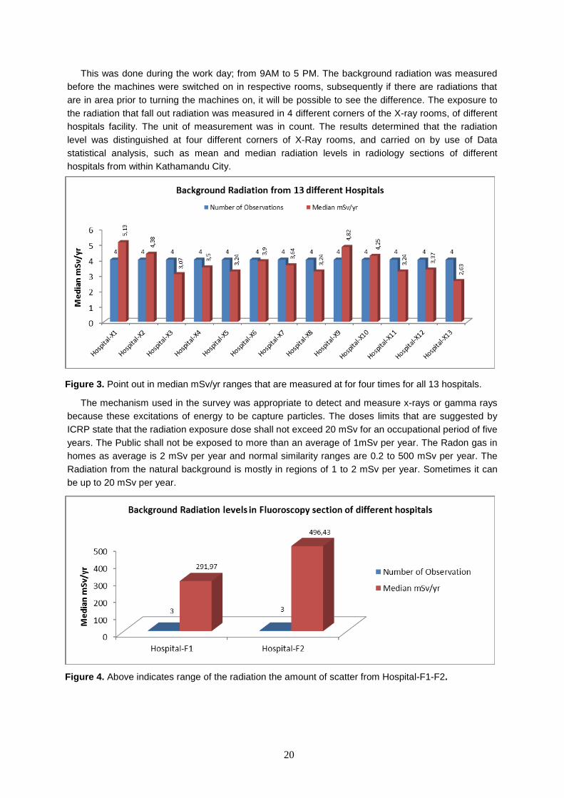
This was done during the work day; from 9AM to 5 PM. The background radiation was measured before the machines were switched on in respective rooms, subsequently if there are radiations that are in area prior to turning the machines on, it will be possible to see the difference. The exposure to the radiation that fall out radiation was measured in 4 different corners of the X-ray rooms, of different hospitals facility. The unit of measurement was in count. The results determined that the radiation level was distinguished at four different corners of X-Ray rooms, and carried on by use of Data statistical analysis, such as mean and median radiation levels in radiology sections of different hospitals from within Kathamandu City.
The mechanism used in the survey was appropriate to detect and measure x-rays or gamma rays because these excitations of energy to be capture particles. The doses limits that are suggested by ICRP state that the radiation exposure dose shall not exceed 20 mSv for an occupational period of five years. The Public shall not be exposed to more than an average of 1mSv per year. The Radon gas in homes as average is 2 mSv per year and normal similarity ranges are 0.2 to 500 mSv per year. The Radiation from the natural background is mostly in regions of 1 to 2 mSv per year. Sometimes it can be up to 20 mSv per year.
Figure 3. Point out in median mSv/yr ranges that are measured at for four times for all 13 hospitals.
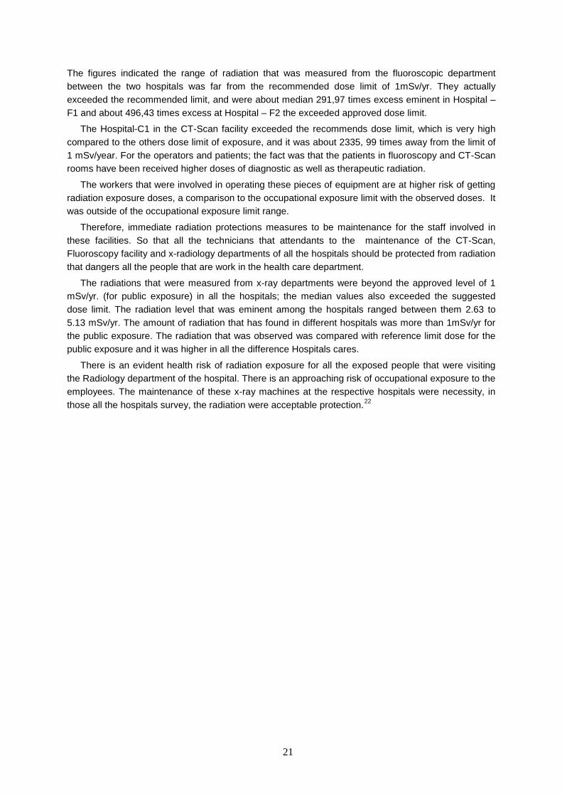
Figure 4. Above indicates range of the radiation the amount of scatter from Hospital-F1-F2.
21
The figures indicated the range of radiation that was measured from the fluoroscopic department between the two hospitals was far from the recommended dose limit of 1mSv/yr. They actually exceeded the recommended limit, and were about median 291,97 times excess eminent in Hospital –F1 and about 496,43 times excess at Hospital – F2 the exceeded approved dose limit.
The Hospital-C1 in the CT-Scan facility exceeded the recommends dose limit, which is very high compared to the others dose limit of exposure, and it was about 2335, 99 times away from the limit of 1 mSv/year. For the operators and patients; the fact was that the patients in fluoroscopy and CT-Scan rooms have been received higher doses of diagnostic as well as therapeutic radiation.
The workers that were involved in operating these pieces of equipment are at higher risk of getting radiation exposure doses, a comparison to the occupational exposure limit with the observed doses. It was outside of the occupational exposure limit range.
Therefore, immediate radiation protections measures to be maintenance for the staff involved in these facilities. So that all the technicians that attendants to the maintenance of the CT-Scan, Fluoroscopy facility and x-radiology departments of all the hospitals should be protected from radiation that dangers all the people that are work in the health care department.
The radiations that were measured from x-ray departments were beyond the approved level of 1 mSv/yr. (for public exposure) in all the hospitals; the median values also exceeded the suggested dose limit. The radiation level that was eminent among the hospitals ranged between them 2.63 to 5.13 mSv/yr. The amount of radiation that has found in different hospitals was more than 1mSv/yr for the public exposure. The radiation that was observed was compared with reference limit dose for the public exposure and it was higher in all the difference Hospitals cares.
There is an evident health risk of radiation exposure for all the exposed people that were visiting the Radiology department of the hospital. There is an approaching risk of occupational exposure to the employees. The maintenance of these x-ray machines at the respective hospitals were necessity, in those all the hospitals survey, the radiation were acceptable protection.22

22
2 Goals
The objectives of this study were to monitor background radiation; as well as observe whether or not the workers were using their TLD appropriately for safety purposes. The individuals that work in the medical imaging department have an obligation to keep the radiation doses as low as possible. They are also obligated to make sure that the radiation monitoring programmers are used appropriately. This is to help prevent high radiation exposures, if for instance, radiation leakages are acknowledged within the radiology department. By observing employee use of TLDs and measuring background radiation levels, this study may assist health care supervisors in their efforts to insure that staff follow the requirement standards for safety. The facility’s goal should be to have minimal individual radiation exposure. Radiation doses should be lowered as regulations required to regulate radiation dose limits are put in place; such as having a groundwork plan. This is the main key to lowering radiation exposure. Utilizing the same operational plan for every procedure will help maintain the same radiation doses.
2.1 Measuring background radiation Measurement of background radiation is performed in the areas where radiology equipment is utilized to assist the employees in recognizing any radiation leakage. This is done with the use of Thermo luminescent Dosimeters (TLD) placed in the working area of radiography.
2.2 Observing how many employees wear TLD The safety standards require workers to wear their TLD badges on a regular basis to insure radiation protection. By observing whether the workers are wearing or not wearing their TLD badges one can evaluate whether they follow the safety standards appropriately or not.

23
3 Materials and methods
Measuring background radiation and observing how many employees in radiography are wearing TLD badges appropriately gathers very useful and helpful information for health care professionals. The study was conducted at Hospitals A, B-Radiation therapy (RT), B-NM, and as well at clinic. The collection of data began on November 22nd of 2013, and ended on February 20th of 2014. The researcher required three-month‘s permission for the placement of the TLD badges to measuring background radiation and observation of whether the workers are wearing or not wear their TLD in radiography departments. Both researches were done at Hospital A, B, B-NM and at clinic. The background radiation measurements began on November 22nd of 2013 to February 20th of 2014; where the researcher was given permission to place the TLD for three-months and observing TLD usage in radiography departments were started after the collection of the TLD back. The researcher began by going to the Icelandic radiation safety workplace, and all the routine special data preparation was done, to make sure it is unpolluted or nothing background radiation ration measure on the TLD to be readable correctly.
Thermo/Scientific device is used to prepare for making TLD readers; which interpret data through the maximum curve, reading temperature rate per cycle, and then cooling it down to gain constancy for measurement of quality assurance in order to make sure the TLD´s measurement is correct for the three-month period. Then the TLDs were placed in all radiography rooms for the background radiation measurement. After three-months of measurement, the TLDs were collected from the entire healthcare center and the research was processed with the same data device that was used in the beginning to read TLD. TLD reader Model 4500 manual and is from Thermo Scientific (Ohio, USA), a type of machine called a MCP ultra-high sensitivity detector which is a solid circular pellets of 4.5 mm diameter with any required thickness in the range 0.9 mm. The detectors consisting of 100% LiF:Mg,Cu,P, Dose range 10μSv -10Sv.From Radcard, Kraków, Poland manufactured under the code name MCP.
The mean and average report from background radiation that has been accumulated over the three-month period of reading was completed with the range in mGy doses; which transforms to threshold converted from nanocoulomb/kg, (nC/kg,microgray (µGy and milligray (mGy)/millisievert (mSv) readings as an unexposed or exposed dose; and is then processed with the Excel data calculation we used, and then converted to the mGy/mSv doses outcome from the background radiations measurements for the entire three months were done. For more details on conversion units view Table 1.
3.1 Health care centers Hospital For the measure of background radiation, Thermo luminescent Dosimeters (TLD) were placed in five heath care centers for the required three-month period. By placing the TLDs in all of the radiography areas within the Hospitals A, B-Radiation therapy, B-NM and at clinic, measurements were performed with the 37 TLDs around radiation areas. Placements of the TLDs were as follows; Angiography, CT scan, Fluoroscopy and X-ray digital rooms. In each, the TLD was placed on the control room windows and in some of the rooms, the opposite side of the same windows TLD was placed; where the radiation the exposure could be high and where employees might be standing or holding patients within an uncovered area, or were covered in X-ray rooms; as well as other areas where employees were most likely involved in assisting, within radiation areas working on biopsy CT scan, Angiography, or Fluoroscopy rooms and become exposed were covered. The following covers the exact placement of the TLDs. the mobile X-ray had TLD placed on it. Bone density scanner, X-ray 01, X-ray 02 and X-ray 03 the TLD´s were placed in controlling rooms only, where employees are managing their jobs on a daily basis.
3.2 How many employees wear TLD on a regular basis? In the entire radiography departments of the Hospitals A, B, B-NM and in the clinic, the researcher observed how many employees wear TLD at all of the appropriate and required times during working

24
hours. The research is done in four different radiography departments for comparison. The Comparison includes employees of doctors and radiographers, as well as students and assistants, who wear TLDs within the radiography departments. Observation was performed on four different days; and each time the researcher went around each facility and checked who was wearing their TLD and who was not. This was done on the same day for each of the centers of health care; with the exception of Hospital-A; which has many more employees; which are everywhere within the departments. Also, Hospital-A has shift change workers, therefore I was checking them all day, and so shift workers are included in the counting of research. In some instances TLD were unable to be counted for because it was covered by clothing or hidden from view.

25
4 Results
The results of the collective information gathered from the hospitals and the clinic where the researcher was able to measure the use of TLDs; and subsequently the background radiation readings of each facility; have led this study to the determination that background radiation leakage has the potential to be a very serious problem; particularly for staff within the facilities that do not use their TLD badges appropriately. These results provide incremental evidence of the standard of health care, guidance management, and the ultimately hidden background radiation levels which were identified in five different healthcare facilities. The results were then processed with a Thermo/Scientific device reader; the TLD; and analyzed by Excel. The device used in the study was suitable to detect and measure X-rays and gamma rays.
4.1 Background radiation Table 3. Shows how many TLD´s were used to measure the background radiation.
TLD used in the Medical field
Places Used TLD
Hospital A 14
Hospital B-RT 9
Hospital B-NM 6
Clinic 8
Total 37
The goal of this study was to establish the basic requirements for protection against the risks of background radiation, so that the research may deliver this important knowledge to employees and the public; those that come for visits or otherwise; as well as to limit the doses received by the staff workers that are working in close proximity with radiation. In some sections of the radiography department it seemed to be handled differently, because some of the background radiation found is higher in some areas of the department. The study’s purpose was to evaluate the association of background radiations from radiography departments in five healthcare centers, and to evaluate if background radiation levels are as should be; however it was higher in some places than others.
Figure 5. Shows background radiation in Hospital A.

26
4.1.1 Background radiation in hospital A The results show that background radiation from some of the rooms is leading to over exposure, and therefore it´s essential for all of the employees that work in the higher radiation area to wear protected shields, stay away from radiation areas when possible, spend less time during operating in those same places and use their TLD´s as recommended safety standards specify.
The results show that in the Angiography room in Hospital A the background radiation was very high during the study period. The control room window was measured at 0,39 mSv, where there should not have been any discernible leakage. On the opposite side of the same window it measured 47,5 mSv. These figures demonstrate the importance for employees to wear protective shields and use their TLD badges to keep a record of how much radiation exposure they are getting over time. The results show that in the CT scan room in Hospital A the background radiation was also high during the study period. In the controlling room window it measured 0,0 mSv and, on the opposite side of the same window it measured 63,39 mSv.
The results show that in the X-ray digital room in Hospital A, the background radiation was high as well; during the study period. In the control room window it measured 0, 0 mSv, but on the opposite side of the same window it measured 27, 17 mSv; where if employees hold a patient they could come into contact with radiation exposure.
The results show that in the Mobile X-ray unit in Hospital A, the background radiation did not have a high radiation level; during the study period. It was measured 0, 73 mSv during the three-month period. Therefor if employees stand far away from the Mobile X-ray during the radiation exposure it would be alright.
Hospital A, in the bone density scanner, X-ray 01, X-ray 02 and X-ray 03; the TLD was placed in control rooms which measured 0,0 mSv, which have protective windows which are lead shielded as they should be. The opposite side of the control rooms measured zero, where the workers stand or sit in order to operate the machine.
4.1.2 Background radiation in Hospital B-RT The results show that in the Fluoroscopy 02 room in Hospital B the background radiation was normal during the study period. In the control room window it measured 0,00 mSv, and on the opposite side of the same window it measured at 10,86 mSv. These figures from Hospital B Fluoroscopy 02 room demonstrate the importance for employees to wear protective shields and use the TLD badges at all times while standing inside the radiation room and assisting doctors or patients.
Figure 6. Shows background radiation in Hospital B.
27
The results show that in the CT scan room in Hospital B the background radiation was much higher than any other CT scan rooms observed during the study period. In the control room window it measured 0,61 mSv; where there should not have been any radiation at all; and, on the opposite side of the same window it measured 93,76 mSv. This was far higher than anywhere else included in the study.
The results show that in the X-ray digital room in Hospital B the background radiation from the control room window measured 0,0 mSv.
The results show that in the Mobile X-ray in Hospital B, the background radiation measured 0,71 mSv during the three-month period.
In the X-ray digital room 01, and the Fluoroscopy 01, the TLD was placed in control rooms which measured 0,0 mSv. It has protective windows with lead shielding, and the measurement from the opposite side of the controlling room’s window measured zero, where workers stand or sit to operate the machine.
The results for the radiation therapy room in the different department of Hospital B-RT showed the background radiation measurement was only by two TLD for the three-month’s study period measured. At the control area, TLD were placed under the desk and were measured at 0,0 mSv where the employees sit in order to operate the machine. This room contains a Linear accelerator machine; a type of high energy beam used to target the cancer cells; which is just done to show a comparison. The TLD were used to study whether or not there is a scatter of high energy radiation around the employees, as well as on the opposite side of the same wall. They were also used a bit further away from the employees; and close to patient treatment areas, where they measured 8,12 mSv.
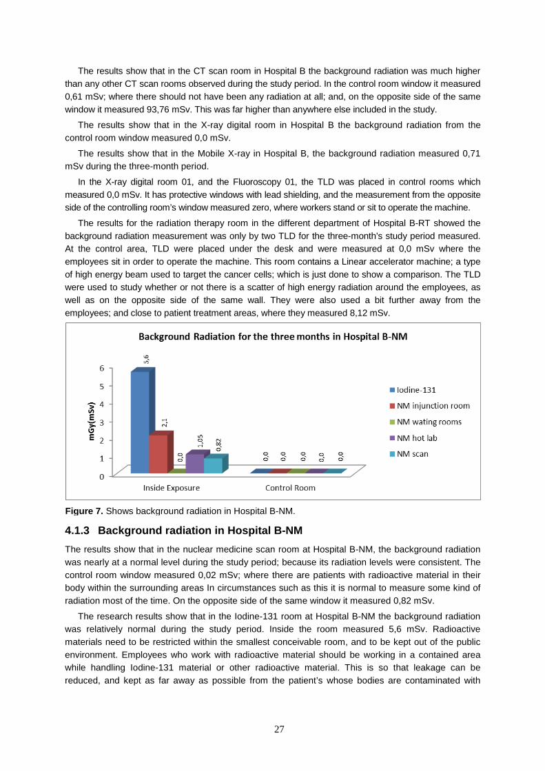
4.1.3 Background radiation in Hospital B-NM The results show that in the nuclear medicine scan room at Hospital B-NM, the background radiation was nearly at a normal level during the study period; because its radiation levels were consistent. The control room window measured 0,02 mSv; where there are patients with radioactive material in their body within the surrounding areas In circumstances such as this it is normal to measure some kind of radiation most of the time. On the opposite side of the same window it measured 0,82 mSv.
The research results show that in the Iodine-131 room at Hospital B-NM the background radiation was relatively normal during the study period. Inside the room measured 5,6 mSv. Radioactive materials need to be restricted within the smallest conceivable room, and to be kept out of the public environment. Employees who work with radioactive material should be working in a contained area while handling Iodine-131 material or other radioactive material. This is so that leakage can be reduced, and kept as far away as possible from the patient’s whose bodies are contaminated with
Figure 7. Shows background radiation in Hospital B-NM.

28
radioactive material and minimizing the time with the patients are exposed, using a protractive shield is the best way to keep it low.
The results show that in the nuclear medicine injection room at Hospital B-NM the background radiation during the study period was stable. The inside of the room measured 2,1 mSv, which can be about 8,4 mSv/year. The following result provides general guidelines for those who handle radiation sources or equipment.
The results shown from Hospital B-NM, from inside the waiting room for nuclear medicine measured 0,00 mSv; during the study period.
The results show that in the nuclear medicine hot lab room at Hospital B-NM, the background radiation during the study period of the inside of the room measured 1,05 mSv, meaning that in one year it can be about 4,2 mSv.
4.1.4 Background radiation in the clinic The results shown from the clinic CT scan room indicate that background radiation was lower than any other CT scan rooms monitored during the study period. In the control room window it measured 0,00 mSv, and on the opposite side of the same window it measured 17,25 mSv
The results shown from the MRI room in the clinic provide information on the background radiation that was measured during the study period. In the control room window it measured 0,09 mSv. This area is used for nuclear medicine injection treatments for patients, which is leading to leakage of nuclear medicine to areas where employees are sitting.
The results show that from the Fluoroscopy in the clinic the background radiation was not very high. During the study period it measured 0,14 mSv, which can be about 0,56mSv/year.
The results of the X-ray room in the clinic measurement of background radiation during this study period measured 0,48 mSv for the three-month period, meaning it can be about 1.92 mSv/year.
The results show that in the Nuclear Medicine hot lab room in the clinic –NM; In the control room window it measured 2,12 mSv, meaning that in could be about 8,48 mSv/year; on the opposite side of the same window it measured 0,82 mSv.
Three months of research in Hospital A, B and B-NM and in the clinic were used to configure this chart; this figure above shows that background radiation measured on average was the highest in Hospital B. Employees and visitors might possibly come into contact with radiation exposure, because radiation leaks were found inside the controlling areas.
Figure 8. Shows background radiation in Clinic.

29
Figure 9. Shows total background radiation of all hospitals as one average.
Figure 10. Shows total background radiation between the hospitals and Clinic in average.

30
4.2 Who Wear Their TLDs Radiation workers who operate x-ray machines, fluoroscopy units, and certain unsealed and sealed radioactive material or are exposed to other sources of gamma or high energy beta radiation are generally required to wear one or more dosimeters.
Table 4. Shows number of employees for each medical institution
How many employees were in work at the time of the counting
Medical institution Average employees Hospital A 18
Hospital B 12
Hospital B-NM 3
Clinic 8
Total 41
4.2.1 How many employees wear TLD badges in Hospital A Hospital A has an average of 18 Employees; where there were doctors, radiographers, and students. The Research was done during work houses and shift change after 3 pm was included, over a day over 4 days.
Hospital A has many more employees than the other radiology departments, and they are moving constantly throughout the departments. Hospital A has shift change employees as well; therefore the researcher was checking them all day. Shift changing for the day employees are included in the counting of research.
Figure 11. Shows how many employees are wearing TLD badges in Hospital A. In some instances TLD was unable to be counted, because it was covered by clothing, or otherwise hidden from view.
Figure 12. Shows how many employees are wearing TLD badge in Hospital B. In some instances TLD was unable to be counted, because it was covered by clothing, or otherwise hidden from view.
31
4.2.2 How many employees wear TLD badges in Hospital B Hospital B has an average of twelve employees; including a staff that consists of; doctors, radiographers and students. The research for this study was performed during work hours; once a day; over 4 days. In Hospital B the employee count was not as high as the others’ radiography departments; in spite of Hospital-B having little more than normal work hours; which mean if some kinds of mergence case happen at night to, they will take such as CT scan. The result being that the majority of the employees are included in the counting of research.
4.2.3 How many employees wear TLD badges in Hospital B-NM Hospital B-NM has an average of three employees; including doctors, radiographers, and assistants. The research was done during work hours; once a day; over 4 days. In Hospital B-NM the employee count was also very low. They operate during normal business hours; and the majority of the employees were accounted for the research.
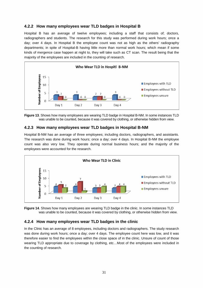
4.2.4 How many employees wear TLD badges in the clinic In the Clinic has an average of 8 employees, including doctors and radiographers. The study research was done during work hours; once a day; over 4 days. The employee count here was low, and it was therefore easier to find the employees within the close space of in the clinic. Unsure of count of those wearing TLD appropriate due to coverage by clothing, etc…Most of the employees were included in the counting of research.
Figure 13. Shows how many employees are wearing TLD badge in Hospital B-NM. In some instances TLD was unable to be counted, because it was covered by clothing, or otherwise hidden from view.
Figure 14. Shows how many employees are wearing TLD badge in the clinic. In some instances TLD was unable to be counted, because it was covered by clothing, or otherwise hidden from view.

32
Figure 16. Shows a pie chart representation of the overall averages of all how many Hospitals A, B, B-NM and clinic employees are wearing their TLD appropriately.
Figure 15. Shows how many employees on average are wearing TLD badge and how many are not in Hospital A, B and B-NM and at clinic.

33
5 Discussion
The background radiation measurement from this healthcare survey shows that all of the facilities measured radiations were tolerable; as well as fairly well protected against the majority of the control rooms. In some of the monitored locations, there is a very real possibility of being exposed to background radiation, due to the potential long term occupational exposure. This is especially true in regards to CT scan rooms, as well as the other rooms where background radiation measurement was very high. As deducted by this survey of background radiation measurement; it is not possible to completely avoid radiation exposure. The best way to reduce risk of radiation exposures is by following rules and regulation guidelines as they are stated. The entire radiology department should have quality control in each facility, and their main goal should be to minimalize individual radiation exposures, and keep up on the maintenance of those divisions. Employees in charge of handling radioactive material should be working in an enclosed space while they are handling the material. This is so that leaks can be reduced. Any potential leaks will be within the protected wall barriers, which keep the radioactive materials from passing through. Due to the increase in the contribution of radiation exposure to employees and to others, the radiation safety standards department should provide constant observation of whether or not health care departments follow rules as required for safety.
5.1 Background radiation The measurement of background radiation in all of the hospitals; and the clinic; was performed with the use of TLDs for the appropriate three-month period. The radiation levels were identified in five different healthcare facilities, and according to the outcomes of radiation leakage measurement, it provides incremental evidence over the safety standards. (There were no other studies based upon similar research for three-months in Radiology rooms; in side controlling windows and opposite sides of those same windows, for the background radiation leakage measurement with using of TLD, therefor there was not much found, in comparison. The same can be said of the observation on who wears their TLD on a regular basis). According to the data shown for the measurement comparisons of annual effective dose limits for employees; the range is very similar to Table 2: with all others documentations such as, Icelandic in 2003. Number 627 regulation documentations19 The 2007 Recommendations of the International Commission on Radiological Protection (ICRP) April 2011 and National Council on Radiation Protection & Measurements (NCRP): radiations protections documentations are all very similar to each other in respect to radiation protection. No person may be exposed to more radiation than the maximum dose, as specified in this Regulation No. 627/2003.
International Commission on Radiological Protection (ICRP) and the Ministry of Health care and Social Security in Iceland detailed Radiation Protection dose, in August 15, 2003 on, National Council on Radiation Protection (NCRP) on 18 recommendations concerning the annual dose limits were comparable to one another. These organizations have estimated the annual average dose limit for the occupational employees is 20 mSv for the duration of 5 years which is allowed some flexibility; however the dose limit should never exceed 50 mSv within that time frame. The dose limit for the public, for the same number of years is 1 mSv. The recommendation of dose limit shall be enforced as shown in Table 2: based upon the data that the NCRP and ICRP estimate that doses rising above 1 mSv per year will be unjustifiable and that the introduction of protection actions for members of the public are needed.19
The annual results of the Icelandic Radiation Safety Authority’s (IRSA) were measured from the individual observation of occupational exposure in 2011 on (p12) as discussed how high the radiation doses were after the collection of TLD back from 454 employees; during which they had been using a personal monitoring TLD at 76 different workplaces. The studies were conducted on 454 individual employees; and around 20% of them received a radiation dose that is above the limit. The average dose limit was 0,12 mSv, and 0,57 mSv for the employees that received a dose above the limits. The

34
highest dose for a worker in the Interventional Cardiology department was 2,9 mSv. This estimated annual average dose limit for the occupational from the background radiations from the five healthcare facilities in this study is highly similar to the above statement; out of the five places observed, three of them were measured as having acceptable levels of exposure. That can still possibly lead to over exposure from radiation leakage.6
The radiations measured in the X-ray departments were above the accepted standard of radiation exposure if someone remained employed in the same department for many years. In some of the hospital control rooms, there was radiation that elevated far beyond the suggested dose limit in areas where there should have been close to zero. In the mobile X-ray in Hospital A, the background radiation was not high. It was measured 0,73 mSv during the three-month period so in one year it can be about 2,92 mSv. If Mobile X-ray machines are going to an emergency area or to an intensive care unit several times within a few days’ time, this can lead to going over the recommended public dose limits on exposures, especially if the same employees are regularly working at the same places and times.
The X-ray digital room in Hospital A had high background radiation as well. In the control room window it measured 0,0 mSv, which is very good. However on the opposite side of the same window it measured 27,17 mSv. This means that in one year it can be about 108 mSv. Therefor it is very important for all of those workers in the X-ray digital rooms to stay away from open areas, and wear protective shields at all times when they are working in an enclosed room where radiation exposure is going on. This is including situations in which they have to hold a patient there during the exposure time. In order to reduce the risk of radiation exposure to themselves they should have the family of the patients hold the patient during times that it is not necessary for them to be present with staff. This is especially true for those younger employees that are of child bearing age. In these cases employees should be wearing suitable protection that covers the whole body part, instead of using the less protective gear and standing away from the open area where the radiation may be targeted. Of course employees should also wear their TLD.
Hospital A in Angiography control room window was measured at 0, 39 mSv; in one year it will be about 1,6 mSv. The opposite side of the same window measured at 47, 5 mSv; in one year it will be about 190 mSv. In the CT scan room of Hospital A, the background radiation was also very high during the study period. In the controlling room window it measured 0,0 mSv and on the opposite side of the same window it measured 63,39 mSv. In one year it will be about 253,6 mSv. That clearly exceeds the dose limit standard regulations; particularly if the same employees would be assisting the doctor during biopsy for many years.
In Hospital B-NM, the background radiation results shown are for the nuclear medicine injection room; during the study period. Inside the room measured 2,1 mSv, which can be about 8,4 mSv/year, this could cause high health risks over longer times of exposure to the employees working in this room, and others employees passing by, so this place clearly needs to be closed off for restriction to these rooms. Radiation leakages that are in the area need to be monitored to alert employees and the public, in order to limit individual exposures. Posting signs around the areas of radiation in clearly noticeable places would help with this as well. In order to minimize your exposure to radioactive materials or radiation-producing devices, always practice recommendation measures to minimize time near a source, maximize distance and shielding between radioactive sources.
The steadfast receiving of an effective dose equivalent is similar to effective dose equivalent, but applies to long-term irradiation of individual organs or tissues resulting from inhalation or ingestion of long-lived radioactive material. In these situations, the total dose is delivered slowly over long periods of time; such as over the many years put into a professional career. The effective dose equivalent is calculated for total life-long effective dose equivalent resulting from an intake that will be dedicated to the individual. This is recognized as the effective dose equivalent will continue to accumulate.
Occupationally exposed individuals must not receive a dose in excess of 30 percent of the annual occupational dose equivalent. An employee pregnancy must be declared, by requesting and
35
submitting the application forms from the Radiation Safety Office. The forms become part of the radiation dosimeters records and are required for compliance with requirement of regulations for dosimeters record keeping; including the dose equivalent to an embryo or fetus during the entire pregnancy. Due to the risks associated with the occupational exposure of a declared pregnant woman, the dose must not exceed 5 mSv. If so avoid situations where the occupational dose limits could be exceeded, abstention from or extreme caution should be used if a declared pregnant worker participates in the following activities; handling radioactive recommendations of regulation Nr 627.
In Hospital B, the background radiation was much higher than in any other CT scan rooms during the study period. Inside the control room window it measured 0,61 mSv; in one year it will be about 2,44 mSv; whereas there should have been none present in the areas where employees sit at all of the time. This is over the standard limit dose, and on the opposite side of the same window it measured 93,76 mSv, this is much higher than anywhere else recorded; it exceeds the dose limit of standards regulations. In one year that can be about 375,0 mSv. Sometimes doctors and radiographers have to take biopsies. Employees are standing in proximity during radiation exposure time, so it seem the employees that work at this place are at risk of going over the limited dose exposure in a short time of period.
Hospital B Fluoroscopy 02 room, in the control room window it measured 0,0 mSv and on the opposite side of the same window it measured 10,86 mSv; where employees are commonly standing inside the radiation room. If the TLD is measured on the window, how high could it be closer to the tube where there is primary radiation going on? The assisting doctors that are working on patients could possibly be exposed to 43,44 mSv in one year. The figure above demonstrates the importance for those employees who are standing inside the radiation areas of CT scan, Fluoroscopy and the Angiography room while assisting doctors or patients, to wear protective shielding and stay away from these areas as much as possible.
At the Clinic; CT scan inside the control room measured 0,05 mSv; in one year it will be about 0,2 mSv. On the opposite side of the same window it measured 16,08 mSv, and in one year it will be about 64,3 mSv. With this amount of background radiation in the area, there is a possibility of over exposure for employees if they are working for many years in the same area. Therefor it´s essential for all of the employees that work in the CT scan room; as well as any other rooms close by; to wear protective shields, and use their TLD´s.
At the Clinic; the area around the MRI machine at the control room window measured 0,09 mSv. This is because this area is used for nuclear medicine injection administration to patients, and is leading to the leakage of radioactive material into the areas where employees are sitting and operating the MRI machine. This estimation shows how many safety restrictions are required in those areas where employees are setting.
The overall data average for the entire healthcare facilities indicated that in; Hospital A, Hospital B, Radiation therapy, Hospital B-NM and in the clinic rooms; measured radiation leakages at the majority of locations. It is critically important to reduce unnecessary radiation exposure from the radiography department.
The highest radiation areas are those where procedures are performed with imaging machines, and are associated with the highest radiation doses, which are similar to the Kathmandu City; The Hospital-C1 in the CT-Scan facility exceeded the recommends dose limit, which is very high compared to the others dose limit of exposure, and it was about 2335, 99 times away from the limit of 1 mSv/year. From Iceland Angiography, fluoroscopy, Hospital A-B and in the Clinic CT; the figures indicate ranges of radiation that were measured higher and from the Kathmandu City Similar radiation rooms that measured high are such as the fluoroscopic between the two hospitals was a far away from the recommended dose limit of 1mSv/yr for the public. This actually exceeded compared to the recommended limit, and was about median 291,97 times excess eminent in Hospital – F1 and about 496,43 times excess at Hospital – F2 the exceeded approved dose limit.21

36
Radioactive materials must be restricted in the smallest possible area. Additionally, all of the rooms where radioactive materials are being handled should be within closer proximity of the radiography patients’ rooms in order to minimalize the contamination of other departments. They should always have all of the basic tools that are required ready before arriving to the zone in which there is a higher dose of radiation; and to be stay out of the areas that have the most radioactive materials.
Background radiation exposure comes from different sources; the body gets it from consuming food and water; and other sources of background radiation include rocks, soil and building materials. Radiation also comes from non-natural sources, such as man-made objects like Medical X-rays. Cosmic radiation can be accumulated through one cross-country air fight.
This research shows the results of some combination of occurring Radiation exposure from Man-made sources such as Medical X-rays. The sources of background Radiation are both man-made and natural from.
X-ray medicine 11%, Nuclear medicine 4%, Consumer products 3%, other 1%, Terrestrial 8%, Internal 11%, Cosmic 8% Radon 54% from US, which appear to be the highest in percentages for Man-made sources and natural.20 Source annual effective doses due to natural radiation from the Nordic countries are classified by mSv/yr.
Table 5. Show Background radiation from the Nordic countries. 10
Background radiation from bedrock, soil and building materials Source Finland Sweden Denmark Norway Iceland
Bedrock, soil building materials 0,5
0,5
0,3
0,5
0,2
Radon at home and at work 2,0
1,9
1,0
1,7
0,2
Radioactive in the body 0,3
0,3
0,3
0,35
0,3
Cosmic 0,3 0,3 0,3 0,3 0,3 Total 3,1 3,0 2,0 2,9 1,0
The above chart shows background radiation exposure categorized as natural and manmade, which when compared from the Nordic and US countries, are higher than Iceland; as well as in Norway because of the peoples high intake of fish and reindeer meat, they have high natural radiation dose of people in Sweden and Finland.
The radiation level that was distinguished amongst the Icelandic health care centers, ranged between the places that were included in this study, from the lowest to the highest was 0,02 to 93,76 mSv for the three-month period.
The studies done from Kathmandu City; on background radiation monitoring that was distinguished among the 13 different hospitals had a range between them 2.63 mSv/yr - 5.13 mSv/yr for normal X-Ray rooms.
The actual limit exceeded the recommended limit, and was about median 291,97 mSv/yr times excess eminent in Hospital-F1 and about 496,43 times excess at Hospital-F2 the exceeded approved dose limit.
The Hospital-C1 in the CT-Scan department exceeded the recommended dose limit, which is very high compared to the others dose limit of exposure, and it was about 2335, 99 times away from the limit of 1 mSv/year.21
If we compared Icelandic background radiation, that was monitored for the three months, the results show that in the CT scan room in Hospital A and Hospital B the background radiation was also high during the study period. In the controlling room window it measured 0,61 mSv and, on the opposite side of the same window it measured Hospital A 63,39 mSv, and Hospital B 93,76 mSv and according to this measures of compared Icelandic are superior.

37
It is the duty of the radiation safety department to analyze background radiation measurements within the medical exposure rooms, including; X-Ray, Bone Density, CT scan, Nuclear Medicine, Fluoroscopy, Angiography and Radiation therapy rooms. Ionizing radiation: radiation from radioactive substances, X-rays or other radiation with similar biological effects. Should be closed area: Work area where employees may be reaching an annual ionizing radiation of more than 30% annual maximum. Work from radioactive substances can spread and contaminate other areas, and they need to take measures to prevent this. Such as external radiation: radiation originating outside the body.19
This is done in order to insure that there is no unwanted background radiation spreading throughout the radiography department. Employees that work within the radiation area must wear appropriate safety protective shielding; including the TLDs; at all times for the same reason.18
The background radiations from above that are analyzed: the difference between the medical and natural background radiations exposure are simple. Background radiation from natural sources fluctuates from place to place, and the medical radiations, such as CT scans have the highest background radiation exposures to employees, as well as if the public gets closer to the source of the radiation exposure. All radiation exposure should be kept to a minimum at all times, with the use of specialized measuring equipment. The amount of radiation in the workplace can be kept to a minimum by using radiation detectors; which can measure the amount of radiation that can be found in the workplace.
5.2 How many employees wear TLD badges The research findings that covered how many employees are not using their TLDs were very disagreeable; and some kind of further reinforcement is needed. The results of this study indicate that improvement is needed. Very few workers wear their TLDs at all times. And that goes for all of the healthcare facilities; although Hospital B-NM measured reasonably better than the other three departments. As far as who wears TLD badges, there was not a lot of information to be found on exactly how many employees wear their TLD on a regular basis.
The overall average for Hospital-A, B, B-NM and in the clinic was rounded out to be three employees with TLDs, and six without TLD. The researcher was unsure about one employee. It is very important that employees take responsibility; if someone is violating rules, they should immediately be reported under any conditions that the employees may lead to causing a violation of the regulations. This is especially true for those employees who are not using their TLD as the regulations stated; the radiology department managers should keep on checking into seriously and if employees are not following rules; and if it happens often; must take action to report them to radiation protections safety organizations.
In regard to those employees who do not wear their TLD on a regular basis; if at all; what will happen if they have been working unknowingly in a high radiation environment? This means there is no record being kept in order to estimate their dose, for longer periods of their working life. Those employees must keep it in mind that it isn’t just for their own benefit to keep the record; it is also used in special cases of accidents where higher radiation is induced in case of dysfunctional equipment or radioactive material spill out, for example. Radiation Safety Officers shall be responsible for the maintenance of records of all regular monitoring being carried out; and for the distribution and maintenance of these records. They will also be responsible for monitoring the knowledgeable record keeping of employees that are working in radiation areas, and if the register report outcome is always 00mSv, the Radiation Safety Officer shall be responsible for insuring that the information is correct. It could be difficult to keep a record, but if some special data software could give notifications if the radiation readings are either too high or consistently at 0,0 for a period of 6 months; or 1 year; it would be easy for them to verify data. This may be one way to gain better control over it. Radiation Safety Officers could make surprise visits to the radiology department; for inspection and ask if they are not wearing their TLD if they are not visible. If employees are not wearing their TLD they should be sent home until they have it with them again.

38
The radiation dose adjustment method is very important; particularly in CT scans and fluoroscopy, because both studies show high radiation doses. The wearing the TLD and use of an apron during usage of the specified equipment is also highly necessary. The automatic exposure controller only permits exposure for short periods. It is of critical importance that the worker follows the correct instructional procedures in order to insure lower dose proportions. Extra precautions must be taken in order to maintain a risk free environment especial for young ages that could get pregnant workers; who will need to wear two TLD at neck level on the outer part of the lead shield, and at waist level. Pregnant employees must also use lead shields and make sure they are not working in high radiation exposure rooms.20

39
6 Conclusions
In regards to the research conducted on the measurements of background radiation; and who wears their TLDs appropriately in hospital-A, hospital B-RT, B-NM and in the clinic; the results were very interesting and informative, making it a very interesting study to have conducted. There is an evident presence of excess background radiation, which may contribute to health risks in the long run; particularly for employees that are working in those areas where radiation exposure remains potentially higher. This is even more so evident in the work rooms where the background radiation is causing higher levels of exposure to the employees within the room. This applies to rooms such as; CT scans, fluoroscopy, MRI, NM and Angiography rooms. In those areas there is also a risk associated with visiting radiology departments; with those amounts of radiation found in different health care centers were possibly increasing the public exposure dose limit.
When employees read these results, it is the hope of the researcher that it will make the employees realize that their work areas are not always as safe as they should be; especially if appropriate safety measures are not in place at all times.
Every employee should have various strategies in mind in order to protect themselves at all times. Habits such as wearing a protective apron or eye glasses; or keeping further distances from high radiation areas and reducing time spent in the radiation zone. This applies especially to employees working with radioactive materials; they have to wear gloves; a mask to prevent getting it in their airway; and use TLD radiation detectors. With this amount of background radiation there is a very real possibility of suffering over exposure if one is working close to the open. Therefore it´s essential for all of the employees that work in the CT scan area to wear protective shields if they have to stand in or around those areas for any reason.
Radiation protection consistencies are necessary for those work places where radiation is high and only evaluated every few months for possible radiation exposure. Measurements are needed throughout all of the radiography areas. It is very important that someone reliable is assigned the responsibility for monitoring every operative machine; in order to insure that they are functioning as they should be at all times. The facility’s goal should keep individual radiation exposure to a minimum.23

40
References
1 Hall EJ. , Giaccia AJ. Radiobiology for the Radiologist. 6th Ed. Philadelphia: Lippincott Williams &
Wilkins; 2006. 2 UK. Parliament. Senate. Statutory Instruments: Health and Safety: The Ionizing Radiation (Medical
Exposure) Regulations 2000.The Senate;2001 jan. Report no.: 2000 No.1059 3 Chhem RK. Radiation protection in medical imaging: A never ending story?. European Journal of
Radiology. 2010;76(1)1-2. doi:10.1016/j.ejrad.2010.06.029. 4 Flakus FN. Radiation detection - Detecting and measuring ionizing radiation - a short history. IAEA
BULLETIN. 1981 Dec;24(4):31-36. 5 Sprawls P. The Physical Principles of Medical Imaging, 2nd Ed. [internet]. Wisconsin: Medical Physics
Publishing; 1995. Chapter 3, Radiation Quantities and Units. [cited 2014 Mar 16]. Available from: http://www.sprawls.org/ppmi2/RADQU/
6 Einarsson G. Geislaálag starfsfólks sem vinnur við jónandi geislun á Íslandi : Yfirlit fyrir árið 2011: Geislavarnir ríkisins;2012 May. Report no.:GR 12:01
7 Coeytaux K, Bay E, Christensen D, Glassman ES, Murdock B, Doucet C. Reported Radiation Overexposure Accidents Worldwide, 1980-2013: A Systematic Review. Plos One. 2015; 10(3): doi: 10.1371/journal.pone.0118709.
8 Kelsey CA. Radiation Information for Hospital Personnel. Radiation Safety Committee. American Association of Physicists in Medicine by the American Institute of Physics; 1995 Apr. Report No. 53
9 Holmberg O, Malone J , Rehani M , McLean D , Czarwinski R. Current issues and actions in radiation protection of patients. European Journal of Radiology . 2010;76(1)15-19. doi: 10.1016/j.ejrad.2010.06.033.
10 Åkerblom G, Mjönes L, Annanmäki M, Magnusson S, Strand T,Ulbak K. Naturally ccurring Radioactivity in the Nordic Countries – Recommendations[internet] : Denmark: The Radiation Protection Authorities in Denmark, Finland, Iceland, Norway and Sweden;2000.p.35 [cited 2015 Apr 10]. Available from: http://www.gr.is/wp-content/media/2013/07/NaturallyOccurringRadioactivity.pdf
11 Langberg MW. Dental X-Rays and your Thyroid. 2011 Apr 12 [cited 2014 Mar 16] in: Langberg MW. [internet] Southfield. 2011. [about 3 screens] . Available from:http://www.drlangberg.com/blog/general-dentistry/dental-x-rays/198
12 Bone Mineral Testing During Menopause [internet]. 2014 [cited 2014 Apr 5]. Available from: http://www.webmd.com/menopause/guide/bone-mineral-testing
13 Magnetic Resonance Imaging (MRI) [internet]. 2014 [cited 2014 Apr 5]. Available from: http://www.webmd.com/a-to-z-guides/magnetic-resonance-imaging-mri
14 Radiation Detection Devices [internet] 2014 Nov 21[cited 2014 Mar 16]. Available from: http://www.remm.nlm.gov/civilian.htm
15 Davis WT, Hutchinson C. When Radiation Therapy Kills and What must be done to Ensure Its Safety [internet] 2014 [cited 2014 Mar 18]. Available from: http://e-medicine.weebly.com/radiation-therapy-review.html
41
16 Brenner DJ, Hall EJ. Computed Tomography — An Increasing Source of Radiation Exposure. N
Engl J Med 2007 Nov 29;357:2277-84. doi: 10.1056/NEJMra072149. 17 ICRP, 2007. The 2007 Recommendations of the International Commission on Radiological
Protection. ICRP Publication 103. Ann. ICRP 37 (2-4). 18 Butler KM, Cool D, Health Physics Society [Internet].2010 Mar 25 [updated 2014 Aug 13; cited
2013 Dec 7]. Available from: https://hps.org/publicinformation/ate/q8900.html. 19 Reglugerð um Hámörk geislunar starfsmanna og almennings vegna starfsemi þar sem notuð er
jónandi geislun. No. 627/2003. 20 Forshier S. Essentials of Radiation Biology and Protection 2nd ed. New York: Delmar; 2011. 21 TLD Personnel Monitoring Service: Users Instruction Manual [internet]. India: Avanttec Labortories
PVT LTD; 2015 [cited 2015 Apr 14]. Available from: http://avanttec.net/TLD%20USER%20MANUAL%20R3.02.pdf
22 Giri K, Giri D, Murthy VK. Radiation Measurement at X-Ray centers of a few Hospitals in Katmandu City, Nepal. Kathmandu University Journal of science, Engineering and Technology. 2007 Aug. 1(4).

42
Appendix 1
Medicine Faculty of Radiography department

43
Appendix 2