bacterial ancestry of actin and tubulin fusinita van den ... opinion micro 2001.… · that...
TRANSCRIPT

634
The structural and functional resemblance between thebacterial cell-division protein FtsZ and eukaryotic tubulin wasthe first indication that the eukaryotic cytoskeleton may have aprokaryotic origin. The bacterial ancestry is made even moreobvious by the findings that the bacterial cell-shape-determining proteins Mreb and Mbl form large spirals insidenon-spherical cells, and that MreB polymerises in vitro intoprotofilaments very similar to actin. Recent advances inresearch on two proteins involved in prokaryotic cytokinesisand cell shape determination that have similar properties to thekey components of the eukaryotic cytoskeleton are discussed.
AddressesMedical Research Council Laboratory of Molecular Biology, Hills Road,Cambridge CB2 2QH, UK *e-mail: [email protected]
Current Opinion in Microbiology 2001, 4:634–638
1369-5274/01/$ — see front matter© 2001 Elsevier Science Ltd. All rights reserved.
AbbreviationsF-actin filamentous actinFtsZ filamentous temperature-sensitive protein ZGTP guanosine triphosphateMre murein cluster eMSP major sperm proteinMT microtubule
IntroductionDespite their apparent internal simplicity, bacteria undergodivision at a remarkable speed and with a high precisionthat requires a dynamic intracellular organisation. Untilrecently, the lack of a cytoskeleton, which for eukaryotes isindispensable to complete mitosis and cytokinesis success-fully, has been one of the defining features of prokaryotes.Besides mitosis, the eukaryotic cytoskeleton is vital formaintenance of cell shape, and for phagocytosis, organellemovement and locomotion. At least some of these processesoccur in bacteria as well, but little is known about theirregulation. Recent results indicate that bacteria containproteins that are similar to cytoskeletal elements ineukaryotic cells. In this review, we will shed light on twokey components of the eukaryotic cytoskeleton that haveremarkable similarities to proteins involved in prokaryoticcytokinesis and cell shape determination.
Eukaryotic cytoskeletonIn eukaryotes, cell shape and the organisation of directedmovements depend on the cytoskeleton. The operation ofthe eukaryotic cytoskeleton is based on microtubules andfilamentous actin that work together. Both tubulin andactin couple intrinsic nucleotide triphosphate hydrolysis topolymer formation [1], whereas passive structures suchas intermediate filaments are dependent on accessoryproteins for polymer formation [2]. Intermediate filaments
are not evolutionarily conserved, hence, in this review, weshall focus on tubulin and actin and their putativehomologues in prokaryotic cells.
The eukaryotic cytoskeleton is not a static structure. Thepolymers of the cytoskeleton are highly dynamic, allowingthe cytoskeleton to rapidly re-organise. Owing to thepolymerisation dynamics, the polymers have the potentialto carry out mechanical work, either via treadmilling(assembly at one end and dissociation at the other end) orvia dynamic instability (stochastic changes in their length)[3,4]. Both actin and tubulin require nucleotides for theirpolymerisation, the hydrolysis of which destabilises thepolymers. This is in contrast to polymer formation ofbacterial flagellin and the tobacco mosaic virus (TMV) coatprotein [1]. Another eukaryotic filament-forming protein ismajor sperm protein (MSP). Although MSP does not showany sequence homology to actin, it replaces actin in thesperm of certain nematodes [5]. The mobility of those cellsis powered by dynamic polymerisation of MSP. (The exactmechanism of MSP polymerisation in vivo is not known.In vitro, ethanol was used to induce reducible polymerisation,and experiments are underway that show that accessoryproteins may be involved in the control of MSP polymeri-sation.) The structure elucidation of the 14 kDa Ascarissuum α-MSP protein showed that it is a member of theimmunoglobin superfamily of proteins [6]. There is noobvious candidate for an MSP-like protein in bacteria.
Dynamic polymerisation is only one mechanism by whichactin and tubulin achieve some of their specific functions.The polar nature of microtubules and actin filamentscontrols the direction of motor proteins and hence enablesthe spatial organisation of the cell. Microtubules (MTs) arethought to be involved in long-range transport of organelles[7]. MTs serve as a track on which motor proteins, such asthose of the kinesin and dynein superfamilies, carry theircargo. They form the mitotic spindle that segregateschromosomes and determine the plane of cleavage. Innon-dividing cells, MTs are involved in the organisation ofthe cytoplasm, in positioning the nucleus and variousorganelles, and in the formation of flagella and cilia [8].Filamentous actin (F-actin) forms the track for myosinmotors and is used for local transport [3,9], as well as forcytokinesis and motility.
Tubulin and FtsZMTs are hollow cylinders (25 nm wide) that normally consistof 13 parallel filaments. Each filament is a longitudinalarray of heterodimers of α- and β-tubulin. Both tubulinsubunits bind the nucleotide guanosine triphosphate(GTP), but only GTP in β-tubulin is hydrolysed, resultingin destabilisation of the filament. The α- and β-tubulinsubunits are 50% identical to each other in sequence. The
Bacterial ancestry of actin and tubulinFusinita van den Ent, Linda Amos and Jan Löwe*

Bacterial ancestry of actin and tubulin van den Ent, Amos and Löwe 635
three-dimensional structure of tubulin, determined byelectron crystallography, reveals remarkable structural sim-ilarity to a bacterial cell-division protein called filamentoustemperature-sensitive protein Z (FtsZ) [10,11]. Despitelow sequence similarity, the three-dimensional structuresof tubulin and FtsZ are extremely similar. Both tubulinand FtsZ have a Rossmann fold in their amino-terminalpart, with the characteristic parallel β-sheet of six strands,and co-ordinate the nucleotide (GTP) in a correspondingmanner (Figure 1). The resemblance extends to thefunctions of tubulin and FtsZ [12]; both proteins exhibitGTP-dependent polymerisation into filamentous structures.GTP hydrolysis regulates the dynamic behaviour of FtsZfilaments and microtubules [8,13,14]. In vitro, tubulin andFtsZ form tubes and sheets that consist of parallel orantiparallel filaments [15,16]. In vivo, the tubulin filamentsare arranged in a parallel fashion, whereas the arrangementof filaments in the Z-ring remains elusive. We believe theFtsZ tubes observed in vitro may be the polymer in vivo,for reasons discussed in [17].
Functional role of FtsZFtsZ is the structure-forming component of the divisome[18], a putative protein complex involved in bacterial celldivision. It forms a ring at the site at which division willoccur. FtsZ is highly conserved and is present in mostbacteria and archaea [19,20]. The Z-ring is also found inchloroplasts ([21•,22,23•,24•,25]; see also the review byKW Osteryoung [pp 639–646] in this issue), which isexpected, as these organelles originated from cyanobacteria.In contrast, FtsZ is absent from most mitochondriaeven though mitochondria originated from prokaryotes[26]. It is only recently that FtsZ has been detected inmitochondria from the alga Mallomonas splendens [27•].(Correct distribution of mitochondria during cytokinesisof higher organisms involves dynamin, which, likeFtsZ, polymerises into a ring-like structure at the site atwhich constriction will occur [28].) Shortly after FtsZ-ringformation, FtsA is located at midcell. FtsA is the keycomponent in the sequential recruitment of othercomponents of the divisome [19,20].
FtsZ: the bacterial ancestor of tubulinThe low sequence identity between FtsZ and tubulin(10%–18% on the amino acid level) may be a reason toargue that both proteins are the result of convergence ratherthan true homology. However, their three-dimensionalstructures are remarkably similar and both proteinsexhibit a similar mechanism in their GTP-dependentpolymerisation [12]. In both tubulin and FtsZ, a loop (T7)from the neighbouring subunit in a protofilament insertsinto the active site and activates GTPase activity, ensuringthat hydrolysis only occurs in the polymeric form. Thestructural and functional properties combined make itunlikely that this evolved twice. Although both tubulinand FtsZ define the division plane, the spatial organizationof tubulin is more divergent than that of FtsZ. In bacteria,FtsZ actually forms the constricting ring, whereas in
eukaryotes, the equatorial plane of the mitotic spindledetermines where the cell divides. Only yeast and plantsshow an FtsZ-like ring, composed of tubulin, early incytokinesis ([29,30]; D Brunner, personal communication).In all eukaryotes, the actual constriction is operated bybipolar myosin that constricts the actin filaments. Themechanism of constriction of the Z-ring in bacteria ispoorly understood. The required force to constrict the cellcould either be applied on FtsZ by motor proteins or couldbe intrinsic to FtsZ, the latter being dependent on aconformational change in the filament [26] or the resultof treadmilling.
The actin family of proteinsThe other major component of the eukaryotic cytoskeletonis filamentous actin (F-actin). F-actin is relatively thinand is composed of two strands that are twisted aroundeach other [31]. Actin filaments are crosslinked into largerstructures to obtain mechanical integrity. They areinvolved in cell locomotion, shape determination, phagocytosis, cytokinesis, rearrangement of surfacecomponents and the movement of organelles. The actincytoskeleton contributes to cell locomotion in two differentways. Polymerization-driven motility accounts for phago-cytosis, shape changes and ruffling of leading lamellipodia(membranous F-actin-containing sheets). Other processesof motility, including muscle contraction and cytokinesis,are based on myosin [32]. An insight into actin-basedmotility is provided by Listeria monocytogenes, an
Figure 1
The three-dimensional structure of a FtsZ dimer, shown on the right,has a similar fold to that of a tubulin dimer, which consists of α-tubulin(bottom left) and β-tubulin (top left). The FtsZ dimer was modeledusing the coordinates with Protein Data Bank (PDB) entry code 1FSZ[15]. The tubulin dimer was based on electron crystallography data[49], PDB entry code 1JFF.
β
α
Current Opinion in Microbiology
Tubulin FtsZ

636 Growth and development
intracellular pathogen that hijacks the actin machinery ofthe eukaryotic host cell and moves itself through the hostcell by activating the host actin assembly.
Actin is a 43 kDa bilobed protein that binds ATP in a cleftbetween its two domains. The crystal structure of actin(Figure 2) has been solved, in complex with differentproteins, to prevent actin polymerization [33–36], andrecently on its own [37]. The actin family of proteinscontains two domains (I and II), each of which can bedivided into two subdomains (A and B), as shown inFigure 2. The larger two subdomains have a common fold,the RnaseH fold, which comprises a mixed β-sheet of fivestrands surrounded by α-helices. This fold is conservedwithin the actin superfamily of proteins (ASHKA), whichincludes Heat shock protein 70 (Hsp70) [38], the bacterialproteins Mreb [39••], FtsA [40•] and StbA, and sugarkinases [32,41]. The presence of those common folds inproteins with entirely different functions was the reason topostulate that these proteins could be the result ofdivergent evolution from a common ancestor [41].According to this hypothesis, a single domain proteinwould have been duplicated and diverged into differentproteins through the evolution of additional structuralfeatures. Hsp70 has an additional substrate-binding
domain of more than 250 residues that is not related to theactin family of proteins. The sugar kinases have a character-istic motif comprising part of their active site thatdistinguishes them from the actin-like and Hsp70-likeproteins. The three-dimensional structure of bacterialcell-division protein FtsA is closely related to actin andHsp70 but, compared with actin or any other member ofthe superfamily, FtsA has one of its smaller subdomainslocated at the opposite side of the molecule [40•]. Thissubdomain is flexible and most likely has a functional rolein cytokinesis. It is intriguing to find two proteins essentialfor bacterial cytokinesis (FtsA and FtsZ) that have struc-tural resemblance to two proteins pivotal for eukaryoticcytokinesis (actin and tubulin, respectively). Nevertheless,FtsA does not form actin-like filaments in vitro, as wastested under various conditions [39••].
MreB: the bacterial ancestor of actinThe actin superfamily of proteins has two more putativemembers found in bacteria: StbA, which is involved inplasmid segregation, and MreB, which is part of thecell-shape determination system in prokaryotes [41]. ThemreB gene is located within the mre (murein cluster e)operon that is associated with cell-shape determination,but not with synthesis of the cell envelope [42–44]. A first
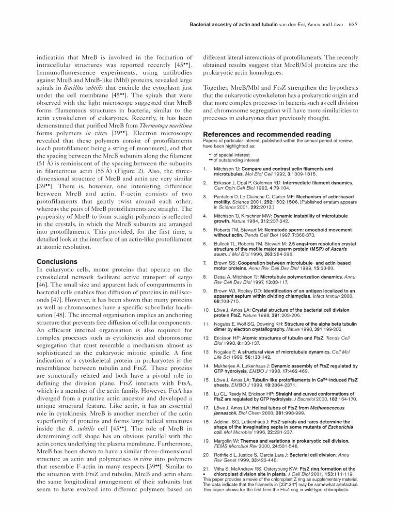
Figure 2
The atomic structure of a MreB protofilament(shown on the right) is reminiscent offilamentous actin (on the left) — both havetheir subunits in the same orientation,resulting in a similar longitudinal repeat. TheMreB protofilament is observed in the crystalstructure [39••] (PDB entry 1JCF) and theF-actin is based on the original model [31].Figure reprinted by permission from Nature(http://www.nature.com) 413:39-44,copyright 2001 Macmillan Magazines Ltd.
55 Å
F-actin MreB
11A3
4IIB
IIA
2 IB
51.1 Å
Bacterial ancestry of actin and tubulin van den Ent, Amos and Löwe 637
indication that MreB is involved in the formation ofintracellular structures was reported recently [45••].Immunofluorescence experiments, using antibodiesagainst MreB and MreB-like (Mbl) proteins, revealed largespirals in Bacillus subtilis that encircle the cytoplasm justunder the cell membrane [45••]. The spirals that wereobserved with the light microscope suggested that MreBforms filamentous structures in bacteria, similar to theactin cytoskeleton of eukaryotes. Recently, it has beendemonstrated that purified MreB from Thermotoga maritimaforms polymers in vitro [39••]. Electron microscopyrevealed that these polymers consist of protofilaments(each protofilament being a string of monomers), and thatthe spacing between the MreB subunits along the filament(51 Å) is reminiscent of the spacing between the subunitsin filamentous actin (55 Å) (Figure 2). Also, the three-dimensional structure of MreB and actin are very similar[39••]. There is, however, one interesting differencebetween MreB and actin. F-actin consists of twoprotofilaments that gently twist around each other,whereas the pairs of MreB protofilaments are straight. Thepropensity of MreB to form straight polymers is reflectedin the crystals, in which the MreB subunits are arrangedinto protofilaments. This provided, for the first time, adetailed look at the interface of an actin-like protofilamentat atomic resolution.
ConclusionsIn eukaryotic cells, motor proteins that operate on thecytoskeletal network facilitate active transport of cargo[46]. The small size and apparent lack of compartments inbacterial cells enables free diffusion of proteins in millisec-onds [47]. However, it has been shown that many proteinsas well as chromosomes have a specific subcellular locali-sation [48]. The internal organisation implies an anchoringstructure that prevents free diffusion of cellular components.An efficient internal organisation is also required forcomplex processes such as cytokinesis and chromosomesegregation that must resemble a mechanism almost assophisticated as the eukaryotic mitotic spindle. A firstindication of a cytoskeletal protein in prokaryotes is theresemblance between tubulin and FtsZ. These proteinsare structurally related and both have a pivotal role indefining the division plane. FtsZ interacts with FtsA,which is a member of the actin family. However, FtsA hasdiverged from a putative actin ancestor and developed aunique structural feature. Like actin, it has an essentialrole in cytokinesis. MreB is another member of the actinsuperfamily of proteins and forms large helical structuresinside the B. subtilis cell [45••]. The role of MreB indetermining cell shape has an obvious parallel with theactin cortex underlying the plasma membrane. Furthermore,MreB has been shown to have a similar three-dimensionalstructure as actin and polymerises in vitro into polymersthat resemble F-actin in many respects [39••]. Similar tothe situation with FtsZ and tubulin, MreB and actin sharethe same longitudinal arrangement of their subunits butseem to have evolved into different polymers based on
different lateral interactions of protofilaments. The recentlyobtained results suggest that MreB/Mbl proteins are theprokaryotic actin homologues.
Together, MreB/Mbl and FtsZ strengthen the hypothesisthat the eukaryotic cytoskeleton has a prokaryotic origin andthat more complex processes in bacteria such as cell divisionand chromosome segregation will have more similarities toprocesses in eukaryotes than previously thought.
References and recommended readingPapers of particular interest, published within the annual period of review,have been highlighted as:
• of special interest••of outstanding interest
1. Mitchison TJ: Compare and contrast actin filaments andmicrotubules. Mol Biol Cell 1992, 3:1309-1315.
2. Eriksson J, Opal P, Goldman RD: Intermediate filament dynamics.Curr Opin Cell Biol 1992, 4:79-104.
3. Pantaloni D, Le Clainche C, Carlier MF: Mechanism of actin-basedmotility. Science 2001, 292:1502-1506. [Published erratum appearsin Science 2001, 292:2012.]
4. Mitchison TJ, Kirschner MW: Dynamic instability of microtubulegrowth. Nature 1984, 312:237-242.
5. Roberts TM, Stewart M: Nematode sperm: amoeboid movementwithout actin. Trends Cell Biol 1997, 7:368-373.
6. Bullock TL, Roberts TM, Stewart M: 2.5 angstrom resolution crystalstructure of the motile major sperm protein (MSP) of Ascarissuum. J Mol Biol 1996, 263:284-296.
7. Brown SS: Cooperation between microtubule- and actin-basedmotor proteins. Annu Rev Cell Dev Biol 1999, 15:63-80.
8. Desai A, Mitchison TJ: Microtubule polymerization dynamics. AnnuRev Cell Dev Biol 1997, 13:83-117.
9. Brown WJ, Rockey DD: Identification of an antigen localized to anapparent septum within dividing chlamydiae. Infect Immun 2000,68:708-715.
10. Löwe J, Amos LA: Crystal structure of the bacterial cell divisionprotein FtsZ. Nature 1998, 391:203-206.
11. Nogales E, Wolf SG, Downing KH: Structure of the alpha beta tubulindimer by electron crystallography. Nature 1998, 391:199-203.
12. Erickson HP: Atomic structures of tubulin and FtsZ. Trends CellBiol 1998, 8:133-137.
13. Nogales E: A structural view of microtubule dynamics. Cell MolLife Sci 1999, 56:133-142.
14. Mukherjee A, Lutkenhaus J: Dynamic assembly of FtsZ regulated byGTP hydrolysis. EMBO J 1998, 17:462-469.
15. Löwe J, Amos LA: Tubulin-like protofilaments in Ca2+-induced FtsZsheets. EMBO J 1999, 18:2364-2371.
16. Lu CL, Reedy M, Erickson HP: Straight and curved conformations ofFtsZ are regulated by GTP hydrolysis. J Bacteriol 2000, 182:164-170.
17. Löwe J, Amos LA: Helical tubes of FtsZ from Methanococcusjannaschii. Biol Chem 2000, 381:993-999.
18. Addinall SG, Lutkenhaus J: FtsZ-spirals and -arcs determine theshape of the invaginating septa in some mutants of Escherichiacoli. Mol Microbiol 1996, 22:231-237.
19. Margolin W: Themes and variations in prokaryotic cell division.FEMS Microbiol Rev 2000, 24:531-548.
20. Rothfield L, Justice S, Garcia-Lara J: Bacterial cell division. AnnuRev Genet 1999, 33:423-448.
21. Vitha S, McAndrew RS, Osteryoung KW: FtsZ ring formation at the • chloroplast division site in plants. J Cell Biol 2001, 153:111-119.This paper provides a movie of the chloroplast Z ring as supplementary material.The data indicate that the filaments in [23•,24•] may be somewhat artefactual.This paper shows for the first time the FtsZ ring in wild-type chloroplasts.

638 Growth and development
22. Stokes KD, McAndrew RS, Figueroa R, Vitha S, Osteryoung KW:Chloroplast division and morphology are differentially affected byoverexpression of FtsZ1 and FtsZ2 genes in Arabidopsis. PlantPhysiol 2000, 124:1668-1677.
23. McFadden GI: Skeletons in the closet: how do chloroplasts stay in• shape? J Cell Biol 2000, 151:F19-F21.See annotation to [24•].
24. Kiessling J, Kruse S, Rensing SA, Harter K, Decker EL, Reski R: • Visualization of a cytoskeleton-like FtsZ network in chloroplasts.
J Cell Biol 2000, 151:945-950.Papers [23•,24•] show an interesting array of filaments underlying chloroplastmembrane when labelled FtsZ was expressed. These papers are the firstreports to show the distribution of overexpressed FtsZ in chloroplasts. Seealso annotation to [21•].
25. Osteryoung KW, Stokes KD, Rutherford SM, Percival AL, Lee WY:Chloroplast division in higher plants requires members of twofunctionally divergent gene families with homology to bacterialFtsZ. Plant Cell 1998, 10:1991-2004.
26. Erickson HP: FtsZ, a tubulin homologue in prokaryote cell division.Trends Cell Biol 1997, 7:362-367.
27. Beech PL, Nheu T, Schultz T, Herbert S, Lithgow T, Gilson PR, • McFadden GI: Mitochondrial FtsZ in a chromophyte alga. Science
2000, 287:1276-1279.Despite the absence of FtsZ from most mitochondria, the mitochondria ofthe alga Mallomonas splendens still contain FtsZ. This is the first paper toshow that some mitochondria still have FtsZ, whereas mitochondria of morecomplex organisms rely on dynamin for their division.
28. Erickson HP: Dynamin and FtsZ: missing links in mitochondrialand bacterial division. J Cell Biol 2000, 148:1103-1105.
29. Hagan IM, Petersen J: The microtubule organizing centers ofSchizosaccharomyces pombe. Curr Top Dev Biol 2000, 49:133-159.
30. Smith LG: Building walls on the right places. Nat Rev Mol Cell Biol2001, 2:33-39.
31. Holmes KC, Popp D, Gebhard W, Kabsch W: Atomic model of theactin filament. Nature 1990, 347:44-49.
32. Kabsch W, Holmes KC: Protein motifs 2. The actin fold. FASEB J1995, 9:167-174.
33. Kabsch W, Mannherz HG, Suck D, Pai EF, Holmes KC: Atomicstructure of the actin DNase I complex. Nature 1990, 347:37-44.
34. McLaughlin PJ, Gooch JT, Mannherz HG, Weeds AG: Structure ofgelsolin segment-1-actin complex and the mechanism of filamentsevering. Nature 1993, 364:685-692.
35. Schutt CE, Myslik JC, Rozycki MD, Goonesekere NCW, Lindberg U: Thestructure of crystalline profilin beta-actin. Nature 1993, 365:810-816.
36. Chik JK, Lindberg U, Schutt CE: The structure of an open state ofbeta-actin at 2.65 angstrom resolution. J Mol Biol 1996, 263:607-623.
37. Otterbein LR, Graceffa P, Dominguez R: The crystal structure ofuncomplexed actin in the ADP state. Science 2001, 293:708-711.
38. Flaherty KM, Delucaflaherty C, McKay DB: 3-dimensional structureof the ATPase fragment of a 70k heat-shock cognate protein.Nature 1990, 346:623-628.
39. van den Ent F, Amos LA, Löwe J: Bacterial origin of the actin •• cytoskeleton. Nature 2001, 413:39-44.The bacterial protein MreB is shown to polymerise into actin-like protofilaments,and its three-dimensional structure reveals its close relationship to actin.The crystals structure shows, for the first time, actin-like protofilaments atatomic detail.
40. van den Ent F, Löwe J: Crystal structure of the cell division protein • FtsA from Thermotoga maritima. EMBO J 2000, 19:5300-5307.FtsA is the second protein in bacterial cytokinesis that is structurally similarto a key component of the eukaryotic cytoskeleton.
41. Bork P, Sander C, Valencia A: An ATPase domain common toprokaryotic cell cycle proteins, sugar kinases, actin, and Hsp70heat shock proteins. Proc Natl Acad Sci USA 1992, 89:7290-7294.
42. Wachi M, Doi M, Tamaki S, Park W, Nakajima-Iijima S, Matsuhashi M:Mutant isolation and molecular cloning of mre genes, whichdetermine cell-shape, sensitivity to mecillinam, and amount ofpenicillin-binding proteins in Escherichia coli. J Bacteriol 1987,169:4935-4940.
43. Doi M, Wachi M, Ishino F, Tomioka S, Ito M, Sakagami Y, Suzuki A,Matsuhashi M: Determinations of the DNA sequence of the mrebgene and of the gene products of the mre region that function information of the rod shape of Escherichia coli cells. J Bacteriol1988, 170:4619-4624.
44. Levin PA, Margolis PS, Setlow P, Losick R, Sun DX: Identification ofBacillus subtilis genes for septum placement and shapedetermination. J Bacteriol 1992, 174:6717-6728.
45. Jones LJF, Carballido-Lopez R, Errington J: Control of cell shape in •• bacteria: helical, actin-like filaments in Bacillus subtilis. Cell 2001,
104:913-922.Immunofluorescence studies using antibodies against MreB and MreB-likeproteins show large structures inside the cell that control cell shape. This isthe first indication that bacteria contain a cytoskeletal-like structure underneaththe cell membrane.
46. Allan V: Membrane traffic motors. FEBS Lett 1995, 369:101-106.
47. Elowitz MB, Surette MG, Wolf PE, Stock JB, Leibler S: Proteinmobility in the cytoplasm of Escherichia coli. J Bacteriol 1999,181:197-203.
48. Shapiro L, Losick R: Dynamic spatial regulation in the bacterial cell.Cell 2000, 100:89-98.
49. Löwe J, Li H, Downing KH, Nogales E: Refined structure ofalpha-beta tubulin from zinc-induced sheets stabilized with taxol.J Mol Biol 2001, in press.