bacterial diseases of turkeys and ducks - cairo...
TRANSCRIPT

Bacterial Diseases of Turkeys and Ducks
Dr Wafaa Abd El-GhanyAssistant Professor of Poultry Diseases
Poultry Diseases DepartmentFac Vet Med
Cairo Univ
Bacterial diseases of Turkeys 1 Mycoplasma gallisepticum (MG)2 Mycoplasma meleagridis (MM)3 Maycoplasma synoviae (MS)4 Mycoplasma iowae (MI)5 Turkey coryza (Bordetellosis)6 Ornithobacterium rhinotracheal (ORT)7 Erysiplas infection8 Avian chlamydiosis9 Fowl cholera10 Avian salmonellosis (Paratyphoid infection Arizona disease and
Pullorum disease)11 Ecoli infection12 Clostridial infections13 Staphylococcus and Streptococcus infections14 Avian Tuberculosis
Bacterial diseases of Ducks
1 Rimerella anatipestifer
2 Botulism (Western duck sickness Limber neck)
3 Paratyphoid infection
4 Avian chlamydiosis
5 Fowl cholera
6 Mycoplasma immitans amp ansaris
7 Avian spiroketosis
8 Ecoli infection
9 Staphylococcus and Streptococcus infections
MYCOPLASMA GALLISEPTICUM
(MG)
(Infectious sinusitis of turkeys)
[Chronic respiratory disease (CRD) of chickens]
Mycoplasma gallisepticum (MG)
MG infections are Respiratory affections ofslow development and long coursecharacterized by Respiratory ralescoughing nasal discharge andconjunctivitis in chickens Swolleninfraorbital sinus is frequently occurred inturkeys
MOI amp Transmission of MG
Vertical infectionFrom infected hens to the embryos causing
embryonic mortalities or infection of day old chicks
Horizontal infectionThrough aerosol (air born) droplet infectionDirect or indirect contact of susceptible birds to
clinical or subclinical infected birds Mechanical (Fomites)workers free living birds utensils visitors cars
feed bags etchellip
Signs of MG
In turkeys (Infectious sinusitis)
1) Unilateral or bilateral swelling of theinfraorbital sinuses with facial swelling
2) Partial or complete closure of eye fromsevere sinus swelling
3) Nasal discharge with foamy eye secretion
4) Tracheal rales coughing and laboredbreathing
Signs of MG
5) Decreased feed intake and weight loss
6) MG can induce encephalitic form in 12-16 weeks old commercial meat turkeywith torticollis and opithotenous
7) Breeder flocks show drop in eggproduction
Lesions of MG
MG without complications
1) Catarrhal rhinitis sinusitis (turkeys)conjunctivitis tracheitis and broncheitis
2) Lung congestion and pneumonia
3) Mild pericarditis perihepatitis andairsacculitis
Lesions of MG
MG with complications
1) Seofibrinous or fibrinous (casous orpurulent) pericarditis perihepatitis andairsacculitis
2) Peritonitis
3) Salpingitis
4) Lung congestion and pneumonia
Swelling of the
infraorbital sinuses
Mild tracheitis with a small amount of mucoid exudate in the tracheal lumen
Congestion of the trachea and catarrhal exudate
Plugs of exudate in the lumen of the
trachea and bronchi
Mild airsacculitis with light presence of
foamAirsacculitis with caseous exudate
Severe airsacculitis with abundant foam and aggregates
of caseous exudate
Severe perihepatitis and
pericarditis
Fibrinous pericarditis and peritonitis
Severe perihepatitis and pericarditis
pericarditis and fibrinous pneumonia
Fibrinous pericarditis
Pneumonia
MYCOPLASMA
MELEAGRIDIS
(MM)
Mycoplasma meleagridis (MM)
It is a specific pathogen of turkeys causingegg transmitted disease (venereal) withprimary lesions in air sacs with decreasehatchability skeletal abnormalities andpoor performance
Airsaculitis deficiency syndrome (TS-65)1) Bowing2) Twisting and shortening of the tarsometatarsal
bone3) Hock joint swelling4) Deformity of the cervical vertebrate5) Lameness 6) Stunting and abnormal feathering 7) Retardation in growth8) Late embryonic mortality9) Respiratory signs
Signs of MM
Lesions of MM
1) Skeletal deformities (tibial dyschondroplasia and chondrodystrophy)
2) Sternal bursitis and synovitis
3) Airsacculitis (thickening of air sac walls with adherence of yellow exudates)
4) Sinusitis
MYCOPLASMA SYNOVIAE
(MS)
Mycoplasma synoviae (MS)
MS induces acute or chronic infectioussynovitis in chickens and turkeys which ischaracterized by exudative synovitis andtenovaginitis or sternal bursitis (breastblister)
MS can also induce upper respiratoryinfection (air sac disease)
Signs of MS
1) Swellings around joints especially hocks footpad and the sternal bursa is enlarged (turkeys) (breast blister)
2) Lameness3) Mild respiratory signs4) Slight drop egg production and egg quality
Retardation in growth5) Pale comb6) Greenish discoloration of dropping with large
amount of ureates7) Mortality 10
Lesions of MS
1) The synovial membranes of tendon sheaths of affected joints and keel bursa have a viscous yellow exudates (fibrinopurelent in turkeys)
2) Articular surfaces become eroded3) Sternal bursitis (breast blister) in turkeys4) Caseous exudates may be found in air
sacs5) Enlarged liver and spleen kidneys usually
swollen mottled and pale
ORGAN Leg of bird
LESIONS Arthritis
SUSPDIS Viral Arthritis (REO)
Swollen hock with yellow exudate
Airsacculitis
Sternal bursitis
Diagnosis of Mycoplasmosis Swabs organs are taken from the respiratory system joint
exudate Samples could be taken from the semen or fertile eggs from
adult suspected breeder flocks Mycoplasma species are fastidious organisms due to that
they difficult to grow and need long time for growth Mycoplasma species need enriched media like PPLO (pleuro-
pneumonia like organism) media or modified Freyrsquos FMmedia
Positive agar culture appears as a characteristic Fried eggshaped appearance (tiny smooth rounded colourlesstranslucent mass dense dark rough central area surroundedby flat translucent hallow zone) colonies under dissecting orsteromicroscope
Serum plate agglutination (SPA) test
On the clean porcelain or glass plate add one drop of MG or MS specific coloured (stained) antigen then add on drop of the serum of the bird
Mix well and rotate the plate in a circular manner
Positive reaction appears with in 2 minutes as the form of agglutination (flocculation or granulation) suspected cases should be confirmed using HI test
flock test
Prevention of Mycoplasmosis
Adopt all biosecurity measures
Egg sanitation and hatchery sanitation
Regular serological flock monitoring
II In case of MM
Examination of the phallus and cloacae ofadult male turkeys for MM serologicallybacteriologically and molecularly before thebreeding season
Using non-infected (non genetically carriers)birds for breeding (mating)
Prevention of Mycoplasmosis
Vaccination1 Moderately virulent strain (F) strain2 685 strain3 Ts-11 strain4 New K 5054 strain5 Naturally occurring virulent strains (S6)6 Inactivated vaccines (bacterins) Vaccination with live MG vaccine between 12
and 16 weeks of age Vaccination can be carried out a 2-4 weeks of
age
Control of mycoplasmosis
1 Separate diseased from healthy birds
2 Hygienic disposal of dead birds (burning or burring)
3 Thorough cleaning and disinfection
4 Treatment of birds Medication helps only to controlclinical signs and lesions and slow the spread ofinfection but it doesnt eliminate the infection
Complete elimination of mycoplasmas from all birds inthe infected flocks using medication is not expected asthe treatment is regarded as a method of short-termamelioration of the disease rather than as long termsolution of the problem
Control of mycoplasmosis
use Teteracyclines Macroilds (erythromycin tylosinspiramycin lincomycin kitasamycin) Pleuromulins(tiamulin) and fluoroquinolones (norfloxacinenrofloxacin and danofloxacin)
Egg treatment
A) Egg inoculation Any antimycoplasmal antibiotics isinjected into the egg cell this method may kill theembryo
B) Egg dipping the eggs are warmed to 37-38C thenimmersed in cold antibiotic solution (1-4C) for 20minutes Due to the temperature differences cooling ofthe egg contents and the antibiotic is pulled throughthe egg shell
Control of mycoplasmosis
C) Pressure differential vacuum (system) by decreasingthe pressure above the egg 25 Cm Hg by vacuumpump then the pressure return slowly to theatmospheric one This method is not completelyeliminate the organism but not affect on thehatchability
D) Heat forced incubator the eggs are heated inforced air incubator for 40C then putted at roomtemperature for cooling at 256C It is the mosteffective method but it reduces the hatchability to 8-12 with high embryonic mortalities
BORDETELLOSIS
(Turkey coryza)
Bordetellosis(Turkey coryza)
It is a highly contagious upper-respiratory tractdisease affect young turkeys caused by Bordetellaavium and is characterized by inflammation anddistortion of the respiratory mucosa
The economic importance including impairedgrowth Mortalities and the losses are resultingfrom Colisepticemia secondary or complicatedinfections and stressors
Transmission of B Avium
Close contact with infected poults or throughexposure to litter or water
Infection is transmitted by aerosol transmission
The severity of bordetellosis is exacerbated byadverse environmental and infectious factors(Temp humidity litter quality Ecoli)
Signs of B Avium
An abrupt onset of sneezing (snick) in a high percentage of 2-6 week-old turkeys over the course of a week
Older turkeys may also develop a dry cough
A clear nasal discharge
During the first 2 weeks of disease the nares and feathers of the head and wings become crusted with wet tenacious brownish exudate and some birds develop submaxillary edema
Signs of B Avium
Foamy exudate at the medial canthus of the eye
Open-mouth breathing and dyspnea and altered vocalization in the second week of clinical signs result when the nasal cavity and upper trachea become partially occluded with mucoid exudate
Poor weight gains In turkeys 2-6 weeks of age morbidity 80-100
whereas the mortality rate is less than 10 High mortality rates (gt40) in young turkeys
frequently are associated with concurrent isolation of Escherichia coli
Lesions of B Avium
Nasal and tracheal exudates varies from serous initially to tenacious and mucoid during the course of disease
Generalized softening and distortion of the cartilaginous rings of the trachea dorsal-ventral compression
In cross-section tracheal rings appear to have thick walls and a diminished lumen
Diagnosis of B Avium
Samples collected from the choanal opening and nostril or by passing a swab into the trachea through the larynx
Isolation and identification of bacterial agent isolation is accomplished on MacConkey agar
Serologic testing has proven to be useful a microagglutination test (MAT)
Prevention of B Avium
Strict biosecurity measures are required toprevent infection of clean flocks
Vaccines available commercially for theprevention of bordetellosis are (ts) mutant of Bavium and a whole cell bacterin
Vaccination of breeder hens with killed bacterinsdelayed the onset and severity of clinicaldisease in challenge-exposed poults
Passive immunization of 3-week-old poults withconvalescent serum
Control of B Avium
Treatment of bordetellosis with antibioticsadministered in the water by injection orby aerosol has produced minimal clinicalimprovement in most cases
Treatment of an infected breeder flockwith tetracycline-HCl and potassiumpenicillin-G for 3 days produced clinicalimprovement within 24 hours
ORNITHOBACTERIUM RIHINOTRACHEAL
(ORT)
ORT Infection amp Transmission
It is a contagious bacterial disease affectschickens and turkeys characterized by respiratorysigns variable mortality and decrease in eggproduction with change in egg quality
The infection occurred by the horizontal routes bycontact through aerosol or drinking water
The vertical transmission occurs by transovarianand oviduct
Signs of ORTIn turkeysA Poults Turkey poults aged 2-8 weeks show mortality
up to 50 Respiratory signs coughing sneezing nasal
discharge and sinusitis Decrease in feed and water intakeB Breeders Decrease in egg production Increase in the number of unhatched eggs
Lesions of ORT
Uni or bilateral pneumonia
Yoghurt like airsaculitis
Pleuritris
Subcutaneous edema of the head
Enlarged liver and spleen with degeneration of heart muscles are additionally seen in turkeys
Thickened opaque air sacs with
profuse foamy white to yellow ldquoyogurt-
likerdquo
Pneumonia and pleuritis in turkeys
Prevention amp control of ORT
1) Application of good sanitary measures especially separation of different ages and species
2) Vaccination
a Inactivated vaccine considered as serotype specific vaccine
b Live vaccine can induce cross protection between some serotypes
Amoxicillin and chlortetracycline in water
Tetracycline and penicillins by injection
ERYSIPELAS
Erysipelas
Erysipelas (Red Skin) occurs in growing turkeys geese chickens quail and Peafowl between 47 months
This acute to chronic disease is caused by the Gram-positive rod shaped bacterium Erysipelothrix rhusiopathiae It is non-spore forming non-acid fast and non-motile
Infection amp transmission of Erysipelas
It can be transmitted through a break in the skin or mucous membranes or fighting between males
It is a soil-borne organism and can also be spread by cannibalism or biting flies
Contaminated fish-meal is also a source of infection
Signsamp lesions of Erysipelas
Signs The incubation period is 2-3 days Swollen snoods (turkeys) and
shock Diarrhea emaciation weakness anemia skin haemorrhage and necrosis can be seen
Fever cyanotic toes and head drop in egg production and or fertility and embryonic mortality can occur
Post mortem lesions Enlarged friable purple-black spleen breast muscle haemorrhage
oral mucous Haemorrhage in muscles spleen lungs fat and small intestine and endocarditis may be seen
Fibrinopurulent Exudate in the joints Thickening of walls or proventriculus or gizzard ulceration and
yellow nodules can occur in the caeca
Swollen snood Turgid snood
Enlarged and mottled spleen (marbled spleen) )
Endocarditis inflammatory process around valves
Diagnosis of Erysipelas
Laboratory isolation from lesions is important and can show smooth colonies colorless to a bluish gray or pin-point size with smooth edges Haemorrhagic swollen spleen and wicked red lesions are diagnosed
It Simulates cholera Salmonella gangrenous dermatitis aspergillosis and E coli
Blood agar tiny pinpoint colonies round and translucent
Prevention and Control of Erysipelas
Prevention Vaccinate birds twice one at 1012 weeks and again at
14-16 weeks Debeak at day one and pooling is done if necessary to
prevent fighting No pigs should be reared near poultry and rotation of
the turkey range to reduce bacteria
Treatment Gallimycin and penicillin can reduce signs Disinfect premises with aerosol phenols or iodine
ARIZONOSIS
Arizonoses
Arizonosis occurs young turkeys It is caused by Bacterium-Salmonella Arizona Gram negative flagellate bacterium Salmonella
Arizona
The effects are the same as for other Salmonella infections- Diarrhoea lameness somnolence (sleepiness) laboured breathing blindness and mortality Mortality up to 100 peaking at 7-10 days Tremors convulsions and twisted necks may also be seen
Infection amp transmission of Arizona
The disease is transmitted in the same way as other salmonella species ie from bird to bird and between farms
Common vectors include birds rodents and sometimes reptiles
Spread via the transovarian route can also occur
Signs and lesions of Arizona
Signs are the same as for other Salmonella Opaque eyes (blindness) tremors convulsions and twisted necks may be seen
Postmortem lesions
Lesions are the same as for Salmonella pullorum which include bacteria septicaemia peritonitis and retained yolk sacs
Congested (filled with blood) duodenum mottled (white necrotic spots) liver caseous plugs in caeca and caseous air sacs can be seen
Round heart (Characteristic)
Diagnosis of Arizona
The organisms must be cultured from post mortem lesions egg yolk etc on brilliant green agar for a definitive diagnosis
Agglutination or ELISA tests using sera from breeders can confirm the presences of S Arizona
It simulates pullorum E coli and typhoid
Prevention amp Control of Arizona
Prevention Biosecurity measures Bacterin for turkey breeders prevents egg transmission Egg and hatchery sanitation are important Breeders should be tested and those that are serologically
positive should be slaughtered Vector control helps control spread of the organismTreatment Treatments will reduce clinical problems but birds will
remain carriers SQ and Ormetoprin in the water and furazolidone in the feed are effective treatments Gentamicin and spectinomycin can be given to day-old-chicks by injection
RIMERELLA (PASTEURELLA) ANATIPESTIFER
(RA)
(DUCK SEPTICAEMIA INFECTIOUS SEROSITIS NEW DUCK SYNDROM)
Riemerella anatipestifer (RA)
It is an acute septicaemic or chroniccontagious disease of growingduckling characterized byserofibrinous pericarditisperihepatitis and airsacculitiscaseous salpingitis and meningitisand high mortality in ducklings(75)
Susceptibility amp Infection of RA
Ducklings aged 1-8 weeks are highly susceptible
The disease in laying birds is rare
Inhalation of infected droplets
Wound infection
Direct and indirect contact
Carrier birds
Signs of RA
Respiratory signs (nasal and ocular discharges sneezing and coughing)
Nervous signs (Ataxia tremors of the head and neck and coma)
Greenish diarrhea
Survival ducks may stunted
Mortality ranged from 5 to75 in relation to other factors as age route of infection and the strain virulence
Lesions of RA
Fibrinous pericarditis Fibrinous perihepatitis Fibrinous airsacculitis Caseous salpingitis fibrinopurulant pneumonia Arthritis Fibrinous meningitis Enlarged and mottled spleen In the chronic form the most predominant lesions in the
skin in the form of necrotic dermatitis on the lower back or around vent with yellowish exudates between skin and the fat layer
Central nervous system infection
Caseous exudate located in the head
region (subcutis)
Submandibulare oedema
Spinal cord compression Vertebral osteomyelitis
(spondylitis)
perihepatitis
Diagnosis of RA
Samples from heart blood brain pericardial exudates air sacs lungs liver and oviduct
Detection of bipolar organism in blood or tissue smear stained by Gimsa
Prevention amp Control of RA Sanitary measures
Avoid stress factors
Prevent contact of the birds with different species sources and ages
Inactivated vaccine must be containing a serotype specific to that cause endemic infection or frequent infection in the farm
Live RA vaccine given by drinking water or spray to day old birds can induces protection till 42 days of age
Sulfamethazine (02 -025) in water or feed sulfaquinoxaline (0025-005) in feed and lincomycin (0011-0022 ) in water are effective
BOTULISM (LIMBER NECK)
WESTERN DUCK SICKNESS
Botulism
Cause
Age group affected
Transmission
Clostridium botulinum
Most avian esp ducks
Ingestion of toxin
Insects or maggots
Feeding on dead decayed carcass
signs of botulism Paralysis of legs wings neck and eye lids are
predominant Paralysis progressed cranially fromlegs to eye lids
Affected bird appear firstly sitting on hocksreluctant to move or appear lame droopedwings extended neck ruffled feathers andfallout with handling
Broiler chickens are showing diarrhea withexcess ureates in the loose droppings
Mortality and morbidity are related to theamount of ingested toxin
Death 12-24 hours
Lesions of botulism
There is no gross or microscopic lesions
Maggots of fly blown carcasses or feathers can be found in crop of dead or affected birds
Control of botulism
No treatment but rapid withdrawal of thetoxicated food
Episons salt as purgative to removedecomposed food from the intestine
use sodium selenite and vitamin AD3E Antibiotics including Bacitracin 100gton in
feed streptomycin 1 gl of water may be ofvalue
Also Inoculation of antitoxin neutralizes onlyfree and extracellular bound toxin and can beused for treatment of valuable birds
MYCOPLASMA IMMITANS
Mycoplasma immitans
Infection with Mycoplasma immitans is seen in ducks and geese
Signs
Swollen sinuses
Post-mortem lesions
Sinusitis
PARATYPHOID
INFECTION (PT)
Paratyphoid (PT) Infection
It is infection of turkeys and duckswith motile salmonella serotypesrather than Salmonella gallinarumpullorum (S Typhimurium Senteritidis S anatum etchellip) resultingin relatively asymptomatic intestinalcarrier or inducing clinical diseaseThe disease in ducks is caused by S
anatum
MOI amp Transmission of PT
Egg shell contamination (false egg bornedisease)
Direct contact
Ingestion of contaminated food and water
Rodents animals wild birds and flies
Human
Contaminated animal protein supplements(Bone meal meet meal and fish meal)
Signs of PT in Ducks
1) Arthritis
2) The bird is unable to move or stand
3) Diarrhea
4) Nervous manifestation
Trembling or shivering
Kicking and violent movement
Move in circle
Keeling over backward with leg paddling (Keeldisease)
1) Necrotic foci on the liver
2) Distended gall bladder
3) Enteritis
4) The ureter is distended with ureates
5) Enlargement and congestion of the internalorgans
6) Pericarditis and perihepatitis
7) Degeneration of the ovary
Lesions of PT in Ducks
Prevention of PT
Sanitation and sound measures in poultryhouses egg store and hatcheries
Administrations of probiotics are exhibitingactivity against various PT salmonellae inthat diminish both intestinal colonizationand subsequent invasion to internal tissues(Competitive Exclusion)
Trials for preparation of killed or livevaccines
AVIAN CHLAMYDIOSIS
(ORNITHOSIS)
Avian chlamydiosis
Avian chlamydiosis is acute fatal or chronic infectiouszonootic respiratory disease of domestic wild andmigratory birds
The disease is characterized by nasal and oculardischarge diarrhea loss of weight drop in eggproduction and the mortality rate reached to 5-30
Presence of intracytoplasmic elementary bodies (LCL)is characteristic to the organism
Giemsa stain intracellular chlamydial inclusion
(reddish-purple)
Infection amp Transmission of Chlamydiosis
Chlamydia Psittaci infection occur horizontally either by inhalation of infected dust or droplet or by ingestion of contaminated feed and water with infected droppings
There is an evidence of low percentage of vertical transmission through eggs (transoverian) in ducks and sea gulls
Chronic carriers transmit the organism without signs
Biting of insects as ticks lice and mites
Wild birds are important in transmission
Signs of chlamydiosis
In turkeysI Acute epidemic Form (toxigenic highly virulent strain)Clinical signs appear as systemic fatal infection with1 Sudden death of 5-302 Fever and anorexia3 Nasal and ocular discharge swollen eye lids
conjunctivitis and sinusitis4 Off food and loss of weight (emaciation)5 Yellow green gelatinous diarrhea6 Rapid drop in egg production (40-50 decline)7 Morbidity rate may be reach 50-80
In ducks and geese (water fowl)
In duckling there are tremors trembling and staggered gait (incoordination)
Ocular and nasal discharge
Greenish watery Diarrhea
Inappetance
Emaciation convulsion and death
Morbidity rate 10-80
Mortality rate 1-30 (depending on the age health status and secondary infection with salmonella species)
Signs of chlamydiosis
Lesions of chlamydiosis
Congestion of the visceral organs
Congestion and pneumonia of the lungs
All the body cavities are filed with fibrinous exudates
Fibrinous pericarditis perihepatitis and airsacculitis and peritonitis
Catarrhal enteritis
Liver and spleen are enlarged dark and covered with gray white foci
Liver necrosisFibrinous pericarditis
liver hepatomegaly and congestion
fibrinous pericarditis
Fibrinous pericarditis
Caseous pericarditisLungs congested and fibrinous exudate
in the pleural cavity
Necrosis and congestion of
spleen
Enlarged congested
liver
Prehepatitis
Airsacculitis
A Thickened
abdominal airsacs
totally covered
with fibrin plaques
B Serous fluid
and fibrin in the
pericardial sac
C Severe
hepatomegaly
Diagnosis of chlamydiosis1 Detection of dark purple blue or pink (acc To the
stain) ICytoplasmic inclusion (LCL) bodies in stained smear (from nasal or ocular exudates serous membranes trachea lungs pancreas liver spleen heart faecal and cloacal swabs) in different stages of elementary body
2 Tissue culture3 Embryonated chicken eggs4 Laboratory animal inoculation Intra peritoneal or intra
cranial or intra nasal inoculation of 3-4 weeks old mice (3-6 mice) could resulted in death of mice after 5-7 days post inoculation Intra peritoneal inoculation resulted in peritonitis with accumulation of fibrinous exudate in the peritoneal cavity with splenomegaly
Prevention of chlamydiosis1 Different avian species and ages must be reared away
from each other2 Newly purchased (imported) birds especially pets must
be quarantined for 35 days (if positive discarded ortreated with tetracyclines till recovery)
3 Prevent the introduction of birds from the enzootic areas4 Avoid the contact with the free living wild birds5 Thorough cleaning and disinfection (iodophors or
formaldhydes)6 Regular testing of the birds using serological tests7 Restrict the movement of people and visitors8 Trials for vaccine preparation
Control of chlamydiosis
1 Oxytetracyclin (15mgkg bwt) in the drinking water for a period for 5 days
2 Chlorotetracyclin
(treatment may be extended to 15-45 days in severe infection)
3 Usage of combined antibiotics to control other associated infections as Salmonellae and Ecoli(quinolones like cipro enro and danofloxacin) for 3-5 days in the drinking water
AVIAN PASTEURELLOSIS
FOWL CHOLERA (FC)
FC
Peracute acute and chronic highly contagious bacterial disease of domestic and wild birds caused by Pasteurella multocida
In the acute form the disease is characterized by rapid course high morbidity and mortality and septicaemic picture while the chronic form is localized and follows the acute form or occur independently due to low virulent mo
Susceptibility of FC
1 Natural hosts are water fowl (ducks and geese) turkeys and chickens
2 Water fowl are the most susceptible birds
3 Turkeys are more susceptible than chickens
4 Adult chickens (more than 16 weeks old) while young turkeys are the most susceptible ages
Infection and transmission of FC
1 Inhalation of infected droplets
2 Ingestion of contaminated food and water by the infected excretions from the mouth and nostrils of diseased or carriers
3 Mechanically clothes feet or hands of workers contaminated equipments feed bags etchellip
4 Wild birds rodents and pets
5 Cannibalism of diseased or dead birds
6 Wound infection
Signs of FC
1 Per acute form
Sudden death without clinical signs
No characteristic post mortem lesions
2 Acute form
High morbidity and mortality rates
Septicaemia (cyanosis of the comb and wattle)
Mucous discharge from the mouth and nostrils
Watery white or greenish mucoid diarrhea
Respiratory manifestations
Drop in egg production
Lesions of FC
Acute form Petechial or echymotic hemorrhages on the
lungs proventriculus abdominal and coronary fats and on serosal membranes as peritoneum and subepicardium and haemorrhagic enteritis
Tracheitis bronchitis and pneumonia (TURKEYS) Oophoritis flaccid regressed or ruptured ova Free yolk material in the abdominal cavity Serofibrinous pericarditis and peritonitis Large amount of viscous mucous in the larynx
crop and intestine
Edematous cyanotic comb and wattles
Mucous excretion
from mouth
Fibrinous pericarditis and perihepatitisEnlarged liver
Small areas of necrosis in the liver (corn meal liver)
Subepicardial hemorrhages and petechiae
on the heart and coronary fat
The left lung has a severe
accumulation of fibrinous exudate on
its pleural surface In turkeys
Hemorrhagic mucosal and serosal lesions
Signs amp lesions of FC
3 Chronic forms
A Wattle form
Unilateral or bilateral swelling of wattle
Caseous exudates in the wattles cut sections
B Otitis media form
Nervous signs as torticollis
Caseous exudates in the middle ears and air spaces of cranial bones
Signs amp lesions of FC
C Arthritic form Swelling of the joints and lameness Caseous exudates in the joints Inflammation of the tendon sheet Hemorrhages and erosions on the articular
cartilageD Ovarian form Drop in egg production Cheesy material around the ovary and
peritoneal cavity
Signs amp lesions of FC
E Roup or catarrhal form
Respiratory signs (rals and dyspnea)
Mucous discharge from the mouth
Nasal discharge
Swelling of the sinuses and facial oedema
Conjunctivitis
Swelling of wattle
Swelling of the wattles
Swelling of the ear canal (middle ear)
Torticollis
Torticollis
strenal
bursitis
Arthritis
Caseous exudate over the hock region and
food pad
Swelling of the sternal bursae
Hyperemia and flaccid mature follicles
Egg-yolk peritonitis
Sinusitis and facial oedema
Yellow caseous exudate in the air spaces of the cranial bones
Diagnosis of FC
Demonstration of bipolar P multocida mo
either from the blood film in acute stage afterstaining with Giemsa Leishmanrsquos stains andseen under microscope
or take impression smear from organs (liver)stain and examine microscopically
Bipolar Pmultocida in between nucleated RBCS in acute infection
Rough colony on the right and a smooth colony on the left
Diagnosis of FC
Laboratory animal inoculationIntraperitoneal or SC inoculation of rabbit or mice
or hamster with blood of infected bird orsuspected material (liver supernatant) (02 ml)induces death of these animals within 24-48 hourspost inoculation with septicaemic pictureImpression smear from blood or tissues (liver)shows bipolar organism
Prevention of FC Adopt all biosecurity measures
Vaccination
P multocida antigen vaccine should be homologous tothat cause fowl cholera or use of autogenous localprepared vaccine in endemic area or when commercialones are not protective
There are many types of vaccines against FC
Whole cell bactrine
Live attenuated vaccine
a CU- vaccine
b Mutants of CU-vaccine as M-9 MN and PM-1 strainsto avoid post vaccinal reaction of the CU
Prevention of FC
In turkeys
Meat type 6-8 weeks bactrine injection or CU via drinking water and repeated every 4-6 weeks till marketing
Breeder flocks must be vaccinated first at 6- 8 weeks and revaccinated 2-5 times till start of egg production according to epidemiology of FC in the area and farm
Control of FC
In chicken and turkeys sulfamerazine 02 in water or 04 in feed or Sulfaquinoxaline 001 ndash005 in water for 3-5 days to stop deaths in 2 days
Streptomycin (150 mg) for IlM injection in turkeys
Chloro- or oxytetracycline 40 mgkgbw by IlM
Chloramphenicol 20 mgkgbw via IlM
Enrofloxacin hellipetc
AVIAN SPIROKETOSIS
Avian spiroketosis
The bacterium Borrelia anserina infects chickens turkey geese ducks pheasants grouse and canaries with morbidity and mortality up to 100
It is transmitted by arthropods eg Argas persicus and occasionally by infected faeces The bacterium is poorly resistant outside host but may be carried by Argas persicus for 430 days
Brachyspira pilosicoli previously known as Serpulina pilosicoli is an intestinal spirochaete that can be associated with inflammation of the large intestine in a broad range of mammals and birds
It has been associated with typhilitis diarrhoea reduced egg production and egg soiling in chickens
Signs amp lesions of avian spiroketosis
Depression and thirst Cyanosis Often diarrhoea with excessive urates Weakness and progressive paralysis Drops in egg production may be seen in both systemic
and intestinal forms Post-mortem lesions Marked splenomegaly Spleen mottled with ecchymotic haemorrhages Liver enlarged with small haemorrhages Necrotic foci Mucoid enteritis
Prevention amp Control of avian spiroketosis
Treatment
Various antibiotics including penicillin
Prevention
Control vectors
Vaccines in some countries
Cause
Age group affected
Transmission
Forms of the disease
Escherichia coli
All types of avian and all ages
Fecal oral route transovarial contamination of the egg shell via fecal material from hen
Colisepticaemia
Airsac disease
Chronic forms (arthritis omphalitis panophthalmitis salpingitis enteritis coligranuloma and SHS)
E coli
Severely congested liver
Foci of necrosis on liver
GANGARINOUS
DERMATITIS
(GD)
GAS OEDEMA DISEASE
WING ROT
Gangrenous dermatitis
Gangrenous (necrotic) dermatitis in broilers and turkeys between 3-7 weeks old
It is caused by a bacteria- Clostridium septicum Escherichia coli Staphylococcus aureus
Disease often occurs in birds which are already immunosuppressed due to a prior infection (Chicken anaemia Infectious Bursal Disease) or mycotoxin ingestion
Transmission is by contact with infected wet caked litter The disease often occurs in immunosuppressed birds
Signs of GD
Loss of feathers
Pale combs and wattles depression
Incoordination
Leg weakness and ataxia (canrsquot move) can be seen
Mortality is low but dead birds decompose quickly
Lesions of GD
Congestion haemorrhage and necrosis of skin with intro lesion bacteria under the microscope
In turkeys with cellulitis of the tail (bubbly tail) edema and vesicle-like lesions were present laterally and ventrally around the tail
Tail feathers were soft blood-filled and broken
Dark and moist skin of the breast
Necrosis of the abdomen skinAffected leg and toes
Necrosis of the wattle
Ulceration and gangrenous formation of skin
Prevention amp Control of GD
Prevention Clean out house and add new litter to prevent the
disease Medicate in starter feed Flavomycin Virginamyciin
Bacitrcin and CTC can reduce bacteria Proper vaccination against IBDV CAV and MDV prevent
mycotoxin formation in the feed and eliminate ALV in the breeders to prevent immunosuppression
Treatment Erythromycin penicillin in the feed to treat signs
Chlortetracycine oxytetracycline copper sulphate can be added to the water to reduce morbidity
ULCERATIVE ENERITIS
(UE)
Ulcerative Enteritis
Turkeys of 6-14 weeks are susceptible to this acute to chronic disease
The agent involved in the aetiology of this disease is clostridium colinum which is spore-forming gram-positive aerobic and non-motile
Mode of transmission
Vectors are faeces soil and litter containing the bacteria
It often accompanies coccidiosis in broilers
Signs amp lesions of UE
High mortality watery diarrhoea ruffled feathers dull listlessness increased thirst emaciation and atrophy of pectoral muscles can occur
Postmortem lesions Yellow irregular ulcers on small intestine and
caeca hemorrhagic enteritis are seen Enlarged haemorrhagic necrotic spleen light
yellow mottling of liver and crop filled with water may occur
Prevention amp Control of UE
Prevention Improved sanitation Adding salt to the soil (500
lbs of salthouse) may kill spores Raising birds on wire andor feeding bacitracin at 50-100 gt will prevent the disease
Treatment NF-180 (50-100 gt) strephomycin (60 gt) and
chlortetracycline vitamins and minerals in water andor lincomycin 2 gt will reduce the signs
Remove dead birds and feed bacitracin (200 gt)
Thank you
ANY QUESTIONS

Signs of MG
5) Decreased feed intake and weight loss
6) MG can induce encephalitic form in 12-16 weeks old commercial meat turkeywith torticollis and opithotenous
7) Breeder flocks show drop in eggproduction
Lesions of MG
MG without complications
1) Catarrhal rhinitis sinusitis (turkeys)conjunctivitis tracheitis and broncheitis
2) Lung congestion and pneumonia
3) Mild pericarditis perihepatitis andairsacculitis
Lesions of MG
MG with complications
1) Seofibrinous or fibrinous (casous orpurulent) pericarditis perihepatitis andairsacculitis
2) Peritonitis
3) Salpingitis
4) Lung congestion and pneumonia
Swelling of the
infraorbital sinuses
Mild tracheitis with a small amount of mucoid exudate in the tracheal lumen
Congestion of the trachea and catarrhal exudate
Plugs of exudate in the lumen of the
trachea and bronchi
Mild airsacculitis with light presence of
foamAirsacculitis with caseous exudate
Severe airsacculitis with abundant foam and aggregates
of caseous exudate
Severe perihepatitis and
pericarditis
Fibrinous pericarditis and peritonitis
Severe perihepatitis and pericarditis
pericarditis and fibrinous pneumonia
Fibrinous pericarditis
Pneumonia
MYCOPLASMA
MELEAGRIDIS
(MM)
Mycoplasma meleagridis (MM)
It is a specific pathogen of turkeys causingegg transmitted disease (venereal) withprimary lesions in air sacs with decreasehatchability skeletal abnormalities andpoor performance
Airsaculitis deficiency syndrome (TS-65)1) Bowing2) Twisting and shortening of the tarsometatarsal
bone3) Hock joint swelling4) Deformity of the cervical vertebrate5) Lameness 6) Stunting and abnormal feathering 7) Retardation in growth8) Late embryonic mortality9) Respiratory signs
Signs of MM
Lesions of MM
1) Skeletal deformities (tibial dyschondroplasia and chondrodystrophy)
2) Sternal bursitis and synovitis
3) Airsacculitis (thickening of air sac walls with adherence of yellow exudates)
4) Sinusitis
MYCOPLASMA SYNOVIAE
(MS)
Mycoplasma synoviae (MS)
MS induces acute or chronic infectioussynovitis in chickens and turkeys which ischaracterized by exudative synovitis andtenovaginitis or sternal bursitis (breastblister)
MS can also induce upper respiratoryinfection (air sac disease)
Signs of MS
1) Swellings around joints especially hocks footpad and the sternal bursa is enlarged (turkeys) (breast blister)
2) Lameness3) Mild respiratory signs4) Slight drop egg production and egg quality
Retardation in growth5) Pale comb6) Greenish discoloration of dropping with large
amount of ureates7) Mortality 10
Lesions of MS
1) The synovial membranes of tendon sheaths of affected joints and keel bursa have a viscous yellow exudates (fibrinopurelent in turkeys)
2) Articular surfaces become eroded3) Sternal bursitis (breast blister) in turkeys4) Caseous exudates may be found in air
sacs5) Enlarged liver and spleen kidneys usually
swollen mottled and pale
ORGAN Leg of bird
LESIONS Arthritis
SUSPDIS Viral Arthritis (REO)
Swollen hock with yellow exudate
Airsacculitis
Sternal bursitis
Diagnosis of Mycoplasmosis Swabs organs are taken from the respiratory system joint
exudate Samples could be taken from the semen or fertile eggs from
adult suspected breeder flocks Mycoplasma species are fastidious organisms due to that
they difficult to grow and need long time for growth Mycoplasma species need enriched media like PPLO (pleuro-
pneumonia like organism) media or modified Freyrsquos FMmedia
Positive agar culture appears as a characteristic Fried eggshaped appearance (tiny smooth rounded colourlesstranslucent mass dense dark rough central area surroundedby flat translucent hallow zone) colonies under dissecting orsteromicroscope
Serum plate agglutination (SPA) test
On the clean porcelain or glass plate add one drop of MG or MS specific coloured (stained) antigen then add on drop of the serum of the bird
Mix well and rotate the plate in a circular manner
Positive reaction appears with in 2 minutes as the form of agglutination (flocculation or granulation) suspected cases should be confirmed using HI test
flock test
Prevention of Mycoplasmosis
Adopt all biosecurity measures
Egg sanitation and hatchery sanitation
Regular serological flock monitoring
II In case of MM
Examination of the phallus and cloacae ofadult male turkeys for MM serologicallybacteriologically and molecularly before thebreeding season
Using non-infected (non genetically carriers)birds for breeding (mating)
Prevention of Mycoplasmosis
Vaccination1 Moderately virulent strain (F) strain2 685 strain3 Ts-11 strain4 New K 5054 strain5 Naturally occurring virulent strains (S6)6 Inactivated vaccines (bacterins) Vaccination with live MG vaccine between 12
and 16 weeks of age Vaccination can be carried out a 2-4 weeks of
age
Control of mycoplasmosis
1 Separate diseased from healthy birds
2 Hygienic disposal of dead birds (burning or burring)
3 Thorough cleaning and disinfection
4 Treatment of birds Medication helps only to controlclinical signs and lesions and slow the spread ofinfection but it doesnt eliminate the infection
Complete elimination of mycoplasmas from all birds inthe infected flocks using medication is not expected asthe treatment is regarded as a method of short-termamelioration of the disease rather than as long termsolution of the problem
Control of mycoplasmosis
use Teteracyclines Macroilds (erythromycin tylosinspiramycin lincomycin kitasamycin) Pleuromulins(tiamulin) and fluoroquinolones (norfloxacinenrofloxacin and danofloxacin)
Egg treatment
A) Egg inoculation Any antimycoplasmal antibiotics isinjected into the egg cell this method may kill theembryo
B) Egg dipping the eggs are warmed to 37-38C thenimmersed in cold antibiotic solution (1-4C) for 20minutes Due to the temperature differences cooling ofthe egg contents and the antibiotic is pulled throughthe egg shell
Control of mycoplasmosis
C) Pressure differential vacuum (system) by decreasingthe pressure above the egg 25 Cm Hg by vacuumpump then the pressure return slowly to theatmospheric one This method is not completelyeliminate the organism but not affect on thehatchability
D) Heat forced incubator the eggs are heated inforced air incubator for 40C then putted at roomtemperature for cooling at 256C It is the mosteffective method but it reduces the hatchability to 8-12 with high embryonic mortalities
BORDETELLOSIS
(Turkey coryza)
Bordetellosis(Turkey coryza)
It is a highly contagious upper-respiratory tractdisease affect young turkeys caused by Bordetellaavium and is characterized by inflammation anddistortion of the respiratory mucosa
The economic importance including impairedgrowth Mortalities and the losses are resultingfrom Colisepticemia secondary or complicatedinfections and stressors
Transmission of B Avium
Close contact with infected poults or throughexposure to litter or water
Infection is transmitted by aerosol transmission
The severity of bordetellosis is exacerbated byadverse environmental and infectious factors(Temp humidity litter quality Ecoli)
Signs of B Avium
An abrupt onset of sneezing (snick) in a high percentage of 2-6 week-old turkeys over the course of a week
Older turkeys may also develop a dry cough
A clear nasal discharge
During the first 2 weeks of disease the nares and feathers of the head and wings become crusted with wet tenacious brownish exudate and some birds develop submaxillary edema
Signs of B Avium
Foamy exudate at the medial canthus of the eye
Open-mouth breathing and dyspnea and altered vocalization in the second week of clinical signs result when the nasal cavity and upper trachea become partially occluded with mucoid exudate
Poor weight gains In turkeys 2-6 weeks of age morbidity 80-100
whereas the mortality rate is less than 10 High mortality rates (gt40) in young turkeys
frequently are associated with concurrent isolation of Escherichia coli
Lesions of B Avium
Nasal and tracheal exudates varies from serous initially to tenacious and mucoid during the course of disease
Generalized softening and distortion of the cartilaginous rings of the trachea dorsal-ventral compression
In cross-section tracheal rings appear to have thick walls and a diminished lumen
Diagnosis of B Avium
Samples collected from the choanal opening and nostril or by passing a swab into the trachea through the larynx
Isolation and identification of bacterial agent isolation is accomplished on MacConkey agar
Serologic testing has proven to be useful a microagglutination test (MAT)
Prevention of B Avium
Strict biosecurity measures are required toprevent infection of clean flocks
Vaccines available commercially for theprevention of bordetellosis are (ts) mutant of Bavium and a whole cell bacterin
Vaccination of breeder hens with killed bacterinsdelayed the onset and severity of clinicaldisease in challenge-exposed poults
Passive immunization of 3-week-old poults withconvalescent serum
Control of B Avium
Treatment of bordetellosis with antibioticsadministered in the water by injection orby aerosol has produced minimal clinicalimprovement in most cases
Treatment of an infected breeder flockwith tetracycline-HCl and potassiumpenicillin-G for 3 days produced clinicalimprovement within 24 hours
ORNITHOBACTERIUM RIHINOTRACHEAL
(ORT)
ORT Infection amp Transmission
It is a contagious bacterial disease affectschickens and turkeys characterized by respiratorysigns variable mortality and decrease in eggproduction with change in egg quality
The infection occurred by the horizontal routes bycontact through aerosol or drinking water
The vertical transmission occurs by transovarianand oviduct
Signs of ORTIn turkeysA Poults Turkey poults aged 2-8 weeks show mortality
up to 50 Respiratory signs coughing sneezing nasal
discharge and sinusitis Decrease in feed and water intakeB Breeders Decrease in egg production Increase in the number of unhatched eggs
Lesions of ORT
Uni or bilateral pneumonia
Yoghurt like airsaculitis
Pleuritris
Subcutaneous edema of the head
Enlarged liver and spleen with degeneration of heart muscles are additionally seen in turkeys
Thickened opaque air sacs with
profuse foamy white to yellow ldquoyogurt-
likerdquo
Pneumonia and pleuritis in turkeys
Prevention amp control of ORT
1) Application of good sanitary measures especially separation of different ages and species
2) Vaccination
a Inactivated vaccine considered as serotype specific vaccine
b Live vaccine can induce cross protection between some serotypes
Amoxicillin and chlortetracycline in water
Tetracycline and penicillins by injection
ERYSIPELAS
Erysipelas
Erysipelas (Red Skin) occurs in growing turkeys geese chickens quail and Peafowl between 47 months
This acute to chronic disease is caused by the Gram-positive rod shaped bacterium Erysipelothrix rhusiopathiae It is non-spore forming non-acid fast and non-motile
Infection amp transmission of Erysipelas
It can be transmitted through a break in the skin or mucous membranes or fighting between males
It is a soil-borne organism and can also be spread by cannibalism or biting flies
Contaminated fish-meal is also a source of infection
Signsamp lesions of Erysipelas
Signs The incubation period is 2-3 days Swollen snoods (turkeys) and
shock Diarrhea emaciation weakness anemia skin haemorrhage and necrosis can be seen
Fever cyanotic toes and head drop in egg production and or fertility and embryonic mortality can occur
Post mortem lesions Enlarged friable purple-black spleen breast muscle haemorrhage
oral mucous Haemorrhage in muscles spleen lungs fat and small intestine and endocarditis may be seen
Fibrinopurulent Exudate in the joints Thickening of walls or proventriculus or gizzard ulceration and
yellow nodules can occur in the caeca
Swollen snood Turgid snood
Enlarged and mottled spleen (marbled spleen) )
Endocarditis inflammatory process around valves
Diagnosis of Erysipelas
Laboratory isolation from lesions is important and can show smooth colonies colorless to a bluish gray or pin-point size with smooth edges Haemorrhagic swollen spleen and wicked red lesions are diagnosed
It Simulates cholera Salmonella gangrenous dermatitis aspergillosis and E coli
Blood agar tiny pinpoint colonies round and translucent
Prevention and Control of Erysipelas
Prevention Vaccinate birds twice one at 1012 weeks and again at
14-16 weeks Debeak at day one and pooling is done if necessary to
prevent fighting No pigs should be reared near poultry and rotation of
the turkey range to reduce bacteria
Treatment Gallimycin and penicillin can reduce signs Disinfect premises with aerosol phenols or iodine
ARIZONOSIS
Arizonoses
Arizonosis occurs young turkeys It is caused by Bacterium-Salmonella Arizona Gram negative flagellate bacterium Salmonella
Arizona
The effects are the same as for other Salmonella infections- Diarrhoea lameness somnolence (sleepiness) laboured breathing blindness and mortality Mortality up to 100 peaking at 7-10 days Tremors convulsions and twisted necks may also be seen
Infection amp transmission of Arizona
The disease is transmitted in the same way as other salmonella species ie from bird to bird and between farms
Common vectors include birds rodents and sometimes reptiles
Spread via the transovarian route can also occur
Signs and lesions of Arizona
Signs are the same as for other Salmonella Opaque eyes (blindness) tremors convulsions and twisted necks may be seen
Postmortem lesions
Lesions are the same as for Salmonella pullorum which include bacteria septicaemia peritonitis and retained yolk sacs
Congested (filled with blood) duodenum mottled (white necrotic spots) liver caseous plugs in caeca and caseous air sacs can be seen
Round heart (Characteristic)
Diagnosis of Arizona
The organisms must be cultured from post mortem lesions egg yolk etc on brilliant green agar for a definitive diagnosis
Agglutination or ELISA tests using sera from breeders can confirm the presences of S Arizona
It simulates pullorum E coli and typhoid
Prevention amp Control of Arizona
Prevention Biosecurity measures Bacterin for turkey breeders prevents egg transmission Egg and hatchery sanitation are important Breeders should be tested and those that are serologically
positive should be slaughtered Vector control helps control spread of the organismTreatment Treatments will reduce clinical problems but birds will
remain carriers SQ and Ormetoprin in the water and furazolidone in the feed are effective treatments Gentamicin and spectinomycin can be given to day-old-chicks by injection
RIMERELLA (PASTEURELLA) ANATIPESTIFER
(RA)
(DUCK SEPTICAEMIA INFECTIOUS SEROSITIS NEW DUCK SYNDROM)
Riemerella anatipestifer (RA)
It is an acute septicaemic or chroniccontagious disease of growingduckling characterized byserofibrinous pericarditisperihepatitis and airsacculitiscaseous salpingitis and meningitisand high mortality in ducklings(75)
Susceptibility amp Infection of RA
Ducklings aged 1-8 weeks are highly susceptible
The disease in laying birds is rare
Inhalation of infected droplets
Wound infection
Direct and indirect contact
Carrier birds
Signs of RA
Respiratory signs (nasal and ocular discharges sneezing and coughing)
Nervous signs (Ataxia tremors of the head and neck and coma)
Greenish diarrhea
Survival ducks may stunted
Mortality ranged from 5 to75 in relation to other factors as age route of infection and the strain virulence
Lesions of RA
Fibrinous pericarditis Fibrinous perihepatitis Fibrinous airsacculitis Caseous salpingitis fibrinopurulant pneumonia Arthritis Fibrinous meningitis Enlarged and mottled spleen In the chronic form the most predominant lesions in the
skin in the form of necrotic dermatitis on the lower back or around vent with yellowish exudates between skin and the fat layer
Central nervous system infection
Caseous exudate located in the head
region (subcutis)
Submandibulare oedema
Spinal cord compression Vertebral osteomyelitis
(spondylitis)
perihepatitis
Diagnosis of RA
Samples from heart blood brain pericardial exudates air sacs lungs liver and oviduct
Detection of bipolar organism in blood or tissue smear stained by Gimsa
Prevention amp Control of RA Sanitary measures
Avoid stress factors
Prevent contact of the birds with different species sources and ages
Inactivated vaccine must be containing a serotype specific to that cause endemic infection or frequent infection in the farm
Live RA vaccine given by drinking water or spray to day old birds can induces protection till 42 days of age
Sulfamethazine (02 -025) in water or feed sulfaquinoxaline (0025-005) in feed and lincomycin (0011-0022 ) in water are effective
BOTULISM (LIMBER NECK)
WESTERN DUCK SICKNESS
Botulism
Cause
Age group affected
Transmission
Clostridium botulinum
Most avian esp ducks
Ingestion of toxin
Insects or maggots
Feeding on dead decayed carcass
signs of botulism Paralysis of legs wings neck and eye lids are
predominant Paralysis progressed cranially fromlegs to eye lids
Affected bird appear firstly sitting on hocksreluctant to move or appear lame droopedwings extended neck ruffled feathers andfallout with handling
Broiler chickens are showing diarrhea withexcess ureates in the loose droppings
Mortality and morbidity are related to theamount of ingested toxin
Death 12-24 hours
Lesions of botulism
There is no gross or microscopic lesions
Maggots of fly blown carcasses or feathers can be found in crop of dead or affected birds
Control of botulism
No treatment but rapid withdrawal of thetoxicated food
Episons salt as purgative to removedecomposed food from the intestine
use sodium selenite and vitamin AD3E Antibiotics including Bacitracin 100gton in
feed streptomycin 1 gl of water may be ofvalue
Also Inoculation of antitoxin neutralizes onlyfree and extracellular bound toxin and can beused for treatment of valuable birds
MYCOPLASMA IMMITANS
Mycoplasma immitans
Infection with Mycoplasma immitans is seen in ducks and geese
Signs
Swollen sinuses
Post-mortem lesions
Sinusitis
PARATYPHOID
INFECTION (PT)
Paratyphoid (PT) Infection
It is infection of turkeys and duckswith motile salmonella serotypesrather than Salmonella gallinarumpullorum (S Typhimurium Senteritidis S anatum etchellip) resultingin relatively asymptomatic intestinalcarrier or inducing clinical diseaseThe disease in ducks is caused by S
anatum
MOI amp Transmission of PT
Egg shell contamination (false egg bornedisease)
Direct contact
Ingestion of contaminated food and water
Rodents animals wild birds and flies
Human
Contaminated animal protein supplements(Bone meal meet meal and fish meal)
Signs of PT in Ducks
1) Arthritis
2) The bird is unable to move or stand
3) Diarrhea
4) Nervous manifestation
Trembling or shivering
Kicking and violent movement
Move in circle
Keeling over backward with leg paddling (Keeldisease)
1) Necrotic foci on the liver
2) Distended gall bladder
3) Enteritis
4) The ureter is distended with ureates
5) Enlargement and congestion of the internalorgans
6) Pericarditis and perihepatitis
7) Degeneration of the ovary
Lesions of PT in Ducks
Prevention of PT
Sanitation and sound measures in poultryhouses egg store and hatcheries
Administrations of probiotics are exhibitingactivity against various PT salmonellae inthat diminish both intestinal colonizationand subsequent invasion to internal tissues(Competitive Exclusion)
Trials for preparation of killed or livevaccines
AVIAN CHLAMYDIOSIS
(ORNITHOSIS)
Avian chlamydiosis
Avian chlamydiosis is acute fatal or chronic infectiouszonootic respiratory disease of domestic wild andmigratory birds
The disease is characterized by nasal and oculardischarge diarrhea loss of weight drop in eggproduction and the mortality rate reached to 5-30
Presence of intracytoplasmic elementary bodies (LCL)is characteristic to the organism
Giemsa stain intracellular chlamydial inclusion
(reddish-purple)
Infection amp Transmission of Chlamydiosis
Chlamydia Psittaci infection occur horizontally either by inhalation of infected dust or droplet or by ingestion of contaminated feed and water with infected droppings
There is an evidence of low percentage of vertical transmission through eggs (transoverian) in ducks and sea gulls
Chronic carriers transmit the organism without signs
Biting of insects as ticks lice and mites
Wild birds are important in transmission
Signs of chlamydiosis
In turkeysI Acute epidemic Form (toxigenic highly virulent strain)Clinical signs appear as systemic fatal infection with1 Sudden death of 5-302 Fever and anorexia3 Nasal and ocular discharge swollen eye lids
conjunctivitis and sinusitis4 Off food and loss of weight (emaciation)5 Yellow green gelatinous diarrhea6 Rapid drop in egg production (40-50 decline)7 Morbidity rate may be reach 50-80
In ducks and geese (water fowl)
In duckling there are tremors trembling and staggered gait (incoordination)
Ocular and nasal discharge
Greenish watery Diarrhea
Inappetance
Emaciation convulsion and death
Morbidity rate 10-80
Mortality rate 1-30 (depending on the age health status and secondary infection with salmonella species)
Signs of chlamydiosis
Lesions of chlamydiosis
Congestion of the visceral organs
Congestion and pneumonia of the lungs
All the body cavities are filed with fibrinous exudates
Fibrinous pericarditis perihepatitis and airsacculitis and peritonitis
Catarrhal enteritis
Liver and spleen are enlarged dark and covered with gray white foci
Liver necrosisFibrinous pericarditis
liver hepatomegaly and congestion
fibrinous pericarditis
Fibrinous pericarditis
Caseous pericarditisLungs congested and fibrinous exudate
in the pleural cavity
Necrosis and congestion of
spleen
Enlarged congested
liver
Prehepatitis
Airsacculitis
A Thickened
abdominal airsacs
totally covered
with fibrin plaques
B Serous fluid
and fibrin in the
pericardial sac
C Severe
hepatomegaly
Diagnosis of chlamydiosis1 Detection of dark purple blue or pink (acc To the
stain) ICytoplasmic inclusion (LCL) bodies in stained smear (from nasal or ocular exudates serous membranes trachea lungs pancreas liver spleen heart faecal and cloacal swabs) in different stages of elementary body
2 Tissue culture3 Embryonated chicken eggs4 Laboratory animal inoculation Intra peritoneal or intra
cranial or intra nasal inoculation of 3-4 weeks old mice (3-6 mice) could resulted in death of mice after 5-7 days post inoculation Intra peritoneal inoculation resulted in peritonitis with accumulation of fibrinous exudate in the peritoneal cavity with splenomegaly
Prevention of chlamydiosis1 Different avian species and ages must be reared away
from each other2 Newly purchased (imported) birds especially pets must
be quarantined for 35 days (if positive discarded ortreated with tetracyclines till recovery)
3 Prevent the introduction of birds from the enzootic areas4 Avoid the contact with the free living wild birds5 Thorough cleaning and disinfection (iodophors or
formaldhydes)6 Regular testing of the birds using serological tests7 Restrict the movement of people and visitors8 Trials for vaccine preparation
Control of chlamydiosis
1 Oxytetracyclin (15mgkg bwt) in the drinking water for a period for 5 days
2 Chlorotetracyclin
(treatment may be extended to 15-45 days in severe infection)
3 Usage of combined antibiotics to control other associated infections as Salmonellae and Ecoli(quinolones like cipro enro and danofloxacin) for 3-5 days in the drinking water
AVIAN PASTEURELLOSIS
FOWL CHOLERA (FC)
FC
Peracute acute and chronic highly contagious bacterial disease of domestic and wild birds caused by Pasteurella multocida
In the acute form the disease is characterized by rapid course high morbidity and mortality and septicaemic picture while the chronic form is localized and follows the acute form or occur independently due to low virulent mo
Susceptibility of FC
1 Natural hosts are water fowl (ducks and geese) turkeys and chickens
2 Water fowl are the most susceptible birds
3 Turkeys are more susceptible than chickens
4 Adult chickens (more than 16 weeks old) while young turkeys are the most susceptible ages
Infection and transmission of FC
1 Inhalation of infected droplets
2 Ingestion of contaminated food and water by the infected excretions from the mouth and nostrils of diseased or carriers
3 Mechanically clothes feet or hands of workers contaminated equipments feed bags etchellip
4 Wild birds rodents and pets
5 Cannibalism of diseased or dead birds
6 Wound infection
Signs of FC
1 Per acute form
Sudden death without clinical signs
No characteristic post mortem lesions
2 Acute form
High morbidity and mortality rates
Septicaemia (cyanosis of the comb and wattle)
Mucous discharge from the mouth and nostrils
Watery white or greenish mucoid diarrhea
Respiratory manifestations
Drop in egg production
Lesions of FC
Acute form Petechial or echymotic hemorrhages on the
lungs proventriculus abdominal and coronary fats and on serosal membranes as peritoneum and subepicardium and haemorrhagic enteritis
Tracheitis bronchitis and pneumonia (TURKEYS) Oophoritis flaccid regressed or ruptured ova Free yolk material in the abdominal cavity Serofibrinous pericarditis and peritonitis Large amount of viscous mucous in the larynx
crop and intestine
Edematous cyanotic comb and wattles
Mucous excretion
from mouth
Fibrinous pericarditis and perihepatitisEnlarged liver
Small areas of necrosis in the liver (corn meal liver)
Subepicardial hemorrhages and petechiae
on the heart and coronary fat
The left lung has a severe
accumulation of fibrinous exudate on
its pleural surface In turkeys
Hemorrhagic mucosal and serosal lesions
Signs amp lesions of FC
3 Chronic forms
A Wattle form
Unilateral or bilateral swelling of wattle
Caseous exudates in the wattles cut sections
B Otitis media form
Nervous signs as torticollis
Caseous exudates in the middle ears and air spaces of cranial bones
Signs amp lesions of FC
C Arthritic form Swelling of the joints and lameness Caseous exudates in the joints Inflammation of the tendon sheet Hemorrhages and erosions on the articular
cartilageD Ovarian form Drop in egg production Cheesy material around the ovary and
peritoneal cavity
Signs amp lesions of FC
E Roup or catarrhal form
Respiratory signs (rals and dyspnea)
Mucous discharge from the mouth
Nasal discharge
Swelling of the sinuses and facial oedema
Conjunctivitis
Swelling of wattle
Swelling of the wattles
Swelling of the ear canal (middle ear)
Torticollis
Torticollis
strenal
bursitis
Arthritis
Caseous exudate over the hock region and
food pad
Swelling of the sternal bursae
Hyperemia and flaccid mature follicles
Egg-yolk peritonitis
Sinusitis and facial oedema
Yellow caseous exudate in the air spaces of the cranial bones
Diagnosis of FC
Demonstration of bipolar P multocida mo
either from the blood film in acute stage afterstaining with Giemsa Leishmanrsquos stains andseen under microscope
or take impression smear from organs (liver)stain and examine microscopically
Bipolar Pmultocida in between nucleated RBCS in acute infection
Rough colony on the right and a smooth colony on the left
Diagnosis of FC
Laboratory animal inoculationIntraperitoneal or SC inoculation of rabbit or mice
or hamster with blood of infected bird orsuspected material (liver supernatant) (02 ml)induces death of these animals within 24-48 hourspost inoculation with septicaemic pictureImpression smear from blood or tissues (liver)shows bipolar organism
Prevention of FC Adopt all biosecurity measures
Vaccination
P multocida antigen vaccine should be homologous tothat cause fowl cholera or use of autogenous localprepared vaccine in endemic area or when commercialones are not protective
There are many types of vaccines against FC
Whole cell bactrine
Live attenuated vaccine
a CU- vaccine
b Mutants of CU-vaccine as M-9 MN and PM-1 strainsto avoid post vaccinal reaction of the CU
Prevention of FC
In turkeys
Meat type 6-8 weeks bactrine injection or CU via drinking water and repeated every 4-6 weeks till marketing
Breeder flocks must be vaccinated first at 6- 8 weeks and revaccinated 2-5 times till start of egg production according to epidemiology of FC in the area and farm
Control of FC
In chicken and turkeys sulfamerazine 02 in water or 04 in feed or Sulfaquinoxaline 001 ndash005 in water for 3-5 days to stop deaths in 2 days
Streptomycin (150 mg) for IlM injection in turkeys
Chloro- or oxytetracycline 40 mgkgbw by IlM
Chloramphenicol 20 mgkgbw via IlM
Enrofloxacin hellipetc
AVIAN SPIROKETOSIS
Avian spiroketosis
The bacterium Borrelia anserina infects chickens turkey geese ducks pheasants grouse and canaries with morbidity and mortality up to 100
It is transmitted by arthropods eg Argas persicus and occasionally by infected faeces The bacterium is poorly resistant outside host but may be carried by Argas persicus for 430 days
Brachyspira pilosicoli previously known as Serpulina pilosicoli is an intestinal spirochaete that can be associated with inflammation of the large intestine in a broad range of mammals and birds
It has been associated with typhilitis diarrhoea reduced egg production and egg soiling in chickens
Signs amp lesions of avian spiroketosis
Depression and thirst Cyanosis Often diarrhoea with excessive urates Weakness and progressive paralysis Drops in egg production may be seen in both systemic
and intestinal forms Post-mortem lesions Marked splenomegaly Spleen mottled with ecchymotic haemorrhages Liver enlarged with small haemorrhages Necrotic foci Mucoid enteritis
Prevention amp Control of avian spiroketosis
Treatment
Various antibiotics including penicillin
Prevention
Control vectors
Vaccines in some countries
Cause
Age group affected
Transmission
Forms of the disease
Escherichia coli
All types of avian and all ages
Fecal oral route transovarial contamination of the egg shell via fecal material from hen
Colisepticaemia
Airsac disease
Chronic forms (arthritis omphalitis panophthalmitis salpingitis enteritis coligranuloma and SHS)
E coli
Severely congested liver
Foci of necrosis on liver
GANGARINOUS
DERMATITIS
(GD)
GAS OEDEMA DISEASE
WING ROT
Gangrenous dermatitis
Gangrenous (necrotic) dermatitis in broilers and turkeys between 3-7 weeks old
It is caused by a bacteria- Clostridium septicum Escherichia coli Staphylococcus aureus
Disease often occurs in birds which are already immunosuppressed due to a prior infection (Chicken anaemia Infectious Bursal Disease) or mycotoxin ingestion
Transmission is by contact with infected wet caked litter The disease often occurs in immunosuppressed birds
Signs of GD
Loss of feathers
Pale combs and wattles depression
Incoordination
Leg weakness and ataxia (canrsquot move) can be seen
Mortality is low but dead birds decompose quickly
Lesions of GD
Congestion haemorrhage and necrosis of skin with intro lesion bacteria under the microscope
In turkeys with cellulitis of the tail (bubbly tail) edema and vesicle-like lesions were present laterally and ventrally around the tail
Tail feathers were soft blood-filled and broken
Dark and moist skin of the breast
Necrosis of the abdomen skinAffected leg and toes
Necrosis of the wattle
Ulceration and gangrenous formation of skin
Prevention amp Control of GD
Prevention Clean out house and add new litter to prevent the
disease Medicate in starter feed Flavomycin Virginamyciin
Bacitrcin and CTC can reduce bacteria Proper vaccination against IBDV CAV and MDV prevent
mycotoxin formation in the feed and eliminate ALV in the breeders to prevent immunosuppression
Treatment Erythromycin penicillin in the feed to treat signs
Chlortetracycine oxytetracycline copper sulphate can be added to the water to reduce morbidity
ULCERATIVE ENERITIS
(UE)
Ulcerative Enteritis
Turkeys of 6-14 weeks are susceptible to this acute to chronic disease
The agent involved in the aetiology of this disease is clostridium colinum which is spore-forming gram-positive aerobic and non-motile
Mode of transmission
Vectors are faeces soil and litter containing the bacteria
It often accompanies coccidiosis in broilers
Signs amp lesions of UE
High mortality watery diarrhoea ruffled feathers dull listlessness increased thirst emaciation and atrophy of pectoral muscles can occur
Postmortem lesions Yellow irregular ulcers on small intestine and
caeca hemorrhagic enteritis are seen Enlarged haemorrhagic necrotic spleen light
yellow mottling of liver and crop filled with water may occur
Prevention amp Control of UE
Prevention Improved sanitation Adding salt to the soil (500
lbs of salthouse) may kill spores Raising birds on wire andor feeding bacitracin at 50-100 gt will prevent the disease
Treatment NF-180 (50-100 gt) strephomycin (60 gt) and
chlortetracycline vitamins and minerals in water andor lincomycin 2 gt will reduce the signs
Remove dead birds and feed bacitracin (200 gt)
Thank you
ANY QUESTIONS

Lesions of MG
MG with complications
1) Seofibrinous or fibrinous (casous orpurulent) pericarditis perihepatitis andairsacculitis
2) Peritonitis
3) Salpingitis
4) Lung congestion and pneumonia
Swelling of the
infraorbital sinuses
Mild tracheitis with a small amount of mucoid exudate in the tracheal lumen
Congestion of the trachea and catarrhal exudate
Plugs of exudate in the lumen of the
trachea and bronchi
Mild airsacculitis with light presence of
foamAirsacculitis with caseous exudate
Severe airsacculitis with abundant foam and aggregates
of caseous exudate
Severe perihepatitis and
pericarditis
Fibrinous pericarditis and peritonitis
Severe perihepatitis and pericarditis
pericarditis and fibrinous pneumonia
Fibrinous pericarditis
Pneumonia
MYCOPLASMA
MELEAGRIDIS
(MM)
Mycoplasma meleagridis (MM)
It is a specific pathogen of turkeys causingegg transmitted disease (venereal) withprimary lesions in air sacs with decreasehatchability skeletal abnormalities andpoor performance
Airsaculitis deficiency syndrome (TS-65)1) Bowing2) Twisting and shortening of the tarsometatarsal
bone3) Hock joint swelling4) Deformity of the cervical vertebrate5) Lameness 6) Stunting and abnormal feathering 7) Retardation in growth8) Late embryonic mortality9) Respiratory signs
Signs of MM
Lesions of MM
1) Skeletal deformities (tibial dyschondroplasia and chondrodystrophy)
2) Sternal bursitis and synovitis
3) Airsacculitis (thickening of air sac walls with adherence of yellow exudates)
4) Sinusitis
MYCOPLASMA SYNOVIAE
(MS)
Mycoplasma synoviae (MS)
MS induces acute or chronic infectioussynovitis in chickens and turkeys which ischaracterized by exudative synovitis andtenovaginitis or sternal bursitis (breastblister)
MS can also induce upper respiratoryinfection (air sac disease)
Signs of MS
1) Swellings around joints especially hocks footpad and the sternal bursa is enlarged (turkeys) (breast blister)
2) Lameness3) Mild respiratory signs4) Slight drop egg production and egg quality
Retardation in growth5) Pale comb6) Greenish discoloration of dropping with large
amount of ureates7) Mortality 10
Lesions of MS
1) The synovial membranes of tendon sheaths of affected joints and keel bursa have a viscous yellow exudates (fibrinopurelent in turkeys)
2) Articular surfaces become eroded3) Sternal bursitis (breast blister) in turkeys4) Caseous exudates may be found in air
sacs5) Enlarged liver and spleen kidneys usually
swollen mottled and pale
ORGAN Leg of bird
LESIONS Arthritis
SUSPDIS Viral Arthritis (REO)
Swollen hock with yellow exudate
Airsacculitis
Sternal bursitis
Diagnosis of Mycoplasmosis Swabs organs are taken from the respiratory system joint
exudate Samples could be taken from the semen or fertile eggs from
adult suspected breeder flocks Mycoplasma species are fastidious organisms due to that
they difficult to grow and need long time for growth Mycoplasma species need enriched media like PPLO (pleuro-
pneumonia like organism) media or modified Freyrsquos FMmedia
Positive agar culture appears as a characteristic Fried eggshaped appearance (tiny smooth rounded colourlesstranslucent mass dense dark rough central area surroundedby flat translucent hallow zone) colonies under dissecting orsteromicroscope
Serum plate agglutination (SPA) test
On the clean porcelain or glass plate add one drop of MG or MS specific coloured (stained) antigen then add on drop of the serum of the bird
Mix well and rotate the plate in a circular manner
Positive reaction appears with in 2 minutes as the form of agglutination (flocculation or granulation) suspected cases should be confirmed using HI test
flock test
Prevention of Mycoplasmosis
Adopt all biosecurity measures
Egg sanitation and hatchery sanitation
Regular serological flock monitoring
II In case of MM
Examination of the phallus and cloacae ofadult male turkeys for MM serologicallybacteriologically and molecularly before thebreeding season
Using non-infected (non genetically carriers)birds for breeding (mating)
Prevention of Mycoplasmosis
Vaccination1 Moderately virulent strain (F) strain2 685 strain3 Ts-11 strain4 New K 5054 strain5 Naturally occurring virulent strains (S6)6 Inactivated vaccines (bacterins) Vaccination with live MG vaccine between 12
and 16 weeks of age Vaccination can be carried out a 2-4 weeks of
age
Control of mycoplasmosis
1 Separate diseased from healthy birds
2 Hygienic disposal of dead birds (burning or burring)
3 Thorough cleaning and disinfection
4 Treatment of birds Medication helps only to controlclinical signs and lesions and slow the spread ofinfection but it doesnt eliminate the infection
Complete elimination of mycoplasmas from all birds inthe infected flocks using medication is not expected asthe treatment is regarded as a method of short-termamelioration of the disease rather than as long termsolution of the problem
Control of mycoplasmosis
use Teteracyclines Macroilds (erythromycin tylosinspiramycin lincomycin kitasamycin) Pleuromulins(tiamulin) and fluoroquinolones (norfloxacinenrofloxacin and danofloxacin)
Egg treatment
A) Egg inoculation Any antimycoplasmal antibiotics isinjected into the egg cell this method may kill theembryo
B) Egg dipping the eggs are warmed to 37-38C thenimmersed in cold antibiotic solution (1-4C) for 20minutes Due to the temperature differences cooling ofthe egg contents and the antibiotic is pulled throughthe egg shell
Control of mycoplasmosis
C) Pressure differential vacuum (system) by decreasingthe pressure above the egg 25 Cm Hg by vacuumpump then the pressure return slowly to theatmospheric one This method is not completelyeliminate the organism but not affect on thehatchability
D) Heat forced incubator the eggs are heated inforced air incubator for 40C then putted at roomtemperature for cooling at 256C It is the mosteffective method but it reduces the hatchability to 8-12 with high embryonic mortalities
BORDETELLOSIS
(Turkey coryza)
Bordetellosis(Turkey coryza)
It is a highly contagious upper-respiratory tractdisease affect young turkeys caused by Bordetellaavium and is characterized by inflammation anddistortion of the respiratory mucosa
The economic importance including impairedgrowth Mortalities and the losses are resultingfrom Colisepticemia secondary or complicatedinfections and stressors
Transmission of B Avium
Close contact with infected poults or throughexposure to litter or water
Infection is transmitted by aerosol transmission
The severity of bordetellosis is exacerbated byadverse environmental and infectious factors(Temp humidity litter quality Ecoli)
Signs of B Avium
An abrupt onset of sneezing (snick) in a high percentage of 2-6 week-old turkeys over the course of a week
Older turkeys may also develop a dry cough
A clear nasal discharge
During the first 2 weeks of disease the nares and feathers of the head and wings become crusted with wet tenacious brownish exudate and some birds develop submaxillary edema
Signs of B Avium
Foamy exudate at the medial canthus of the eye
Open-mouth breathing and dyspnea and altered vocalization in the second week of clinical signs result when the nasal cavity and upper trachea become partially occluded with mucoid exudate
Poor weight gains In turkeys 2-6 weeks of age morbidity 80-100
whereas the mortality rate is less than 10 High mortality rates (gt40) in young turkeys
frequently are associated with concurrent isolation of Escherichia coli
Lesions of B Avium
Nasal and tracheal exudates varies from serous initially to tenacious and mucoid during the course of disease
Generalized softening and distortion of the cartilaginous rings of the trachea dorsal-ventral compression
In cross-section tracheal rings appear to have thick walls and a diminished lumen
Diagnosis of B Avium
Samples collected from the choanal opening and nostril or by passing a swab into the trachea through the larynx
Isolation and identification of bacterial agent isolation is accomplished on MacConkey agar
Serologic testing has proven to be useful a microagglutination test (MAT)
Prevention of B Avium
Strict biosecurity measures are required toprevent infection of clean flocks
Vaccines available commercially for theprevention of bordetellosis are (ts) mutant of Bavium and a whole cell bacterin
Vaccination of breeder hens with killed bacterinsdelayed the onset and severity of clinicaldisease in challenge-exposed poults
Passive immunization of 3-week-old poults withconvalescent serum
Control of B Avium
Treatment of bordetellosis with antibioticsadministered in the water by injection orby aerosol has produced minimal clinicalimprovement in most cases
Treatment of an infected breeder flockwith tetracycline-HCl and potassiumpenicillin-G for 3 days produced clinicalimprovement within 24 hours
ORNITHOBACTERIUM RIHINOTRACHEAL
(ORT)
ORT Infection amp Transmission
It is a contagious bacterial disease affectschickens and turkeys characterized by respiratorysigns variable mortality and decrease in eggproduction with change in egg quality
The infection occurred by the horizontal routes bycontact through aerosol or drinking water
The vertical transmission occurs by transovarianand oviduct
Signs of ORTIn turkeysA Poults Turkey poults aged 2-8 weeks show mortality
up to 50 Respiratory signs coughing sneezing nasal
discharge and sinusitis Decrease in feed and water intakeB Breeders Decrease in egg production Increase in the number of unhatched eggs
Lesions of ORT
Uni or bilateral pneumonia
Yoghurt like airsaculitis
Pleuritris
Subcutaneous edema of the head
Enlarged liver and spleen with degeneration of heart muscles are additionally seen in turkeys
Thickened opaque air sacs with
profuse foamy white to yellow ldquoyogurt-
likerdquo
Pneumonia and pleuritis in turkeys
Prevention amp control of ORT
1) Application of good sanitary measures especially separation of different ages and species
2) Vaccination
a Inactivated vaccine considered as serotype specific vaccine
b Live vaccine can induce cross protection between some serotypes
Amoxicillin and chlortetracycline in water
Tetracycline and penicillins by injection
ERYSIPELAS
Erysipelas
Erysipelas (Red Skin) occurs in growing turkeys geese chickens quail and Peafowl between 47 months
This acute to chronic disease is caused by the Gram-positive rod shaped bacterium Erysipelothrix rhusiopathiae It is non-spore forming non-acid fast and non-motile
Infection amp transmission of Erysipelas
It can be transmitted through a break in the skin or mucous membranes or fighting between males
It is a soil-borne organism and can also be spread by cannibalism or biting flies
Contaminated fish-meal is also a source of infection
Signsamp lesions of Erysipelas
Signs The incubation period is 2-3 days Swollen snoods (turkeys) and
shock Diarrhea emaciation weakness anemia skin haemorrhage and necrosis can be seen
Fever cyanotic toes and head drop in egg production and or fertility and embryonic mortality can occur
Post mortem lesions Enlarged friable purple-black spleen breast muscle haemorrhage
oral mucous Haemorrhage in muscles spleen lungs fat and small intestine and endocarditis may be seen
Fibrinopurulent Exudate in the joints Thickening of walls or proventriculus or gizzard ulceration and
yellow nodules can occur in the caeca
Swollen snood Turgid snood
Enlarged and mottled spleen (marbled spleen) )
Endocarditis inflammatory process around valves
Diagnosis of Erysipelas
Laboratory isolation from lesions is important and can show smooth colonies colorless to a bluish gray or pin-point size with smooth edges Haemorrhagic swollen spleen and wicked red lesions are diagnosed
It Simulates cholera Salmonella gangrenous dermatitis aspergillosis and E coli
Blood agar tiny pinpoint colonies round and translucent
Prevention and Control of Erysipelas
Prevention Vaccinate birds twice one at 1012 weeks and again at
14-16 weeks Debeak at day one and pooling is done if necessary to
prevent fighting No pigs should be reared near poultry and rotation of
the turkey range to reduce bacteria
Treatment Gallimycin and penicillin can reduce signs Disinfect premises with aerosol phenols or iodine
ARIZONOSIS
Arizonoses
Arizonosis occurs young turkeys It is caused by Bacterium-Salmonella Arizona Gram negative flagellate bacterium Salmonella
Arizona
The effects are the same as for other Salmonella infections- Diarrhoea lameness somnolence (sleepiness) laboured breathing blindness and mortality Mortality up to 100 peaking at 7-10 days Tremors convulsions and twisted necks may also be seen
Infection amp transmission of Arizona
The disease is transmitted in the same way as other salmonella species ie from bird to bird and between farms
Common vectors include birds rodents and sometimes reptiles
Spread via the transovarian route can also occur
Signs and lesions of Arizona
Signs are the same as for other Salmonella Opaque eyes (blindness) tremors convulsions and twisted necks may be seen
Postmortem lesions
Lesions are the same as for Salmonella pullorum which include bacteria septicaemia peritonitis and retained yolk sacs
Congested (filled with blood) duodenum mottled (white necrotic spots) liver caseous plugs in caeca and caseous air sacs can be seen
Round heart (Characteristic)
Diagnosis of Arizona
The organisms must be cultured from post mortem lesions egg yolk etc on brilliant green agar for a definitive diagnosis
Agglutination or ELISA tests using sera from breeders can confirm the presences of S Arizona
It simulates pullorum E coli and typhoid
Prevention amp Control of Arizona
Prevention Biosecurity measures Bacterin for turkey breeders prevents egg transmission Egg and hatchery sanitation are important Breeders should be tested and those that are serologically
positive should be slaughtered Vector control helps control spread of the organismTreatment Treatments will reduce clinical problems but birds will
remain carriers SQ and Ormetoprin in the water and furazolidone in the feed are effective treatments Gentamicin and spectinomycin can be given to day-old-chicks by injection
RIMERELLA (PASTEURELLA) ANATIPESTIFER
(RA)
(DUCK SEPTICAEMIA INFECTIOUS SEROSITIS NEW DUCK SYNDROM)
Riemerella anatipestifer (RA)
It is an acute septicaemic or chroniccontagious disease of growingduckling characterized byserofibrinous pericarditisperihepatitis and airsacculitiscaseous salpingitis and meningitisand high mortality in ducklings(75)
Susceptibility amp Infection of RA
Ducklings aged 1-8 weeks are highly susceptible
The disease in laying birds is rare
Inhalation of infected droplets
Wound infection
Direct and indirect contact
Carrier birds
Signs of RA
Respiratory signs (nasal and ocular discharges sneezing and coughing)
Nervous signs (Ataxia tremors of the head and neck and coma)
Greenish diarrhea
Survival ducks may stunted
Mortality ranged from 5 to75 in relation to other factors as age route of infection and the strain virulence
Lesions of RA
Fibrinous pericarditis Fibrinous perihepatitis Fibrinous airsacculitis Caseous salpingitis fibrinopurulant pneumonia Arthritis Fibrinous meningitis Enlarged and mottled spleen In the chronic form the most predominant lesions in the
skin in the form of necrotic dermatitis on the lower back or around vent with yellowish exudates between skin and the fat layer
Central nervous system infection
Caseous exudate located in the head
region (subcutis)
Submandibulare oedema
Spinal cord compression Vertebral osteomyelitis
(spondylitis)
perihepatitis
Diagnosis of RA
Samples from heart blood brain pericardial exudates air sacs lungs liver and oviduct
Detection of bipolar organism in blood or tissue smear stained by Gimsa
Prevention amp Control of RA Sanitary measures
Avoid stress factors
Prevent contact of the birds with different species sources and ages
Inactivated vaccine must be containing a serotype specific to that cause endemic infection or frequent infection in the farm
Live RA vaccine given by drinking water or spray to day old birds can induces protection till 42 days of age
Sulfamethazine (02 -025) in water or feed sulfaquinoxaline (0025-005) in feed and lincomycin (0011-0022 ) in water are effective
BOTULISM (LIMBER NECK)
WESTERN DUCK SICKNESS
Botulism
Cause
Age group affected
Transmission
Clostridium botulinum
Most avian esp ducks
Ingestion of toxin
Insects or maggots
Feeding on dead decayed carcass
signs of botulism Paralysis of legs wings neck and eye lids are
predominant Paralysis progressed cranially fromlegs to eye lids
Affected bird appear firstly sitting on hocksreluctant to move or appear lame droopedwings extended neck ruffled feathers andfallout with handling
Broiler chickens are showing diarrhea withexcess ureates in the loose droppings
Mortality and morbidity are related to theamount of ingested toxin
Death 12-24 hours
Lesions of botulism
There is no gross or microscopic lesions
Maggots of fly blown carcasses or feathers can be found in crop of dead or affected birds
Control of botulism
No treatment but rapid withdrawal of thetoxicated food
Episons salt as purgative to removedecomposed food from the intestine
use sodium selenite and vitamin AD3E Antibiotics including Bacitracin 100gton in
feed streptomycin 1 gl of water may be ofvalue
Also Inoculation of antitoxin neutralizes onlyfree and extracellular bound toxin and can beused for treatment of valuable birds
MYCOPLASMA IMMITANS
Mycoplasma immitans
Infection with Mycoplasma immitans is seen in ducks and geese
Signs
Swollen sinuses
Post-mortem lesions
Sinusitis
PARATYPHOID
INFECTION (PT)
Paratyphoid (PT) Infection
It is infection of turkeys and duckswith motile salmonella serotypesrather than Salmonella gallinarumpullorum (S Typhimurium Senteritidis S anatum etchellip) resultingin relatively asymptomatic intestinalcarrier or inducing clinical diseaseThe disease in ducks is caused by S
anatum
MOI amp Transmission of PT
Egg shell contamination (false egg bornedisease)
Direct contact
Ingestion of contaminated food and water
Rodents animals wild birds and flies
Human
Contaminated animal protein supplements(Bone meal meet meal and fish meal)
Signs of PT in Ducks
1) Arthritis
2) The bird is unable to move or stand
3) Diarrhea
4) Nervous manifestation
Trembling or shivering
Kicking and violent movement
Move in circle
Keeling over backward with leg paddling (Keeldisease)
1) Necrotic foci on the liver
2) Distended gall bladder
3) Enteritis
4) The ureter is distended with ureates
5) Enlargement and congestion of the internalorgans
6) Pericarditis and perihepatitis
7) Degeneration of the ovary
Lesions of PT in Ducks
Prevention of PT
Sanitation and sound measures in poultryhouses egg store and hatcheries
Administrations of probiotics are exhibitingactivity against various PT salmonellae inthat diminish both intestinal colonizationand subsequent invasion to internal tissues(Competitive Exclusion)
Trials for preparation of killed or livevaccines
AVIAN CHLAMYDIOSIS
(ORNITHOSIS)
Avian chlamydiosis
Avian chlamydiosis is acute fatal or chronic infectiouszonootic respiratory disease of domestic wild andmigratory birds
The disease is characterized by nasal and oculardischarge diarrhea loss of weight drop in eggproduction and the mortality rate reached to 5-30
Presence of intracytoplasmic elementary bodies (LCL)is characteristic to the organism
Giemsa stain intracellular chlamydial inclusion
(reddish-purple)
Infection amp Transmission of Chlamydiosis
Chlamydia Psittaci infection occur horizontally either by inhalation of infected dust or droplet or by ingestion of contaminated feed and water with infected droppings
There is an evidence of low percentage of vertical transmission through eggs (transoverian) in ducks and sea gulls
Chronic carriers transmit the organism without signs
Biting of insects as ticks lice and mites
Wild birds are important in transmission
Signs of chlamydiosis
In turkeysI Acute epidemic Form (toxigenic highly virulent strain)Clinical signs appear as systemic fatal infection with1 Sudden death of 5-302 Fever and anorexia3 Nasal and ocular discharge swollen eye lids
conjunctivitis and sinusitis4 Off food and loss of weight (emaciation)5 Yellow green gelatinous diarrhea6 Rapid drop in egg production (40-50 decline)7 Morbidity rate may be reach 50-80
In ducks and geese (water fowl)
In duckling there are tremors trembling and staggered gait (incoordination)
Ocular and nasal discharge
Greenish watery Diarrhea
Inappetance
Emaciation convulsion and death
Morbidity rate 10-80
Mortality rate 1-30 (depending on the age health status and secondary infection with salmonella species)
Signs of chlamydiosis
Lesions of chlamydiosis
Congestion of the visceral organs
Congestion and pneumonia of the lungs
All the body cavities are filed with fibrinous exudates
Fibrinous pericarditis perihepatitis and airsacculitis and peritonitis
Catarrhal enteritis
Liver and spleen are enlarged dark and covered with gray white foci
Liver necrosisFibrinous pericarditis
liver hepatomegaly and congestion
fibrinous pericarditis
Fibrinous pericarditis
Caseous pericarditisLungs congested and fibrinous exudate
in the pleural cavity
Necrosis and congestion of
spleen
Enlarged congested
liver
Prehepatitis
Airsacculitis
A Thickened
abdominal airsacs
totally covered
with fibrin plaques
B Serous fluid
and fibrin in the
pericardial sac
C Severe
hepatomegaly
Diagnosis of chlamydiosis1 Detection of dark purple blue or pink (acc To the
stain) ICytoplasmic inclusion (LCL) bodies in stained smear (from nasal or ocular exudates serous membranes trachea lungs pancreas liver spleen heart faecal and cloacal swabs) in different stages of elementary body
2 Tissue culture3 Embryonated chicken eggs4 Laboratory animal inoculation Intra peritoneal or intra
cranial or intra nasal inoculation of 3-4 weeks old mice (3-6 mice) could resulted in death of mice after 5-7 days post inoculation Intra peritoneal inoculation resulted in peritonitis with accumulation of fibrinous exudate in the peritoneal cavity with splenomegaly
Prevention of chlamydiosis1 Different avian species and ages must be reared away
from each other2 Newly purchased (imported) birds especially pets must
be quarantined for 35 days (if positive discarded ortreated with tetracyclines till recovery)
3 Prevent the introduction of birds from the enzootic areas4 Avoid the contact with the free living wild birds5 Thorough cleaning and disinfection (iodophors or
formaldhydes)6 Regular testing of the birds using serological tests7 Restrict the movement of people and visitors8 Trials for vaccine preparation
Control of chlamydiosis
1 Oxytetracyclin (15mgkg bwt) in the drinking water for a period for 5 days
2 Chlorotetracyclin
(treatment may be extended to 15-45 days in severe infection)
3 Usage of combined antibiotics to control other associated infections as Salmonellae and Ecoli(quinolones like cipro enro and danofloxacin) for 3-5 days in the drinking water
AVIAN PASTEURELLOSIS
FOWL CHOLERA (FC)
FC
Peracute acute and chronic highly contagious bacterial disease of domestic and wild birds caused by Pasteurella multocida
In the acute form the disease is characterized by rapid course high morbidity and mortality and septicaemic picture while the chronic form is localized and follows the acute form or occur independently due to low virulent mo
Susceptibility of FC
1 Natural hosts are water fowl (ducks and geese) turkeys and chickens
2 Water fowl are the most susceptible birds
3 Turkeys are more susceptible than chickens
4 Adult chickens (more than 16 weeks old) while young turkeys are the most susceptible ages
Infection and transmission of FC
1 Inhalation of infected droplets
2 Ingestion of contaminated food and water by the infected excretions from the mouth and nostrils of diseased or carriers
3 Mechanically clothes feet or hands of workers contaminated equipments feed bags etchellip
4 Wild birds rodents and pets
5 Cannibalism of diseased or dead birds
6 Wound infection
Signs of FC
1 Per acute form
Sudden death without clinical signs
No characteristic post mortem lesions
2 Acute form
High morbidity and mortality rates
Septicaemia (cyanosis of the comb and wattle)
Mucous discharge from the mouth and nostrils
Watery white or greenish mucoid diarrhea
Respiratory manifestations
Drop in egg production
Lesions of FC
Acute form Petechial or echymotic hemorrhages on the
lungs proventriculus abdominal and coronary fats and on serosal membranes as peritoneum and subepicardium and haemorrhagic enteritis
Tracheitis bronchitis and pneumonia (TURKEYS) Oophoritis flaccid regressed or ruptured ova Free yolk material in the abdominal cavity Serofibrinous pericarditis and peritonitis Large amount of viscous mucous in the larynx
crop and intestine
Edematous cyanotic comb and wattles
Mucous excretion
from mouth
Fibrinous pericarditis and perihepatitisEnlarged liver
Small areas of necrosis in the liver (corn meal liver)
Subepicardial hemorrhages and petechiae
on the heart and coronary fat
The left lung has a severe
accumulation of fibrinous exudate on
its pleural surface In turkeys
Hemorrhagic mucosal and serosal lesions
Signs amp lesions of FC
3 Chronic forms
A Wattle form
Unilateral or bilateral swelling of wattle
Caseous exudates in the wattles cut sections
B Otitis media form
Nervous signs as torticollis
Caseous exudates in the middle ears and air spaces of cranial bones
Signs amp lesions of FC
C Arthritic form Swelling of the joints and lameness Caseous exudates in the joints Inflammation of the tendon sheet Hemorrhages and erosions on the articular
cartilageD Ovarian form Drop in egg production Cheesy material around the ovary and
peritoneal cavity
Signs amp lesions of FC
E Roup or catarrhal form
Respiratory signs (rals and dyspnea)
Mucous discharge from the mouth
Nasal discharge
Swelling of the sinuses and facial oedema
Conjunctivitis
Swelling of wattle
Swelling of the wattles
Swelling of the ear canal (middle ear)
Torticollis
Torticollis
strenal
bursitis
Arthritis
Caseous exudate over the hock region and
food pad
Swelling of the sternal bursae
Hyperemia and flaccid mature follicles
Egg-yolk peritonitis
Sinusitis and facial oedema
Yellow caseous exudate in the air spaces of the cranial bones
Diagnosis of FC
Demonstration of bipolar P multocida mo
either from the blood film in acute stage afterstaining with Giemsa Leishmanrsquos stains andseen under microscope
or take impression smear from organs (liver)stain and examine microscopically
Bipolar Pmultocida in between nucleated RBCS in acute infection
Rough colony on the right and a smooth colony on the left
Diagnosis of FC
Laboratory animal inoculationIntraperitoneal or SC inoculation of rabbit or mice
or hamster with blood of infected bird orsuspected material (liver supernatant) (02 ml)induces death of these animals within 24-48 hourspost inoculation with septicaemic pictureImpression smear from blood or tissues (liver)shows bipolar organism
Prevention of FC Adopt all biosecurity measures
Vaccination
P multocida antigen vaccine should be homologous tothat cause fowl cholera or use of autogenous localprepared vaccine in endemic area or when commercialones are not protective
There are many types of vaccines against FC
Whole cell bactrine
Live attenuated vaccine
a CU- vaccine
b Mutants of CU-vaccine as M-9 MN and PM-1 strainsto avoid post vaccinal reaction of the CU
Prevention of FC
In turkeys
Meat type 6-8 weeks bactrine injection or CU via drinking water and repeated every 4-6 weeks till marketing
Breeder flocks must be vaccinated first at 6- 8 weeks and revaccinated 2-5 times till start of egg production according to epidemiology of FC in the area and farm
Control of FC
In chicken and turkeys sulfamerazine 02 in water or 04 in feed or Sulfaquinoxaline 001 ndash005 in water for 3-5 days to stop deaths in 2 days
Streptomycin (150 mg) for IlM injection in turkeys
Chloro- or oxytetracycline 40 mgkgbw by IlM
Chloramphenicol 20 mgkgbw via IlM
Enrofloxacin hellipetc
AVIAN SPIROKETOSIS
Avian spiroketosis
The bacterium Borrelia anserina infects chickens turkey geese ducks pheasants grouse and canaries with morbidity and mortality up to 100
It is transmitted by arthropods eg Argas persicus and occasionally by infected faeces The bacterium is poorly resistant outside host but may be carried by Argas persicus for 430 days
Brachyspira pilosicoli previously known as Serpulina pilosicoli is an intestinal spirochaete that can be associated with inflammation of the large intestine in a broad range of mammals and birds
It has been associated with typhilitis diarrhoea reduced egg production and egg soiling in chickens
Signs amp lesions of avian spiroketosis
Depression and thirst Cyanosis Often diarrhoea with excessive urates Weakness and progressive paralysis Drops in egg production may be seen in both systemic
and intestinal forms Post-mortem lesions Marked splenomegaly Spleen mottled with ecchymotic haemorrhages Liver enlarged with small haemorrhages Necrotic foci Mucoid enteritis
Prevention amp Control of avian spiroketosis
Treatment
Various antibiotics including penicillin
Prevention
Control vectors
Vaccines in some countries
Cause
Age group affected
Transmission
Forms of the disease
Escherichia coli
All types of avian and all ages
Fecal oral route transovarial contamination of the egg shell via fecal material from hen
Colisepticaemia
Airsac disease
Chronic forms (arthritis omphalitis panophthalmitis salpingitis enteritis coligranuloma and SHS)
E coli
Severely congested liver
Foci of necrosis on liver
GANGARINOUS
DERMATITIS
(GD)
GAS OEDEMA DISEASE
WING ROT
Gangrenous dermatitis
Gangrenous (necrotic) dermatitis in broilers and turkeys between 3-7 weeks old
It is caused by a bacteria- Clostridium septicum Escherichia coli Staphylococcus aureus
Disease often occurs in birds which are already immunosuppressed due to a prior infection (Chicken anaemia Infectious Bursal Disease) or mycotoxin ingestion
Transmission is by contact with infected wet caked litter The disease often occurs in immunosuppressed birds
Signs of GD
Loss of feathers
Pale combs and wattles depression
Incoordination
Leg weakness and ataxia (canrsquot move) can be seen
Mortality is low but dead birds decompose quickly
Lesions of GD
Congestion haemorrhage and necrosis of skin with intro lesion bacteria under the microscope
In turkeys with cellulitis of the tail (bubbly tail) edema and vesicle-like lesions were present laterally and ventrally around the tail
Tail feathers were soft blood-filled and broken
Dark and moist skin of the breast
Necrosis of the abdomen skinAffected leg and toes
Necrosis of the wattle
Ulceration and gangrenous formation of skin
Prevention amp Control of GD
Prevention Clean out house and add new litter to prevent the
disease Medicate in starter feed Flavomycin Virginamyciin
Bacitrcin and CTC can reduce bacteria Proper vaccination against IBDV CAV and MDV prevent
mycotoxin formation in the feed and eliminate ALV in the breeders to prevent immunosuppression
Treatment Erythromycin penicillin in the feed to treat signs
Chlortetracycine oxytetracycline copper sulphate can be added to the water to reduce morbidity
ULCERATIVE ENERITIS
(UE)
Ulcerative Enteritis
Turkeys of 6-14 weeks are susceptible to this acute to chronic disease
The agent involved in the aetiology of this disease is clostridium colinum which is spore-forming gram-positive aerobic and non-motile
Mode of transmission
Vectors are faeces soil and litter containing the bacteria
It often accompanies coccidiosis in broilers
Signs amp lesions of UE
High mortality watery diarrhoea ruffled feathers dull listlessness increased thirst emaciation and atrophy of pectoral muscles can occur
Postmortem lesions Yellow irregular ulcers on small intestine and
caeca hemorrhagic enteritis are seen Enlarged haemorrhagic necrotic spleen light
yellow mottling of liver and crop filled with water may occur
Prevention amp Control of UE
Prevention Improved sanitation Adding salt to the soil (500
lbs of salthouse) may kill spores Raising birds on wire andor feeding bacitracin at 50-100 gt will prevent the disease
Treatment NF-180 (50-100 gt) strephomycin (60 gt) and
chlortetracycline vitamins and minerals in water andor lincomycin 2 gt will reduce the signs
Remove dead birds and feed bacitracin (200 gt)
Thank you
ANY QUESTIONS
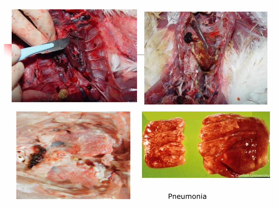
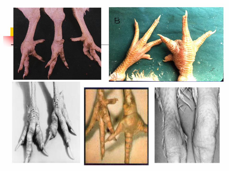

Prevention of Mycoplasmosis
Adopt all biosecurity measures
Egg sanitation and hatchery sanitation
Regular serological flock monitoring
II In case of MM
Examination of the phallus and cloacae ofadult male turkeys for MM serologicallybacteriologically and molecularly before thebreeding season
Using non-infected (non genetically carriers)birds for breeding (mating)
Prevention of Mycoplasmosis
Vaccination1 Moderately virulent strain (F) strain2 685 strain3 Ts-11 strain4 New K 5054 strain5 Naturally occurring virulent strains (S6)6 Inactivated vaccines (bacterins) Vaccination with live MG vaccine between 12
and 16 weeks of age Vaccination can be carried out a 2-4 weeks of
age
Control of mycoplasmosis
1 Separate diseased from healthy birds
2 Hygienic disposal of dead birds (burning or burring)
3 Thorough cleaning and disinfection
4 Treatment of birds Medication helps only to controlclinical signs and lesions and slow the spread ofinfection but it doesnt eliminate the infection
Complete elimination of mycoplasmas from all birds inthe infected flocks using medication is not expected asthe treatment is regarded as a method of short-termamelioration of the disease rather than as long termsolution of the problem
Control of mycoplasmosis
use Teteracyclines Macroilds (erythromycin tylosinspiramycin lincomycin kitasamycin) Pleuromulins(tiamulin) and fluoroquinolones (norfloxacinenrofloxacin and danofloxacin)
Egg treatment
A) Egg inoculation Any antimycoplasmal antibiotics isinjected into the egg cell this method may kill theembryo
B) Egg dipping the eggs are warmed to 37-38C thenimmersed in cold antibiotic solution (1-4C) for 20minutes Due to the temperature differences cooling ofthe egg contents and the antibiotic is pulled throughthe egg shell
Control of mycoplasmosis
C) Pressure differential vacuum (system) by decreasingthe pressure above the egg 25 Cm Hg by vacuumpump then the pressure return slowly to theatmospheric one This method is not completelyeliminate the organism but not affect on thehatchability
D) Heat forced incubator the eggs are heated inforced air incubator for 40C then putted at roomtemperature for cooling at 256C It is the mosteffective method but it reduces the hatchability to 8-12 with high embryonic mortalities
BORDETELLOSIS
(Turkey coryza)
Bordetellosis(Turkey coryza)
It is a highly contagious upper-respiratory tractdisease affect young turkeys caused by Bordetellaavium and is characterized by inflammation anddistortion of the respiratory mucosa
The economic importance including impairedgrowth Mortalities and the losses are resultingfrom Colisepticemia secondary or complicatedinfections and stressors
Transmission of B Avium
Close contact with infected poults or throughexposure to litter or water
Infection is transmitted by aerosol transmission
The severity of bordetellosis is exacerbated byadverse environmental and infectious factors(Temp humidity litter quality Ecoli)
Signs of B Avium
An abrupt onset of sneezing (snick) in a high percentage of 2-6 week-old turkeys over the course of a week
Older turkeys may also develop a dry cough
A clear nasal discharge
During the first 2 weeks of disease the nares and feathers of the head and wings become crusted with wet tenacious brownish exudate and some birds develop submaxillary edema
Signs of B Avium
Foamy exudate at the medial canthus of the eye
Open-mouth breathing and dyspnea and altered vocalization in the second week of clinical signs result when the nasal cavity and upper trachea become partially occluded with mucoid exudate
Poor weight gains In turkeys 2-6 weeks of age morbidity 80-100
whereas the mortality rate is less than 10 High mortality rates (gt40) in young turkeys
frequently are associated with concurrent isolation of Escherichia coli
Lesions of B Avium
Nasal and tracheal exudates varies from serous initially to tenacious and mucoid during the course of disease
Generalized softening and distortion of the cartilaginous rings of the trachea dorsal-ventral compression
In cross-section tracheal rings appear to have thick walls and a diminished lumen
Diagnosis of B Avium
Samples collected from the choanal opening and nostril or by passing a swab into the trachea through the larynx
Isolation and identification of bacterial agent isolation is accomplished on MacConkey agar
Serologic testing has proven to be useful a microagglutination test (MAT)
Prevention of B Avium
Strict biosecurity measures are required toprevent infection of clean flocks
Vaccines available commercially for theprevention of bordetellosis are (ts) mutant of Bavium and a whole cell bacterin
Vaccination of breeder hens with killed bacterinsdelayed the onset and severity of clinicaldisease in challenge-exposed poults
Passive immunization of 3-week-old poults withconvalescent serum
Control of B Avium
Treatment of bordetellosis with antibioticsadministered in the water by injection orby aerosol has produced minimal clinicalimprovement in most cases
Treatment of an infected breeder flockwith tetracycline-HCl and potassiumpenicillin-G for 3 days produced clinicalimprovement within 24 hours
ORNITHOBACTERIUM RIHINOTRACHEAL
(ORT)
ORT Infection amp Transmission
It is a contagious bacterial disease affectschickens and turkeys characterized by respiratorysigns variable mortality and decrease in eggproduction with change in egg quality
The infection occurred by the horizontal routes bycontact through aerosol or drinking water
The vertical transmission occurs by transovarianand oviduct
Signs of ORTIn turkeysA Poults Turkey poults aged 2-8 weeks show mortality
up to 50 Respiratory signs coughing sneezing nasal
discharge and sinusitis Decrease in feed and water intakeB Breeders Decrease in egg production Increase in the number of unhatched eggs
Lesions of ORT
Uni or bilateral pneumonia
Yoghurt like airsaculitis
Pleuritris
Subcutaneous edema of the head
Enlarged liver and spleen with degeneration of heart muscles are additionally seen in turkeys
Thickened opaque air sacs with
profuse foamy white to yellow ldquoyogurt-
likerdquo
Pneumonia and pleuritis in turkeys
Prevention amp control of ORT
1) Application of good sanitary measures especially separation of different ages and species
2) Vaccination
a Inactivated vaccine considered as serotype specific vaccine
b Live vaccine can induce cross protection between some serotypes
Amoxicillin and chlortetracycline in water
Tetracycline and penicillins by injection
ERYSIPELAS
Erysipelas
Erysipelas (Red Skin) occurs in growing turkeys geese chickens quail and Peafowl between 47 months
This acute to chronic disease is caused by the Gram-positive rod shaped bacterium Erysipelothrix rhusiopathiae It is non-spore forming non-acid fast and non-motile
Infection amp transmission of Erysipelas
It can be transmitted through a break in the skin or mucous membranes or fighting between males
It is a soil-borne organism and can also be spread by cannibalism or biting flies
Contaminated fish-meal is also a source of infection
Signsamp lesions of Erysipelas
Signs The incubation period is 2-3 days Swollen snoods (turkeys) and
shock Diarrhea emaciation weakness anemia skin haemorrhage and necrosis can be seen
Fever cyanotic toes and head drop in egg production and or fertility and embryonic mortality can occur
Post mortem lesions Enlarged friable purple-black spleen breast muscle haemorrhage
oral mucous Haemorrhage in muscles spleen lungs fat and small intestine and endocarditis may be seen
Fibrinopurulent Exudate in the joints Thickening of walls or proventriculus or gizzard ulceration and
yellow nodules can occur in the caeca
Swollen snood Turgid snood
Enlarged and mottled spleen (marbled spleen) )
Endocarditis inflammatory process around valves
Diagnosis of Erysipelas
Laboratory isolation from lesions is important and can show smooth colonies colorless to a bluish gray or pin-point size with smooth edges Haemorrhagic swollen spleen and wicked red lesions are diagnosed
It Simulates cholera Salmonella gangrenous dermatitis aspergillosis and E coli
Blood agar tiny pinpoint colonies round and translucent
Prevention and Control of Erysipelas
Prevention Vaccinate birds twice one at 1012 weeks and again at
14-16 weeks Debeak at day one and pooling is done if necessary to
prevent fighting No pigs should be reared near poultry and rotation of
the turkey range to reduce bacteria
Treatment Gallimycin and penicillin can reduce signs Disinfect premises with aerosol phenols or iodine
ARIZONOSIS
Arizonoses
Arizonosis occurs young turkeys It is caused by Bacterium-Salmonella Arizona Gram negative flagellate bacterium Salmonella
Arizona
The effects are the same as for other Salmonella infections- Diarrhoea lameness somnolence (sleepiness) laboured breathing blindness and mortality Mortality up to 100 peaking at 7-10 days Tremors convulsions and twisted necks may also be seen
Infection amp transmission of Arizona
The disease is transmitted in the same way as other salmonella species ie from bird to bird and between farms
Common vectors include birds rodents and sometimes reptiles
Spread via the transovarian route can also occur
Signs and lesions of Arizona
Signs are the same as for other Salmonella Opaque eyes (blindness) tremors convulsions and twisted necks may be seen
Postmortem lesions
Lesions are the same as for Salmonella pullorum which include bacteria septicaemia peritonitis and retained yolk sacs
Congested (filled with blood) duodenum mottled (white necrotic spots) liver caseous plugs in caeca and caseous air sacs can be seen
Round heart (Characteristic)
Diagnosis of Arizona
The organisms must be cultured from post mortem lesions egg yolk etc on brilliant green agar for a definitive diagnosis
Agglutination or ELISA tests using sera from breeders can confirm the presences of S Arizona
It simulates pullorum E coli and typhoid
Prevention amp Control of Arizona
Prevention Biosecurity measures Bacterin for turkey breeders prevents egg transmission Egg and hatchery sanitation are important Breeders should be tested and those that are serologically
positive should be slaughtered Vector control helps control spread of the organismTreatment Treatments will reduce clinical problems but birds will
remain carriers SQ and Ormetoprin in the water and furazolidone in the feed are effective treatments Gentamicin and spectinomycin can be given to day-old-chicks by injection
RIMERELLA (PASTEURELLA) ANATIPESTIFER
(RA)
(DUCK SEPTICAEMIA INFECTIOUS SEROSITIS NEW DUCK SYNDROM)
Riemerella anatipestifer (RA)
It is an acute septicaemic or chroniccontagious disease of growingduckling characterized byserofibrinous pericarditisperihepatitis and airsacculitiscaseous salpingitis and meningitisand high mortality in ducklings(75)
Susceptibility amp Infection of RA
Ducklings aged 1-8 weeks are highly susceptible
The disease in laying birds is rare
Inhalation of infected droplets
Wound infection
Direct and indirect contact
Carrier birds
Signs of RA
Respiratory signs (nasal and ocular discharges sneezing and coughing)
Nervous signs (Ataxia tremors of the head and neck and coma)
Greenish diarrhea
Survival ducks may stunted
Mortality ranged from 5 to75 in relation to other factors as age route of infection and the strain virulence
Lesions of RA
Fibrinous pericarditis Fibrinous perihepatitis Fibrinous airsacculitis Caseous salpingitis fibrinopurulant pneumonia Arthritis Fibrinous meningitis Enlarged and mottled spleen In the chronic form the most predominant lesions in the
skin in the form of necrotic dermatitis on the lower back or around vent with yellowish exudates between skin and the fat layer
Central nervous system infection
Caseous exudate located in the head
region (subcutis)
Submandibulare oedema
Spinal cord compression Vertebral osteomyelitis
(spondylitis)
perihepatitis
Diagnosis of RA
Samples from heart blood brain pericardial exudates air sacs lungs liver and oviduct
Detection of bipolar organism in blood or tissue smear stained by Gimsa
Prevention amp Control of RA Sanitary measures
Avoid stress factors
Prevent contact of the birds with different species sources and ages
Inactivated vaccine must be containing a serotype specific to that cause endemic infection or frequent infection in the farm
Live RA vaccine given by drinking water or spray to day old birds can induces protection till 42 days of age
Sulfamethazine (02 -025) in water or feed sulfaquinoxaline (0025-005) in feed and lincomycin (0011-0022 ) in water are effective
BOTULISM (LIMBER NECK)
WESTERN DUCK SICKNESS
Botulism
Cause
Age group affected
Transmission
Clostridium botulinum
Most avian esp ducks
Ingestion of toxin
Insects or maggots
Feeding on dead decayed carcass
signs of botulism Paralysis of legs wings neck and eye lids are
predominant Paralysis progressed cranially fromlegs to eye lids
Affected bird appear firstly sitting on hocksreluctant to move or appear lame droopedwings extended neck ruffled feathers andfallout with handling
Broiler chickens are showing diarrhea withexcess ureates in the loose droppings
Mortality and morbidity are related to theamount of ingested toxin
Death 12-24 hours
Lesions of botulism
There is no gross or microscopic lesions
Maggots of fly blown carcasses or feathers can be found in crop of dead or affected birds
Control of botulism
No treatment but rapid withdrawal of thetoxicated food
Episons salt as purgative to removedecomposed food from the intestine
use sodium selenite and vitamin AD3E Antibiotics including Bacitracin 100gton in
feed streptomycin 1 gl of water may be ofvalue
Also Inoculation of antitoxin neutralizes onlyfree and extracellular bound toxin and can beused for treatment of valuable birds
MYCOPLASMA IMMITANS
Mycoplasma immitans
Infection with Mycoplasma immitans is seen in ducks and geese
Signs
Swollen sinuses
Post-mortem lesions
Sinusitis
PARATYPHOID
INFECTION (PT)
Paratyphoid (PT) Infection
It is infection of turkeys and duckswith motile salmonella serotypesrather than Salmonella gallinarumpullorum (S Typhimurium Senteritidis S anatum etchellip) resultingin relatively asymptomatic intestinalcarrier or inducing clinical diseaseThe disease in ducks is caused by S
anatum
MOI amp Transmission of PT
Egg shell contamination (false egg bornedisease)
Direct contact
Ingestion of contaminated food and water
Rodents animals wild birds and flies
Human
Contaminated animal protein supplements(Bone meal meet meal and fish meal)
Signs of PT in Ducks
1) Arthritis
2) The bird is unable to move or stand
3) Diarrhea
4) Nervous manifestation
Trembling or shivering
Kicking and violent movement
Move in circle
Keeling over backward with leg paddling (Keeldisease)
1) Necrotic foci on the liver
2) Distended gall bladder
3) Enteritis
4) The ureter is distended with ureates
5) Enlargement and congestion of the internalorgans
6) Pericarditis and perihepatitis
7) Degeneration of the ovary
Lesions of PT in Ducks
Prevention of PT
Sanitation and sound measures in poultryhouses egg store and hatcheries
Administrations of probiotics are exhibitingactivity against various PT salmonellae inthat diminish both intestinal colonizationand subsequent invasion to internal tissues(Competitive Exclusion)
Trials for preparation of killed or livevaccines
AVIAN CHLAMYDIOSIS
(ORNITHOSIS)
Avian chlamydiosis
Avian chlamydiosis is acute fatal or chronic infectiouszonootic respiratory disease of domestic wild andmigratory birds
The disease is characterized by nasal and oculardischarge diarrhea loss of weight drop in eggproduction and the mortality rate reached to 5-30
Presence of intracytoplasmic elementary bodies (LCL)is characteristic to the organism
Giemsa stain intracellular chlamydial inclusion
(reddish-purple)
Infection amp Transmission of Chlamydiosis
Chlamydia Psittaci infection occur horizontally either by inhalation of infected dust or droplet or by ingestion of contaminated feed and water with infected droppings
There is an evidence of low percentage of vertical transmission through eggs (transoverian) in ducks and sea gulls
Chronic carriers transmit the organism without signs
Biting of insects as ticks lice and mites
Wild birds are important in transmission
Signs of chlamydiosis
In turkeysI Acute epidemic Form (toxigenic highly virulent strain)Clinical signs appear as systemic fatal infection with1 Sudden death of 5-302 Fever and anorexia3 Nasal and ocular discharge swollen eye lids
conjunctivitis and sinusitis4 Off food and loss of weight (emaciation)5 Yellow green gelatinous diarrhea6 Rapid drop in egg production (40-50 decline)7 Morbidity rate may be reach 50-80
In ducks and geese (water fowl)
In duckling there are tremors trembling and staggered gait (incoordination)
Ocular and nasal discharge
Greenish watery Diarrhea
Inappetance
Emaciation convulsion and death
Morbidity rate 10-80
Mortality rate 1-30 (depending on the age health status and secondary infection with salmonella species)
Signs of chlamydiosis
Lesions of chlamydiosis
Congestion of the visceral organs
Congestion and pneumonia of the lungs
All the body cavities are filed with fibrinous exudates
Fibrinous pericarditis perihepatitis and airsacculitis and peritonitis
Catarrhal enteritis
Liver and spleen are enlarged dark and covered with gray white foci
Liver necrosisFibrinous pericarditis
liver hepatomegaly and congestion
fibrinous pericarditis
Fibrinous pericarditis
Caseous pericarditisLungs congested and fibrinous exudate
in the pleural cavity
Necrosis and congestion of
spleen
Enlarged congested
liver
Prehepatitis
Airsacculitis
A Thickened
abdominal airsacs
totally covered
with fibrin plaques
B Serous fluid
and fibrin in the
pericardial sac
C Severe
hepatomegaly
Diagnosis of chlamydiosis1 Detection of dark purple blue or pink (acc To the
stain) ICytoplasmic inclusion (LCL) bodies in stained smear (from nasal or ocular exudates serous membranes trachea lungs pancreas liver spleen heart faecal and cloacal swabs) in different stages of elementary body
2 Tissue culture3 Embryonated chicken eggs4 Laboratory animal inoculation Intra peritoneal or intra
cranial or intra nasal inoculation of 3-4 weeks old mice (3-6 mice) could resulted in death of mice after 5-7 days post inoculation Intra peritoneal inoculation resulted in peritonitis with accumulation of fibrinous exudate in the peritoneal cavity with splenomegaly
Prevention of chlamydiosis1 Different avian species and ages must be reared away
from each other2 Newly purchased (imported) birds especially pets must
be quarantined for 35 days (if positive discarded ortreated with tetracyclines till recovery)
3 Prevent the introduction of birds from the enzootic areas4 Avoid the contact with the free living wild birds5 Thorough cleaning and disinfection (iodophors or
formaldhydes)6 Regular testing of the birds using serological tests7 Restrict the movement of people and visitors8 Trials for vaccine preparation
Control of chlamydiosis
1 Oxytetracyclin (15mgkg bwt) in the drinking water for a period for 5 days
2 Chlorotetracyclin
(treatment may be extended to 15-45 days in severe infection)
3 Usage of combined antibiotics to control other associated infections as Salmonellae and Ecoli(quinolones like cipro enro and danofloxacin) for 3-5 days in the drinking water
AVIAN PASTEURELLOSIS
FOWL CHOLERA (FC)
FC
Peracute acute and chronic highly contagious bacterial disease of domestic and wild birds caused by Pasteurella multocida
In the acute form the disease is characterized by rapid course high morbidity and mortality and septicaemic picture while the chronic form is localized and follows the acute form or occur independently due to low virulent mo
Susceptibility of FC
1 Natural hosts are water fowl (ducks and geese) turkeys and chickens
2 Water fowl are the most susceptible birds
3 Turkeys are more susceptible than chickens
4 Adult chickens (more than 16 weeks old) while young turkeys are the most susceptible ages
Infection and transmission of FC
1 Inhalation of infected droplets
2 Ingestion of contaminated food and water by the infected excretions from the mouth and nostrils of diseased or carriers
3 Mechanically clothes feet or hands of workers contaminated equipments feed bags etchellip
4 Wild birds rodents and pets
5 Cannibalism of diseased or dead birds
6 Wound infection
Signs of FC
1 Per acute form
Sudden death without clinical signs
No characteristic post mortem lesions
2 Acute form
High morbidity and mortality rates
Septicaemia (cyanosis of the comb and wattle)
Mucous discharge from the mouth and nostrils
Watery white or greenish mucoid diarrhea
Respiratory manifestations
Drop in egg production
Lesions of FC
Acute form Petechial or echymotic hemorrhages on the
lungs proventriculus abdominal and coronary fats and on serosal membranes as peritoneum and subepicardium and haemorrhagic enteritis
Tracheitis bronchitis and pneumonia (TURKEYS) Oophoritis flaccid regressed or ruptured ova Free yolk material in the abdominal cavity Serofibrinous pericarditis and peritonitis Large amount of viscous mucous in the larynx
crop and intestine
Edematous cyanotic comb and wattles
Mucous excretion
from mouth
Fibrinous pericarditis and perihepatitisEnlarged liver
Small areas of necrosis in the liver (corn meal liver)
Subepicardial hemorrhages and petechiae
on the heart and coronary fat
The left lung has a severe
accumulation of fibrinous exudate on
its pleural surface In turkeys
Hemorrhagic mucosal and serosal lesions
Signs amp lesions of FC
3 Chronic forms
A Wattle form
Unilateral or bilateral swelling of wattle
Caseous exudates in the wattles cut sections
B Otitis media form
Nervous signs as torticollis
Caseous exudates in the middle ears and air spaces of cranial bones
Signs amp lesions of FC
C Arthritic form Swelling of the joints and lameness Caseous exudates in the joints Inflammation of the tendon sheet Hemorrhages and erosions on the articular
cartilageD Ovarian form Drop in egg production Cheesy material around the ovary and
peritoneal cavity
Signs amp lesions of FC
E Roup or catarrhal form
Respiratory signs (rals and dyspnea)
Mucous discharge from the mouth
Nasal discharge
Swelling of the sinuses and facial oedema
Conjunctivitis
Swelling of wattle
Swelling of the wattles
Swelling of the ear canal (middle ear)
Torticollis
Torticollis
strenal
bursitis
Arthritis
Caseous exudate over the hock region and
food pad
Swelling of the sternal bursae
Hyperemia and flaccid mature follicles
Egg-yolk peritonitis
Sinusitis and facial oedema
Yellow caseous exudate in the air spaces of the cranial bones
Diagnosis of FC
Demonstration of bipolar P multocida mo
either from the blood film in acute stage afterstaining with Giemsa Leishmanrsquos stains andseen under microscope
or take impression smear from organs (liver)stain and examine microscopically
Bipolar Pmultocida in between nucleated RBCS in acute infection
Rough colony on the right and a smooth colony on the left
Diagnosis of FC
Laboratory animal inoculationIntraperitoneal or SC inoculation of rabbit or mice
or hamster with blood of infected bird orsuspected material (liver supernatant) (02 ml)induces death of these animals within 24-48 hourspost inoculation with septicaemic pictureImpression smear from blood or tissues (liver)shows bipolar organism
Prevention of FC Adopt all biosecurity measures
Vaccination
P multocida antigen vaccine should be homologous tothat cause fowl cholera or use of autogenous localprepared vaccine in endemic area or when commercialones are not protective
There are many types of vaccines against FC
Whole cell bactrine
Live attenuated vaccine
a CU- vaccine
b Mutants of CU-vaccine as M-9 MN and PM-1 strainsto avoid post vaccinal reaction of the CU
Prevention of FC
In turkeys
Meat type 6-8 weeks bactrine injection or CU via drinking water and repeated every 4-6 weeks till marketing
Breeder flocks must be vaccinated first at 6- 8 weeks and revaccinated 2-5 times till start of egg production according to epidemiology of FC in the area and farm
Control of FC
In chicken and turkeys sulfamerazine 02 in water or 04 in feed or Sulfaquinoxaline 001 ndash005 in water for 3-5 days to stop deaths in 2 days
Streptomycin (150 mg) for IlM injection in turkeys
Chloro- or oxytetracycline 40 mgkgbw by IlM
Chloramphenicol 20 mgkgbw via IlM
Enrofloxacin hellipetc
AVIAN SPIROKETOSIS
Avian spiroketosis
The bacterium Borrelia anserina infects chickens turkey geese ducks pheasants grouse and canaries with morbidity and mortality up to 100
It is transmitted by arthropods eg Argas persicus and occasionally by infected faeces The bacterium is poorly resistant outside host but may be carried by Argas persicus for 430 days
Brachyspira pilosicoli previously known as Serpulina pilosicoli is an intestinal spirochaete that can be associated with inflammation of the large intestine in a broad range of mammals and birds
It has been associated with typhilitis diarrhoea reduced egg production and egg soiling in chickens
Signs amp lesions of avian spiroketosis
Depression and thirst Cyanosis Often diarrhoea with excessive urates Weakness and progressive paralysis Drops in egg production may be seen in both systemic
and intestinal forms Post-mortem lesions Marked splenomegaly Spleen mottled with ecchymotic haemorrhages Liver enlarged with small haemorrhages Necrotic foci Mucoid enteritis
Prevention amp Control of avian spiroketosis
Treatment
Various antibiotics including penicillin
Prevention
Control vectors
Vaccines in some countries
Cause
Age group affected
Transmission
Forms of the disease
Escherichia coli
All types of avian and all ages
Fecal oral route transovarial contamination of the egg shell via fecal material from hen
Colisepticaemia
Airsac disease
Chronic forms (arthritis omphalitis panophthalmitis salpingitis enteritis coligranuloma and SHS)
E coli
Severely congested liver
Foci of necrosis on liver
GANGARINOUS
DERMATITIS
(GD)
GAS OEDEMA DISEASE
WING ROT
Gangrenous dermatitis
Gangrenous (necrotic) dermatitis in broilers and turkeys between 3-7 weeks old
It is caused by a bacteria- Clostridium septicum Escherichia coli Staphylococcus aureus
Disease often occurs in birds which are already immunosuppressed due to a prior infection (Chicken anaemia Infectious Bursal Disease) or mycotoxin ingestion
Transmission is by contact with infected wet caked litter The disease often occurs in immunosuppressed birds
Signs of GD
Loss of feathers
Pale combs and wattles depression
Incoordination
Leg weakness and ataxia (canrsquot move) can be seen
Mortality is low but dead birds decompose quickly
Lesions of GD
Congestion haemorrhage and necrosis of skin with intro lesion bacteria under the microscope
In turkeys with cellulitis of the tail (bubbly tail) edema and vesicle-like lesions were present laterally and ventrally around the tail
Tail feathers were soft blood-filled and broken
Dark and moist skin of the breast
Necrosis of the abdomen skinAffected leg and toes
Necrosis of the wattle
Ulceration and gangrenous formation of skin
Prevention amp Control of GD
Prevention Clean out house and add new litter to prevent the
disease Medicate in starter feed Flavomycin Virginamyciin
Bacitrcin and CTC can reduce bacteria Proper vaccination against IBDV CAV and MDV prevent
mycotoxin formation in the feed and eliminate ALV in the breeders to prevent immunosuppression
Treatment Erythromycin penicillin in the feed to treat signs
Chlortetracycine oxytetracycline copper sulphate can be added to the water to reduce morbidity
ULCERATIVE ENERITIS
(UE)
Ulcerative Enteritis
Turkeys of 6-14 weeks are susceptible to this acute to chronic disease
The agent involved in the aetiology of this disease is clostridium colinum which is spore-forming gram-positive aerobic and non-motile
Mode of transmission
Vectors are faeces soil and litter containing the bacteria
It often accompanies coccidiosis in broilers
Signs amp lesions of UE
High mortality watery diarrhoea ruffled feathers dull listlessness increased thirst emaciation and atrophy of pectoral muscles can occur
Postmortem lesions Yellow irregular ulcers on small intestine and
caeca hemorrhagic enteritis are seen Enlarged haemorrhagic necrotic spleen light
yellow mottling of liver and crop filled with water may occur
Prevention amp Control of UE
Prevention Improved sanitation Adding salt to the soil (500
lbs of salthouse) may kill spores Raising birds on wire andor feeding bacitracin at 50-100 gt will prevent the disease
Treatment NF-180 (50-100 gt) strephomycin (60 gt) and
chlortetracycline vitamins and minerals in water andor lincomycin 2 gt will reduce the signs
Remove dead birds and feed bacitracin (200 gt)
Thank you
ANY QUESTIONS
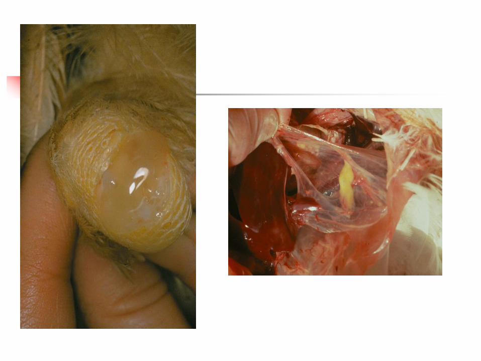
Prevention of Mycoplasmosis
Vaccination1 Moderately virulent strain (F) strain2 685 strain3 Ts-11 strain4 New K 5054 strain5 Naturally occurring virulent strains (S6)6 Inactivated vaccines (bacterins) Vaccination with live MG vaccine between 12
and 16 weeks of age Vaccination can be carried out a 2-4 weeks of
age
Control of mycoplasmosis
1 Separate diseased from healthy birds
2 Hygienic disposal of dead birds (burning or burring)
3 Thorough cleaning and disinfection
4 Treatment of birds Medication helps only to controlclinical signs and lesions and slow the spread ofinfection but it doesnt eliminate the infection
Complete elimination of mycoplasmas from all birds inthe infected flocks using medication is not expected asthe treatment is regarded as a method of short-termamelioration of the disease rather than as long termsolution of the problem
Control of mycoplasmosis
use Teteracyclines Macroilds (erythromycin tylosinspiramycin lincomycin kitasamycin) Pleuromulins(tiamulin) and fluoroquinolones (norfloxacinenrofloxacin and danofloxacin)
Egg treatment
A) Egg inoculation Any antimycoplasmal antibiotics isinjected into the egg cell this method may kill theembryo
B) Egg dipping the eggs are warmed to 37-38C thenimmersed in cold antibiotic solution (1-4C) for 20minutes Due to the temperature differences cooling ofthe egg contents and the antibiotic is pulled throughthe egg shell
Control of mycoplasmosis
C) Pressure differential vacuum (system) by decreasingthe pressure above the egg 25 Cm Hg by vacuumpump then the pressure return slowly to theatmospheric one This method is not completelyeliminate the organism but not affect on thehatchability
D) Heat forced incubator the eggs are heated inforced air incubator for 40C then putted at roomtemperature for cooling at 256C It is the mosteffective method but it reduces the hatchability to 8-12 with high embryonic mortalities
BORDETELLOSIS
(Turkey coryza)
Bordetellosis(Turkey coryza)
It is a highly contagious upper-respiratory tractdisease affect young turkeys caused by Bordetellaavium and is characterized by inflammation anddistortion of the respiratory mucosa
The economic importance including impairedgrowth Mortalities and the losses are resultingfrom Colisepticemia secondary or complicatedinfections and stressors
Transmission of B Avium
Close contact with infected poults or throughexposure to litter or water
Infection is transmitted by aerosol transmission
The severity of bordetellosis is exacerbated byadverse environmental and infectious factors(Temp humidity litter quality Ecoli)
Signs of B Avium
An abrupt onset of sneezing (snick) in a high percentage of 2-6 week-old turkeys over the course of a week
Older turkeys may also develop a dry cough
A clear nasal discharge
During the first 2 weeks of disease the nares and feathers of the head and wings become crusted with wet tenacious brownish exudate and some birds develop submaxillary edema
Signs of B Avium
Foamy exudate at the medial canthus of the eye
Open-mouth breathing and dyspnea and altered vocalization in the second week of clinical signs result when the nasal cavity and upper trachea become partially occluded with mucoid exudate
Poor weight gains In turkeys 2-6 weeks of age morbidity 80-100
whereas the mortality rate is less than 10 High mortality rates (gt40) in young turkeys
frequently are associated with concurrent isolation of Escherichia coli
Lesions of B Avium
Nasal and tracheal exudates varies from serous initially to tenacious and mucoid during the course of disease
Generalized softening and distortion of the cartilaginous rings of the trachea dorsal-ventral compression
In cross-section tracheal rings appear to have thick walls and a diminished lumen
Diagnosis of B Avium
Samples collected from the choanal opening and nostril or by passing a swab into the trachea through the larynx
Isolation and identification of bacterial agent isolation is accomplished on MacConkey agar
Serologic testing has proven to be useful a microagglutination test (MAT)
Prevention of B Avium
Strict biosecurity measures are required toprevent infection of clean flocks
Vaccines available commercially for theprevention of bordetellosis are (ts) mutant of Bavium and a whole cell bacterin
Vaccination of breeder hens with killed bacterinsdelayed the onset and severity of clinicaldisease in challenge-exposed poults
Passive immunization of 3-week-old poults withconvalescent serum
Control of B Avium
Treatment of bordetellosis with antibioticsadministered in the water by injection orby aerosol has produced minimal clinicalimprovement in most cases
Treatment of an infected breeder flockwith tetracycline-HCl and potassiumpenicillin-G for 3 days produced clinicalimprovement within 24 hours
ORNITHOBACTERIUM RIHINOTRACHEAL
(ORT)
ORT Infection amp Transmission
It is a contagious bacterial disease affectschickens and turkeys characterized by respiratorysigns variable mortality and decrease in eggproduction with change in egg quality
The infection occurred by the horizontal routes bycontact through aerosol or drinking water
The vertical transmission occurs by transovarianand oviduct
Signs of ORTIn turkeysA Poults Turkey poults aged 2-8 weeks show mortality
up to 50 Respiratory signs coughing sneezing nasal
discharge and sinusitis Decrease in feed and water intakeB Breeders Decrease in egg production Increase in the number of unhatched eggs
Lesions of ORT
Uni or bilateral pneumonia
Yoghurt like airsaculitis
Pleuritris
Subcutaneous edema of the head
Enlarged liver and spleen with degeneration of heart muscles are additionally seen in turkeys
Thickened opaque air sacs with
profuse foamy white to yellow ldquoyogurt-
likerdquo
Pneumonia and pleuritis in turkeys
Prevention amp control of ORT
1) Application of good sanitary measures especially separation of different ages and species
2) Vaccination
a Inactivated vaccine considered as serotype specific vaccine
b Live vaccine can induce cross protection between some serotypes
Amoxicillin and chlortetracycline in water
Tetracycline and penicillins by injection
ERYSIPELAS
Erysipelas
Erysipelas (Red Skin) occurs in growing turkeys geese chickens quail and Peafowl between 47 months
This acute to chronic disease is caused by the Gram-positive rod shaped bacterium Erysipelothrix rhusiopathiae It is non-spore forming non-acid fast and non-motile
Infection amp transmission of Erysipelas
It can be transmitted through a break in the skin or mucous membranes or fighting between males
It is a soil-borne organism and can also be spread by cannibalism or biting flies
Contaminated fish-meal is also a source of infection
Signsamp lesions of Erysipelas
Signs The incubation period is 2-3 days Swollen snoods (turkeys) and
shock Diarrhea emaciation weakness anemia skin haemorrhage and necrosis can be seen
Fever cyanotic toes and head drop in egg production and or fertility and embryonic mortality can occur
Post mortem lesions Enlarged friable purple-black spleen breast muscle haemorrhage
oral mucous Haemorrhage in muscles spleen lungs fat and small intestine and endocarditis may be seen
Fibrinopurulent Exudate in the joints Thickening of walls or proventriculus or gizzard ulceration and
yellow nodules can occur in the caeca
Swollen snood Turgid snood
Enlarged and mottled spleen (marbled spleen) )
Endocarditis inflammatory process around valves
Diagnosis of Erysipelas
Laboratory isolation from lesions is important and can show smooth colonies colorless to a bluish gray or pin-point size with smooth edges Haemorrhagic swollen spleen and wicked red lesions are diagnosed
It Simulates cholera Salmonella gangrenous dermatitis aspergillosis and E coli
Blood agar tiny pinpoint colonies round and translucent
Prevention and Control of Erysipelas
Prevention Vaccinate birds twice one at 1012 weeks and again at
14-16 weeks Debeak at day one and pooling is done if necessary to
prevent fighting No pigs should be reared near poultry and rotation of
the turkey range to reduce bacteria
Treatment Gallimycin and penicillin can reduce signs Disinfect premises with aerosol phenols or iodine
ARIZONOSIS
Arizonoses
Arizonosis occurs young turkeys It is caused by Bacterium-Salmonella Arizona Gram negative flagellate bacterium Salmonella
Arizona
The effects are the same as for other Salmonella infections- Diarrhoea lameness somnolence (sleepiness) laboured breathing blindness and mortality Mortality up to 100 peaking at 7-10 days Tremors convulsions and twisted necks may also be seen
Infection amp transmission of Arizona
The disease is transmitted in the same way as other salmonella species ie from bird to bird and between farms
Common vectors include birds rodents and sometimes reptiles
Spread via the transovarian route can also occur
Signs and lesions of Arizona
Signs are the same as for other Salmonella Opaque eyes (blindness) tremors convulsions and twisted necks may be seen
Postmortem lesions
Lesions are the same as for Salmonella pullorum which include bacteria septicaemia peritonitis and retained yolk sacs
Congested (filled with blood) duodenum mottled (white necrotic spots) liver caseous plugs in caeca and caseous air sacs can be seen
Round heart (Characteristic)
Diagnosis of Arizona
The organisms must be cultured from post mortem lesions egg yolk etc on brilliant green agar for a definitive diagnosis
Agglutination or ELISA tests using sera from breeders can confirm the presences of S Arizona
It simulates pullorum E coli and typhoid
Prevention amp Control of Arizona
Prevention Biosecurity measures Bacterin for turkey breeders prevents egg transmission Egg and hatchery sanitation are important Breeders should be tested and those that are serologically
positive should be slaughtered Vector control helps control spread of the organismTreatment Treatments will reduce clinical problems but birds will
remain carriers SQ and Ormetoprin in the water and furazolidone in the feed are effective treatments Gentamicin and spectinomycin can be given to day-old-chicks by injection
RIMERELLA (PASTEURELLA) ANATIPESTIFER
(RA)
(DUCK SEPTICAEMIA INFECTIOUS SEROSITIS NEW DUCK SYNDROM)
Riemerella anatipestifer (RA)
It is an acute septicaemic or chroniccontagious disease of growingduckling characterized byserofibrinous pericarditisperihepatitis and airsacculitiscaseous salpingitis and meningitisand high mortality in ducklings(75)
Susceptibility amp Infection of RA
Ducklings aged 1-8 weeks are highly susceptible
The disease in laying birds is rare
Inhalation of infected droplets
Wound infection
Direct and indirect contact
Carrier birds
Signs of RA
Respiratory signs (nasal and ocular discharges sneezing and coughing)
Nervous signs (Ataxia tremors of the head and neck and coma)
Greenish diarrhea
Survival ducks may stunted
Mortality ranged from 5 to75 in relation to other factors as age route of infection and the strain virulence
Lesions of RA
Fibrinous pericarditis Fibrinous perihepatitis Fibrinous airsacculitis Caseous salpingitis fibrinopurulant pneumonia Arthritis Fibrinous meningitis Enlarged and mottled spleen In the chronic form the most predominant lesions in the
skin in the form of necrotic dermatitis on the lower back or around vent with yellowish exudates between skin and the fat layer
Central nervous system infection
Caseous exudate located in the head
region (subcutis)
Submandibulare oedema
Spinal cord compression Vertebral osteomyelitis
(spondylitis)
perihepatitis
Diagnosis of RA
Samples from heart blood brain pericardial exudates air sacs lungs liver and oviduct
Detection of bipolar organism in blood or tissue smear stained by Gimsa
Prevention amp Control of RA Sanitary measures
Avoid stress factors
Prevent contact of the birds with different species sources and ages
Inactivated vaccine must be containing a serotype specific to that cause endemic infection or frequent infection in the farm
Live RA vaccine given by drinking water or spray to day old birds can induces protection till 42 days of age
Sulfamethazine (02 -025) in water or feed sulfaquinoxaline (0025-005) in feed and lincomycin (0011-0022 ) in water are effective
BOTULISM (LIMBER NECK)
WESTERN DUCK SICKNESS
Botulism
Cause
Age group affected
Transmission
Clostridium botulinum
Most avian esp ducks
Ingestion of toxin
Insects or maggots
Feeding on dead decayed carcass
signs of botulism Paralysis of legs wings neck and eye lids are
predominant Paralysis progressed cranially fromlegs to eye lids
Affected bird appear firstly sitting on hocksreluctant to move or appear lame droopedwings extended neck ruffled feathers andfallout with handling
Broiler chickens are showing diarrhea withexcess ureates in the loose droppings
Mortality and morbidity are related to theamount of ingested toxin
Death 12-24 hours
Lesions of botulism
There is no gross or microscopic lesions
Maggots of fly blown carcasses or feathers can be found in crop of dead or affected birds
Control of botulism
No treatment but rapid withdrawal of thetoxicated food
Episons salt as purgative to removedecomposed food from the intestine
use sodium selenite and vitamin AD3E Antibiotics including Bacitracin 100gton in
feed streptomycin 1 gl of water may be ofvalue
Also Inoculation of antitoxin neutralizes onlyfree and extracellular bound toxin and can beused for treatment of valuable birds
MYCOPLASMA IMMITANS
Mycoplasma immitans
Infection with Mycoplasma immitans is seen in ducks and geese
Signs
Swollen sinuses
Post-mortem lesions
Sinusitis
PARATYPHOID
INFECTION (PT)
Paratyphoid (PT) Infection
It is infection of turkeys and duckswith motile salmonella serotypesrather than Salmonella gallinarumpullorum (S Typhimurium Senteritidis S anatum etchellip) resultingin relatively asymptomatic intestinalcarrier or inducing clinical diseaseThe disease in ducks is caused by S
anatum
MOI amp Transmission of PT
Egg shell contamination (false egg bornedisease)
Direct contact
Ingestion of contaminated food and water
Rodents animals wild birds and flies
Human
Contaminated animal protein supplements(Bone meal meet meal and fish meal)
Signs of PT in Ducks
1) Arthritis
2) The bird is unable to move or stand
3) Diarrhea
4) Nervous manifestation
Trembling or shivering
Kicking and violent movement
Move in circle
Keeling over backward with leg paddling (Keeldisease)
1) Necrotic foci on the liver
2) Distended gall bladder
3) Enteritis
4) The ureter is distended with ureates
5) Enlargement and congestion of the internalorgans
6) Pericarditis and perihepatitis
7) Degeneration of the ovary
Lesions of PT in Ducks
Prevention of PT
Sanitation and sound measures in poultryhouses egg store and hatcheries
Administrations of probiotics are exhibitingactivity against various PT salmonellae inthat diminish both intestinal colonizationand subsequent invasion to internal tissues(Competitive Exclusion)
Trials for preparation of killed or livevaccines
AVIAN CHLAMYDIOSIS
(ORNITHOSIS)
Avian chlamydiosis
Avian chlamydiosis is acute fatal or chronic infectiouszonootic respiratory disease of domestic wild andmigratory birds
The disease is characterized by nasal and oculardischarge diarrhea loss of weight drop in eggproduction and the mortality rate reached to 5-30
Presence of intracytoplasmic elementary bodies (LCL)is characteristic to the organism
Giemsa stain intracellular chlamydial inclusion
(reddish-purple)
Infection amp Transmission of Chlamydiosis
Chlamydia Psittaci infection occur horizontally either by inhalation of infected dust or droplet or by ingestion of contaminated feed and water with infected droppings
There is an evidence of low percentage of vertical transmission through eggs (transoverian) in ducks and sea gulls
Chronic carriers transmit the organism without signs
Biting of insects as ticks lice and mites
Wild birds are important in transmission
Signs of chlamydiosis
In turkeysI Acute epidemic Form (toxigenic highly virulent strain)Clinical signs appear as systemic fatal infection with1 Sudden death of 5-302 Fever and anorexia3 Nasal and ocular discharge swollen eye lids
conjunctivitis and sinusitis4 Off food and loss of weight (emaciation)5 Yellow green gelatinous diarrhea6 Rapid drop in egg production (40-50 decline)7 Morbidity rate may be reach 50-80
In ducks and geese (water fowl)
In duckling there are tremors trembling and staggered gait (incoordination)
Ocular and nasal discharge
Greenish watery Diarrhea
Inappetance
Emaciation convulsion and death
Morbidity rate 10-80
Mortality rate 1-30 (depending on the age health status and secondary infection with salmonella species)
Signs of chlamydiosis
Lesions of chlamydiosis
Congestion of the visceral organs
Congestion and pneumonia of the lungs
All the body cavities are filed with fibrinous exudates
Fibrinous pericarditis perihepatitis and airsacculitis and peritonitis
Catarrhal enteritis
Liver and spleen are enlarged dark and covered with gray white foci
Liver necrosisFibrinous pericarditis
liver hepatomegaly and congestion
fibrinous pericarditis
Fibrinous pericarditis
Caseous pericarditisLungs congested and fibrinous exudate
in the pleural cavity
Necrosis and congestion of
spleen
Enlarged congested
liver
Prehepatitis
Airsacculitis
A Thickened
abdominal airsacs
totally covered
with fibrin plaques
B Serous fluid
and fibrin in the
pericardial sac
C Severe
hepatomegaly
Diagnosis of chlamydiosis1 Detection of dark purple blue or pink (acc To the
stain) ICytoplasmic inclusion (LCL) bodies in stained smear (from nasal or ocular exudates serous membranes trachea lungs pancreas liver spleen heart faecal and cloacal swabs) in different stages of elementary body
2 Tissue culture3 Embryonated chicken eggs4 Laboratory animal inoculation Intra peritoneal or intra
cranial or intra nasal inoculation of 3-4 weeks old mice (3-6 mice) could resulted in death of mice after 5-7 days post inoculation Intra peritoneal inoculation resulted in peritonitis with accumulation of fibrinous exudate in the peritoneal cavity with splenomegaly
Prevention of chlamydiosis1 Different avian species and ages must be reared away
from each other2 Newly purchased (imported) birds especially pets must
be quarantined for 35 days (if positive discarded ortreated with tetracyclines till recovery)
3 Prevent the introduction of birds from the enzootic areas4 Avoid the contact with the free living wild birds5 Thorough cleaning and disinfection (iodophors or
formaldhydes)6 Regular testing of the birds using serological tests7 Restrict the movement of people and visitors8 Trials for vaccine preparation
Control of chlamydiosis
1 Oxytetracyclin (15mgkg bwt) in the drinking water for a period for 5 days
2 Chlorotetracyclin
(treatment may be extended to 15-45 days in severe infection)
3 Usage of combined antibiotics to control other associated infections as Salmonellae and Ecoli(quinolones like cipro enro and danofloxacin) for 3-5 days in the drinking water
AVIAN PASTEURELLOSIS
FOWL CHOLERA (FC)
FC
Peracute acute and chronic highly contagious bacterial disease of domestic and wild birds caused by Pasteurella multocida
In the acute form the disease is characterized by rapid course high morbidity and mortality and septicaemic picture while the chronic form is localized and follows the acute form or occur independently due to low virulent mo
Susceptibility of FC
1 Natural hosts are water fowl (ducks and geese) turkeys and chickens
2 Water fowl are the most susceptible birds
3 Turkeys are more susceptible than chickens
4 Adult chickens (more than 16 weeks old) while young turkeys are the most susceptible ages
Infection and transmission of FC
1 Inhalation of infected droplets
2 Ingestion of contaminated food and water by the infected excretions from the mouth and nostrils of diseased or carriers
3 Mechanically clothes feet or hands of workers contaminated equipments feed bags etchellip
4 Wild birds rodents and pets
5 Cannibalism of diseased or dead birds
6 Wound infection
Signs of FC
1 Per acute form
Sudden death without clinical signs
No characteristic post mortem lesions
2 Acute form
High morbidity and mortality rates
Septicaemia (cyanosis of the comb and wattle)
Mucous discharge from the mouth and nostrils
Watery white or greenish mucoid diarrhea
Respiratory manifestations
Drop in egg production
Lesions of FC
Acute form Petechial or echymotic hemorrhages on the
lungs proventriculus abdominal and coronary fats and on serosal membranes as peritoneum and subepicardium and haemorrhagic enteritis
Tracheitis bronchitis and pneumonia (TURKEYS) Oophoritis flaccid regressed or ruptured ova Free yolk material in the abdominal cavity Serofibrinous pericarditis and peritonitis Large amount of viscous mucous in the larynx
crop and intestine
Edematous cyanotic comb and wattles
Mucous excretion
from mouth
Fibrinous pericarditis and perihepatitisEnlarged liver
Small areas of necrosis in the liver (corn meal liver)
Subepicardial hemorrhages and petechiae
on the heart and coronary fat
The left lung has a severe
accumulation of fibrinous exudate on
its pleural surface In turkeys
Hemorrhagic mucosal and serosal lesions
Signs amp lesions of FC
3 Chronic forms
A Wattle form
Unilateral or bilateral swelling of wattle
Caseous exudates in the wattles cut sections
B Otitis media form
Nervous signs as torticollis
Caseous exudates in the middle ears and air spaces of cranial bones
Signs amp lesions of FC
C Arthritic form Swelling of the joints and lameness Caseous exudates in the joints Inflammation of the tendon sheet Hemorrhages and erosions on the articular
cartilageD Ovarian form Drop in egg production Cheesy material around the ovary and
peritoneal cavity
Signs amp lesions of FC
E Roup or catarrhal form
Respiratory signs (rals and dyspnea)
Mucous discharge from the mouth
Nasal discharge
Swelling of the sinuses and facial oedema
Conjunctivitis
Swelling of wattle
Swelling of the wattles
Swelling of the ear canal (middle ear)
Torticollis
Torticollis
strenal
bursitis
Arthritis
Caseous exudate over the hock region and
food pad
Swelling of the sternal bursae
Hyperemia and flaccid mature follicles
Egg-yolk peritonitis
Sinusitis and facial oedema
Yellow caseous exudate in the air spaces of the cranial bones
Diagnosis of FC
Demonstration of bipolar P multocida mo
either from the blood film in acute stage afterstaining with Giemsa Leishmanrsquos stains andseen under microscope
or take impression smear from organs (liver)stain and examine microscopically
Bipolar Pmultocida in between nucleated RBCS in acute infection
Rough colony on the right and a smooth colony on the left
Diagnosis of FC
Laboratory animal inoculationIntraperitoneal or SC inoculation of rabbit or mice
or hamster with blood of infected bird orsuspected material (liver supernatant) (02 ml)induces death of these animals within 24-48 hourspost inoculation with septicaemic pictureImpression smear from blood or tissues (liver)shows bipolar organism
Prevention of FC Adopt all biosecurity measures
Vaccination
P multocida antigen vaccine should be homologous tothat cause fowl cholera or use of autogenous localprepared vaccine in endemic area or when commercialones are not protective
There are many types of vaccines against FC
Whole cell bactrine
Live attenuated vaccine
a CU- vaccine
b Mutants of CU-vaccine as M-9 MN and PM-1 strainsto avoid post vaccinal reaction of the CU
Prevention of FC
In turkeys
Meat type 6-8 weeks bactrine injection or CU via drinking water and repeated every 4-6 weeks till marketing
Breeder flocks must be vaccinated first at 6- 8 weeks and revaccinated 2-5 times till start of egg production according to epidemiology of FC in the area and farm
Control of FC
In chicken and turkeys sulfamerazine 02 in water or 04 in feed or Sulfaquinoxaline 001 ndash005 in water for 3-5 days to stop deaths in 2 days
Streptomycin (150 mg) for IlM injection in turkeys
Chloro- or oxytetracycline 40 mgkgbw by IlM
Chloramphenicol 20 mgkgbw via IlM
Enrofloxacin hellipetc
AVIAN SPIROKETOSIS
Avian spiroketosis
The bacterium Borrelia anserina infects chickens turkey geese ducks pheasants grouse and canaries with morbidity and mortality up to 100
It is transmitted by arthropods eg Argas persicus and occasionally by infected faeces The bacterium is poorly resistant outside host but may be carried by Argas persicus for 430 days
Brachyspira pilosicoli previously known as Serpulina pilosicoli is an intestinal spirochaete that can be associated with inflammation of the large intestine in a broad range of mammals and birds
It has been associated with typhilitis diarrhoea reduced egg production and egg soiling in chickens
Signs amp lesions of avian spiroketosis
Depression and thirst Cyanosis Often diarrhoea with excessive urates Weakness and progressive paralysis Drops in egg production may be seen in both systemic
and intestinal forms Post-mortem lesions Marked splenomegaly Spleen mottled with ecchymotic haemorrhages Liver enlarged with small haemorrhages Necrotic foci Mucoid enteritis
Prevention amp Control of avian spiroketosis
Treatment
Various antibiotics including penicillin
Prevention
Control vectors
Vaccines in some countries
Cause
Age group affected
Transmission
Forms of the disease
Escherichia coli
All types of avian and all ages
Fecal oral route transovarial contamination of the egg shell via fecal material from hen
Colisepticaemia
Airsac disease
Chronic forms (arthritis omphalitis panophthalmitis salpingitis enteritis coligranuloma and SHS)
E coli
Severely congested liver
Foci of necrosis on liver
GANGARINOUS
DERMATITIS
(GD)
GAS OEDEMA DISEASE
WING ROT
Gangrenous dermatitis
Gangrenous (necrotic) dermatitis in broilers and turkeys between 3-7 weeks old
It is caused by a bacteria- Clostridium septicum Escherichia coli Staphylococcus aureus
Disease often occurs in birds which are already immunosuppressed due to a prior infection (Chicken anaemia Infectious Bursal Disease) or mycotoxin ingestion
Transmission is by contact with infected wet caked litter The disease often occurs in immunosuppressed birds
Signs of GD
Loss of feathers
Pale combs and wattles depression
Incoordination
Leg weakness and ataxia (canrsquot move) can be seen
Mortality is low but dead birds decompose quickly
Lesions of GD
Congestion haemorrhage and necrosis of skin with intro lesion bacteria under the microscope
In turkeys with cellulitis of the tail (bubbly tail) edema and vesicle-like lesions were present laterally and ventrally around the tail
Tail feathers were soft blood-filled and broken
Dark and moist skin of the breast
Necrosis of the abdomen skinAffected leg and toes
Necrosis of the wattle
Ulceration and gangrenous formation of skin
Prevention amp Control of GD
Prevention Clean out house and add new litter to prevent the
disease Medicate in starter feed Flavomycin Virginamyciin
Bacitrcin and CTC can reduce bacteria Proper vaccination against IBDV CAV and MDV prevent
mycotoxin formation in the feed and eliminate ALV in the breeders to prevent immunosuppression
Treatment Erythromycin penicillin in the feed to treat signs
Chlortetracycine oxytetracycline copper sulphate can be added to the water to reduce morbidity
ULCERATIVE ENERITIS
(UE)
Ulcerative Enteritis
Turkeys of 6-14 weeks are susceptible to this acute to chronic disease
The agent involved in the aetiology of this disease is clostridium colinum which is spore-forming gram-positive aerobic and non-motile
Mode of transmission
Vectors are faeces soil and litter containing the bacteria
It often accompanies coccidiosis in broilers
Signs amp lesions of UE
High mortality watery diarrhoea ruffled feathers dull listlessness increased thirst emaciation and atrophy of pectoral muscles can occur
Postmortem lesions Yellow irregular ulcers on small intestine and
caeca hemorrhagic enteritis are seen Enlarged haemorrhagic necrotic spleen light
yellow mottling of liver and crop filled with water may occur
Prevention amp Control of UE
Prevention Improved sanitation Adding salt to the soil (500
lbs of salthouse) may kill spores Raising birds on wire andor feeding bacitracin at 50-100 gt will prevent the disease
Treatment NF-180 (50-100 gt) strephomycin (60 gt) and
chlortetracycline vitamins and minerals in water andor lincomycin 2 gt will reduce the signs
Remove dead birds and feed bacitracin (200 gt)
Thank you
ANY QUESTIONS

Control of mycoplasmosis
C) Pressure differential vacuum (system) by decreasingthe pressure above the egg 25 Cm Hg by vacuumpump then the pressure return slowly to theatmospheric one This method is not completelyeliminate the organism but not affect on thehatchability
D) Heat forced incubator the eggs are heated inforced air incubator for 40C then putted at roomtemperature for cooling at 256C It is the mosteffective method but it reduces the hatchability to 8-12 with high embryonic mortalities
BORDETELLOSIS
(Turkey coryza)
Bordetellosis(Turkey coryza)
It is a highly contagious upper-respiratory tractdisease affect young turkeys caused by Bordetellaavium and is characterized by inflammation anddistortion of the respiratory mucosa
The economic importance including impairedgrowth Mortalities and the losses are resultingfrom Colisepticemia secondary or complicatedinfections and stressors
Transmission of B Avium
Close contact with infected poults or throughexposure to litter or water
Infection is transmitted by aerosol transmission
The severity of bordetellosis is exacerbated byadverse environmental and infectious factors(Temp humidity litter quality Ecoli)
Signs of B Avium
An abrupt onset of sneezing (snick) in a high percentage of 2-6 week-old turkeys over the course of a week
Older turkeys may also develop a dry cough
A clear nasal discharge
During the first 2 weeks of disease the nares and feathers of the head and wings become crusted with wet tenacious brownish exudate and some birds develop submaxillary edema
Signs of B Avium
Foamy exudate at the medial canthus of the eye
Open-mouth breathing and dyspnea and altered vocalization in the second week of clinical signs result when the nasal cavity and upper trachea become partially occluded with mucoid exudate
Poor weight gains In turkeys 2-6 weeks of age morbidity 80-100
whereas the mortality rate is less than 10 High mortality rates (gt40) in young turkeys
frequently are associated with concurrent isolation of Escherichia coli
Lesions of B Avium
Nasal and tracheal exudates varies from serous initially to tenacious and mucoid during the course of disease
Generalized softening and distortion of the cartilaginous rings of the trachea dorsal-ventral compression
In cross-section tracheal rings appear to have thick walls and a diminished lumen
Diagnosis of B Avium
Samples collected from the choanal opening and nostril or by passing a swab into the trachea through the larynx
Isolation and identification of bacterial agent isolation is accomplished on MacConkey agar
Serologic testing has proven to be useful a microagglutination test (MAT)
Prevention of B Avium
Strict biosecurity measures are required toprevent infection of clean flocks
Vaccines available commercially for theprevention of bordetellosis are (ts) mutant of Bavium and a whole cell bacterin
Vaccination of breeder hens with killed bacterinsdelayed the onset and severity of clinicaldisease in challenge-exposed poults
Passive immunization of 3-week-old poults withconvalescent serum
Control of B Avium
Treatment of bordetellosis with antibioticsadministered in the water by injection orby aerosol has produced minimal clinicalimprovement in most cases
Treatment of an infected breeder flockwith tetracycline-HCl and potassiumpenicillin-G for 3 days produced clinicalimprovement within 24 hours
ORNITHOBACTERIUM RIHINOTRACHEAL
(ORT)
ORT Infection amp Transmission
It is a contagious bacterial disease affectschickens and turkeys characterized by respiratorysigns variable mortality and decrease in eggproduction with change in egg quality
The infection occurred by the horizontal routes bycontact through aerosol or drinking water
The vertical transmission occurs by transovarianand oviduct
Signs of ORTIn turkeysA Poults Turkey poults aged 2-8 weeks show mortality
up to 50 Respiratory signs coughing sneezing nasal
discharge and sinusitis Decrease in feed and water intakeB Breeders Decrease in egg production Increase in the number of unhatched eggs
Lesions of ORT
Uni or bilateral pneumonia
Yoghurt like airsaculitis
Pleuritris
Subcutaneous edema of the head
Enlarged liver and spleen with degeneration of heart muscles are additionally seen in turkeys
Thickened opaque air sacs with
profuse foamy white to yellow ldquoyogurt-
likerdquo
Pneumonia and pleuritis in turkeys
Prevention amp control of ORT
1) Application of good sanitary measures especially separation of different ages and species
2) Vaccination
a Inactivated vaccine considered as serotype specific vaccine
b Live vaccine can induce cross protection between some serotypes
Amoxicillin and chlortetracycline in water
Tetracycline and penicillins by injection
ERYSIPELAS
Erysipelas
Erysipelas (Red Skin) occurs in growing turkeys geese chickens quail and Peafowl between 47 months
This acute to chronic disease is caused by the Gram-positive rod shaped bacterium Erysipelothrix rhusiopathiae It is non-spore forming non-acid fast and non-motile
Infection amp transmission of Erysipelas
It can be transmitted through a break in the skin or mucous membranes or fighting between males
It is a soil-borne organism and can also be spread by cannibalism or biting flies
Contaminated fish-meal is also a source of infection
Signsamp lesions of Erysipelas
Signs The incubation period is 2-3 days Swollen snoods (turkeys) and
shock Diarrhea emaciation weakness anemia skin haemorrhage and necrosis can be seen
Fever cyanotic toes and head drop in egg production and or fertility and embryonic mortality can occur
Post mortem lesions Enlarged friable purple-black spleen breast muscle haemorrhage
oral mucous Haemorrhage in muscles spleen lungs fat and small intestine and endocarditis may be seen
Fibrinopurulent Exudate in the joints Thickening of walls or proventriculus or gizzard ulceration and
yellow nodules can occur in the caeca
Swollen snood Turgid snood
Enlarged and mottled spleen (marbled spleen) )
Endocarditis inflammatory process around valves
Diagnosis of Erysipelas
Laboratory isolation from lesions is important and can show smooth colonies colorless to a bluish gray or pin-point size with smooth edges Haemorrhagic swollen spleen and wicked red lesions are diagnosed
It Simulates cholera Salmonella gangrenous dermatitis aspergillosis and E coli
Blood agar tiny pinpoint colonies round and translucent
Prevention and Control of Erysipelas
Prevention Vaccinate birds twice one at 1012 weeks and again at
14-16 weeks Debeak at day one and pooling is done if necessary to
prevent fighting No pigs should be reared near poultry and rotation of
the turkey range to reduce bacteria
Treatment Gallimycin and penicillin can reduce signs Disinfect premises with aerosol phenols or iodine
ARIZONOSIS
Arizonoses
Arizonosis occurs young turkeys It is caused by Bacterium-Salmonella Arizona Gram negative flagellate bacterium Salmonella
Arizona
The effects are the same as for other Salmonella infections- Diarrhoea lameness somnolence (sleepiness) laboured breathing blindness and mortality Mortality up to 100 peaking at 7-10 days Tremors convulsions and twisted necks may also be seen
Infection amp transmission of Arizona
The disease is transmitted in the same way as other salmonella species ie from bird to bird and between farms
Common vectors include birds rodents and sometimes reptiles
Spread via the transovarian route can also occur
Signs and lesions of Arizona
Signs are the same as for other Salmonella Opaque eyes (blindness) tremors convulsions and twisted necks may be seen
Postmortem lesions
Lesions are the same as for Salmonella pullorum which include bacteria septicaemia peritonitis and retained yolk sacs
Congested (filled with blood) duodenum mottled (white necrotic spots) liver caseous plugs in caeca and caseous air sacs can be seen
Round heart (Characteristic)
Diagnosis of Arizona
The organisms must be cultured from post mortem lesions egg yolk etc on brilliant green agar for a definitive diagnosis
Agglutination or ELISA tests using sera from breeders can confirm the presences of S Arizona
It simulates pullorum E coli and typhoid
Prevention amp Control of Arizona
Prevention Biosecurity measures Bacterin for turkey breeders prevents egg transmission Egg and hatchery sanitation are important Breeders should be tested and those that are serologically
positive should be slaughtered Vector control helps control spread of the organismTreatment Treatments will reduce clinical problems but birds will
remain carriers SQ and Ormetoprin in the water and furazolidone in the feed are effective treatments Gentamicin and spectinomycin can be given to day-old-chicks by injection
RIMERELLA (PASTEURELLA) ANATIPESTIFER
(RA)
(DUCK SEPTICAEMIA INFECTIOUS SEROSITIS NEW DUCK SYNDROM)
Riemerella anatipestifer (RA)
It is an acute septicaemic or chroniccontagious disease of growingduckling characterized byserofibrinous pericarditisperihepatitis and airsacculitiscaseous salpingitis and meningitisand high mortality in ducklings(75)
Susceptibility amp Infection of RA
Ducklings aged 1-8 weeks are highly susceptible
The disease in laying birds is rare
Inhalation of infected droplets
Wound infection
Direct and indirect contact
Carrier birds
Signs of RA
Respiratory signs (nasal and ocular discharges sneezing and coughing)
Nervous signs (Ataxia tremors of the head and neck and coma)
Greenish diarrhea
Survival ducks may stunted
Mortality ranged from 5 to75 in relation to other factors as age route of infection and the strain virulence
Lesions of RA
Fibrinous pericarditis Fibrinous perihepatitis Fibrinous airsacculitis Caseous salpingitis fibrinopurulant pneumonia Arthritis Fibrinous meningitis Enlarged and mottled spleen In the chronic form the most predominant lesions in the
skin in the form of necrotic dermatitis on the lower back or around vent with yellowish exudates between skin and the fat layer
Central nervous system infection
Caseous exudate located in the head
region (subcutis)
Submandibulare oedema
Spinal cord compression Vertebral osteomyelitis
(spondylitis)
perihepatitis
Diagnosis of RA
Samples from heart blood brain pericardial exudates air sacs lungs liver and oviduct
Detection of bipolar organism in blood or tissue smear stained by Gimsa
Prevention amp Control of RA Sanitary measures
Avoid stress factors
Prevent contact of the birds with different species sources and ages
Inactivated vaccine must be containing a serotype specific to that cause endemic infection or frequent infection in the farm
Live RA vaccine given by drinking water or spray to day old birds can induces protection till 42 days of age
Sulfamethazine (02 -025) in water or feed sulfaquinoxaline (0025-005) in feed and lincomycin (0011-0022 ) in water are effective
BOTULISM (LIMBER NECK)
WESTERN DUCK SICKNESS
Botulism
Cause
Age group affected
Transmission
Clostridium botulinum
Most avian esp ducks
Ingestion of toxin
Insects or maggots
Feeding on dead decayed carcass
signs of botulism Paralysis of legs wings neck and eye lids are
predominant Paralysis progressed cranially fromlegs to eye lids
Affected bird appear firstly sitting on hocksreluctant to move or appear lame droopedwings extended neck ruffled feathers andfallout with handling
Broiler chickens are showing diarrhea withexcess ureates in the loose droppings
Mortality and morbidity are related to theamount of ingested toxin
Death 12-24 hours
Lesions of botulism
There is no gross or microscopic lesions
Maggots of fly blown carcasses or feathers can be found in crop of dead or affected birds
Control of botulism
No treatment but rapid withdrawal of thetoxicated food
Episons salt as purgative to removedecomposed food from the intestine
use sodium selenite and vitamin AD3E Antibiotics including Bacitracin 100gton in
feed streptomycin 1 gl of water may be ofvalue
Also Inoculation of antitoxin neutralizes onlyfree and extracellular bound toxin and can beused for treatment of valuable birds
MYCOPLASMA IMMITANS
Mycoplasma immitans
Infection with Mycoplasma immitans is seen in ducks and geese
Signs
Swollen sinuses
Post-mortem lesions
Sinusitis
PARATYPHOID
INFECTION (PT)
Paratyphoid (PT) Infection
It is infection of turkeys and duckswith motile salmonella serotypesrather than Salmonella gallinarumpullorum (S Typhimurium Senteritidis S anatum etchellip) resultingin relatively asymptomatic intestinalcarrier or inducing clinical diseaseThe disease in ducks is caused by S
anatum
MOI amp Transmission of PT
Egg shell contamination (false egg bornedisease)
Direct contact
Ingestion of contaminated food and water
Rodents animals wild birds and flies
Human
Contaminated animal protein supplements(Bone meal meet meal and fish meal)
Signs of PT in Ducks
1) Arthritis
2) The bird is unable to move or stand
3) Diarrhea
4) Nervous manifestation
Trembling or shivering
Kicking and violent movement
Move in circle
Keeling over backward with leg paddling (Keeldisease)
1) Necrotic foci on the liver
2) Distended gall bladder
3) Enteritis
4) The ureter is distended with ureates
5) Enlargement and congestion of the internalorgans
6) Pericarditis and perihepatitis
7) Degeneration of the ovary
Lesions of PT in Ducks
Prevention of PT
Sanitation and sound measures in poultryhouses egg store and hatcheries
Administrations of probiotics are exhibitingactivity against various PT salmonellae inthat diminish both intestinal colonizationand subsequent invasion to internal tissues(Competitive Exclusion)
Trials for preparation of killed or livevaccines
AVIAN CHLAMYDIOSIS
(ORNITHOSIS)
Avian chlamydiosis
Avian chlamydiosis is acute fatal or chronic infectiouszonootic respiratory disease of domestic wild andmigratory birds
The disease is characterized by nasal and oculardischarge diarrhea loss of weight drop in eggproduction and the mortality rate reached to 5-30
Presence of intracytoplasmic elementary bodies (LCL)is characteristic to the organism
Giemsa stain intracellular chlamydial inclusion
(reddish-purple)
Infection amp Transmission of Chlamydiosis
Chlamydia Psittaci infection occur horizontally either by inhalation of infected dust or droplet or by ingestion of contaminated feed and water with infected droppings
There is an evidence of low percentage of vertical transmission through eggs (transoverian) in ducks and sea gulls
Chronic carriers transmit the organism without signs
Biting of insects as ticks lice and mites
Wild birds are important in transmission
Signs of chlamydiosis
In turkeysI Acute epidemic Form (toxigenic highly virulent strain)Clinical signs appear as systemic fatal infection with1 Sudden death of 5-302 Fever and anorexia3 Nasal and ocular discharge swollen eye lids
conjunctivitis and sinusitis4 Off food and loss of weight (emaciation)5 Yellow green gelatinous diarrhea6 Rapid drop in egg production (40-50 decline)7 Morbidity rate may be reach 50-80
In ducks and geese (water fowl)
In duckling there are tremors trembling and staggered gait (incoordination)
Ocular and nasal discharge
Greenish watery Diarrhea
Inappetance
Emaciation convulsion and death
Morbidity rate 10-80
Mortality rate 1-30 (depending on the age health status and secondary infection with salmonella species)
Signs of chlamydiosis
Lesions of chlamydiosis
Congestion of the visceral organs
Congestion and pneumonia of the lungs
All the body cavities are filed with fibrinous exudates
Fibrinous pericarditis perihepatitis and airsacculitis and peritonitis
Catarrhal enteritis
Liver and spleen are enlarged dark and covered with gray white foci
Liver necrosisFibrinous pericarditis
liver hepatomegaly and congestion
fibrinous pericarditis
Fibrinous pericarditis
Caseous pericarditisLungs congested and fibrinous exudate
in the pleural cavity
Necrosis and congestion of
spleen
Enlarged congested
liver
Prehepatitis
Airsacculitis
A Thickened
abdominal airsacs
totally covered
with fibrin plaques
B Serous fluid
and fibrin in the
pericardial sac
C Severe
hepatomegaly
Diagnosis of chlamydiosis1 Detection of dark purple blue or pink (acc To the
stain) ICytoplasmic inclusion (LCL) bodies in stained smear (from nasal or ocular exudates serous membranes trachea lungs pancreas liver spleen heart faecal and cloacal swabs) in different stages of elementary body
2 Tissue culture3 Embryonated chicken eggs4 Laboratory animal inoculation Intra peritoneal or intra
cranial or intra nasal inoculation of 3-4 weeks old mice (3-6 mice) could resulted in death of mice after 5-7 days post inoculation Intra peritoneal inoculation resulted in peritonitis with accumulation of fibrinous exudate in the peritoneal cavity with splenomegaly
Prevention of chlamydiosis1 Different avian species and ages must be reared away
from each other2 Newly purchased (imported) birds especially pets must
be quarantined for 35 days (if positive discarded ortreated with tetracyclines till recovery)
3 Prevent the introduction of birds from the enzootic areas4 Avoid the contact with the free living wild birds5 Thorough cleaning and disinfection (iodophors or
formaldhydes)6 Regular testing of the birds using serological tests7 Restrict the movement of people and visitors8 Trials for vaccine preparation
Control of chlamydiosis
1 Oxytetracyclin (15mgkg bwt) in the drinking water for a period for 5 days
2 Chlorotetracyclin
(treatment may be extended to 15-45 days in severe infection)
3 Usage of combined antibiotics to control other associated infections as Salmonellae and Ecoli(quinolones like cipro enro and danofloxacin) for 3-5 days in the drinking water
AVIAN PASTEURELLOSIS
FOWL CHOLERA (FC)
FC
Peracute acute and chronic highly contagious bacterial disease of domestic and wild birds caused by Pasteurella multocida
In the acute form the disease is characterized by rapid course high morbidity and mortality and septicaemic picture while the chronic form is localized and follows the acute form or occur independently due to low virulent mo
Susceptibility of FC
1 Natural hosts are water fowl (ducks and geese) turkeys and chickens
2 Water fowl are the most susceptible birds
3 Turkeys are more susceptible than chickens
4 Adult chickens (more than 16 weeks old) while young turkeys are the most susceptible ages
Infection and transmission of FC
1 Inhalation of infected droplets
2 Ingestion of contaminated food and water by the infected excretions from the mouth and nostrils of diseased or carriers
3 Mechanically clothes feet or hands of workers contaminated equipments feed bags etchellip
4 Wild birds rodents and pets
5 Cannibalism of diseased or dead birds
6 Wound infection
Signs of FC
1 Per acute form
Sudden death without clinical signs
No characteristic post mortem lesions
2 Acute form
High morbidity and mortality rates
Septicaemia (cyanosis of the comb and wattle)
Mucous discharge from the mouth and nostrils
Watery white or greenish mucoid diarrhea
Respiratory manifestations
Drop in egg production
Lesions of FC
Acute form Petechial or echymotic hemorrhages on the
lungs proventriculus abdominal and coronary fats and on serosal membranes as peritoneum and subepicardium and haemorrhagic enteritis
Tracheitis bronchitis and pneumonia (TURKEYS) Oophoritis flaccid regressed or ruptured ova Free yolk material in the abdominal cavity Serofibrinous pericarditis and peritonitis Large amount of viscous mucous in the larynx
crop and intestine
Edematous cyanotic comb and wattles
Mucous excretion
from mouth
Fibrinous pericarditis and perihepatitisEnlarged liver
Small areas of necrosis in the liver (corn meal liver)
Subepicardial hemorrhages and petechiae
on the heart and coronary fat
The left lung has a severe
accumulation of fibrinous exudate on
its pleural surface In turkeys
Hemorrhagic mucosal and serosal lesions
Signs amp lesions of FC
3 Chronic forms
A Wattle form
Unilateral or bilateral swelling of wattle
Caseous exudates in the wattles cut sections
B Otitis media form
Nervous signs as torticollis
Caseous exudates in the middle ears and air spaces of cranial bones
Signs amp lesions of FC
C Arthritic form Swelling of the joints and lameness Caseous exudates in the joints Inflammation of the tendon sheet Hemorrhages and erosions on the articular
cartilageD Ovarian form Drop in egg production Cheesy material around the ovary and
peritoneal cavity
Signs amp lesions of FC
E Roup or catarrhal form
Respiratory signs (rals and dyspnea)
Mucous discharge from the mouth
Nasal discharge
Swelling of the sinuses and facial oedema
Conjunctivitis
Swelling of wattle
Swelling of the wattles
Swelling of the ear canal (middle ear)
Torticollis
Torticollis
strenal
bursitis
Arthritis
Caseous exudate over the hock region and
food pad
Swelling of the sternal bursae
Hyperemia and flaccid mature follicles
Egg-yolk peritonitis
Sinusitis and facial oedema
Yellow caseous exudate in the air spaces of the cranial bones
Diagnosis of FC
Demonstration of bipolar P multocida mo
either from the blood film in acute stage afterstaining with Giemsa Leishmanrsquos stains andseen under microscope
or take impression smear from organs (liver)stain and examine microscopically
Bipolar Pmultocida in between nucleated RBCS in acute infection
Rough colony on the right and a smooth colony on the left
Diagnosis of FC
Laboratory animal inoculationIntraperitoneal or SC inoculation of rabbit or mice
or hamster with blood of infected bird orsuspected material (liver supernatant) (02 ml)induces death of these animals within 24-48 hourspost inoculation with septicaemic pictureImpression smear from blood or tissues (liver)shows bipolar organism
Prevention of FC Adopt all biosecurity measures
Vaccination
P multocida antigen vaccine should be homologous tothat cause fowl cholera or use of autogenous localprepared vaccine in endemic area or when commercialones are not protective
There are many types of vaccines against FC
Whole cell bactrine
Live attenuated vaccine
a CU- vaccine
b Mutants of CU-vaccine as M-9 MN and PM-1 strainsto avoid post vaccinal reaction of the CU
Prevention of FC
In turkeys
Meat type 6-8 weeks bactrine injection or CU via drinking water and repeated every 4-6 weeks till marketing
Breeder flocks must be vaccinated first at 6- 8 weeks and revaccinated 2-5 times till start of egg production according to epidemiology of FC in the area and farm
Control of FC
In chicken and turkeys sulfamerazine 02 in water or 04 in feed or Sulfaquinoxaline 001 ndash005 in water for 3-5 days to stop deaths in 2 days
Streptomycin (150 mg) for IlM injection in turkeys
Chloro- or oxytetracycline 40 mgkgbw by IlM
Chloramphenicol 20 mgkgbw via IlM
Enrofloxacin hellipetc
AVIAN SPIROKETOSIS
Avian spiroketosis
The bacterium Borrelia anserina infects chickens turkey geese ducks pheasants grouse and canaries with morbidity and mortality up to 100
It is transmitted by arthropods eg Argas persicus and occasionally by infected faeces The bacterium is poorly resistant outside host but may be carried by Argas persicus for 430 days
Brachyspira pilosicoli previously known as Serpulina pilosicoli is an intestinal spirochaete that can be associated with inflammation of the large intestine in a broad range of mammals and birds
It has been associated with typhilitis diarrhoea reduced egg production and egg soiling in chickens
Signs amp lesions of avian spiroketosis
Depression and thirst Cyanosis Often diarrhoea with excessive urates Weakness and progressive paralysis Drops in egg production may be seen in both systemic
and intestinal forms Post-mortem lesions Marked splenomegaly Spleen mottled with ecchymotic haemorrhages Liver enlarged with small haemorrhages Necrotic foci Mucoid enteritis
Prevention amp Control of avian spiroketosis
Treatment
Various antibiotics including penicillin
Prevention
Control vectors
Vaccines in some countries
Cause
Age group affected
Transmission
Forms of the disease
Escherichia coli
All types of avian and all ages
Fecal oral route transovarial contamination of the egg shell via fecal material from hen
Colisepticaemia
Airsac disease
Chronic forms (arthritis omphalitis panophthalmitis salpingitis enteritis coligranuloma and SHS)
E coli
Severely congested liver
Foci of necrosis on liver
GANGARINOUS
DERMATITIS
(GD)
GAS OEDEMA DISEASE
WING ROT
Gangrenous dermatitis
Gangrenous (necrotic) dermatitis in broilers and turkeys between 3-7 weeks old
It is caused by a bacteria- Clostridium septicum Escherichia coli Staphylococcus aureus
Disease often occurs in birds which are already immunosuppressed due to a prior infection (Chicken anaemia Infectious Bursal Disease) or mycotoxin ingestion
Transmission is by contact with infected wet caked litter The disease often occurs in immunosuppressed birds
Signs of GD
Loss of feathers
Pale combs and wattles depression
Incoordination
Leg weakness and ataxia (canrsquot move) can be seen
Mortality is low but dead birds decompose quickly
Lesions of GD
Congestion haemorrhage and necrosis of skin with intro lesion bacteria under the microscope
In turkeys with cellulitis of the tail (bubbly tail) edema and vesicle-like lesions were present laterally and ventrally around the tail
Tail feathers were soft blood-filled and broken
Dark and moist skin of the breast
Necrosis of the abdomen skinAffected leg and toes
Necrosis of the wattle
Ulceration and gangrenous formation of skin
Prevention amp Control of GD
Prevention Clean out house and add new litter to prevent the
disease Medicate in starter feed Flavomycin Virginamyciin
Bacitrcin and CTC can reduce bacteria Proper vaccination against IBDV CAV and MDV prevent
mycotoxin formation in the feed and eliminate ALV in the breeders to prevent immunosuppression
Treatment Erythromycin penicillin in the feed to treat signs
Chlortetracycine oxytetracycline copper sulphate can be added to the water to reduce morbidity
ULCERATIVE ENERITIS
(UE)
Ulcerative Enteritis
Turkeys of 6-14 weeks are susceptible to this acute to chronic disease
The agent involved in the aetiology of this disease is clostridium colinum which is spore-forming gram-positive aerobic and non-motile
Mode of transmission
Vectors are faeces soil and litter containing the bacteria
It often accompanies coccidiosis in broilers
Signs amp lesions of UE
High mortality watery diarrhoea ruffled feathers dull listlessness increased thirst emaciation and atrophy of pectoral muscles can occur
Postmortem lesions Yellow irregular ulcers on small intestine and
caeca hemorrhagic enteritis are seen Enlarged haemorrhagic necrotic spleen light
yellow mottling of liver and crop filled with water may occur
Prevention amp Control of UE
Prevention Improved sanitation Adding salt to the soil (500
lbs of salthouse) may kill spores Raising birds on wire andor feeding bacitracin at 50-100 gt will prevent the disease
Treatment NF-180 (50-100 gt) strephomycin (60 gt) and
chlortetracycline vitamins and minerals in water andor lincomycin 2 gt will reduce the signs
Remove dead birds and feed bacitracin (200 gt)
Thank you
ANY QUESTIONS

Signs of B Avium
Foamy exudate at the medial canthus of the eye
Open-mouth breathing and dyspnea and altered vocalization in the second week of clinical signs result when the nasal cavity and upper trachea become partially occluded with mucoid exudate
Poor weight gains In turkeys 2-6 weeks of age morbidity 80-100
whereas the mortality rate is less than 10 High mortality rates (gt40) in young turkeys
frequently are associated with concurrent isolation of Escherichia coli
Lesions of B Avium
Nasal and tracheal exudates varies from serous initially to tenacious and mucoid during the course of disease
Generalized softening and distortion of the cartilaginous rings of the trachea dorsal-ventral compression
In cross-section tracheal rings appear to have thick walls and a diminished lumen
Diagnosis of B Avium
Samples collected from the choanal opening and nostril or by passing a swab into the trachea through the larynx
Isolation and identification of bacterial agent isolation is accomplished on MacConkey agar
Serologic testing has proven to be useful a microagglutination test (MAT)
Prevention of B Avium
Strict biosecurity measures are required toprevent infection of clean flocks
Vaccines available commercially for theprevention of bordetellosis are (ts) mutant of Bavium and a whole cell bacterin
Vaccination of breeder hens with killed bacterinsdelayed the onset and severity of clinicaldisease in challenge-exposed poults
Passive immunization of 3-week-old poults withconvalescent serum
Control of B Avium
Treatment of bordetellosis with antibioticsadministered in the water by injection orby aerosol has produced minimal clinicalimprovement in most cases
Treatment of an infected breeder flockwith tetracycline-HCl and potassiumpenicillin-G for 3 days produced clinicalimprovement within 24 hours
ORNITHOBACTERIUM RIHINOTRACHEAL
(ORT)
ORT Infection amp Transmission
It is a contagious bacterial disease affectschickens and turkeys characterized by respiratorysigns variable mortality and decrease in eggproduction with change in egg quality
The infection occurred by the horizontal routes bycontact through aerosol or drinking water
The vertical transmission occurs by transovarianand oviduct
Signs of ORTIn turkeysA Poults Turkey poults aged 2-8 weeks show mortality
up to 50 Respiratory signs coughing sneezing nasal
discharge and sinusitis Decrease in feed and water intakeB Breeders Decrease in egg production Increase in the number of unhatched eggs
Lesions of ORT
Uni or bilateral pneumonia
Yoghurt like airsaculitis
Pleuritris
Subcutaneous edema of the head
Enlarged liver and spleen with degeneration of heart muscles are additionally seen in turkeys
Thickened opaque air sacs with
profuse foamy white to yellow ldquoyogurt-
likerdquo
Pneumonia and pleuritis in turkeys
Prevention amp control of ORT
1) Application of good sanitary measures especially separation of different ages and species
2) Vaccination
a Inactivated vaccine considered as serotype specific vaccine
b Live vaccine can induce cross protection between some serotypes
Amoxicillin and chlortetracycline in water
Tetracycline and penicillins by injection
ERYSIPELAS
Erysipelas
Erysipelas (Red Skin) occurs in growing turkeys geese chickens quail and Peafowl between 47 months
This acute to chronic disease is caused by the Gram-positive rod shaped bacterium Erysipelothrix rhusiopathiae It is non-spore forming non-acid fast and non-motile
Infection amp transmission of Erysipelas
It can be transmitted through a break in the skin or mucous membranes or fighting between males
It is a soil-borne organism and can also be spread by cannibalism or biting flies
Contaminated fish-meal is also a source of infection
Signsamp lesions of Erysipelas
Signs The incubation period is 2-3 days Swollen snoods (turkeys) and
shock Diarrhea emaciation weakness anemia skin haemorrhage and necrosis can be seen
Fever cyanotic toes and head drop in egg production and or fertility and embryonic mortality can occur
Post mortem lesions Enlarged friable purple-black spleen breast muscle haemorrhage
oral mucous Haemorrhage in muscles spleen lungs fat and small intestine and endocarditis may be seen
Fibrinopurulent Exudate in the joints Thickening of walls or proventriculus or gizzard ulceration and
yellow nodules can occur in the caeca
Swollen snood Turgid snood
Enlarged and mottled spleen (marbled spleen) )
Endocarditis inflammatory process around valves
Diagnosis of Erysipelas
Laboratory isolation from lesions is important and can show smooth colonies colorless to a bluish gray or pin-point size with smooth edges Haemorrhagic swollen spleen and wicked red lesions are diagnosed
It Simulates cholera Salmonella gangrenous dermatitis aspergillosis and E coli
Blood agar tiny pinpoint colonies round and translucent
Prevention and Control of Erysipelas
Prevention Vaccinate birds twice one at 1012 weeks and again at
14-16 weeks Debeak at day one and pooling is done if necessary to
prevent fighting No pigs should be reared near poultry and rotation of
the turkey range to reduce bacteria
Treatment Gallimycin and penicillin can reduce signs Disinfect premises with aerosol phenols or iodine
ARIZONOSIS
Arizonoses
Arizonosis occurs young turkeys It is caused by Bacterium-Salmonella Arizona Gram negative flagellate bacterium Salmonella
Arizona
The effects are the same as for other Salmonella infections- Diarrhoea lameness somnolence (sleepiness) laboured breathing blindness and mortality Mortality up to 100 peaking at 7-10 days Tremors convulsions and twisted necks may also be seen
Infection amp transmission of Arizona
The disease is transmitted in the same way as other salmonella species ie from bird to bird and between farms
Common vectors include birds rodents and sometimes reptiles
Spread via the transovarian route can also occur
Signs and lesions of Arizona
Signs are the same as for other Salmonella Opaque eyes (blindness) tremors convulsions and twisted necks may be seen
Postmortem lesions
Lesions are the same as for Salmonella pullorum which include bacteria septicaemia peritonitis and retained yolk sacs
Congested (filled with blood) duodenum mottled (white necrotic spots) liver caseous plugs in caeca and caseous air sacs can be seen
Round heart (Characteristic)
Diagnosis of Arizona
The organisms must be cultured from post mortem lesions egg yolk etc on brilliant green agar for a definitive diagnosis
Agglutination or ELISA tests using sera from breeders can confirm the presences of S Arizona
It simulates pullorum E coli and typhoid
Prevention amp Control of Arizona
Prevention Biosecurity measures Bacterin for turkey breeders prevents egg transmission Egg and hatchery sanitation are important Breeders should be tested and those that are serologically
positive should be slaughtered Vector control helps control spread of the organismTreatment Treatments will reduce clinical problems but birds will
remain carriers SQ and Ormetoprin in the water and furazolidone in the feed are effective treatments Gentamicin and spectinomycin can be given to day-old-chicks by injection
RIMERELLA (PASTEURELLA) ANATIPESTIFER
(RA)
(DUCK SEPTICAEMIA INFECTIOUS SEROSITIS NEW DUCK SYNDROM)
Riemerella anatipestifer (RA)
It is an acute septicaemic or chroniccontagious disease of growingduckling characterized byserofibrinous pericarditisperihepatitis and airsacculitiscaseous salpingitis and meningitisand high mortality in ducklings(75)
Susceptibility amp Infection of RA
Ducklings aged 1-8 weeks are highly susceptible
The disease in laying birds is rare
Inhalation of infected droplets
Wound infection
Direct and indirect contact
Carrier birds
Signs of RA
Respiratory signs (nasal and ocular discharges sneezing and coughing)
Nervous signs (Ataxia tremors of the head and neck and coma)
Greenish diarrhea
Survival ducks may stunted
Mortality ranged from 5 to75 in relation to other factors as age route of infection and the strain virulence
Lesions of RA
Fibrinous pericarditis Fibrinous perihepatitis Fibrinous airsacculitis Caseous salpingitis fibrinopurulant pneumonia Arthritis Fibrinous meningitis Enlarged and mottled spleen In the chronic form the most predominant lesions in the
skin in the form of necrotic dermatitis on the lower back or around vent with yellowish exudates between skin and the fat layer
Central nervous system infection
Caseous exudate located in the head
region (subcutis)
Submandibulare oedema
Spinal cord compression Vertebral osteomyelitis
(spondylitis)
perihepatitis
Diagnosis of RA
Samples from heart blood brain pericardial exudates air sacs lungs liver and oviduct
Detection of bipolar organism in blood or tissue smear stained by Gimsa
Prevention amp Control of RA Sanitary measures
Avoid stress factors
Prevent contact of the birds with different species sources and ages
Inactivated vaccine must be containing a serotype specific to that cause endemic infection or frequent infection in the farm
Live RA vaccine given by drinking water or spray to day old birds can induces protection till 42 days of age
Sulfamethazine (02 -025) in water or feed sulfaquinoxaline (0025-005) in feed and lincomycin (0011-0022 ) in water are effective
BOTULISM (LIMBER NECK)
WESTERN DUCK SICKNESS
Botulism
Cause
Age group affected
Transmission
Clostridium botulinum
Most avian esp ducks
Ingestion of toxin
Insects or maggots
Feeding on dead decayed carcass
signs of botulism Paralysis of legs wings neck and eye lids are
predominant Paralysis progressed cranially fromlegs to eye lids
Affected bird appear firstly sitting on hocksreluctant to move or appear lame droopedwings extended neck ruffled feathers andfallout with handling
Broiler chickens are showing diarrhea withexcess ureates in the loose droppings
Mortality and morbidity are related to theamount of ingested toxin
Death 12-24 hours
Lesions of botulism
There is no gross or microscopic lesions
Maggots of fly blown carcasses or feathers can be found in crop of dead or affected birds
Control of botulism
No treatment but rapid withdrawal of thetoxicated food
Episons salt as purgative to removedecomposed food from the intestine
use sodium selenite and vitamin AD3E Antibiotics including Bacitracin 100gton in
feed streptomycin 1 gl of water may be ofvalue
Also Inoculation of antitoxin neutralizes onlyfree and extracellular bound toxin and can beused for treatment of valuable birds
MYCOPLASMA IMMITANS
Mycoplasma immitans
Infection with Mycoplasma immitans is seen in ducks and geese
Signs
Swollen sinuses
Post-mortem lesions
Sinusitis
PARATYPHOID
INFECTION (PT)
Paratyphoid (PT) Infection
It is infection of turkeys and duckswith motile salmonella serotypesrather than Salmonella gallinarumpullorum (S Typhimurium Senteritidis S anatum etchellip) resultingin relatively asymptomatic intestinalcarrier or inducing clinical diseaseThe disease in ducks is caused by S
anatum
MOI amp Transmission of PT
Egg shell contamination (false egg bornedisease)
Direct contact
Ingestion of contaminated food and water
Rodents animals wild birds and flies
Human
Contaminated animal protein supplements(Bone meal meet meal and fish meal)
Signs of PT in Ducks
1) Arthritis
2) The bird is unable to move or stand
3) Diarrhea
4) Nervous manifestation
Trembling or shivering
Kicking and violent movement
Move in circle
Keeling over backward with leg paddling (Keeldisease)
1) Necrotic foci on the liver
2) Distended gall bladder
3) Enteritis
4) The ureter is distended with ureates
5) Enlargement and congestion of the internalorgans
6) Pericarditis and perihepatitis
7) Degeneration of the ovary
Lesions of PT in Ducks
Prevention of PT
Sanitation and sound measures in poultryhouses egg store and hatcheries
Administrations of probiotics are exhibitingactivity against various PT salmonellae inthat diminish both intestinal colonizationand subsequent invasion to internal tissues(Competitive Exclusion)
Trials for preparation of killed or livevaccines
AVIAN CHLAMYDIOSIS
(ORNITHOSIS)
Avian chlamydiosis
Avian chlamydiosis is acute fatal or chronic infectiouszonootic respiratory disease of domestic wild andmigratory birds
The disease is characterized by nasal and oculardischarge diarrhea loss of weight drop in eggproduction and the mortality rate reached to 5-30
Presence of intracytoplasmic elementary bodies (LCL)is characteristic to the organism
Giemsa stain intracellular chlamydial inclusion
(reddish-purple)
Infection amp Transmission of Chlamydiosis
Chlamydia Psittaci infection occur horizontally either by inhalation of infected dust or droplet or by ingestion of contaminated feed and water with infected droppings
There is an evidence of low percentage of vertical transmission through eggs (transoverian) in ducks and sea gulls
Chronic carriers transmit the organism without signs
Biting of insects as ticks lice and mites
Wild birds are important in transmission
Signs of chlamydiosis
In turkeysI Acute epidemic Form (toxigenic highly virulent strain)Clinical signs appear as systemic fatal infection with1 Sudden death of 5-302 Fever and anorexia3 Nasal and ocular discharge swollen eye lids
conjunctivitis and sinusitis4 Off food and loss of weight (emaciation)5 Yellow green gelatinous diarrhea6 Rapid drop in egg production (40-50 decline)7 Morbidity rate may be reach 50-80
In ducks and geese (water fowl)
In duckling there are tremors trembling and staggered gait (incoordination)
Ocular and nasal discharge
Greenish watery Diarrhea
Inappetance
Emaciation convulsion and death
Morbidity rate 10-80
Mortality rate 1-30 (depending on the age health status and secondary infection with salmonella species)
Signs of chlamydiosis
Lesions of chlamydiosis
Congestion of the visceral organs
Congestion and pneumonia of the lungs
All the body cavities are filed with fibrinous exudates
Fibrinous pericarditis perihepatitis and airsacculitis and peritonitis
Catarrhal enteritis
Liver and spleen are enlarged dark and covered with gray white foci
Liver necrosisFibrinous pericarditis
liver hepatomegaly and congestion
fibrinous pericarditis
Fibrinous pericarditis
Caseous pericarditisLungs congested and fibrinous exudate
in the pleural cavity
Necrosis and congestion of
spleen
Enlarged congested
liver
Prehepatitis
Airsacculitis
A Thickened
abdominal airsacs
totally covered
with fibrin plaques
B Serous fluid
and fibrin in the
pericardial sac
C Severe
hepatomegaly
Diagnosis of chlamydiosis1 Detection of dark purple blue or pink (acc To the
stain) ICytoplasmic inclusion (LCL) bodies in stained smear (from nasal or ocular exudates serous membranes trachea lungs pancreas liver spleen heart faecal and cloacal swabs) in different stages of elementary body
2 Tissue culture3 Embryonated chicken eggs4 Laboratory animal inoculation Intra peritoneal or intra
cranial or intra nasal inoculation of 3-4 weeks old mice (3-6 mice) could resulted in death of mice after 5-7 days post inoculation Intra peritoneal inoculation resulted in peritonitis with accumulation of fibrinous exudate in the peritoneal cavity with splenomegaly
Prevention of chlamydiosis1 Different avian species and ages must be reared away
from each other2 Newly purchased (imported) birds especially pets must
be quarantined for 35 days (if positive discarded ortreated with tetracyclines till recovery)
3 Prevent the introduction of birds from the enzootic areas4 Avoid the contact with the free living wild birds5 Thorough cleaning and disinfection (iodophors or
formaldhydes)6 Regular testing of the birds using serological tests7 Restrict the movement of people and visitors8 Trials for vaccine preparation
Control of chlamydiosis
1 Oxytetracyclin (15mgkg bwt) in the drinking water for a period for 5 days
2 Chlorotetracyclin
(treatment may be extended to 15-45 days in severe infection)
3 Usage of combined antibiotics to control other associated infections as Salmonellae and Ecoli(quinolones like cipro enro and danofloxacin) for 3-5 days in the drinking water
AVIAN PASTEURELLOSIS
FOWL CHOLERA (FC)
FC
Peracute acute and chronic highly contagious bacterial disease of domestic and wild birds caused by Pasteurella multocida
In the acute form the disease is characterized by rapid course high morbidity and mortality and septicaemic picture while the chronic form is localized and follows the acute form or occur independently due to low virulent mo
Susceptibility of FC
1 Natural hosts are water fowl (ducks and geese) turkeys and chickens
2 Water fowl are the most susceptible birds
3 Turkeys are more susceptible than chickens
4 Adult chickens (more than 16 weeks old) while young turkeys are the most susceptible ages
Infection and transmission of FC
1 Inhalation of infected droplets
2 Ingestion of contaminated food and water by the infected excretions from the mouth and nostrils of diseased or carriers
3 Mechanically clothes feet or hands of workers contaminated equipments feed bags etchellip
4 Wild birds rodents and pets
5 Cannibalism of diseased or dead birds
6 Wound infection
Signs of FC
1 Per acute form
Sudden death without clinical signs
No characteristic post mortem lesions
2 Acute form
High morbidity and mortality rates
Septicaemia (cyanosis of the comb and wattle)
Mucous discharge from the mouth and nostrils
Watery white or greenish mucoid diarrhea
Respiratory manifestations
Drop in egg production
Lesions of FC
Acute form Petechial or echymotic hemorrhages on the
lungs proventriculus abdominal and coronary fats and on serosal membranes as peritoneum and subepicardium and haemorrhagic enteritis
Tracheitis bronchitis and pneumonia (TURKEYS) Oophoritis flaccid regressed or ruptured ova Free yolk material in the abdominal cavity Serofibrinous pericarditis and peritonitis Large amount of viscous mucous in the larynx
crop and intestine
Edematous cyanotic comb and wattles
Mucous excretion
from mouth
Fibrinous pericarditis and perihepatitisEnlarged liver
Small areas of necrosis in the liver (corn meal liver)
Subepicardial hemorrhages and petechiae
on the heart and coronary fat
The left lung has a severe
accumulation of fibrinous exudate on
its pleural surface In turkeys
Hemorrhagic mucosal and serosal lesions
Signs amp lesions of FC
3 Chronic forms
A Wattle form
Unilateral or bilateral swelling of wattle
Caseous exudates in the wattles cut sections
B Otitis media form
Nervous signs as torticollis
Caseous exudates in the middle ears and air spaces of cranial bones
Signs amp lesions of FC
C Arthritic form Swelling of the joints and lameness Caseous exudates in the joints Inflammation of the tendon sheet Hemorrhages and erosions on the articular
cartilageD Ovarian form Drop in egg production Cheesy material around the ovary and
peritoneal cavity
Signs amp lesions of FC
E Roup or catarrhal form
Respiratory signs (rals and dyspnea)
Mucous discharge from the mouth
Nasal discharge
Swelling of the sinuses and facial oedema
Conjunctivitis
Swelling of wattle
Swelling of the wattles
Swelling of the ear canal (middle ear)
Torticollis
Torticollis
strenal
bursitis
Arthritis
Caseous exudate over the hock region and
food pad
Swelling of the sternal bursae
Hyperemia and flaccid mature follicles
Egg-yolk peritonitis
Sinusitis and facial oedema
Yellow caseous exudate in the air spaces of the cranial bones
Diagnosis of FC
Demonstration of bipolar P multocida mo
either from the blood film in acute stage afterstaining with Giemsa Leishmanrsquos stains andseen under microscope
or take impression smear from organs (liver)stain and examine microscopically
Bipolar Pmultocida in between nucleated RBCS in acute infection
Rough colony on the right and a smooth colony on the left
Diagnosis of FC
Laboratory animal inoculationIntraperitoneal or SC inoculation of rabbit or mice
or hamster with blood of infected bird orsuspected material (liver supernatant) (02 ml)induces death of these animals within 24-48 hourspost inoculation with septicaemic pictureImpression smear from blood or tissues (liver)shows bipolar organism
Prevention of FC Adopt all biosecurity measures
Vaccination
P multocida antigen vaccine should be homologous tothat cause fowl cholera or use of autogenous localprepared vaccine in endemic area or when commercialones are not protective
There are many types of vaccines against FC
Whole cell bactrine
Live attenuated vaccine
a CU- vaccine
b Mutants of CU-vaccine as M-9 MN and PM-1 strainsto avoid post vaccinal reaction of the CU
Prevention of FC
In turkeys
Meat type 6-8 weeks bactrine injection or CU via drinking water and repeated every 4-6 weeks till marketing
Breeder flocks must be vaccinated first at 6- 8 weeks and revaccinated 2-5 times till start of egg production according to epidemiology of FC in the area and farm
Control of FC
In chicken and turkeys sulfamerazine 02 in water or 04 in feed or Sulfaquinoxaline 001 ndash005 in water for 3-5 days to stop deaths in 2 days
Streptomycin (150 mg) for IlM injection in turkeys
Chloro- or oxytetracycline 40 mgkgbw by IlM
Chloramphenicol 20 mgkgbw via IlM
Enrofloxacin hellipetc
AVIAN SPIROKETOSIS
Avian spiroketosis
The bacterium Borrelia anserina infects chickens turkey geese ducks pheasants grouse and canaries with morbidity and mortality up to 100
It is transmitted by arthropods eg Argas persicus and occasionally by infected faeces The bacterium is poorly resistant outside host but may be carried by Argas persicus for 430 days
Brachyspira pilosicoli previously known as Serpulina pilosicoli is an intestinal spirochaete that can be associated with inflammation of the large intestine in a broad range of mammals and birds
It has been associated with typhilitis diarrhoea reduced egg production and egg soiling in chickens
Signs amp lesions of avian spiroketosis
Depression and thirst Cyanosis Often diarrhoea with excessive urates Weakness and progressive paralysis Drops in egg production may be seen in both systemic
and intestinal forms Post-mortem lesions Marked splenomegaly Spleen mottled with ecchymotic haemorrhages Liver enlarged with small haemorrhages Necrotic foci Mucoid enteritis
Prevention amp Control of avian spiroketosis
Treatment
Various antibiotics including penicillin
Prevention
Control vectors
Vaccines in some countries
Cause
Age group affected
Transmission
Forms of the disease
Escherichia coli
All types of avian and all ages
Fecal oral route transovarial contamination of the egg shell via fecal material from hen
Colisepticaemia
Airsac disease
Chronic forms (arthritis omphalitis panophthalmitis salpingitis enteritis coligranuloma and SHS)
E coli
Severely congested liver
Foci of necrosis on liver
GANGARINOUS
DERMATITIS
(GD)
GAS OEDEMA DISEASE
WING ROT
Gangrenous dermatitis
Gangrenous (necrotic) dermatitis in broilers and turkeys between 3-7 weeks old
It is caused by a bacteria- Clostridium septicum Escherichia coli Staphylococcus aureus
Disease often occurs in birds which are already immunosuppressed due to a prior infection (Chicken anaemia Infectious Bursal Disease) or mycotoxin ingestion
Transmission is by contact with infected wet caked litter The disease often occurs in immunosuppressed birds
Signs of GD
Loss of feathers
Pale combs and wattles depression
Incoordination
Leg weakness and ataxia (canrsquot move) can be seen
Mortality is low but dead birds decompose quickly
Lesions of GD
Congestion haemorrhage and necrosis of skin with intro lesion bacteria under the microscope
In turkeys with cellulitis of the tail (bubbly tail) edema and vesicle-like lesions were present laterally and ventrally around the tail
Tail feathers were soft blood-filled and broken
Dark and moist skin of the breast
Necrosis of the abdomen skinAffected leg and toes
Necrosis of the wattle
Ulceration and gangrenous formation of skin
Prevention amp Control of GD
Prevention Clean out house and add new litter to prevent the
disease Medicate in starter feed Flavomycin Virginamyciin
Bacitrcin and CTC can reduce bacteria Proper vaccination against IBDV CAV and MDV prevent
mycotoxin formation in the feed and eliminate ALV in the breeders to prevent immunosuppression
Treatment Erythromycin penicillin in the feed to treat signs
Chlortetracycine oxytetracycline copper sulphate can be added to the water to reduce morbidity
ULCERATIVE ENERITIS
(UE)
Ulcerative Enteritis
Turkeys of 6-14 weeks are susceptible to this acute to chronic disease
The agent involved in the aetiology of this disease is clostridium colinum which is spore-forming gram-positive aerobic and non-motile
Mode of transmission
Vectors are faeces soil and litter containing the bacteria
It often accompanies coccidiosis in broilers
Signs amp lesions of UE
High mortality watery diarrhoea ruffled feathers dull listlessness increased thirst emaciation and atrophy of pectoral muscles can occur
Postmortem lesions Yellow irregular ulcers on small intestine and
caeca hemorrhagic enteritis are seen Enlarged haemorrhagic necrotic spleen light
yellow mottling of liver and crop filled with water may occur
Prevention amp Control of UE
Prevention Improved sanitation Adding salt to the soil (500
lbs of salthouse) may kill spores Raising birds on wire andor feeding bacitracin at 50-100 gt will prevent the disease
Treatment NF-180 (50-100 gt) strephomycin (60 gt) and
chlortetracycline vitamins and minerals in water andor lincomycin 2 gt will reduce the signs
Remove dead birds and feed bacitracin (200 gt)
Thank you
ANY QUESTIONS
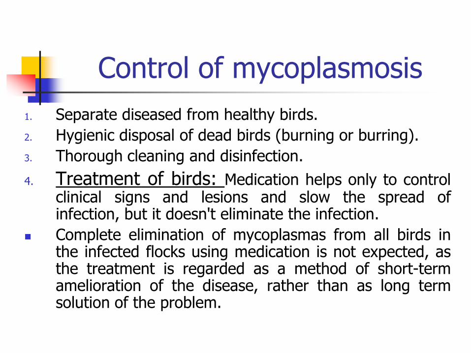
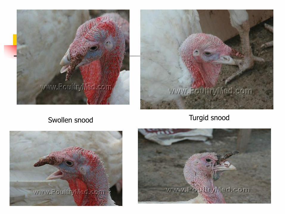

Signs of chlamydiosis
In turkeysI Acute epidemic Form (toxigenic highly virulent strain)Clinical signs appear as systemic fatal infection with1 Sudden death of 5-302 Fever and anorexia3 Nasal and ocular discharge swollen eye lids
conjunctivitis and sinusitis4 Off food and loss of weight (emaciation)5 Yellow green gelatinous diarrhea6 Rapid drop in egg production (40-50 decline)7 Morbidity rate may be reach 50-80
In ducks and geese (water fowl)
In duckling there are tremors trembling and staggered gait (incoordination)
Ocular and nasal discharge
Greenish watery Diarrhea
Inappetance
Emaciation convulsion and death
Morbidity rate 10-80
Mortality rate 1-30 (depending on the age health status and secondary infection with salmonella species)
Signs of chlamydiosis
Lesions of chlamydiosis
Congestion of the visceral organs
Congestion and pneumonia of the lungs
All the body cavities are filed with fibrinous exudates
Fibrinous pericarditis perihepatitis and airsacculitis and peritonitis
Catarrhal enteritis
Liver and spleen are enlarged dark and covered with gray white foci
Liver necrosisFibrinous pericarditis
liver hepatomegaly and congestion
fibrinous pericarditis
Fibrinous pericarditis
Caseous pericarditisLungs congested and fibrinous exudate
in the pleural cavity
Necrosis and congestion of
spleen
Enlarged congested
liver
Prehepatitis
Airsacculitis
A Thickened
abdominal airsacs
totally covered
with fibrin plaques
B Serous fluid
and fibrin in the
pericardial sac
C Severe
hepatomegaly
Diagnosis of chlamydiosis1 Detection of dark purple blue or pink (acc To the
stain) ICytoplasmic inclusion (LCL) bodies in stained smear (from nasal or ocular exudates serous membranes trachea lungs pancreas liver spleen heart faecal and cloacal swabs) in different stages of elementary body
2 Tissue culture3 Embryonated chicken eggs4 Laboratory animal inoculation Intra peritoneal or intra
cranial or intra nasal inoculation of 3-4 weeks old mice (3-6 mice) could resulted in death of mice after 5-7 days post inoculation Intra peritoneal inoculation resulted in peritonitis with accumulation of fibrinous exudate in the peritoneal cavity with splenomegaly
Prevention of chlamydiosis1 Different avian species and ages must be reared away
from each other2 Newly purchased (imported) birds especially pets must
be quarantined for 35 days (if positive discarded ortreated with tetracyclines till recovery)
3 Prevent the introduction of birds from the enzootic areas4 Avoid the contact with the free living wild birds5 Thorough cleaning and disinfection (iodophors or
formaldhydes)6 Regular testing of the birds using serological tests7 Restrict the movement of people and visitors8 Trials for vaccine preparation
Control of chlamydiosis
1 Oxytetracyclin (15mgkg bwt) in the drinking water for a period for 5 days
2 Chlorotetracyclin
(treatment may be extended to 15-45 days in severe infection)
3 Usage of combined antibiotics to control other associated infections as Salmonellae and Ecoli(quinolones like cipro enro and danofloxacin) for 3-5 days in the drinking water
AVIAN PASTEURELLOSIS
FOWL CHOLERA (FC)
FC
Peracute acute and chronic highly contagious bacterial disease of domestic and wild birds caused by Pasteurella multocida
In the acute form the disease is characterized by rapid course high morbidity and mortality and septicaemic picture while the chronic form is localized and follows the acute form or occur independently due to low virulent mo
Susceptibility of FC
1 Natural hosts are water fowl (ducks and geese) turkeys and chickens
2 Water fowl are the most susceptible birds
3 Turkeys are more susceptible than chickens
4 Adult chickens (more than 16 weeks old) while young turkeys are the most susceptible ages
Infection and transmission of FC
1 Inhalation of infected droplets
2 Ingestion of contaminated food and water by the infected excretions from the mouth and nostrils of diseased or carriers
3 Mechanically clothes feet or hands of workers contaminated equipments feed bags etchellip
4 Wild birds rodents and pets
5 Cannibalism of diseased or dead birds
6 Wound infection
Signs of FC
1 Per acute form
Sudden death without clinical signs
No characteristic post mortem lesions
2 Acute form
High morbidity and mortality rates
Septicaemia (cyanosis of the comb and wattle)
Mucous discharge from the mouth and nostrils
Watery white or greenish mucoid diarrhea
Respiratory manifestations
Drop in egg production
Lesions of FC
Acute form Petechial or echymotic hemorrhages on the
lungs proventriculus abdominal and coronary fats and on serosal membranes as peritoneum and subepicardium and haemorrhagic enteritis
Tracheitis bronchitis and pneumonia (TURKEYS) Oophoritis flaccid regressed or ruptured ova Free yolk material in the abdominal cavity Serofibrinous pericarditis and peritonitis Large amount of viscous mucous in the larynx
crop and intestine
Edematous cyanotic comb and wattles
Mucous excretion
from mouth
Fibrinous pericarditis and perihepatitisEnlarged liver
Small areas of necrosis in the liver (corn meal liver)
Subepicardial hemorrhages and petechiae
on the heart and coronary fat
The left lung has a severe
accumulation of fibrinous exudate on
its pleural surface In turkeys
Hemorrhagic mucosal and serosal lesions
Signs amp lesions of FC
3 Chronic forms
A Wattle form
Unilateral or bilateral swelling of wattle
Caseous exudates in the wattles cut sections
B Otitis media form
Nervous signs as torticollis
Caseous exudates in the middle ears and air spaces of cranial bones
Signs amp lesions of FC
C Arthritic form Swelling of the joints and lameness Caseous exudates in the joints Inflammation of the tendon sheet Hemorrhages and erosions on the articular
cartilageD Ovarian form Drop in egg production Cheesy material around the ovary and
peritoneal cavity
Signs amp lesions of FC
E Roup or catarrhal form
Respiratory signs (rals and dyspnea)
Mucous discharge from the mouth
Nasal discharge
Swelling of the sinuses and facial oedema
Conjunctivitis
Swelling of wattle
Swelling of the wattles
Swelling of the ear canal (middle ear)
Torticollis
Torticollis
strenal
bursitis
Arthritis
Caseous exudate over the hock region and
food pad
Swelling of the sternal bursae
Hyperemia and flaccid mature follicles
Egg-yolk peritonitis
Sinusitis and facial oedema
Yellow caseous exudate in the air spaces of the cranial bones
Diagnosis of FC
Demonstration of bipolar P multocida mo
either from the blood film in acute stage afterstaining with Giemsa Leishmanrsquos stains andseen under microscope
or take impression smear from organs (liver)stain and examine microscopically
Bipolar Pmultocida in between nucleated RBCS in acute infection
Rough colony on the right and a smooth colony on the left
Diagnosis of FC
Laboratory animal inoculationIntraperitoneal or SC inoculation of rabbit or mice
or hamster with blood of infected bird orsuspected material (liver supernatant) (02 ml)induces death of these animals within 24-48 hourspost inoculation with septicaemic pictureImpression smear from blood or tissues (liver)shows bipolar organism
Prevention of FC Adopt all biosecurity measures
Vaccination
P multocida antigen vaccine should be homologous tothat cause fowl cholera or use of autogenous localprepared vaccine in endemic area or when commercialones are not protective
There are many types of vaccines against FC
Whole cell bactrine
Live attenuated vaccine
a CU- vaccine
b Mutants of CU-vaccine as M-9 MN and PM-1 strainsto avoid post vaccinal reaction of the CU
Prevention of FC
In turkeys
Meat type 6-8 weeks bactrine injection or CU via drinking water and repeated every 4-6 weeks till marketing
Breeder flocks must be vaccinated first at 6- 8 weeks and revaccinated 2-5 times till start of egg production according to epidemiology of FC in the area and farm
Control of FC
In chicken and turkeys sulfamerazine 02 in water or 04 in feed or Sulfaquinoxaline 001 ndash005 in water for 3-5 days to stop deaths in 2 days
Streptomycin (150 mg) for IlM injection in turkeys
Chloro- or oxytetracycline 40 mgkgbw by IlM
Chloramphenicol 20 mgkgbw via IlM
Enrofloxacin hellipetc
AVIAN SPIROKETOSIS
Avian spiroketosis
The bacterium Borrelia anserina infects chickens turkey geese ducks pheasants grouse and canaries with morbidity and mortality up to 100
It is transmitted by arthropods eg Argas persicus and occasionally by infected faeces The bacterium is poorly resistant outside host but may be carried by Argas persicus for 430 days
Brachyspira pilosicoli previously known as Serpulina pilosicoli is an intestinal spirochaete that can be associated with inflammation of the large intestine in a broad range of mammals and birds
It has been associated with typhilitis diarrhoea reduced egg production and egg soiling in chickens
Signs amp lesions of avian spiroketosis
Depression and thirst Cyanosis Often diarrhoea with excessive urates Weakness and progressive paralysis Drops in egg production may be seen in both systemic
and intestinal forms Post-mortem lesions Marked splenomegaly Spleen mottled with ecchymotic haemorrhages Liver enlarged with small haemorrhages Necrotic foci Mucoid enteritis
Prevention amp Control of avian spiroketosis
Treatment
Various antibiotics including penicillin
Prevention
Control vectors
Vaccines in some countries
Cause
Age group affected
Transmission
Forms of the disease
Escherichia coli
All types of avian and all ages
Fecal oral route transovarial contamination of the egg shell via fecal material from hen
Colisepticaemia
Airsac disease
Chronic forms (arthritis omphalitis panophthalmitis salpingitis enteritis coligranuloma and SHS)
E coli
Severely congested liver
Foci of necrosis on liver
GANGARINOUS
DERMATITIS
(GD)
GAS OEDEMA DISEASE
WING ROT
Gangrenous dermatitis
Gangrenous (necrotic) dermatitis in broilers and turkeys between 3-7 weeks old
It is caused by a bacteria- Clostridium septicum Escherichia coli Staphylococcus aureus
Disease often occurs in birds which are already immunosuppressed due to a prior infection (Chicken anaemia Infectious Bursal Disease) or mycotoxin ingestion
Transmission is by contact with infected wet caked litter The disease often occurs in immunosuppressed birds
Signs of GD
Loss of feathers
Pale combs and wattles depression
Incoordination
Leg weakness and ataxia (canrsquot move) can be seen
Mortality is low but dead birds decompose quickly
Lesions of GD
Congestion haemorrhage and necrosis of skin with intro lesion bacteria under the microscope
In turkeys with cellulitis of the tail (bubbly tail) edema and vesicle-like lesions were present laterally and ventrally around the tail
Tail feathers were soft blood-filled and broken
Dark and moist skin of the breast
Necrosis of the abdomen skinAffected leg and toes
Necrosis of the wattle
Ulceration and gangrenous formation of skin
Prevention amp Control of GD
Prevention Clean out house and add new litter to prevent the
disease Medicate in starter feed Flavomycin Virginamyciin
Bacitrcin and CTC can reduce bacteria Proper vaccination against IBDV CAV and MDV prevent
mycotoxin formation in the feed and eliminate ALV in the breeders to prevent immunosuppression
Treatment Erythromycin penicillin in the feed to treat signs
Chlortetracycine oxytetracycline copper sulphate can be added to the water to reduce morbidity
ULCERATIVE ENERITIS
(UE)
Ulcerative Enteritis
Turkeys of 6-14 weeks are susceptible to this acute to chronic disease
The agent involved in the aetiology of this disease is clostridium colinum which is spore-forming gram-positive aerobic and non-motile
Mode of transmission
Vectors are faeces soil and litter containing the bacteria
It often accompanies coccidiosis in broilers
Signs amp lesions of UE
High mortality watery diarrhoea ruffled feathers dull listlessness increased thirst emaciation and atrophy of pectoral muscles can occur
Postmortem lesions Yellow irregular ulcers on small intestine and
caeca hemorrhagic enteritis are seen Enlarged haemorrhagic necrotic spleen light
yellow mottling of liver and crop filled with water may occur
Prevention amp Control of UE
Prevention Improved sanitation Adding salt to the soil (500
lbs of salthouse) may kill spores Raising birds on wire andor feeding bacitracin at 50-100 gt will prevent the disease
Treatment NF-180 (50-100 gt) strephomycin (60 gt) and
chlortetracycline vitamins and minerals in water andor lincomycin 2 gt will reduce the signs
Remove dead birds and feed bacitracin (200 gt)
Thank you
ANY QUESTIONS
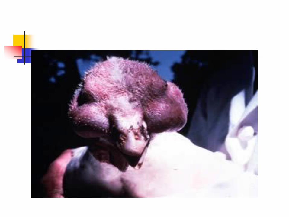

Signs amp lesions of FC
C Arthritic form Swelling of the joints and lameness Caseous exudates in the joints Inflammation of the tendon sheet Hemorrhages and erosions on the articular
cartilageD Ovarian form Drop in egg production Cheesy material around the ovary and
peritoneal cavity
Signs amp lesions of FC
E Roup or catarrhal form
Respiratory signs (rals and dyspnea)
Mucous discharge from the mouth
Nasal discharge
Swelling of the sinuses and facial oedema
Conjunctivitis
Swelling of wattle
Swelling of the wattles
Swelling of the ear canal (middle ear)
Torticollis
Torticollis
strenal
bursitis
Arthritis
Caseous exudate over the hock region and
food pad
Swelling of the sternal bursae
Hyperemia and flaccid mature follicles
Egg-yolk peritonitis
Sinusitis and facial oedema
Yellow caseous exudate in the air spaces of the cranial bones
Diagnosis of FC
Demonstration of bipolar P multocida mo
either from the blood film in acute stage afterstaining with Giemsa Leishmanrsquos stains andseen under microscope
or take impression smear from organs (liver)stain and examine microscopically
Bipolar Pmultocida in between nucleated RBCS in acute infection
Rough colony on the right and a smooth colony on the left
Diagnosis of FC
Laboratory animal inoculationIntraperitoneal or SC inoculation of rabbit or mice
or hamster with blood of infected bird orsuspected material (liver supernatant) (02 ml)induces death of these animals within 24-48 hourspost inoculation with septicaemic pictureImpression smear from blood or tissues (liver)shows bipolar organism
Prevention of FC Adopt all biosecurity measures
Vaccination
P multocida antigen vaccine should be homologous tothat cause fowl cholera or use of autogenous localprepared vaccine in endemic area or when commercialones are not protective
There are many types of vaccines against FC
Whole cell bactrine
Live attenuated vaccine
a CU- vaccine
b Mutants of CU-vaccine as M-9 MN and PM-1 strainsto avoid post vaccinal reaction of the CU
Prevention of FC
In turkeys
Meat type 6-8 weeks bactrine injection or CU via drinking water and repeated every 4-6 weeks till marketing
Breeder flocks must be vaccinated first at 6- 8 weeks and revaccinated 2-5 times till start of egg production according to epidemiology of FC in the area and farm
Control of FC
In chicken and turkeys sulfamerazine 02 in water or 04 in feed or Sulfaquinoxaline 001 ndash005 in water for 3-5 days to stop deaths in 2 days
Streptomycin (150 mg) for IlM injection in turkeys
Chloro- or oxytetracycline 40 mgkgbw by IlM
Chloramphenicol 20 mgkgbw via IlM
Enrofloxacin hellipetc
AVIAN SPIROKETOSIS
Avian spiroketosis
The bacterium Borrelia anserina infects chickens turkey geese ducks pheasants grouse and canaries with morbidity and mortality up to 100
It is transmitted by arthropods eg Argas persicus and occasionally by infected faeces The bacterium is poorly resistant outside host but may be carried by Argas persicus for 430 days
Brachyspira pilosicoli previously known as Serpulina pilosicoli is an intestinal spirochaete that can be associated with inflammation of the large intestine in a broad range of mammals and birds
It has been associated with typhilitis diarrhoea reduced egg production and egg soiling in chickens
Signs amp lesions of avian spiroketosis
Depression and thirst Cyanosis Often diarrhoea with excessive urates Weakness and progressive paralysis Drops in egg production may be seen in both systemic
and intestinal forms Post-mortem lesions Marked splenomegaly Spleen mottled with ecchymotic haemorrhages Liver enlarged with small haemorrhages Necrotic foci Mucoid enteritis
Prevention amp Control of avian spiroketosis
Treatment
Various antibiotics including penicillin
Prevention
Control vectors
Vaccines in some countries
Cause
Age group affected
Transmission
Forms of the disease
Escherichia coli
All types of avian and all ages
Fecal oral route transovarial contamination of the egg shell via fecal material from hen
Colisepticaemia
Airsac disease
Chronic forms (arthritis omphalitis panophthalmitis salpingitis enteritis coligranuloma and SHS)
E coli
Severely congested liver
Foci of necrosis on liver
GANGARINOUS
DERMATITIS
(GD)
GAS OEDEMA DISEASE
WING ROT
Gangrenous dermatitis
Gangrenous (necrotic) dermatitis in broilers and turkeys between 3-7 weeks old
It is caused by a bacteria- Clostridium septicum Escherichia coli Staphylococcus aureus
Disease often occurs in birds which are already immunosuppressed due to a prior infection (Chicken anaemia Infectious Bursal Disease) or mycotoxin ingestion
Transmission is by contact with infected wet caked litter The disease often occurs in immunosuppressed birds
Signs of GD
Loss of feathers
Pale combs and wattles depression
Incoordination
Leg weakness and ataxia (canrsquot move) can be seen
Mortality is low but dead birds decompose quickly
Lesions of GD
Congestion haemorrhage and necrosis of skin with intro lesion bacteria under the microscope
In turkeys with cellulitis of the tail (bubbly tail) edema and vesicle-like lesions were present laterally and ventrally around the tail
Tail feathers were soft blood-filled and broken
Dark and moist skin of the breast
Necrosis of the abdomen skinAffected leg and toes
Necrosis of the wattle
Ulceration and gangrenous formation of skin
Prevention amp Control of GD
Prevention Clean out house and add new litter to prevent the
disease Medicate in starter feed Flavomycin Virginamyciin
Bacitrcin and CTC can reduce bacteria Proper vaccination against IBDV CAV and MDV prevent
mycotoxin formation in the feed and eliminate ALV in the breeders to prevent immunosuppression
Treatment Erythromycin penicillin in the feed to treat signs
Chlortetracycine oxytetracycline copper sulphate can be added to the water to reduce morbidity
ULCERATIVE ENERITIS
(UE)
Ulcerative Enteritis
Turkeys of 6-14 weeks are susceptible to this acute to chronic disease
The agent involved in the aetiology of this disease is clostridium colinum which is spore-forming gram-positive aerobic and non-motile
Mode of transmission
Vectors are faeces soil and litter containing the bacteria
It often accompanies coccidiosis in broilers
Signs amp lesions of UE
High mortality watery diarrhoea ruffled feathers dull listlessness increased thirst emaciation and atrophy of pectoral muscles can occur
Postmortem lesions Yellow irregular ulcers on small intestine and
caeca hemorrhagic enteritis are seen Enlarged haemorrhagic necrotic spleen light
yellow mottling of liver and crop filled with water may occur
Prevention amp Control of UE
Prevention Improved sanitation Adding salt to the soil (500
lbs of salthouse) may kill spores Raising birds on wire andor feeding bacitracin at 50-100 gt will prevent the disease
Treatment NF-180 (50-100 gt) strephomycin (60 gt) and
chlortetracycline vitamins and minerals in water andor lincomycin 2 gt will reduce the signs
Remove dead birds and feed bacitracin (200 gt)
Thank you
ANY QUESTIONS
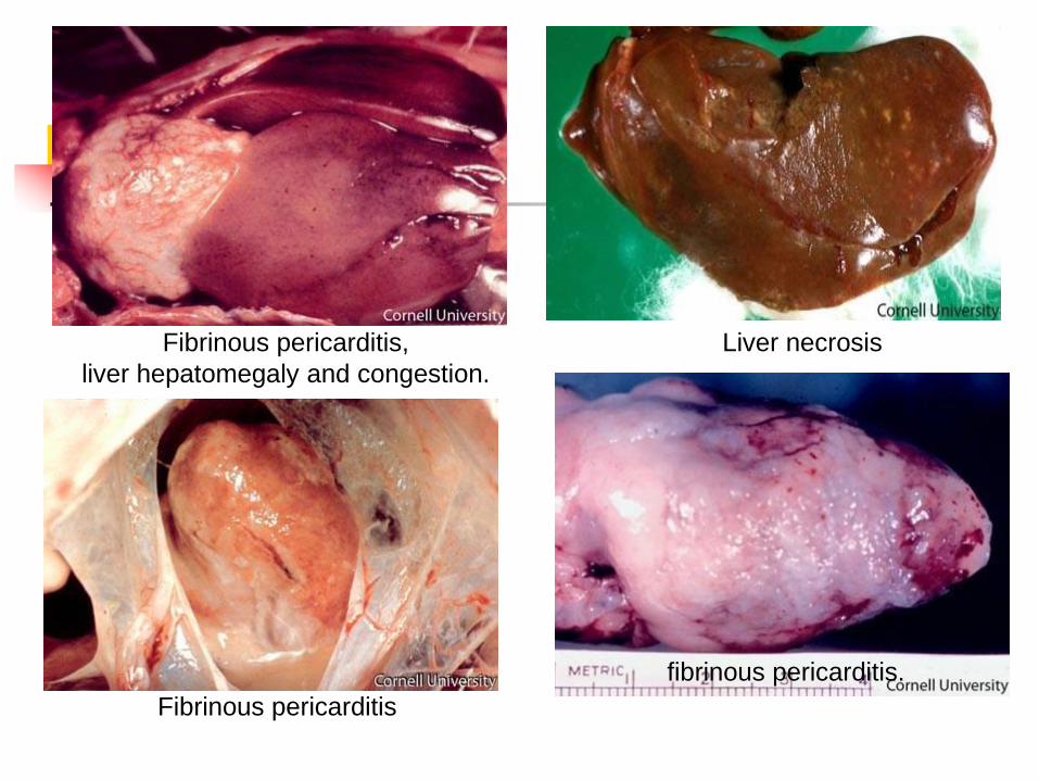
Signs amp lesions of FC
E Roup or catarrhal form
Respiratory signs (rals and dyspnea)
Mucous discharge from the mouth
Nasal discharge
Swelling of the sinuses and facial oedema
Conjunctivitis
Swelling of wattle
Swelling of the wattles
Swelling of the ear canal (middle ear)
Torticollis
Torticollis
strenal
bursitis
Arthritis
Caseous exudate over the hock region and
food pad
Swelling of the sternal bursae
Hyperemia and flaccid mature follicles
Egg-yolk peritonitis
Sinusitis and facial oedema
Yellow caseous exudate in the air spaces of the cranial bones
Diagnosis of FC
Demonstration of bipolar P multocida mo
either from the blood film in acute stage afterstaining with Giemsa Leishmanrsquos stains andseen under microscope
or take impression smear from organs (liver)stain and examine microscopically
Bipolar Pmultocida in between nucleated RBCS in acute infection
Rough colony on the right and a smooth colony on the left
Diagnosis of FC
Laboratory animal inoculationIntraperitoneal or SC inoculation of rabbit or mice
or hamster with blood of infected bird orsuspected material (liver supernatant) (02 ml)induces death of these animals within 24-48 hourspost inoculation with septicaemic pictureImpression smear from blood or tissues (liver)shows bipolar organism
Prevention of FC Adopt all biosecurity measures
Vaccination
P multocida antigen vaccine should be homologous tothat cause fowl cholera or use of autogenous localprepared vaccine in endemic area or when commercialones are not protective
There are many types of vaccines against FC
Whole cell bactrine
Live attenuated vaccine
a CU- vaccine
b Mutants of CU-vaccine as M-9 MN and PM-1 strainsto avoid post vaccinal reaction of the CU
Prevention of FC
In turkeys
Meat type 6-8 weeks bactrine injection or CU via drinking water and repeated every 4-6 weeks till marketing
Breeder flocks must be vaccinated first at 6- 8 weeks and revaccinated 2-5 times till start of egg production according to epidemiology of FC in the area and farm
Control of FC
In chicken and turkeys sulfamerazine 02 in water or 04 in feed or Sulfaquinoxaline 001 ndash005 in water for 3-5 days to stop deaths in 2 days
Streptomycin (150 mg) for IlM injection in turkeys
Chloro- or oxytetracycline 40 mgkgbw by IlM
Chloramphenicol 20 mgkgbw via IlM
Enrofloxacin hellipetc
AVIAN SPIROKETOSIS
Avian spiroketosis
The bacterium Borrelia anserina infects chickens turkey geese ducks pheasants grouse and canaries with morbidity and mortality up to 100
It is transmitted by arthropods eg Argas persicus and occasionally by infected faeces The bacterium is poorly resistant outside host but may be carried by Argas persicus for 430 days
Brachyspira pilosicoli previously known as Serpulina pilosicoli is an intestinal spirochaete that can be associated with inflammation of the large intestine in a broad range of mammals and birds
It has been associated with typhilitis diarrhoea reduced egg production and egg soiling in chickens
Signs amp lesions of avian spiroketosis
Depression and thirst Cyanosis Often diarrhoea with excessive urates Weakness and progressive paralysis Drops in egg production may be seen in both systemic
and intestinal forms Post-mortem lesions Marked splenomegaly Spleen mottled with ecchymotic haemorrhages Liver enlarged with small haemorrhages Necrotic foci Mucoid enteritis
Prevention amp Control of avian spiroketosis
Treatment
Various antibiotics including penicillin
Prevention
Control vectors
Vaccines in some countries
Cause
Age group affected
Transmission
Forms of the disease
Escherichia coli
All types of avian and all ages
Fecal oral route transovarial contamination of the egg shell via fecal material from hen
Colisepticaemia
Airsac disease
Chronic forms (arthritis omphalitis panophthalmitis salpingitis enteritis coligranuloma and SHS)
E coli
Severely congested liver
Foci of necrosis on liver
GANGARINOUS
DERMATITIS
(GD)
GAS OEDEMA DISEASE
WING ROT
Gangrenous dermatitis
Gangrenous (necrotic) dermatitis in broilers and turkeys between 3-7 weeks old
It is caused by a bacteria- Clostridium septicum Escherichia coli Staphylococcus aureus
Disease often occurs in birds which are already immunosuppressed due to a prior infection (Chicken anaemia Infectious Bursal Disease) or mycotoxin ingestion
Transmission is by contact with infected wet caked litter The disease often occurs in immunosuppressed birds
Signs of GD
Loss of feathers
Pale combs and wattles depression
Incoordination
Leg weakness and ataxia (canrsquot move) can be seen
Mortality is low but dead birds decompose quickly
Lesions of GD
Congestion haemorrhage and necrosis of skin with intro lesion bacteria under the microscope
In turkeys with cellulitis of the tail (bubbly tail) edema and vesicle-like lesions were present laterally and ventrally around the tail
Tail feathers were soft blood-filled and broken
Dark and moist skin of the breast
Necrosis of the abdomen skinAffected leg and toes
Necrosis of the wattle
Ulceration and gangrenous formation of skin
Prevention amp Control of GD
Prevention Clean out house and add new litter to prevent the
disease Medicate in starter feed Flavomycin Virginamyciin
Bacitrcin and CTC can reduce bacteria Proper vaccination against IBDV CAV and MDV prevent
mycotoxin formation in the feed and eliminate ALV in the breeders to prevent immunosuppression
Treatment Erythromycin penicillin in the feed to treat signs
Chlortetracycine oxytetracycline copper sulphate can be added to the water to reduce morbidity
ULCERATIVE ENERITIS
(UE)
Ulcerative Enteritis
Turkeys of 6-14 weeks are susceptible to this acute to chronic disease
The agent involved in the aetiology of this disease is clostridium colinum which is spore-forming gram-positive aerobic and non-motile
Mode of transmission
Vectors are faeces soil and litter containing the bacteria
It often accompanies coccidiosis in broilers
Signs amp lesions of UE
High mortality watery diarrhoea ruffled feathers dull listlessness increased thirst emaciation and atrophy of pectoral muscles can occur
Postmortem lesions Yellow irregular ulcers on small intestine and
caeca hemorrhagic enteritis are seen Enlarged haemorrhagic necrotic spleen light
yellow mottling of liver and crop filled with water may occur
Prevention amp Control of UE
Prevention Improved sanitation Adding salt to the soil (500
lbs of salthouse) may kill spores Raising birds on wire andor feeding bacitracin at 50-100 gt will prevent the disease
Treatment NF-180 (50-100 gt) strephomycin (60 gt) and
chlortetracycline vitamins and minerals in water andor lincomycin 2 gt will reduce the signs
Remove dead birds and feed bacitracin (200 gt)
Thank you
ANY QUESTIONS

Diagnosis of FC
Demonstration of bipolar P multocida mo
either from the blood film in acute stage afterstaining with Giemsa Leishmanrsquos stains andseen under microscope
or take impression smear from organs (liver)stain and examine microscopically
Bipolar Pmultocida in between nucleated RBCS in acute infection
Rough colony on the right and a smooth colony on the left
Diagnosis of FC
Laboratory animal inoculationIntraperitoneal or SC inoculation of rabbit or mice
or hamster with blood of infected bird orsuspected material (liver supernatant) (02 ml)induces death of these animals within 24-48 hourspost inoculation with septicaemic pictureImpression smear from blood or tissues (liver)shows bipolar organism
Prevention of FC Adopt all biosecurity measures
Vaccination
P multocida antigen vaccine should be homologous tothat cause fowl cholera or use of autogenous localprepared vaccine in endemic area or when commercialones are not protective
There are many types of vaccines against FC
Whole cell bactrine
Live attenuated vaccine
a CU- vaccine
b Mutants of CU-vaccine as M-9 MN and PM-1 strainsto avoid post vaccinal reaction of the CU
Prevention of FC
In turkeys
Meat type 6-8 weeks bactrine injection or CU via drinking water and repeated every 4-6 weeks till marketing
Breeder flocks must be vaccinated first at 6- 8 weeks and revaccinated 2-5 times till start of egg production according to epidemiology of FC in the area and farm
Control of FC
In chicken and turkeys sulfamerazine 02 in water or 04 in feed or Sulfaquinoxaline 001 ndash005 in water for 3-5 days to stop deaths in 2 days
Streptomycin (150 mg) for IlM injection in turkeys
Chloro- or oxytetracycline 40 mgkgbw by IlM
Chloramphenicol 20 mgkgbw via IlM
Enrofloxacin hellipetc
AVIAN SPIROKETOSIS
Avian spiroketosis
The bacterium Borrelia anserina infects chickens turkey geese ducks pheasants grouse and canaries with morbidity and mortality up to 100
It is transmitted by arthropods eg Argas persicus and occasionally by infected faeces The bacterium is poorly resistant outside host but may be carried by Argas persicus for 430 days
Brachyspira pilosicoli previously known as Serpulina pilosicoli is an intestinal spirochaete that can be associated with inflammation of the large intestine in a broad range of mammals and birds
It has been associated with typhilitis diarrhoea reduced egg production and egg soiling in chickens
Signs amp lesions of avian spiroketosis
Depression and thirst Cyanosis Often diarrhoea with excessive urates Weakness and progressive paralysis Drops in egg production may be seen in both systemic
and intestinal forms Post-mortem lesions Marked splenomegaly Spleen mottled with ecchymotic haemorrhages Liver enlarged with small haemorrhages Necrotic foci Mucoid enteritis
Prevention amp Control of avian spiroketosis
Treatment
Various antibiotics including penicillin
Prevention
Control vectors
Vaccines in some countries
Cause
Age group affected
Transmission
Forms of the disease
Escherichia coli
All types of avian and all ages
Fecal oral route transovarial contamination of the egg shell via fecal material from hen
Colisepticaemia
Airsac disease
Chronic forms (arthritis omphalitis panophthalmitis salpingitis enteritis coligranuloma and SHS)
E coli
Severely congested liver
Foci of necrosis on liver
GANGARINOUS
DERMATITIS
(GD)
GAS OEDEMA DISEASE
WING ROT
Gangrenous dermatitis
Gangrenous (necrotic) dermatitis in broilers and turkeys between 3-7 weeks old
It is caused by a bacteria- Clostridium septicum Escherichia coli Staphylococcus aureus
Disease often occurs in birds which are already immunosuppressed due to a prior infection (Chicken anaemia Infectious Bursal Disease) or mycotoxin ingestion
Transmission is by contact with infected wet caked litter The disease often occurs in immunosuppressed birds
Signs of GD
Loss of feathers
Pale combs and wattles depression
Incoordination
Leg weakness and ataxia (canrsquot move) can be seen
Mortality is low but dead birds decompose quickly
Lesions of GD
Congestion haemorrhage and necrosis of skin with intro lesion bacteria under the microscope
In turkeys with cellulitis of the tail (bubbly tail) edema and vesicle-like lesions were present laterally and ventrally around the tail
Tail feathers were soft blood-filled and broken
Dark and moist skin of the breast
Necrosis of the abdomen skinAffected leg and toes
Necrosis of the wattle
Ulceration and gangrenous formation of skin
Prevention amp Control of GD
Prevention Clean out house and add new litter to prevent the
disease Medicate in starter feed Flavomycin Virginamyciin
Bacitrcin and CTC can reduce bacteria Proper vaccination against IBDV CAV and MDV prevent
mycotoxin formation in the feed and eliminate ALV in the breeders to prevent immunosuppression
Treatment Erythromycin penicillin in the feed to treat signs
Chlortetracycine oxytetracycline copper sulphate can be added to the water to reduce morbidity
ULCERATIVE ENERITIS
(UE)
Ulcerative Enteritis
Turkeys of 6-14 weeks are susceptible to this acute to chronic disease
The agent involved in the aetiology of this disease is clostridium colinum which is spore-forming gram-positive aerobic and non-motile
Mode of transmission
Vectors are faeces soil and litter containing the bacteria
It often accompanies coccidiosis in broilers
Signs amp lesions of UE
High mortality watery diarrhoea ruffled feathers dull listlessness increased thirst emaciation and atrophy of pectoral muscles can occur
Postmortem lesions Yellow irregular ulcers on small intestine and
caeca hemorrhagic enteritis are seen Enlarged haemorrhagic necrotic spleen light
yellow mottling of liver and crop filled with water may occur
Prevention amp Control of UE
Prevention Improved sanitation Adding salt to the soil (500
lbs of salthouse) may kill spores Raising birds on wire andor feeding bacitracin at 50-100 gt will prevent the disease
Treatment NF-180 (50-100 gt) strephomycin (60 gt) and
chlortetracycline vitamins and minerals in water andor lincomycin 2 gt will reduce the signs
Remove dead birds and feed bacitracin (200 gt)
Thank you
ANY QUESTIONS

Thank you
ANY QUESTIONS

ANY QUESTIONS