bacterial-invertebrate symbioses: from anasphalt
TRANSCRIPT

Bacterial-invertebrate
symbioses: from an asphalt
cold seep to shallow waters
Dissertation
zur Erlangung des Grades eines
Doktors der Naturwissenschaften
- Dr. rer. nat. -
dem Fachbereich Biologie/Chemie der
Universitat Bremen
vorgelegt von
Luciana Raggi Hoyos
Bremen
September 2010

Die vorliegende Arbeit wurde in der Zeit von April 2007 bis August 2010 in
der Symbiose Gruppe am Max-Planck Institut fur marine Mikrobiologie in
Bremen angefertigt.
1. Gutachterin: Dr. Nicole Dubilier
2. Gutachter: Prof. Dr. Ulrich Fischer
Tag des Promotionskolloquiums: 18. Oktober 2010

To Pablo
to my family

‘La simbiosis, la union de distintos organismos para formar nuevos
colectivos, ha resultado ser la mas importante fuerza de cambio sobre la
Tierra’
L. Margulis & D. Sagan, 1995
‘La union hace la fuerza’
- Frase popular

Abstract
Symbiotic associations are complex partnerships that can lead to new metabolic
capabilities and the establishment of novel organisms. The diversity of these as-
sociations is very broad and there are still many mysteries about the origin and
the exact relationship between the organisms that are involved in a symbiosis
(host and symbiont). Some of these associations are essential to the hosts, such
as the chemosynthetic symbioses occurring in invertebrates of the deep-sea. In
others the host probably would rather not be the host, as in the case of parasitic
microbes. My PhD research focuses on symbiotic and parasitic associations in
chemosynthetic and non-chemosynthetic invertebrates. This thesis describes and
discusses three different aspects of associations between bacteria and marine in-
vertebrates. The first aspect focuses on chemosynthetic associations from a unique
asphalt seep called Chapopote in the Gulf of Mexico (GoM). Phylogenetic analyses
of host genes (cytochrome-c-oxidase subunit I) and bacterial genes (16S rRNA) in
two Bathymodiolus mussel species and an Escarpia tubeworm showed that both
the hosts and their chemosynthetic symbionts are very similar to their congeners
from the northern GoM. Unexpectedly, a novel symbiont most closely related to
hydrocarbon degrading bacteria of the genus Cycloclasticus was discovered in B.
heckerae. Stable carbon isotope values in B. heckerae tissues of lipids typical for
Cycloclasticus spp. were consistently heavier by 2.5� than other lipids indicating
that the novel symbiont might use isotopically heavy hydrocarbons from the as-
phalt seep as an energy and carbon source. The discovery of a novel symbiont that
may be able to metabolize hydrocarbons is particularly intriguing because until
now only methane and reduced sulfur compounds have been identified as energy
sources in chemosynthetic symbioses. The large amounts of hydrocarbons available
at Chapopote would provide these mussel symbioses with a rich source of nutri-
tion. The second aspect of this thesis deals with bacteria that infect the nuclei of
vi

marine invertebrates and were recently found to be widespread in deep-sea Bathy-
modiolus mussels. Because of their potentially lethal effect on bivalve populations,
I looked for the presence of intranuclear bacteria in economically important and
commercially available bivalve species, i.e. oysters (Crassostrea gigas), razor clams
(Siliqua patula and Ensis directus), blue mussels (Mytilus edulis), Manila clams
(Venerupis philippinarum), and common cockles (Cerastoderma edule). Fluores-
cence in situ hybridization (FISH) revealed the presence of intranuclear bacteria in
all investigated bivalves except oysters and blue mussels. Preliminary tests with
real-time PCR showed massive amounts of intranuclear bacteria in some of the
bivalve species, raising the question if these might affect not only the health of
the bivalves but possibly also of the humans that eat them. In the third and
final aspect of my thesis, I examined the general diversity of bacteria in the gill
tissues of deep-sea and shallow-water mussels and clams. Comparative 16S rRNA
sequence analysis and cultivation experiments revealed a much higher diversity
than previously recognized. This thesis shows that bivalves are ideal models for
studying the microbiota of marine invertebrates because of the high diversity of
both highly specific and more generalized symbiotic and parasitic bacteria in their
gill tissues.

Zusammenfassung
Symbiotische Assoziationen sind komplexe Partnerschaften, die zu neuen metabo-
lischen Fahigkeiten und der Etablierung neuartiger Organismen fuhren konnen.
Die Vielfalt dieser Assoziationen ist sehr hoch, und in vielen Fallen bleiben ihr
Ursprung und die genaue Beziehung zwischen den in die Symbiose eingebunde-
nen Organismen (Wirt und Symbiont) ungeklart. Einige dieser Verbindungen sind
unverzichtbar fur den Wirt, wie etwa die chemosynthetische Symbionten, die bei
Invertebraten in der Tiefsee vorkommen. In einigen anderen ware der Wirt wohl
lieber nicht der Wirt, wie im Fall von parasitischen Mikroorganismen. Die For-
schung meiner Dissertation konzentriert sich auf symbiotische und parasitische
Assoziationen in chemosynthetischen und nicht-chemosynthetischen Wirbellosen.
Die vorliegende Arbeit beschreibt und diskutiert drei verschiedene Aspek-
te der Assoziationen zwischen Bakterien und marinen Invertebraten. Der erste
Aspekt konzentriert sich auf chemosynthetische Assoziationen an einem einzigarti-
gen Asphaltvulkan, dem Chapopote im Golf von Mexico (GoM). Phylogenetische
Analysen von Wirtsgenen (Cytochrom-c-Oxidase Untereinheit I) und bakteriel-
len Genen (16S rRNA) in zwei Bathymodiolus-Muschelarten und einem Escarpia-
Rohrenwurm haben gezeigt, dass sowohl die Wirte als auch ihre chemosynthe-
tischen Symbionten ihren Artverwandten aus dem nordlichen GoM sehr ahnlich
sind. Unerwarteterweise wurde in B. heckerae ein neuer Symbiont entdeckt, der
am nachsten mit den Kohlenwasserstoffe abbauenden Bakterien des Genus Cy-
cloclasticus verwandt ist. Die stabilen Kohlenstoffisotope der fur Cycloclasticus
typischen Lipide in den Geweben von B. heckerae waren durchgangig um 2.5�schwerer als bei anderen Lipiden. Dies deutet darauf hin, dass der neuartige Sym-
biont isotopenschwere Kohlenwasserstoffe aus dem Asphaltvulkan als Energie- und
Kohlenstoffquelle nutzen konnte. Die Entdeckung eines neuartigen Symbionten,
der in der Lage sein konnte, Kohlenwasserstoffe zu metabolisieren, ist besonders
viii

faszinierend, da bisher nur Methan und reduzierte Schwefelverbindungen als Ener-
giequelle in chemosynthetischen Symbiosen identifiziert worden sind. Die großen
Mengen von Kohlenwasserstoffen, die bei Chapopote verfugbar sind, wurden dieser
Muschelsymbiose eine reichhaltige Nahrstoffquelle zur Verfugung stellen.
Der zweite Aspekt dieser Arbeit beschaftigt sich mit Bakterien, die die Zellker-
ne von marinen Invertebraten infizieren und vor Kurzem weit verbreitet in Bathy-
modiolus-Muscheln der Tiefsee gefunden wurden. Wegen ihrer potentiell todlichen
Auswirkungen auf Bivalven-Populationen habe ich besonders nach der Prasenz von
intranuklearen Bakterien in okonomisch bedeutsamen und kommerziell erhaltlichen
Muschelspezies gesucht, d.h. in Austern (Crassostrea gigas), Schwertmuscheln (Si-
liqua patula und Ensis directus), Miesmuscheln (Mytilus edulis), Venusmuscheln
(Venerupis philippinarum) und Herzmuscheln (Cerastoderma edule). Die Fluores-
zenz-in-situ-Hybridisierung (FISH) brachte intranukleare Bakterien in allen unter-
suchten Muscheln zum Vorschein, außer in Austern und Miesmuscheln. Vorlaufige
Tests mit Hilfe der Real-time PCR zeigten hohe Mengen von intranuklearen Bak-
terien in einigen der Bivalvenspezies, was die Frage aufwirft, ob diese nicht nur die
Gesundheit der Muscheln, sondern moglicherweise auch die der sie verzehrenden
Menschen beeintrachtigen konnten.
Im dritten und letzten Aspekt meiner Doktorarbeit habe ich die allgemeine
Diversitat von Bakterien in den Kiemengeweben von Tiefsee- und Flachwassermu-
scheln untersucht. Vergleichende 16S rRNA-Sequenzanalyse und Kultivierungsex-
perimente haben eine deutlich hohere Diversitat enthullt, als vorher bekannt war.
Diese Dissertation zeigt, dass Bivalvia aufgrund der hohen Diversitat von sowohl
hochspezifischen als auch generalisierten symbiotischen und parasitischen Bakteri-
en in ihren Kiemengeweben ideale Modellorganismen sind, um die Mikrobiota von
marinen Invertebraten zu studieren.

Contents
About the structure of this thesis . . . . . . . . . . . . . . . . . . . xvi
I Introduction 2
1 Invertebrate-bacteria associations 3
1.1 The different models . . . . . . . . . . . . . . . . . . . . . . . 4
1.1.1 Insects . . . . . . . . . . . . . . . . . . . . . . . . . . . 5
1.1.2 Squid . . . . . . . . . . . . . . . . . . . . . . . . . . . 9
1.1.3 Gutless oligochaetes . . . . . . . . . . . . . . . . . . . 10
1.1.4 Vesicomyid clams . . . . . . . . . . . . . . . . . . . . . 10
1.2 Summary: The role of symbioses . . . . . . . . . . . . . . . . 13
Concept - Box 1: Symbiosis and symbiology . . . . . . . . . . . . . 14
2 Habitats 15
2.1 Deep-sea cold seeps . . . . . . . . . . . . . . . . . . . . . . . . 15
2.1.1 Gulf of Mexico . . . . . . . . . . . . . . . . . . . . . . 16
2.1.2 Chapopote . . . . . . . . . . . . . . . . . . . . . . . . 18
2.2 Shallow-water coastal zone . . . . . . . . . . . . . . . . . . . . 18
3 Hosts 20
3.1 Deep-sea Bathymodiolus mussels . . . . . . . . . . . . . . . . . 20
3.2 Deep-sea Escarpia tubeworms . . . . . . . . . . . . . . . . . . 22
3.3 Shallow-water bivalves . . . . . . . . . . . . . . . . . . . . . . 27
Concept - Box 2: Immunology of bivalves . . . . . . . . . . . . . . . 28
x

TABLE OF CONTENTS
4 Bacterial Symbionts 29
4.1 Chemosynthetic symbionts . . . . . . . . . . . . . . . . . . . . 29
4.1.1 Thiotrophic symbionts . . . . . . . . . . . . . . . . . . 31
4.1.2 Methanotrophic symbionts . . . . . . . . . . . . . . . . 31
4.2 Hydrocarbon degraders . . . . . . . . . . . . . . . . . . . . . . 34
4.3 Intranuclear parasites . . . . . . . . . . . . . . . . . . . . . . . 36
5 Methods of study 39
5.1 Cultivation . . . . . . . . . . . . . . . . . . . . . . . . . . . . 39
5.2 Molecular markers: 16S rRNA, aprA, pmoA . . . . . . . . . . 39
Aims 41
II Results and Discussion 44
6 Studies from an asphalt cold seep 45
6.1 Phylogeny of tubeworms and mussels from Chapopote . . . . . 46
6.2 Phylogeny of chemosynthetic Bathymodiolus and Escarpia sym-
bionts . . . . . . . . . . . . . . . . . . . . . . . . . . . . . . . 46
6.3 Novel symbionts in Bathymodiolus mussels . . . . . . . . . . . 50
6.4 Host-bacteria specificity . . . . . . . . . . . . . . . . . . . . . 53
6.5 Metabolism of the symbioses . . . . . . . . . . . . . . . . . . . 54
6.6 Summary . . . . . . . . . . . . . . . . . . . . . . . . . . . . . 56
7 Bacteria associated with bivalves 58
7.1 Intranuclear bacteria . . . . . . . . . . . . . . . . . . . . . . . 58
7.2 Diversity of bacteria associated with bivalves . . . . . . . . . . 60
7.3 Bacterial cultivation . . . . . . . . . . . . . . . . . . . . . . . 66
7.4 Summary and Outlook . . . . . . . . . . . . . . . . . . . . . . 68
III Manuscripts 72
Resulting manuscripts from this thesis work and contributions: 74
xi

TABLE OF CONTENTS
Manuscript I: Bacterial symbionts of Bathymodiolus mussels and
Escarpia tubeworms from Chapopote, an asphalt seep in the
southern Gulf of Mexico . . . . . . . . . . . . . . . . . . . . . 76
Manuscript II: An intranuclear bacterial parasite in shallow water
bivalves . . . . . . . . . . . . . . . . . . . . . . . . . . . . . . 117
Manuscript III: Minireview: Bacterial diversity of shallow-water
bivalves . . . . . . . . . . . . . . . . . . . . . . . . . . . . . . 134
Manuscript IV: Widespread occurrence of an intranuclear parasite
in bathymodiolin mussels . . . . . . . . . . . . . . . . . . . . . 148
IV Concluding remarks 168
8 General Summary, Conclusions and Outlook 169
8.1 Symbiont diversity in Chapopote . . . . . . . . . . . . . . . . 169
8.2 The S and P concept . . . . . . . . . . . . . . . . . . . . . . . 170
8.3 Conclusions . . . . . . . . . . . . . . . . . . . . . . . . . . . . 172
Bibliography 173
Glossary 195
Acknowledgements 197
xii

List of Figures
1.1 Aphid-Buchnera symbiosis . . . . . . . . . . . . . . . . . . . . 7
1.2 Squid-Vibrio symbiosis . . . . . . . . . . . . . . . . . . . . . . 8
1.3 Gutless oligochaete symbiosis . . . . . . . . . . . . . . . . . . 11
1.4 Calyptogena-thiotrophs symbiosis . . . . . . . . . . . . . . . . 12
2.1 Gulf of Mexico - Chapopote . . . . . . . . . . . . . . . . . . . 16
2.2 Diapirism - salt domes . . . . . . . . . . . . . . . . . . . . . . 17
2.3 Bivalves in their habitat tiers . . . . . . . . . . . . . . . . . . 19
3.1 Phylogeny of Bathymodiolus mussels . . . . . . . . . . . . . . 21
3.2 Phylogeny of vestimentiferan tubeworms . . . . . . . . . . . . 23
4.1 Symbiosis in bathymodiolin mussels . . . . . . . . . . . . . . . 30
4.2 Sulfur oxidation . . . . . . . . . . . . . . . . . . . . . . . . . . 32
4.3 Methane oxidation . . . . . . . . . . . . . . . . . . . . . . . . 33
4.4 Thiotrophic and methanotrophic phylogeny . . . . . . . . . . . 35
4.5 Intranuclear bacteria in Bathymodiolus spp. . . . . . . . . . . 37
6.1 Escarpia and symbionts phylogeny . . . . . . . . . . . . . . . 47
6.2 Bathymodiolus and symbionts phylogeny . . . . . . . . . . . . 48
6.3 Escarpia tubeworms and bathymodiolin mussels symbioses . . 51
6.4 Isotopic values of the mussels and tubeworms . . . . . . . . . 54
6.5 Metabolic marker genes . . . . . . . . . . . . . . . . . . . . . 57
7.1 Bacteria 16S rRNA tree . . . . . . . . . . . . . . . . . . . . . 59
7.2 Gammaproteobacterial diversity . . . . . . . . . . . . . . . . . 62
xiii

LIST OF FIGURES
7.3 Alpha- and Epsilonproteobacterial diversity . . . . . . . . . . 63
7.4 Bacteroidetes diversity . . . . . . . . . . . . . . . . . . . . . . 64
7.5 Spirochaete and Fusobacterial diversity . . . . . . . . . . . . . 67
7.6 NIX-clade and probes in a 16S rRNA tree . . . . . . . . . . . 69
xiv

List of Tables
3.1 Bathymodiolus mussels and their symbionts . . . . . . . . . . 22
3.2 Escarpia tubeworms and their symbionts . . . . . . . . . . . . 25
7.1 Bacterial diversity studies in bivalves . . . . . . . . . . . . . . 65
xv

Preface
About the structure of this thesis
This thesis is composed of four general parts. Part I is the Introduction,
where all the concepts on which this thesis is based are summarized. Within
this part Chapter 1 describes the main models of symbiosis in a general
context. Chapters 2-5 describe the habitats, hosts, symbionts and methods
relevant to this thesis. The aims of this thesis are explained in Chapter 6.
Part II is the summary and discussion of the results obtained during the PhD
period. Three manuscripts are anticipated as a result of this thesis work and
they are included in Part III. Part IV is the conclusion of the thesis. Herein
I summarize and bring up the outlook of my area of investigation. The main
objective of this thesis is accomplished in the moment you reader have fun
learning about symbiosis through these pages.
xvi

Abbreviations
APR - Dissimilatory adenosine-5’-phosphosulfate reductase
aprA - Gene coding for the alpha-subunit of the APR
APS - Adenosine-5’-phosphosulfate
BLAST - Basic Local Alignment Search Tool
CARD - Catalyzed reporter deposition
CH4 - Methane
CO2 - Carbon dioxide
COI - Mitochondrial cytochrome c oxidase subunit I
CTAB - Hexadecyl-trimethyl-ammonium bromide
DAPI - 4’6-diamidino-2-phenylindole
DNA - Deoxyribonucleic acid
EDTA - Ethylendiamintetraacetic acid
FA - Formamide
FISH - Fluorescence in situ hybridization
GoM - Gulf of Mexico
H2S - Hydrogen sulfide
mRNA - Messanger RNA
MTPH - Methyl-toluene-phenol hydroxylase
NIX - Nuclear inclusion X
PCR - Polymerase chain reaction
pmoA - Gene coding for the pMMO active subunit
pMMO - Particulate methane monooxygenase
RNA - Ribonucleic acid
ROV - Remotely Operated Vehicle
rRNA - Ribosomal RNA
xviii

ABBREVIATIONS
SO2−4 - Sulfate
Taq - Thermus aquaticus
xix


Part I
Introduction
2

Chapter 1
Invertebrate-bacteria associations
Symbiotic bacteria are widespread within almost all invertebrate animals.
Insects are the most studied group and they overwhelm the pool of described
invertebrate species, 1 million species are formally described but it is esti-
mated that there are about 3 to 30 million species (Gaston 1994). Far less
is known about the biodiversity of marine species than terrestrial ones but
it is estimated that there are 1-10 million species of only deep-sea inverte-
brates (May 1992), and that marine invertebrates have the greatest phylo-
genetic diversity among animals (Brusca and Brusca 1990, McFall-Ngai and
Ruby 2000). Thus, it is likely that the greatest variety of animal-bacterial
symbioses occurs within this group. Marine bacteria-invertebrate associ-
ations have been greatly studied in marine annelids like Riftia pachyptila
(Cavanaugh et al. 1981, Di Meo et al. 2000, Bright and Sorgo 2003, Bright
and Bulgheresi 2010), Olavius algarvensis (Dubilier et al. 2001, Ruehland
et al. 2008) or Escarpia and Lamellibrachia vestimentiferans (reviewed by
McMullin et al. 2003, Bright and Bulgheresi 2010), in sponges (Vacelet and
Donadey 1977, Friedrich et al. 1999, Radjasa and Sabdono 2009), and among
the mollusks, the squid Euprymna scolopes (McFall-Ngai and Kimbell 2001,
McFall-Ngai et al. 2010), clams (Southward 2009, Fisher 1990, Newton et al.
2007), mytilids (Distel1994, Nelson et al. 1995, Van Dover and Trask 2000,
Duperron et al. 2009) and other mussels such as Lyrodus pedicellatus (Distel
et al. 2002). Three types of metabolic interactions have been recognized in
symbioses in general, and also in bacteria-marine invertebrates symbioses in
particular: ‘phototrophic’ - where bacteria like cyanobacteria live associated
3

INTRODUCTION
to sponges, ascidians, or echiuroid worms and gain energy from light (Usher
2008); ‘heterotrophic’ - where bacteria use organic compounds as carbon
source. Examples are sponges (e.g. Friedrich et al. 1999) and Osedax spp.
symbioses (e.g. Rouse et al. 2004); and ‘chemosynthetic’ - where bacteria
convert one or more carbon molecules (usually carbon dioxide or methane)
and nutrients into organic matter using methane (methanotrophs) or inor-
ganic compounds such as hydrogen sulphide (thiotrophs) as electron donors
(for a review on chemosynthesis see Dubilier et al. 2008). Chemoautotrophic
bacteria (as thiotrophs) would use CO2 as carbon source. If we track back
and observe the symbiotic associations in the whole invertebrate group we
find that the insect symbiosis research is the oldest within the symbiology
studies (Hertig and Wolbach 1924, Buchner 1965). This is the cutting edge
area and I think we should learn about it and discuss general results com-
pared with insect models. Then, we will be able to standardize names and
concepts and expand the symbiology studies with a better foundation.
1.1 The different models
This section summarizes some of the most important models of invertebrate
symbiosis. They are the most studied models and the most advanced in the
sense of information and understanding; therefore they are the most com-
plete. I have chosen examples to include one of each case of symbiosis: het-
erotrophic, chemoautotrophic, mixed, intracellular, extracellular, obligatory,
and facultative (see Glossary for explanation of concepts). The focus of this
thesis is on marine symbioses, however I start by introducing a terrestrial
model because of its great importance to symbiology studies: the insect-
bacteria symbioses. I do not choose only one insect model because I would
like to show the symbiosis diversity in insect studies. These diverse associa-
tions are not strict and are even dynamic, making their study more difficult
and challenging, however concepts are broad and well accepted. For example
the ‘S’ concept, about facultative symbionts, (more details in Section 1.1.1)
is a very dynamic concept that leaves the door open to include many differ-
ent associations and gives importance to the non-obligatory associations. We
4

INTRODUCTION
find in insect-bacteria associations all the main different symbioses described
so far: intracellular or extracellular, obligate or facultative, mutualistic, com-
mensalistic or parasitic. Some of these bacteria have been cultivated, which
permits a better understanding about the transmission process, the ecological
importance, and the physiological intricacies of the different symbioses. We
know now that bacterial symbionts influence many physiological functions
of insects. In conclusion, insect studies teach us many biochemical path-
ways used by the insect-bacteria association, the experimental design used
for their study, and the ecological importance that they might have. Perhaps
we would find all these functions in bacterial symbionts from marine organ-
isms but the studies are far too few in comparison. Comparisons of marine
and terrestrial symbioses should improve our understanding of both. In next
section (Section 1.1.2) I go directly to the marine systems and introduce the
heterotrophic symbiosis of squid-Vibrio bacteria. As this bacterium has also
been cultivated, the study at the molecular and physiological level is remark-
able, being perhaps the most understood marine symbioses at the molecular
level. In Section 1.1.3 I do a synthesis of the gutless worm symbiosis, as
this might be one of the most studied models where there is the presence of
both chemoautotrophic and non-chemoautotrophic bacteria. This is a very
particular symbiosis because it is a well studied extracellular but endogenous
marine symbiosis. Finally in section 1.1.4 I introduce vesicomyids clams as
they maintain a very well studied chemoautotrophic symbiosis. It is a one-
to-one (bynarian) host-symbiont obligatory association and they are a group
close-related to the main group of interest in this thesis, the Bathymodiolus
mussels, which I will be introducing in Section 3.1. Also, they belong to the
bivalves, which are the focus of the third manuscript.
1.1.1 Insects
Insects are the largest described group of eukaryotic organisms where symbi-
otic microorganisms are universally present. It is believed that they have the
most diversified symbiotic associations, both inside and outside their bodies
(Bourtzis and Miller 2003). Symbionts influence insect nutrition, develop-
5

INTRODUCTION
ment, reproduction and speciation, immunological responses, and habitat
selection (Bourtzis and Miller 2003, Siozios et al. 2008, Bourtzis 2008, Buch-
ner 1965), making insects the most versatile organisms on Earth. Insect
symbionts are classified under two categories: ‘primary’ (P) and ‘secondary’
(S) symbionts, based on characteristic traits that for S-symbionts are com-
plex and therefore difficult to define. P-symbionts are large bacteria hosted
in specialized host cells (bacteriocytes), transmitted in a vertical mode (from
parents to offspring), and have a coevolutionary history with their hosts.
Insects with P-endosymbionts have a nutrient-poor diet, therefore their sym-
bionts are nutritionally important to gain essential amino acids, vitamins,
and other cofactors. S-symbionts are a very heterogeneous group because
they are usually incidental infections with a highly variable function (Bau-
mann 2005, Bourtzis 2008). Both positive and negative effects on the host
have been observed in symbiotic associations involving secondary symbionts.
Some of the positive effects are the capacity of infected hosts to survive heat
stress, develop resistance to parasitic wasps, or exhibit altered host plant
preference (e.g. Montllor et al. 2002, Oliver et al. 2005, Oliver et al. 2003,
Scarborough et al. 2005, Tsuchida et al. 2004). In other cases, the facultative
symbionts affect growth, reproduction, and longevity of the host (Chen et al.
2000; Min and Benzer 1997, Stouthamer et al. 1999). The importance of the
S-symbiont is undetermined in part because of the dynamism that a sym-
biosis can have, e.g. ‘replacement’ can occur in aphids: an S-symbiont can
take over the nutritional role of the disappeared P-endosymbiont (Koga et al.
2003). Furthermore, bacteria likeWolbachia that are members of the obligate
intracellular rickettsiales forge not only parasitic relationships with arthro-
pods, but also mutualistic relationships, primarily with nematodes (Mercot
and Poinsot 2009). To date only S-symbionts have been cultivated (e.g.
Burkhordelia of the broad-headed bug Riptortus clavatus (Heteroptera: Aly-
didae)). Nevertheless, non-cultivating methods like whole genome sequencing
let us now gain insight into several species, and even more completely, the
genome sequencing of both, the host and symbiotic bacteria. As example we
have now the aphid-Buchnera association that have been sequenced twice
(International Aphid Genomics Consortium 2010, Shigenobu et al. 2000).
6

INTRODUCTION
Figure 1.1: Aphid-Buchnera symbiosis. In the inset, the aphid scheme showingBuchnera in green. Left image showing symbiont-containing bacteriocytes withinaphid abdomen revealed by FISH specific probes. Blue is a general DNA stain,highlighting aphid nuclei, in green Buchnera bacteria (P-symbionts) and in redRegiella (S-symbionts). In the right image, a micrograph showing elongate Regiellacells within a bacteriocyte (pink arrows) and nerby bactetiocytes containing Buch-nera (green arrows). Black arrows indicate the bacteriome cell membrane. Scalebars are in microns. (From IAC 2010)
Buchnera genome analysis uncovered a large number of genes that likely
code for amino acid biosynthesis genes and almost none for non-essential
amino acids. It also revealed that obligate bacterial endosymbionts of in-
sects have lost many genes and are among the smallest of known bacterial
genomes. Another interesting observation is the absence of immunological
response elements in the host, as it is the immune deficiency (IMD) pathway,
which is present in other non-symbiotic insects and controls the recognition
of Gram-negative bacteria. Also, the host lacks peptidoglycan recognition
proteins (PGRPs), that detect certain pathogens and trigger immunological
responses. In parallel with this genomes analysis there has been also great
progress in the study of molecular processes that govern host-bacteria phys-
iology, many genetic studies show the importance and evolution of certain
genes. In conclusion the area of the insect symbiology is an important area
with most advanced research.
7

INTRODUCTION
Figure 1.2: The light organ of the Euprymna scolopes squid is located in the ventralpart of the body. The internal components of the squid light organ at hatchinghave very well developed appendages to which bacteria are attracted. Appendagesregress when Vibrio fisheri has successfully colonized the crypt epithelium. Theimage below-right depicts the progression of the colonization. (a) Mucus is secretedin appendages as a positive feedback response to bacteria peptidoglycans. (b) Onlyviable Gram-negative bacteria form dense aggregations. (c) Motile or non-motileV. fischeri out-compete other bacteria and become dominant in the aggregation.(d) V. fischeri are the only bacteria able to migrate through the pores and colonizethe host tissue. (e) Symbiotic V. fischeri become non-motile and induce host-epithelial cell swelling. Only bioluminescent V. fischeri will sustain long-termcolonization of the crypt epithelium. (E. scolopes photo: E. Roettinger. Schemesfrom Nyholm and McFall-Ngai 2004)
8

INTRODUCTION
1.1.2 Squid
The symbiotic association between the Hawaiian bobtail squid Euprymna
scolopes and the bioluminescent bacterium Vibrio fischeri has been utilized
as a model system for understanding many symbiologically essential ques-
tions, i.e., the effects of beneficial bacteria on animal development, the trans-
mission hypothesis, and the role of the immune system in the acquisition and
maintenance of symbiosis. V. fischeri is a heterotroph and it is found in free-
living stage. When associated to the light-organ crypt (Fig. 1.2), its host
provides to the bacteria carbon and nitrogen in the form of peptides and
proteins (Graf and Ruby 1998). Over-population of the crypt spaces is con-
trolled by a daily venting event at dawn, involving the expulsion of 95% of the
crypt contents via the pores each dawn (Lee and Ruby 1994). The remaining
crypt symbionts will then multiply to repopulate the crypts over the follow-
ing day, completing the day-dawn cycle that the symbiosis has. The light
produced by the symbiont is emitted downward, and the squid can manip-
ulate the intensity of the light to match the intensity of down-welling moon
and starlight, thus masking its silhouette to evade bottom-dwelling preda-
tors (Jones and Nishiguchi 2004). Whole genome sequencing (Ruby et al.
2005, Mandel et al. 2009) has brought also many insights into the poten-
tial functions of symbiosis. V. fischeri is an extracellular bacterial symbiont
and it is transmitted horizontally (taken newly from the environment in each
generation). The acquisition occurs thanks to the activation of the juvenile
ciliated special tissue by bacterial peptidoglycans. After hatching, the host
tissue enters in contact with many microbe membrane-associated molecules
and starts secreting mucus abundantly. This mucus permits adhesion of bac-
teria, especially Gram negative, but at the end of 2-3 hours the aggregation
is dominated by V. fischeri (McFall-Ngai et al. 2010). From here on, sym-
bionts induce morphological changes in the host tissue and in the behaviour
of hemocytes that flow through the host’s hemolymph. It is not clear what
the function of these hemocytes may be, but it seems they have some type
of memory that make them distinguish symbionts from host cells and clear
other bacteria by phagocytosis (Nyholm et al. 2009, McFall-Ngai et al. 2010).
9

INTRODUCTION
Molecular signalization pathways are still unknown but very active research
on the matter is underway.
1.1.3 Gutless oligochaetes
As their name indicates, gutless oligochaetes have no mouth or gut, there-
fore, these worms (2-50 mm long and 0.1 - 0.3 mm thick) depend obligatorily
on symbiotic bacteria for nutrition (Dubilier et al. 2008). The symbionts are
extracellular but occur endogenously between the cuticule and epidermis. An
oligochaete like Olavius algarvensis can harbor as many as six co-occurring
symbionts that belong to the Gamma-, Delta-, or Alphaproteobacteria, and a
spirochaete has also been found (Blazejak et al. 2006, Ruehland et al. 2008).
Enzyme assays, immunohistochemistry, and labeled carbon experiments in-
dicate that at least some of the bacterial symbionts are thiotrophic, using
reduced sulfur compounds as electron donors and fixing CO2 autotrophically
to generate organic carbon compounds (Dubilier et al. 2006, Ruehland et al.
2008). A metagenomic analysis performed in the oligochaete Olavius algar-
vensis showed that most probable the symbionts are engaged in a syntrophic
sulfur cycle where Deltaproteobacteria are sulfate reducers and produce the
reduced sulfur compounds that thiotrophic gammaproteobacteria oxidize as
their primary energy source (Dubilier et al. 2001, Woyke et al. 2006). It is
been proposed that some of the symbionts in these worms have a vertical
transmission (Dubilier et al. 2006). Furthermore, the genome is not reduced
but contains a high number of transposable elements. This may mean that
symbionts are vertically transmitted and they are in an early stage of genome
reduction (Dubilier et al. 2008).
1.1.4 Vesicomyid clams
Large vesicomyid clams (e.g. Calyptogena spp., “Ectenagena” extenta) have
only a vestigial digestive tract, thus they depend nutritionally on their intra-
cellular gammaproteobacteria symbionts. Individual bacteria are contained
in a membrane-bound vacuole, and these are housed within host bacterio-
cytes. Symbionts are chemoautotrophs using energy from sulfide oxidation.
10

INTRODUCTION
Figure 1.3: Gutless oligochaete symbiosis. As they lack a digestive system, gutlessoligochaetes host bacterial symbionts to get their nutrition. (a) One of the modelgutless oligochaete Olavius algarvensis (Photo: N.Dubilier). (b) Transmissionelectron micrograph of symbiont-containing region below the worm cuticle (cu).Small and large symbiont morphotypes are shown with smaller and larger arrows,respectively. Scale bar: 2 μm. (From Dubilier et al. 1995). (c and d) FISHidentification of bacterial symbionts with specific probes. Two of the six containedphylotypes are localized, Gamma 1 (green) and Gamma 3 (red). Scale bars: 20μm in (c) and 10 μm in (d). (From Ruehland et al. 2008)
It seems that vesicomyids synthesize a di-globular, non-heme molecule that
runs within the blood serum and binds free sulfide, perhaps via Zn2+ residues
(Childress et al. 1993; Franck et al. 2000), to provide their symbionts with the
required electron donor. The transfer of nutritional compounds to the host
is still not clear, but detection of lysozymes in the gills of the vent bivalve
Calyptogena magnifica (Fiala-Medioni et al. 1994) could be an evidence of
the digestion of the symbionts by the host. Symbionts are transmitted ver-
tically between host generations via the egg (Cary and Giovannoni 1993);
this model is supported by the phylogenetic coupling of mitochondrial with
symbiont genes (Hurtado et al. 2003). As symbionts have not been cultured,
11

INTRODUCTION
Figure 1.4: Calyptogena-thiotrophs symbiosis. A large �20 cm Calyptogena clamis shown in left image (photo: www.exploretheabyss.com). In the right (A) Trans-mission electron micrograph of gill filament, showing coccoid-shaped symbioticbacteria within a bacteriocyte and intercalary cells lacking symbionts; b: bacte-ria; mv: microvilli (of both cell-types); nb: nucleus of bacteriocyte; ni: nucleus ofintercalary cell. (B) Higher magnification. Ultrastructure typical Gram-negativebacteria and peribacterial membrane (arrow). Scale bars: A, 5 μm, B, 0.25 μm.(From Cavanaugh 1985).
metagenomics have been used to sequence the bacterial genomes. Two differ-
ent whole genome sequencings were performed by Kuwahara et al. 2007 and
Newton et al. 2007: after a small process of tissue homogenization, filtration
and host DNA digestion, bacterial cells were separated from the host which
permitted submission of the bacterial DNA to a whole genome sequence anal-
ysis with little host DNA interference. The symbiont genomes sequencing
has shown that these are the smallest genomes within autotrophic bacteria,
and also has given the possibility of linking the symbiosis metabolism and
transmission hypothesis, with the potential implicated genes, as well as a
better overview of the diversification and genomic evolution. One of the lat-
est descriptions shows that vesicomyid symbionts have two different sulfur
oxidation pathways, one for thiosulfate and one for sulfide, which could be
an adaptation to the resource competition between tubeworms and bivalves
(Harada et al. 2009).
12

INTRODUCTION
1.2 Summary: The role of symbioses
Symbiosis is a way to obtain shelter, nutrients (needed compounds), or en-
ergy. But this is not the only level of importance that an association between
organisms has: it also stamps evolutionary traces on both sides, and some-
times it brings a new organism into play. We know now many of the roles
that symbionts have in some of the well studied symbioses, however there
are still many unclear pathways and many mysterious processes, such as how
nutrients are transmitted or how obligatory an association is. After physio-
logical, stable isotopic, enzymatic, and molecular studies, we know now that
essential amino acids, vitamins, and other cofactors are transmitted from
symbionts to insects; also that C1-elements are transferred from methan-
otrophs to mussels, snails, and tubeworms (for a review of methanotrophs
see Petersen and Dubilier 2009) in an organic source form, and that new
fixed carbon compounds are provided by chemoautotrophic (sulfur oxidiz-
ers) symbionts to their hosts that vary from ciliates to arthropods, including
nematodes, mollusks, and annelids (see Dubilier et al. 2008 for a review).
13

INTRODUCTION
Box 1. Symbiosis Concept and Symbiology. Symbiosis is a greekword (συμβιωση) meaning: coexistence. As there are inconsistencies inthe use of the term symbiosis, this thesis will refer to the concept asdefined here. This word was first introduced as a biological descriptor inthe second half of the 19th century. The study of lichens made explicitthe need for a term to describe the coexistence of different organismsthat result in a ‘”new”’ entity with distinct morphological, genetic andmetabolic capabilities. The concept was brought into use by two dif-ferent lichen biologists, the Swiss, Simon Schwendener (1829-1919) andthe German, Anton de Bary (1831-1888), who was likely the first to usethe term ‘”symbiosis”’ (de Bary 1878). Sometime before, the Germanbotanist, Albert Bernhard Frank (1839-1900), proposed the term ‘”sym-biotism”’ (Frank 1877) but his work was less widely read than that of deBary’s. Around the same time, the terms mutualism and commensalismwere coined by P.J. Van Beneden (1845-1910) referring to a ‘benefit toboth organisms’ or a ‘benefit to one of the associated organisms, with nobenefit or harm to the other’, respectively. Symbiosis with a mutualisticconcept is used nowadays very often thanks to the historical connec-tion to sociology, economy, politics, philosophy and other non-scientificendevours (Margulis and Fester 1991). However, to strictly define anassociation between organisms as parasitic, mutualistic, or otherwise, isnot an easy matter, as certain associations are not stable and the def-inition of a benefit is not straightforward. Furthermore, the molecularmechanisms enabling the establishment of a parasitic, mutualistic, or anyother association, are often similar (Hentschel et al. 2000), thus it is mostlogical to study these associations as ‘symbiosis’ sensu lato: This thesisshall be considered a symbiological study, contributing with novel de-scription and understanding of two different systems: an endosymbioticand an endonuclear one. Though not commonly used in literature, theterm symbiology is the ‘study of the symbioses’ (Read 1970). I con-sider symbiology to refer to and focus on the different symbiotic systemsand the mechanisms that govern them. A symbiological study aims todescribe the different members of an association, their role, and theirrelationship, to model the symbiosis on a holistic system level. This con-tributes to the understanding of the physiology, ecology and evolution ofthe organisms involved in the association.
14
Chapter 2
Habitats
In this chapter I will introduce the habitats that are of interest to this thesis,
from a general view to the specific habitat. Two main marine ecosystems
are reviewed: deep-sea cold seeps and shallow-water coastal zone. These
habitats differ greatly, but they both host a stable bivalve community.
2.1 Deep-sea cold seeps
As a general description, a ‘cold seep’ is a site where there is seepage of
hydrocarbons (in gas or liquid state), other gases such as hydrogen sulfide,
carbon dioxide, and also brines, which combine to make the environment very
energy-rich. The main hydrocarbon gas in most seeps is methane. There is
not yet an unequivocal explanation about how the seepage composition in
cold seeps is so highly charged in methane, it seems that phase partitioning
and fractionation during upward migration of hydrocarbons, and interaction
with water, minerals, and catalytically active transition metals in sedimen-
tary basins determine the final gas and oil composition (Seewald 2003). This
would be the explanation for the petrogenic origin of methane. Nevertheless,
there is active microbial activity in the subsurface that would be respon-
sible for the biogenic methane and sulfide supply. Methanogenic archaea
and sulfate reducers, carry out diverse anoxic processes: methane genera-
tion, anaerobic methane oxidation (AOM) and sulfide production. These
processes determine habitat geochemistry.
15

INTRODUCTION
Figure 2.1: Gulf of Mexico. The most studied sites are mapped with their respec-tive Bathymodiolus and tubeworm fauna. The two pictures in the right show themegafauna in Chapopote site (MARUM Copyright).
2.1.1 Gulf of Mexico
The Gulf of Mexico (GoM) is the ninth largest body of water in the world,
with an oval shape and a diameter of 1500 Km. It connects with the At-
lantic Ocean through the Florida Strait between the U.S.A. and Cuba, and
with the Caribbean Sea via the Yucatan Channel between Mexico and Cuba.
The GoM seafloor is composed principally of evaporates, red sediment, in-
trusive, and metamorphic rocks. Underneath, a few kilometers below the
surface floor, a huge deposit of hydrocarbons is found. These deposits date
from the upper Jurassic period and are considered to jointly represent one of
the biggest reserves in the world (Nehring 1991). Saline deposits are found
towards the surface sediment, creating a very dynamic floor. Diapirism, or
saline-density movements, commonly occur throughout the GoM. Cold seeps
(methane and hydrocarbon seepage) are widespread in the GoM as a result
of its special tectonics and geological history. Here, hydrocarbon seepage
and gas hydrates are two of the common settings. Methane and sulfide,
chemosynthetic life-sustaining elements, are normally present providing rich
16

INTRODUCTION
Figure 2.2: Salt domes. Diapirism, or saline-density movements, commonly occurthroughout the GoM allowing hydrocarbons to seep to the surface. Source rocksare deeply buried beneath the allochthonous salt. Fluids migrate upward throughholes in the salt thrust (arrows). Within basins, salt and related faluts provideconduits for vertical migration of fluids to reservoirs and to seafloor. (From Sassenet al. 2004)
energy sources for chemosynthetic bacteria (Lanoil et al. 2001, Orcutt et al.
2005). The biology of the seeps in the northern Gulf of Mexico is well stud-
ied (e.g. Fisher 1993, Cavanaugh et al. 1987, Cavanaugh 1993, Carney 1994,
Cordes et al. 2005, 2007), but in the southern part the studies are scarcer,
making interesting a comparison between the symbiotic fauna of the northern
and southern sites. In the southern GoM, off the Mexican state of Campeche
there is a region called Campeche Knolls with a depth of almost 3000 m. This
area has a hummocky (many low ridges present) topography derived from
diapirism. Traps or paths of hydrocarbons in this zone are found frequently
seeping to the sediment surface and water column (Ewing 1991, Zhao and
Lerche 1993). A very unique area of the deep ocean was discovered here
in November 2003: among the large deserts of soft sediment a monticule of
solidified asphalt was found (MacDonald et al. 2004). Lava-like fluids and
well-developed metazoan communities living in between the asphalt layers
were observed. The locality was called Chapopote (“tar” in Spanish).
17

INTRODUCTION
2.1.2 Chapopote
Chapopote is a unique cold seep where asphalt, gas hydrates, and hydrocar-
bons are present all together in a deep-sea environment (2930 meters depth).
It seems that the asphalt flows out from time to time, making the habitat
very dynamic. It is suggested that shifts in this habitat occur in relatively
short time periods (MacDonald et al. 2004), consequently the biological com-
munity would have to re-structure constantly. Nevertheless, shrimps, tube-
worms, bivalves, and other fauna are abundant and coexist in this amazing
environment. What makes this site a special habitat with unique parameters
is that the oil has more asphaltenes that make it heavier, with a density
higher than water. Thus, the oil stays in the seafloor, while in other settings
the oil leaks upward to the water surface. And it is not just that it stays in
the depths but it stays in an oxic area and hydrocarbons can be aerobically
oxidized. Also, this is a system with new substrate for the megafauna to
settle, as there are not just carbonates but also solid asphalt formations.
2.2 Shallow-water coastal zone
The “coastal zone” is a transitional area in which terrestrial environments
influence marine environments and marine influence terrestrial ones (Carter
1988). This is a zone conformed mainly of shallow water habitats that are
characterized depending on their geographic location, and biogeochemical
parameters. Important parameters are the depth, grain size (fine-grained
or coarse-grained), sedimentology (soft bottom, carbonate concretions), and
hydrodynamics. Five trophic guilds are recognized within the shallow wa-
ter mollusks: suspension-feeders, deposit-feeders, carnivores, woodborers and
chemoautotrophs; and these are distributed within six habitat tiers: epifau-
nal cemented, epifaunal byssate, semi-infaunal, shallow infaunal, deep infau-
nal and boring (Stanley 1970, Grill and Zuschin 2001) (Fig. 2.3). In contrast
to the deep sea, where there is lack of organic matter from photosynthetic
primary production, shallow waters are organic rich at the pelagic and ben-
thic level. Additionally, loads of organic matter and nutrients from human
practices are deposited in these ecosystems (Andersson et al. 2005). For all
18

INTRODUCTION
Figure 2.3: Bivalves in their habitat tiers. (a) epifaunal cemented (e.g. oysters),(b) epifaunal byssate (e.g. mytilids), (c) semi-infaunal (e.g. modiolids), (d) shal-low infaunal (e.g. venerids), (e) deep infaunal (e.g. mactrids, razor clams), (f)deep infaunal (lucinids) - that have a tube system which is formed with their ex-tendable foot to obtain hydrogen sulfide from underlying sediments, (g) boring(e.g. lithophagins). (From Grill and Zuschin 2001).
of the above reasons, life of bivalves in this ecosystem becomes very dynamic,
as they should be constantly adapting to the changing conditions. Yet, they
might not succeed and diseases would diminish bivalve populations (see also
Section 3.3: shallow-water bivalves).
19

Chapter 3
Hosts
3.1 Deep-sea Bathymodiolus mussels
Deep-sea mytilid mussels of the genus Bathymodiolus have been found and
studied all around the world. The presence of this genus is limited to hy-
drothermal vents and cold seeps (Distel et al. 2000, Miyazaki et al. 2010).
Bathymodiolin mussels (Bathymodiolus spp. and relatives) rely for their nu-
trition on endosymbionts harbored in bacteriocytes, specialized cells of the
gill tissue. Some bathymodiolin species host thiotrophic symbionts, methan-
otrophs or both (see Section 4.1 for more details on symbionts). They retain
the ability to filter-feed which, in combination with their symbiotic associ-
ations, contributes to their broad geographic success (Fisher et al. 1987).
In the Gulf of Mexico seeps, five bathymodiolin species have been described,
three of them belonging to the Bathymodiolus genus: B. childressi, B. brooksi,
B. heckerae (Gustafson et al. 1998). B. childressi mussels have been found
all along the Louisiana slope including the Alaminos Canyon. B. brooksi has
been found in the Atwater Canyon and co-existing with B. childressi in the
Alaminos Canyon, and with B. heckerae in the West Florida Escarpment.
B. heckerae mussels have been reported from the West Florida Escarpment,
and also out of the GoM in Blake Ridge, off East Florida (see Figure 2.1
for species location in the GoM). Bathymodiolin mussels harbor different en-
dosymbionts. Whereas B. childressi has only methanotrophs (Fisher et al.
1987, Distel and Cavanaugh 1994, Duperron et al. 2007), B. brooksi and B.
heckerae possess a dual symbiosis of thiotrophic and methanotrophic bacte-
20

INTRODUCTION
Figure 3.1: Phylogeny of Bathymodiolus mussels based on COI and ND4 sequences.The scale bar indicates 0.01 substitutions per site. (empty circles) hydrothermalvent; (full circles) cold-water seep; (squares) wood/whale bone; (triangles) shallow.(From Miyazaki et al. 2010).
ria (Cavanaugh et al. 1987, Fisher 1993, Duperron et al. 2007). Recently,
more than two phylotypes of bacteria were observed, namely in B. heckerae
which harbors four co-ocurring symbionts, a methanotroph, two phylogenet-
ically distinct thiotrophs, and a methylotroph-related one (Duperron et al.
2007). To date, phylogeny and distribution of Bathymodiolus spp. mussels
and their symbionts from the GoM have only been described in species from
northern locations (see Table 3.1), and it is not known how mussels and their
symbionts from the southern GoM are related to the former ones. Mitochon-
drial cytochrome c oxidase subunit I (COI) gene has been used to determine
the phylogeny within Bathymodiolus species (Miyazaki et al. 2004, Iwasaki
et al. 2006, Jones et al. 2006) with a good definition. However analysis with
several concatenated genes as ND4 and 28S rRNA (e.g. Won et al. 2008,
21

INTRODUCTION
Miyazaki et al. 2010) promise to give better phylogenetic histories.
Table 3.1: Distribution of Bathymodiolus mussels and their symbionts. T in-dicates thiotrophic and M methanotrophic and their relative abundance. HV–hydrothermal vent; CS–cold seep. (modified from DeChaine and Cavanaugh 2005and Duperron et al. 2005
Zone Species Symb Hab Reference
PACIFIC
East P. Rise B. thermophius T HV Fiala-Medioni et al. 1986
North Fiji B. brevior T HV Distel and Cavanaugh
1994, Dubilier et al. 1998
Japan B. japonicus M HV and CS Hashimoto and Okutani
1994
B. platifrons M HV and CS Fujiwara et al. 2000, Barry
et al. 2002
B. septemdierum T HV Fujiwara et al. 2000
B. sp. T HV McKiness et al. 2005
ATLANTIC
Mid-Atlantic
Ridge
B. azoricus T>M HV Fiala-Medioni et al. 2002
B. puteoserpentis T>M HV Distel et al. 1995
Gulf of Mexico
& Blake Ridge
B. childressi M CS Fisher et al. 1987 Distel
and Cavanaugh 1994
B. heckerae M>T CS Cavanaugh et al. 1987,
Salerno et al. 2005, Duper-
ron et al. 2007
B. brooksi M>T CS Fisher 1993, Duperron
et al. 2007
Gabon Margin Bathymodiolus sp. M + T CS Duperron et al. 2005
Barbados B. boomerang M + T CS von Cosel and Olu 1998
3.2 Deep-sea Escarpia tubeworms
Adult vestimentiferan tubeworms lack a digestive tract and depend on their
chemoautotrophic symbionts for nourishment. They host their symbionts
in a specialized organ, the trophosome, a highly irrigated tissue complexed
with bacteriocytes. Tubeworm taxonomy has been intensively investigated
22

INTRODUCTION
Ridgeia spp.
Oasisia alvinae, U74069, AY646013
95
Riftia pachyptila, U74053, AY645989
Tevnia jerichonana, U74075, AY645993
Seepiophila jonesi
Escarpia southwardae, (Zaire Margin), AY326303
Escarpia laminata, (Florida Escarpment, GoM), AY129131
Escarpia laminata, (Alaminos Canyon, GoM), AY129128Escarpia spicata, (Santa Catalina Basin whale fall), U84262
Escarpia spicata, (Guaymas vent), U74064
Escarpia spicata, (Guaymas seep), U74065
Escarpia laminata, (Alaminos Canyon, GoM), AY129129
Escarpia laminata, (West Florida Esc, GoM), U74063
Escarpia laminata, (Alaminos Canyon, GoM), AY129130
Escarpia laminata, (Chapopote Knoll, GoM)Escarpia sp., (Louisiana Slope, GoM), AY129134
100
96
87
Paraescarpia cf., (Nanaki Trough, Japan), D50594
Lamellibrachia spp.
95
82
82
Osedax japonicus, AB259569
0.10ST
NEV
SPE ES DL
OC
WF
Figure 3.2: Phylogeny of vestimentiferan tubeworms based on COI sequences.Tubeworms from the three different deep-sea habitats are shown: vents (violet),cold seeps (blue) and wood-fall (W - in brown). New specimen from this studyappears in bold. The tree was built based on all the sequences publicly available,using RAxML, with 100 bootstrap replicates and rooted on Osedax japonicus.Scale bar indicates 10% estimated base substitution.
in recent years (e.g. McHugh 2000, Halanych et al. 2001, Rousset et al. 2007,
McMullin et al. 2003). The current classification places all tubeworms in-
side the vestimentiferan group which belongs to the family Siboglinidae (Mc-
Mullin et al. 2003). They have many morphological and molecular features
in common, such as no mouth or functional gut, a trophosome tissue full of
symbiotic bacteria and closely related COI sequences. Tubeworms are found
generally in highly sulfidic habitats on continental margins, hydrothermal
vents, and cold seeps, with seeps inhabited mainly by escarpids and lamel-
librachids. Both groups are widely distributed in all ocean basins but the
Indian. Vestimentiferan tubeworms from the northern Gulf of Mexico have
23

INTRODUCTION
been well studied, for example by McMullin et al. 2003, who made an exten-
sive study of the phylogeny and biogeography of these tubeworms and their
symbionts using the 18S rRNA, COI, and 16S rRNA genes. A lot of data
was generated from this study which showed that there was no congruence
or clear pattern between both host and symbiont phylogeny. Two escarpid
species Escarpia laminata and Seepiophila jonesi, are characteristic in the
GoM basin, as is the lamellibrachid Lamellibrachia luymesi (Nelson et al.
1995, McMullin et al. 2003). Therefore, we expect to find these tubeworms
or closely related species at Chapopote. No molecular studies, as far as we
know, have been performed with species in the southern GoM and thus it is
of interest to compare these southern tubeworms and their symbionts from
an asphaltic location, with the not too distant northern tubeworms. Vent
vestimentiferan symbionts are related and belong to the Gammaproteobac-
teria group. Seep symbionts are phylogenetically more diverse; nevertheless,
lamellibrachid and escarpid symbionts form a cluster with the sulfide-oxidizer
symbiont bacteria of the vent vestimentiferans within the Gammaproteobac-
teria (Fig. 3.2).
24

INTRODUCTION
Table3.2:DistributionofEscarpia
tubew
ormsan
dtheirsymbionts.G1,G2andG3indicate
thiotrophicsymbionts
belonging
tothedifferentgroupssuggestedbyMcM
ullin
etal2003.W
F–woodfall;HV–hydrothermalvent;CS–cold
seep
Zone
Host
sp.
Acc
Ref
Symb
Acc
Ref
Hab
Depth
Site
Eastern
Pacific
E.spicata
U84262
Feldman1998
G2
U77482
Feldman1997
WF
1240
Santa
Catalina
E.sp
icata
U74065
Black
1997
G1
DQ232903
Vrijenhoek
etal
2007
CS
1653
GuaymasTF
E.spicata
U74064
Black
1997
VAF165908,9
DiMeo
etal
2000
V2020
GuaymasST
L.barh
ami
Black
1997
G1
DQ232902
Vrijenhoek
etal
2007
CS
1653
GuaymasTF
L.barh
ami
U74055
Black
1997
G1
AY129113
Nelsonand
Fisher
2000;
McM
ullin
et2003
V2400
JuandeFuca
,
Middle
Valley
L.barh
ami
AY129137,8
McM
ullin
etal.
2003
G2
AY129093,4
McM
ullin
etal.
2003
CS
1000
MontereyCanyon
Western
Pacific
P.echinospica
N/D
Southward
2002
N/D
V1660-1900
ManusBasin
P.echinospica
D50594
Kojima1997
N/D
CS
1200
NankaiTrough
P.echinospica
D50595
Kojima1997
N/D
V680-1000
OkinawaTrough
New
esca
rpid
D50593
Kojima1997
N/D
CS
300
NankaiTrough
L.columna
U74061
Black
1997
G1
U77481
Feldman1997
V1890
S.LauBasin
Gulfof
Mex
ico
E.laminata
AY129128-
30
McM
ullin
etal.
2003
G2
AY129102,8,9Nelsonand
Fisher
2000;
McM
ullin
et2003
CS
2200
AlaminosCanyon
Continued
onnex
tpage...
25

INTRODUCTION
Table
3.2
–co
ntinued
Zone
Host
sp.
Acc
Ref
Symb
Acc
Ref
Hab
Depth
Site
E.laminata
U74063,
AY129131
Black
1997
G1
AY129106,7
Nelsonand
Fisher
2000;
McM
ullin
etal.
2003
CS
3300
FloridaEscarp
-
men
t
Secondesca
rpid
AY129134
McM
ullin
et
2003
G3
AY129089
McM
ullin
et2003
CS
540-640
LouisianaSlope
E.laminata
This
study
G1
This
study
CS
2915
Chapopote
Lamellibrach
iasp
.G1
U77479
Feldman1997
CS
�1500
Green
Canyon
L.cf.luymesi
G3
AY129100
Nelsonand
Fisher
2000
CS
�1500
Green
Canyon
Lamellibrach
iasp
.G3
AY129110
Nelsonand
Fisher
2000
CS
540
580
Bush
Hill
S.jonesi
G3
AY129092
Nelsonand
Fisher
2000
CS
Gard
enBanks
EAtlantic
E.southwardae
AY3263034
Andersen2004
N/D
ZaireMargin
26
INTRODUCTION
3.3 Shallow-water bivalves
Shallow water bivalves are widespread along coastal habitats. They are filter-
feeding animals that draw water in over their gills, extracting organic matter
from the water in which they live. An oyster can filter up to five liters of
water per hour (Prieur et al. 1990). Suspended matter (phytoplankton, zoo-
plankton, algae, and other nutrients and particles) is trapped in the mucus of
a gill, and from there is transported to the mouth, where it is eaten, digested,
and expelled as feces or pseudofeces. Due to this filter-feeding mechanism, a
high quantity of bacteria accumulates in the gill tissue. Bivalves harvested
for human consumption are submitted to a depuration process, where wa-
ter is run through their gills, to reduce the amount of particles and bacteria
present on this tissue. Bacterial communities of bivalves have been character-
ized, but mainly from the human health standpoint of view, biasing research
toward the study of the pathogenic bacteria diversity. There are also re-
search efforts in analyzing the potential of bivalve-bacteria associations as
producers of metabolites with antimicrobial agents (e.g. Zheng et al. 2005,
Lemos et al. 1985, Ivanova et al. 1998, Burgess 1999). Zheng et al. (2005)
described that more cultivated bacteria associated with invertebrates (20%)
have antimicrobial activity than bacteria isolated from seaweed (11%), water
(7%), or sediment (5%). These bacteria are not considered ‘true symbionts’
but only associated bacteria. However, depending on the symbiosis defini-
tion used (i.e. the sensu lato concept) these bacteria could be considered
real symbionts as their nutrition would be based on the vitamins, polysac-
charides, and fatty acids from the host tissue; and on the other hand they
would be excreting products such as amino acids and toxins, propitious to
their host’s development (Zheng et al. 2005, Armstrong et al. 2001).
27

INTRODUCTION
Box 2. Immunology of bivalves. Molecular studies of bivalve recog-nition systems for bacteria are limited. Nevertheless, there is alreadysome knowledge about the bivalve immunological system and the re-sponses that take place when confronting bacteria or pathogens. Bi-valves possess various levels of defense mechanisms, and in general, theyhave very effective humoral and cellular defense responses. The firstlevel includes natural physicochemical barriers, such as the exoskeletons,cockles, cuticles and mucus. The second level of defense includes circu-lating hemocytes and soluble factors in the hemolymph. Antimicrobialpeptides secreted by hemocytes have been identified: mytilins, myticins,and defensis (Gestal et al. 2007). However, the main activity of thehemocytes is the phagocytosis, as invertebrates lack leukocytes, mono-cytes, or macrophages. And while they have not evolved a complex im-munology (Canesi et al. 2002), hemocytes can have a response (chemo-taxis or chemokinesis) to molecules or metabolites of bacteria (certainlipopolysaccharides, formylated compounds, peptides, or lectins that areundoubtedly involved in cell recognition by opsonization) and togetherwith the other hemolymph factors, trigger a wide range of defense mech-anisms (Canesi et al. 2002). After nonself-recognition (by ligand-receptorinteractions not characterized to date) the foreign bacterium or the par-ticle is internalized into a primary phagosome. Lysosomal granules fusewith this phagosome to form the secondary phagosome and shortly afterintegrate to mould a vacuole. Digestive glands provide enzymes to thesevacuoles, and these enzymes have been observed, for example, in Mytilusedulis to possess N-acetyl-muramyl-hidrolases, lysozymes capable of de-grading bacteria cell walls (Birkbeck et al. 1987). Different bacterialsensitivities suggest that the role of surface interactions between bacte-ria and hemolymph components is crucial in determining the fate of theinvading microorganism in the tissue (Prieur et al. 1990, Rinkevich andMuller 1996).
28

Chapter 4
Bacterial Symbionts
4.1 Chemosynthetic symbionts
Chemosynthetic symbiosis was discovered almost 30 years ago in marine in-
vertebrates, in particular within the megafauna from the hydrothermal vents
and cold seeps where it was observed that primary production is not based
on photo- but chemosynthesis (Cavanaugh et al. 1981). It has been inferred
that these symbioses (thiotrophic and methanotrophic) are based on a mutu-
alistic association where the host provides the substrates to the symbiont and
the symbiont pays in return by providing organic carbon. Chemosynthetic
symbioses are widespread in marine invertebrates, and the relationship varies
depending on the host organism. For example, in shrimps the association is
epibiotic (e.g. Segonzac et al. 1993, Petersen et al. 2010); with clams (e.g.
Van Dover and Trask 2000), mussels (e.g. DeChaine and Cavanaugh 2005),
and tubeworms (e.g. Cavanaugh 1985) it is intracellular; and finally extra-
cellular symbionts are found in gutless oligochaetes (e.g Dubilier et al. 2001,
Ruehland et al. 2008) and sponges (e.g Vacelet and Donadey 1977, Friedrich
et al. 1999). Intracellular symbiosis is very specific and few symbiont phylo-
types, based on electron microscopy, 16S rRNA sequence analysis, and FISH,
are detected in each host, meaning that the diversity is limited and probably
species-specific. The relative abundance of each phylotype is also variable.
The studies in bathymodiolin mussels where thiotrophic, methanotrophic, or
both bacterial symbionts are present with different relative abundances of
each phylotype are shown in Table 3.1.
29

INTRODUCTION
Figure 4.1: Bathymodiolin mussels host in their gills thiotrophic, methanotrophicor both types of bacteria. Top-left image depicts the location of gills, how the waterflow through them (blue arrows) and a transversal cut normally used for micro-scopical preparations (Scheme from: http://homes.bio.psu.edu). In the top-righta transversal cut of a Bathymodiolus that harbours a dual symbiosis is hybridizedwith FISH specific probes for thiotrophic (green) and for methanotrophic bacterialsymbionts (red). Scale bar: 10mum. (Photo: L.Raggi). Bottom-left image is ascanning electron micrograph showing an opened bacteriocyte revealing abundantintracellular bacteria. (Photo: Fisher et al. 1987). Bottom-right is a transmissionelectron micrograph showing small morphotype (thitrophs) and large morphotypebacteria (methanotrophs). Scale bar: 1 mum. (From Duperron et al. 2005).
30

INTRODUCTION
4.1.1 Thiotrophic symbionts
Thiotrophic or sulfur oxidizer bacteria (also called chemoautotrophic) are
able to get their energy from sulfide or other inorganic sulfur compounds,
oxidizing it with oxygen or nitrate. The ATP that is produced fuels au-
totrophic CO2 fixation (Figure 4.2). Although different phylotypes are found
in each host the majority belong to the Gammaproteobacteria group (Figure
4.4). In the recently sequenced chemosynthetic endosymbiont genomes of the
clams Calyptogena magnifica (Candidatus Ruthia magnifica) and C. okutanni
(Candidatus Vesicomyosocius okutanii), a large number of biosynthetic path-
ways were present (Newton et al. 2007, Kuwahara et al. 2007). The sulfur
oxidation process has been analyzed by means of genes and their transcripts
by Harada and collegues (2009) and it seems oxidation pathways function si-
multaneously. They proposed that thiotrophic symbionts oxidize sulfide and
thiosulfate. Sulfide is oxidized to sulfite by reversible dissimilatory sulfite
reductase (rdsr). Sulfite is oxidized to sulfate by adenosine 5-phosphosulfate
(APS) reductase (apr) and ATP sulfurylase (sat). By means of the sulfur-
oxidizing multienzyme system (sox ), thiosulfate is oxidized to elemental sul-
fur, which is then reduced to sulfide by dissimilatory sulfite reductase (dsr).
In addition, thiosulfate may also be oxidized into sulfate by another compo-
nent of sox (Figure 4.2). The enzyme APR is present in both the reductive
and the oxidative sulfur pathways, catalizing the transformation between
APS and sulfite, in both directions. The aprA gene encodes for the alpha
subunit of this enzyme, and it has become a marker gene to identify the pres-
ence of thiotrophic bacteria in a symbiotic system. Thiotrophic bacteria are
potentially providing their host with the majority of its nutrition (Newton
et al. 2007, Harada et al. 2009).
4.1.2 Methanotrophic symbionts
Aerobic methanotrophs are bacteria that use methane as both an energy
(electrondonor) and a carbon source (for review see Cavanaugh et al. 2006,
McDonald et al. 2008 and Petersen and Dubilier 2009). They are included
in the broader class of the methylotrophs, which are defined as oxidizers of
31

INTRODUCTION
Key of compounds:
H2S, HS-
S o
S O2-32
SO2-3
APS
SO 2-4
sulfide
sulfur
thiosulfate
sulfiteadenosine 5’-phosphosulfate
sulfate
H+ protons
CO2 fixation
H2S
rdsr
SO2-3 AMP
e-
APS
aprapr
HS-
H+
ADP
ATP
satPPi
ATP
SO2-4
NADH
sor
S o
S O2-32
sox
sqr
dsr
1/2 O2
H2O
Reve
rse e
lect
ron f
low
Ele
ctro
n f
low
Figure 4.2: Oxidation of reduced sulfur compounds by sulfur chemolitotrophs(thiotrophs). Sulfide is oxidized to sulfite by reversible dissimilatory sulfite re-ductase rdsr. Sulfite is oxidized to sulfate by adenosine 5-phosphosulfate (APS)reductase apr and ATP sulfurylase sat. By means of the sulfur-oxidizing mul-tienzyme system sox, thiosulfate is oxidized to elemental sulfur, which is thenreduced to sulfide by dissimilatory sulfite reductase dsr. In addition, thiosulfatemay also be oxidized into sulfate by another component of sox. Almost all sulfur-oxidation pathways are present in a thiotrophic symbiont except for the sulfiteoxidoreductase pathway (green arrow), which does not have sulfite as intermedi-ate compound. From all pathways electrons from sulfur compounds feed into anelectron transport chain (through membrane proteins: flavoprotein (FP), quinone(Q) and cytochromes bc1, c, aa) and drive a proton motive force that results inATP production and a reverse electron flow that produce reducing power (NADH)for C fixation.(Image modified from: Harada et al. 2009, Newton et al. 2007,Madigan and Martinko 2009).
32

INTRODUCTION
Figure 4.3: Aerobic methane oxidation by methanotrophs. Methane (CH4) is con-verted to methanol (CH3OH) by the enzyme methane monooxygenase. A protonmotive force is established from electron flow in the membrane, and this fuels AT-Pase. Methanotrophs assimilate either all or one-half of their carbon (dependingon the pathway used) at the oxidation state of formaldehyde (CH2O). (Madiganand Martinko 2009).
C1 compounds, such as methanol, formate, and carbon monoxide (Bowman
2006). The gene coding for the active subunit of the particulate methane
monooxygenase (pmoA) is an indicator of the aerobic methane oxidation
pathway. The particulate methane monooxygenase (pMMO) is a membrane
bound copper and iron containing enzyme and it is the first enzyme in the
aerobic oxidation of methane pathway (Figure 4.3). It has been found in all
methane-oxidizing bacteria investigated so far (Elsaied et al. 2006, Nerces-
sian et al. 2005) except for the genus Methylocella (Theisen et al. 2005). It
catalyzes the transformation of methane into methanol. Methanol is further
converted to formaldehyde, and this is easily recognized in the biosynthesis
pathways. The symbiont transfers the assimilated carbon rapidly to the host
(Fisher et al. 1987, Fisher and Childress 1992, Streams et al. 1997) and the
isotopic signature of the tissue (principally membrane lipids) becomes very
negative, close to the values of the biogenic methane (Jahnke et al. 1995,
33

INTRODUCTION
Pond et al. 1998, MacAvoy et al. 2002).
4.2 Hydrocarbon degraders
No hydrocarbon degrader symbiont has yet been described. However 79 bac-
terial species have been described that degrade hydrocarbons and use them as
the sole carbon and energy source (Prince 2005). Crude oil or petroleum is a
complex mixture (perhaps the most complex organic substance on Earth)
of more than 17,000 compounds that can be classified into four groups:
saturated and aromatic hydrocarbons, and non-hydrocarbon components:
resins, and asphaltenes (Head et al. 2006). There are two types of isolated
bacteria that use hydrocarbons almost exclusively as their carbon source,
the ones that use a variety of saturated hydrocarbons: Alcanivorax spp.,
Oleiphilus spp., Oleispira spp., Thalassolitus spp., and Planomicrobium spp.;
and Cycloclasticus spp. that use a range of polycyclic aromatic hydrocarbons
(PAH). However, there are a good number of bacteria that degrade PAH but
not as their only source belonging to the genus (Pseudomonas, Aeromonas,
Flavobacterium, Beijerinckia, Alcaligenes, Micrococcus, Vibrio, andMycobac-
terium). Cycloclasticus are thus unique and are commonly found blooming
in oil spills (Kasai et al. 2002, Maruyama et al. 2003). The first Cycloclasticus
sp. bacterium was isolated in 1995 being Methylobacter, Methylomonas and
the sulfur-oxidizing symbionts isolated from marine invertebrates Lucinoma
aequizonata and Thyasira flexuosa the closest relatives (Dyksterhouse et al.
1995). Fatty acid composition of isolated Cycloclasticus is not peculiar as
their predominant fatty acids are 16ω7cis and 16:0, which are characteristic
of general bacteria. However an unidentified fatty acid peak with a carbon
length of 11.798 was observed by Dyksterhouse et al. (1995). Methane has
not been observed to be degraded by Cycloclasticus but biphenyl, naphtalene,
anthracene, phenanthrene, salicylate, toluene, benzoate, acetate, propionate,
and glutamate were degraded and utilized as sole carbon source, after obser-
vations both in culture and in the environment (Dyksterhouse et al. 1995,
Kasai et al. 2002, 2003, Demaneche et al. 2004).
34

INTRODUCTION
Escherichia coliHalomonas elongataPseudomonas mendocinaHydrogenovibrio marinusThiomicrospira thyasiraeCalyptogena sp. Florida symbiontCalyptogena fossajaponica symbiontCalyptogena magnifica symbiont
61
Calyptogena elongata symbiontVesicomya sp. Gulf of Mexico symbiontEctenagena extenta symbiontVesicomya gigas symbiontCalyptogena kilmeri symbiont
6199
98
51
99
Calyptogena pacifica symbiontVesicomya lepta symbiont
98
Calyptogena phaseoliformis symbiontVesicomya sp. Blake Ridge symbiont
65
Idas sp. symbiont
100
Gigantidas gladius symbiontAdipicola crypta symbiont
26
41
Bathymodiolus brevior symbiontBathymodiolus hirtus symbiontBathymodiolus septemdierum symbiont
90
85
Bathymodiolus sp. Juan de Fuca symbiont
31
Bathymodiolus azoricus symbiontBathymodiolus puteoserpentis symbiont
34
97
Bathymodiolus brooksi symbiont
25
Bathymodiolus thermophilus symbiont
26
uncultured Beggiatoa sp.
100
Beggiatoa alba
70
Thyasira flexuosa symbiontLucinoma aequizonata symbiontCodakia costata gill symbiontLucina nassula symbiontLucina floridana symbiont
9693
98
Escarpia spicata symbiontRidgeia piscesae symbiontTevnia jerichonana symbiontRiftia pachyptila symbiont
92
96
Lamellibrachia columna symbiontLamellibrachia barhami symbiontLamellibrachia cf. luymesi symbiontSeepiophila jonesi symbiont
100100
39
100
Rhabdochromatium marinumThiocystis gelatinosaOlavius algarvensis symbiontInanidrilus leukodermatus symbiontStilbonema sp. symbiontOlavius crassitunicatus symbiontLaxus cosmopolitus symbiontOlavius ilvae symbiontOlavius loisae symbiont
2664
46
39
45
99
100
42
100
45
25
Bathymodiolus platifrons symbiontBathymodiolus brooksi symbiontBathymodiolus puteoserpentis symbiontBathymodiolus childressi symbiontBathymodiolus japonicus symbiontBathymodiolus azoricus symbiontBathymodiolus sp. Gabon margin symbiontBathymodiolus heckerae symbiont
1003071
61
2637
Methylomonas methanica Methylomonas fodinarumMethylomonas scandinavica
99
Methylobacter capsulatusMethylobacter luteus
100
56
100
94
43
100
66
34
100
43
75
88
0.10
C
M
Figure 4.4: Phylogeny of thiotrophic and methanotrophic endosymbionts hostedby marine invertebrates and free-living gammaproteobacteria. Tree inferred from16S rRNA gene sequences and based on 1000 maximum parsimony replicates. Thetwo usual phylotypes present in Bathymodiolus spp. are boxed and lettered (C andM) for chemoautotrophic and methanotrophic respectively. All symbiotic bacteriaare labelled ‘symbiont’ while free-living are designated by taxonomic name alone.(Modified from DeChaine et al. 2006). 35

INTRODUCTION
4.3 Intranuclear parasites
Endonuclear organisms have been observed and studied since the nineteenth
century (reviewed by Gortz 1983). The first studied organisms were eukary-
otes, such as flagellates present in ciliates nuclei. Nuclear bacteria in ciliates
were observed later and enough evidence to corroborate they were bacteria
was summarized by Preer (1975). Endonuclear symbiosis commonly occurs
in ciliates (Gortz 2006). It was not until 1986 that Elston (1986) described
an endonuclear pathogenic bacterium in the gills of the razor clam (Siliqua
patula) as unprecedented in metazoans, calling it ‘nuclear inclusion x’ (NIX).
Kerk et al. (1992) isolated the RNA of NIX bacteria to analyze the 16S rRNA
gene and they concluded it was a novel genus within the Gammaproteobac-
teria. Zielinski et al. (2009) found an intranuclear bacterium in gill tissues of
Bathymodiolus mussels calling it Candidatus Endonucleobacter bathymodi-
olin. The 16S rRNA gene sequences were not found in a regular clone library
with bacterial universal primers but with specific primers. A global distribu-
tion study showed the presence of the endonuclear bacteria in B. puteoser-
pentis (from the Mid-Atlantic Ridge), B. azoricus (the collection site is not
specified), B. brooksi (from the northern GoM), B. heckerae (from the south-
ern GoM) and other Bathymodiolus spp. from the Mid-Atlantic Ridge and
Pacific Antarctic Ridge. All 16S rRNA gene sequences group together within
the Gammaproteobacteria phylum. Closely related sequences to the ones
found in mussels have been found in other invertebrates as sponges, corals,
ascidians and sea slugs. It is suggested that this bacterium is host-specific,
as only one sequence of 16S rRNA is found in each host species. In Zielin-
ski et al. (2009) a name for the mussels phylotype is proposed: Candidatus
E. bathymodioli. How pathogenic these bacteria are for the mussels is still
unknown. Interestingly, the endonuclear bacteria are not found in the bacte-
riocyte nuclei but exclusively in intercalary cell nuclei, that are free of other
symbionts. This suggests that the methanotrophic and thiotrophic sym-
bionts might prevent the infection in bacteriocyte cells and thereby prevent
host death. In sponges, an endonuclear bacterium has been clearly observed
and its morphology is very similar to the one observed in the Bathymodiolus
36

INTRODUCTION
Figure 4.5: ‘Ca. E. bathymodioli’ in various mussel tissues and developmentalstages. BD. Non-ciliated gill tissue with intranuclear bacterium in intercalary cellswhich alternate with bacteriocytes. E. Gut tissue. In images BE intranuclearbacteria are shown in green and eukaryotic tissue is represented in yellow. Nucleiand bacterial endosymbiotic DNA in bacteriocytes appear in blue. F. Non-ciliatedgill tissue with intranuclear bacteria; intranuclear bacteria appear in bright yel-low, whereas eukaryotic tissue is represented by a yellowish to brownish colour.Chemoautotrophic and methanotrophic bacterial endosymbionts in bacteriocytesare shown in green and red respectively. Nuclei were stained with DAPI andappear in blue. GP. Developmental stages of Ca. E. bathymodioli in B. puteoser-pentis gill tissues. The intranuclear bacterium appears in green, the nucleus inblue. Images HM result from projection of a stack of several two-dimensionallayers onto one single layer reflecting the overall three-dimensional structure ona two-dimensional plane. GJ. Series showing growth from a single short rod toa single filament in Stages 1 and 2. K. Two overlapping filaments or filament inthe process of longitudinal binary fission in transition from Stage 2 to Stage 3. L.Two separate filaments (Stages 23). M. Filamentous assembly consisting of eitherone single long coiled filament or several filaments (Stage 3). N. Stacks of shorterfilaments (Stage 4) resulting from transverse fissions of coiled filaments. O. Longrods resulting from division of Stage 4 filaments. (Zielinski et al. 2009).
37

INTRODUCTION
mussels (Friedrich et al. 1999). No further studies about this bacterium in
sponges has been done yet. Zielinski et al. (2009) proposed a developmental
cycle for these novel bacteria in bathymodiolin mussels (Figure 4.5). It has
been reported that mussels inhabiting different habitats might be infested
with parasites in different abundances due to differences in their physio-
logical condition (Smith et al. 2000, Bergquist et al. 2004). Also, massive
mortalities have been reported without apparent explanation. Thus, it is of
interest to study the distribution and abundance of the endonuclear bacteria
in mussels coming from habitats with different environmental factors and
with economical importance as are the shallow-water bivalves.
38
Chapter 5
Methods of study
5.1 Cultivation
Most of what we know about physiology of organisms is based on laboratory
cultures. Cultivation is limited because so little is known about the needs for
growing a specific organism. Each organism has different needs. A culture
medium has to have all the nutrients, metals, and extra organic compounds
that the organism to grow requires. There are “selective”, “differential”, and
“enriched” media that define the isolation of a particular species. For the
intranuclear bacteria that I attempted to grow in this present work, we deal
with bacteria that grow in a high organic content environment: the bivalve
tissue. Therefore, enriched media were the preferred ones for the assay to
grow bivalve intranuclear bacteria. Three different media were used in solid
(with 1.5 % Agar) and liquid presentations: Marine Medium 2216 (Difco),
Minimum Medium with and without CTAB (0.1% yeast, 0.01% peptone,
1.5% agar, 100m CTAB, dissolved in sea water), and WL Nutrient Medium
(Difco) in 3.5% NaCl. To start the cultures bivalve sample was homogenated
with sterile sea water by using a homogenizer and was serially diluted with
sterile sea water. Then, an aliquot of each dilution was spread onto the
isolation medium plate or in bottles with liquid medium.
5.2 Molecular markers: 16S rRNA, aprA, pmoA
The study of symbiosis is not an easy task, especially because the system is
usually not separable and should be studied as a whole. Thus, independent-
39

METHODS OF STUDY
culture methods are essential to the study of symbioses. Molecular studies of
bacterial diversity probably started with the work of Lane et al. 1985 where
an easy method was proposed to rapidly analyze the 16S rRNA genes of a
non-isolated group of bacteria. This method permits the analysis of each
organism’s molecules separately, giving then the possibility of studying a
host and a symbiont simultaneously. The in situ hibridization (ISH) method
(Giovannoni et al. 1988) and then later the fluorescence in situ hibridiza-
tion (FISH) method (Amann et al. 1995) completed the analysis (full 16S
rRNA cycle analysis), because based on the same molecule is possible to lo-
calize specific bacteria, in this case the specific symbiont inside the host (e.g.
Dubilier et al. 1999, Blazejak et al. 2006, Duperron et al. 2007).
To investigate more about energy sources, the amplification and sequenc-
ing of metabolic marker genes was performed. The already well investigated
genes in mussel and tubeworm symbionts are the genes of the active subunit
of the particulate methane oxigenase (pmoA), marker of the methanotrophy,
and of the subunit A of the adenosyl-phosphate reductase (aprA) marker of
the thiotrophy. In addition in this thesis, assays to amplify metabolic marker
genes of hydrocarbon degradation, like mono- and di-oxygenases were per-
formed (see Manuscript I for more detail in material and methods).
40

41

Aims
There are still many basic questions to be answered in the microbial symbi-
ology studies, questions inherited from the microbial ecology fundamentals.
Who is out there, or better said: which symbionts are out there? Where and
how many are there? What is their function? This thesis is a study aimed
to find answers to these questions.
The main objective of this thesis is to describe and analyze the symbiont-
host diversity in what it seems to be three different symbiosis scenarios: a
mutualistic, a parasitic, and a probable commensal. The first case is illus-
trated by the chemosynthetic Bathymodiolus mussels and tubeworms sym-
bioses. The second, by the intranuclear bacteria found in Bathymodiolus spp.
and shallow-water bivalves. What I call the third symbiosis consists of all
gill-associated bacteria found in all the studied bivalve species.
Chemosynthetic Symbiosis
In order to gain greater insight into this broad topic and re-investigate Bathy-
modiolus sp. and cold seep tubeworm symbioses, I had the opportunity to
be involved in a collaboration that my supervisor Dr. Nicole Dubilier had
forged with Dr. Antje Boetius, and be able to investigate the megafauna
and its associated symbiotic microbiota present in a newly discovered cold
seep that has the unique characteristic of presenting asphalt flows, giving a
series of new parameters that influence the life of the mentioned organisms.
These new settings that I have described in Section 2.1.2 are the explana-
tion for the discovery of a new hydrocarbon-degrading symbiont present in
Bathymodiolus heckerae. The results of this investigation are presented in
Chapter 6 and synthesized in the Manuscript I. During the search for this
42

Aims
new symbiont mentioned above in other sites and other Bathymodiolus spp.,
I observed as yet undescribed so far epibiontic Epsilonproteobacteria lying
on apical filaments of the Bathymodiolus childressi gills. Epsilonproteobacte-
ria have been described as sulfur oxidizing symbionts in invertebrate species
(e.g. Rimicaris exoculata, Alvinoconcha hessli). I have included these results
in a review of the bivalve microbiota in the Manuscript III.
Intranuclear bacteria and other associated bacteria in bivalves
Based on the results of the study by Zielinski et al (2009) where I had the op-
portunity to participate in researching the presence of intranuclear bacteria in
Bathymodiolus spp. (Manuscript IV) and the state of the art that I described
in Section 4.3, I investigated the economically more important shallow-water
bivalves, focusing on the distribution of intranuclear bacteria. The objective
of this study was to determine whether these bacteria are broadly present in
bivalves and to develop a method to screen for these intranuclear bacteria.
The results of this investigation are shown in Manuscript II. After microbi-
ological and molecular studies, a high diversity of bacteria was found in the
studied bivalves. Some of these bacteria were isolated and many of them
were observed with FISH methods in association with the bivalve gill tissue.
The results of these investigations are summarized in Manuscript III, giving
an overview of past studies and showing how shallow-water bivalves could be
used as a model for studying bacteria-invertebrate associations.
43

Part II
Results and Discussion
44

Chapter 6
Studies from an asphalt cold seep
Chemosynthetic life was discovered in Chapopote, southern Gulf of Mex-
ico (GoM) associated to lava-like flows of solidified asphalt, oil seeps and
gas hydrate deposits were also present (MacDonald et al. 2004). The site
is colonized by animals with chemosynthetic symbionts such as vestimen-
tiferan tubeworms, mussels, and clams. Morphological and molecular analy-
ses (COI gene) of 4 mussel individuals and 4 tubeworms, two mussel species
are present at Chapopote, Bathymodiolus heckerae and B. brooksi, and a
single Escarpia tubeworm species. Comparative 16S rRNA sequence analy-
sis and FISH showed that all three host species harbor intracellular sulfur-
oxidizing symbionts that are highly similar or identical to the symbionts
found in the same host species from northern GoM sites. The mussels also
harbor methane-oxidizing symbionts, and these are identical to their north-
ern GoM conspecifics. Unexpectedly, we discovered a novel symbiont in B.
heckerae, that is closely related to hydrocarbon degrading bacteria of the
genus Cycloclasticus. We found in B. heckerae the phenol hydroxylase gene
and stable carbon isotope analyses of lipids typical for heterotrophic bacteria
were consistently heavier in B. heckerae by 3 than in B. brooksi, indicating
that the novel symbiont might use isotopically heavy hydrocarbons from the
asphalt seep as an energy and carbon source. The discovery of a novel sym-
biont that may be able to metabolize hydrocarbons is particularly intriguing
because until now only methane and reduced sulfur compounds have been
identified as energy sources in chemosynthetic symbioses. The large amounts
of hydrocarbons available at Chapopote would provide these mussel sym-
45

RESULTS AND DISCUSSION
bioses with a rich source of nutrition. In this chapter I present all the results
obtained throughout the investigation of this subject.
6.1 Phylogeny of tubeworms and mussels from Chapopote
Phylogenetic resolution with the COI gene worked well for bathymodiolin
mussels, integrating the Chapopote individuals into defined groups of B.
heckeare and B. brooksi (Figure 6.2 a) species. However, as observed before
(for review see McMullin et al. 2003), the resolution of this gene is not suffi-
cient for determining tubeworm species (Figure 6.1 a), especially within the
escarpids which have a very similar COI sequence. In spite of that, it gives
a good definition of the tubeworm genera, in this case Escarpia. The phylo-
genetic analysis of the tubeworms including other molecular markers (as the
18S and ND4 mitochondrial gene) would be needed to differentiate between
Escarpia species. However, vestimentiferan tubeworms have a remarkable
plasticity (Black et al. 1997) and therefore E. laminata, E. southwardae and
E. spicata could be the same species. To resolve this, a population genetic
study would be required.
6.2 Phylogeny of chemosynthetic Bathymodiolus and Escarpia
symbionts
In the two tubeworm individuals that were analyzed by 16S rRNA, the pres-
ence of a single thiotrophic bacterial phylotype falling in the Group 1 (defined
by McMullin et al. 2003) was found (Figure 6.1b). The Escarpia sp. sym-
biont 16S rRNA sequence of this study was identical to the E. laminata
from the Florida Escarpment in the northern GoM (McMullin et al. 2003),
and E. spicata from the Guaymas seep (Vrijenhoek et al. 2007). Two Es-
carpia tubeworms from two collection sites in Chapopote were analyzed with
FISH specific probes designed for this study (see details sites and probes in
Manuscript I). Symbionts were localized in only one of the analyzed tube-
worms. High abundance through the whole transversal worm tissue could
be detected in a patchy distribution (Figure 6.3 c). DAPI signals correlated
with the symbiont signals. The explanation for the lack of FISH signals in
46

RESULTS AND DISCUSSION
Escarpia southwardae (Zaire Margin), AY326303Escarpia laminata (Alaminos Canyon, GoM), AY129128Escarpia laminata (Florida Escarpment, GoM), AY129131Escarpia spicata (Santa Catalina Basin whale fall), U84262Tubeworm 4 (Chapopote, GoM)Escarpia spicata (Guaymas seep), U74065Escarpia spicata (Guaymas vent), U74064Tubeworm 2 (Chapopote, GoM)Escarpia laminata, (Alaminos Canyon, GoM), AY129129Escarpia laminata, (West Florida Esc, GoM), U74063Escarpia laminata, (Alaminos Canyon, GoM), AY129130Tubeworm 3 (Chapopote, GoM)Tubeworm 1 (Chapopote, GoM)
Escarpia sp. (Louisiana Slope, GoM), AY129134
98
Seepiophila jonesi
90
100
Paraescarpia cf., echinospica (Nanaki Trough, Japan), D50594
Lamellibrachia spp.
100
100
Riftia pachyptila
Tevnia jerichonata
100
100
99
96
Oasisia alvinae
100
99
Ridgeia spp.
100
1000.10
Seepiophila jonesi endosymbiont (GB, nGoM), AY129092Lamellibrachia cf. luymesi endosymbiont (Green Canyon, nGoM), AY129100Lamellibrachia sp. endosymbiont (Bush Hill, nGoM), AY129110
unclassified escarpiid symbiont (GB, nGoM), AY129088Lamellibrachia barhami endosymbiont (Monterey Canyon, EP), AY129094Escarpia laminata endosymbiont (Atwater Canyon, nGoM), AY129102
Escarpia spicata endosymbiont (Whale fall, SCB), U77482Lamellibrachia columna endosymbiont (Lau Basin, WP), U77481
Lamellibrachia barhami endosymbiont (Guaymas seep), DQ232902Escarpia spicata endosymbiont (Guaymas seep), DQ232903Escarpia laminata tubeworms endosymbiont (Chapopote, sGoM)Lamellibrachia barhami endosymbiont (Middle Valley, NEP), AY129113Lamellibrachia sp. endosymbiont (Green Canyon, nGoM), U77479Escarpia laminata endosymbiont (Florida Escarpment, nGoM), AY129106
Vent group
Escarpia spicata endosymbiont, AF165908Escarpia spicata endosymbiont, AF165909Solemya terraeregina gill symbiont, U62131
Thyasira flexuosa gill symbiont, L01575 0.10
Group 3
Group 2
Group 1
FIG 2. Phylogenetic affiliation of Escarpia tubeworms and their bacterial symbionts. (a) Tree based on COI gene sequences. Maximum-likelihood tree showing vestimentiferan tubeworm species from vent and seep environments including the 4 individuals of this study (sequences highlighted in gray). Only bootstrap values greater than 70 % are shown. (b) Tree based on 16S rRNA gene sequences. Maximum-likelihood tree shows within the gamma-proteobacteria phylum, thiotrophic symbionts of seep and vent vestimentiferans. Only one phylotype was present in the two investigated tubeworms (in bold), and fell in group 1 (McMullin at al 2003) within Escarpia spicata, Escarpia laminata and Lamellibrachia spp.
(a) COI
(b) 16S rRNA
Figure 6.1: Phylogenetic affiliation of Escarpia tubeworms and their bacterialsymbionts. (a) Tree based on COI gene sequences. Maximum-likelihood treeshowing vestimentiferan tubeworm species from vent and seep environments in-cluding the 4 individuals of this study (sequences highlighted in gray). Only boot-strap values greater than 70% are shown. (b) Tree based on 16S rRNA genesequences. Maximum-likelihood tree shows within the Gammaproteobacteria phy-lum, thiotrophic symbionts of seep and vent vestimentiferans. Only one phylotypewas present in the two investigated tubeworm individuals (in bold), falling in group1 (McMullin et al. 2003) with Escarpia spicata, Escarpia laminata and Lamelli-brachia spp. symbionts.
47

RESULTS AND DISCUSSION
Figure 6.2: Legend on the next page
48

RESULTS AND DISCUSSION
Figure 6.2: Phylogenetic affiliation of Bathymodiolus mussels and their bacterialsymbionts. (a) Tree based on COI gene sequences. Maximum likelihood tree show-ing Bathymodiolus spp. from vent and seep environments including the 4 individ-uals of this study (highlighted in gray). Only bootstrap values greater than 70%are shown. (b) Phylogenetic reconstruction of bacterial symbionts of Bathymodio-lus mussels based on 16S rRNA gene sequences. Maximum-likelihood tree showswithin the Gammaproteobacteria phylum thiotrophic, Cycloclasticus-related, Psy-chromonas-related and methanotrophic bacteria. The phylotypes investigated inthis study are shown in bold. Note that B. heckerae individuals have two differentthiotrophic phylotypes, one Cycloclasticus-related and one methanotrophic phy-lotypes, and B. brooksi present only one thiotrophic, one Psychromonas-related,and one methanotrophic phylotypes.
the second tubeworm analyzed might be because based on ROV images, the
sample was coming from a tubeworm community that looked dead, therefore
their symbionts might not be very active any more or tubeworms might be
loosing them. TEM observations of the sample would be necessary to corrob-
orate the absence of the symbionts. We might even observe many symbionts
being digested.
The 16S rRNA analysis of the bathymodiolin mussels showed that in
B. heckerae, two different thiotrophic bacteria phylotypes (TI and TII) and
one methanotrophic (M) were present (Figure 6.2. In B. brooksi, only one
thiotrophic (TI) and one methanotrophic (M) were recognized. With FISH
specific probes, each phylotype of thiotrophs and methanotrophs were lo-
calized in both individuals of each species (Figure 6.3). When comparing
to the mussels from the northern GoM (Duperron et al. 2007) they have
more thiotrophic bacteria based on the clone libraries and also with FISH
observations. In B. heckerae there is a clear dominance of TII symbiont over
TI and methanotrophs (Figure 6.3 e-g). This is a very interesting differ-
ence with the other Gulf of Mexico mussels investigated to date (Cavanaugh
1993, Fisher 1993, Duperron et al. 2007) where the dominant phylotype has
always been the methanotrophic one. Although there are not punctual mea-
surements for sulfide, or other sulfur sources, or methane compounds in the
collection site, we know that there is absence of sulfide in the water column
and there is high methane concentration (A. Boetius, personal comm). In
49

RESULTS AND DISCUSSION
consequence we suspect that the sulfide might be diffusing with difficulties
from below the asphalt and being consumed by the thiotrophic bacteria as
soon as it reaches the mussels. The presence of both thiotrophic phylotypes
present in one bacteriocytes (Figure 6.3 e and f), supports the idea that
each phylotype consumes a different sulfur source, however we can not dis-
card that they could be competing for the same resource and that is why in
some bacteriocytes one phylotype seem to dominate. It has been shown a
positive correlation between the amount of compounds present in the envi-
ronment and the quantity of each symbiont in bathymodiolin mussels (Trask
and Van Dover 1999, Fiala-Medioni et al. 2002, Salerno et al. 2005), however
it could also depend on the host needs, if the sulfur compounds provided
by thiotrophs (i.e. vitamins, amino acids, carbon compounds from CO2 as-
similation, etc) are low, they could stimulate augmentation of thiotrophic
bacterial content in the tissue to be able to increase the sulfur-compounds
uptake. Nevertheless, it is clear that the total and relative abundance of the
different phylotypes depends to a great extent on the biogeochemistry of the
environment (Duperron et al. 2007).
6.3 Novel symbionts in Bathymodiolus mussels
In addition to the symbionts described above, bacterial phylotypes that have
far never been found in close association with animals were found. A Psy-
chromonas-related species was found in the 16S rRNA clone library of B.
brooksi. With specific probes for this bacterial genus the presence of Psy-
chromonas-related bacteria in the mussel tissue was confirmed (Figure 6.3
k). Like the previously described thiotrophic and methanotrophic bacteria,
the Psychromonas-related phylotype also appeared to be present within the
host tissue, however they do not seem to be intracellular like the first ones
(Figure 6.3). They were far less abundant than the thiotrophic and methan-
otrophic symbionts, and could only be found by FISH in one of the two B.
brooksi individuals. Psychromonas are heterotrophic gammaproteobacteria
frequently found in cold-water sediments (Ivanova et al. 2004, Xu et al. 2003).
50

RESULTS AND DISCUSSION
Figure 6.3: Legend on the next page
51

RESULTS AND DISCUSSION
Figure 6.3: Bathymodiolus mussels and Escarpia tubeworms from Chapopote.FISH images of bacteriocytes in the mussel gill filaments and in the tubewormtrophosome. (a) Bathymodiolus brooksi and B. heckerae mussels together with es-carpid tubeworms on the asphalt bottom at Chapopote, southern Gulf of Mexico.Each species harbors its own specific bacterial phylotypes. (b) Escarpia tubewormsfrom this study bear chemoautotrophic tubeworm symbionts. (c) Localization ofthe symbionts (arrows) with a FISH specific probe through a tubeworm cross-section. (d-g) B. heckerae mussel and respective FISH images: B. heckerae shellhas an elongated shape (d); its filamentous gills house methanotrophs (not shownhere) and two different chemoautotrophic bacterial phylotypes. The host nucleiare in blue, thiotrophs TI in red, and thiotrophs TII in yellow (e, f). A new hy-drocarbon degrader (Cycloclasticus-related) symbiont in green, co-exists with themethanotrophic bacteria in blue and the thiotrophs in pink (g). (h-k) B. brooksimussel (h) and respective FISH images: The shape of the B. brooksi shell is rounderand it is smaller than B. heckerae. B. brooksi gill filaments (autofluorescence ofthe tissue is purple) house a methanotrophic bacterial phylotyope, in red, and athiotrophic one, in green (i). A detail of (i) shows host nuclei in blue, methan-otrophs in red and thiotrophs in green (j). A Psychromonas-related bacteria wasassociated with B. brooksi gill tissues (k). Scale bars: (c, e, i) = 50μm; (d, h) =5 cm; (f, g, k) = 5 μm; (j) = 10 μm.
Only once, another Psychromonas-related phylotype has been observed as-
sociated with an animal, in the bones of a whale fall (Goffredi et al. 2004).
The presence of these bacteria seem to be related to a high organic matter
content in the environment, which in this case it exists at the Chapopote site
and would explain their presence in B. brooksi tissue.
A Cycloclasticus-related bacterium was found in B. heckerae 16S rRNA
clone library. With specific FISH probes I observed Cycloclasticus-related
bacteria co-existing in the same bacteriocytes as the thio- and methan-
otrophic endosymbionts in both B. heckerae individuals (Figure 6.3g). With
the PHLIP image software it was calculated that the Cycloclasticus-related
bacteria make up 6% of the total endosymbiont biovolume (results from D.
Fink, MPI Bremen). I suggest that the hydrocarbons in the environment
make Cycloclasticus sp. presence possible, as they have been found also in
oily sediments from shallow and deep waters (see section 4.2. However, this
is the first time that Cycloclasticus sp. is observed as an intracellular bac-
terium within animal tissues. The membrane molecules of this bacterium
52

RESULTS AND DISCUSSION
might be similar to the symbionts to the point that B. heckerae mussels let
them get inside them. The association might provide this bacterium the ad-
vantage of being in an aerobic habitat with available nutrients (as the flow
of water through the gills would bring the necessary resources) and for the
mussel this would provide a new nutrition source from the degradation of
aromatic compounds.
6.4 Host-bacteria specificity
The two different systems that I have been working with, Escarpia tube-
worms and Bathymodiolus mussels, show contrasting patterns in their sym-
biont specificity. A lack of host-specificity can be observed in Escarpia tube-
worms: the same symbiont phylotype is broadly distributed within different
tubeworm species. And the same tubeworm species from different habitats
or geographic locations can host a different bacterial phylotype (see Figure
6.1 b). This might be explained by a horizontal transmission of symbionts,
that in other tubeworms like Ridgeia piscesae and Riftia pachyptila has been
corroborated (Bright and Sorgo 2003, Nussbaumer et al. 2006). However,
a horizontal system could also have a very specific symbiont selection, like
in the squid-Vibrio symbiosis (see section 1.1.2). For the tubeworms, this
could mean that the molecular recognition process is not highly specific, al-
lowing promiscuity of bacterial phylotypes within the different hosts. This
is in contrast to the mussels where each mussel species hosts specific bacte-
rial phylotypes (Fig 7.2b). It might certainly be that some symbionts, like
the thiotrophs TI and methanotrophs from this study, are transmitted ver-
tically explaining why symbionts are exactly the same (based on 16S rRNA)
in mussels from distant places as northern and southern Gulf of Mexico,
but others like thiotroph TII (where northern and southern phylotypes are
close-related but not identical) are perhaps transmitted environmentally. An
alternative explanation to the vertical transmission is a very specific host-
symbiont recognition system, where methanotrophs and thiotrophs in each
host-species might have particular molecules and they would be recognized
and interiorized. Future studies focusing in the study of recognition molecules
53

RESULTS AND DISCUSSION
Figure 6.4: Stable carbon isotope measurements of lipids extracted from two B.heckerae (heck), two B. brooksi (brook), and 4 Escarpia sp. (esc) tissue. Carbonisotope values of lipids (circles and bars) and bulk tissue (red diamonds).The valuesof methane (CH4) and heavier hydrocarbons (CnHx) that are characteristic of thesite are shonw.
(e.g.lectins) might reveal the mechanism for the host-symbiont specificity,
and might also reveal how novel associations e.g. B. heckerae-Cyclocasticus
can be established.
6.5 Metabolism of the symbioses
Genes aprA (subunit A gene of the adenosyl-phosphate reductase [APR])
and pmoA (active subunit gene of the particulate methane monooxygenase
[pMMO]) were analyzed. An aprA gene sequence was found in each host
species (6.5 a). After the 16S rRNA clone library there are two thiotrophic
bacterial phylotypes, thus two different aprA sequences were expected. Most
likely the aprA clone library was not screened enough to find the second
thiotrophic phylotype, that it is perhaps the low abundant phylotype (TI).
Based on 16S rRNA and aprA analyses both investigated tubeworms contain
only one identical thiotrophic phylotype, and each mussel species also bear
an own thiotrophic phylotype. One pmoA sequence was also present and
unique in each mussel species (6.5 b).
54

RESULTS AND DISCUSSION
The values of this study for the δ13C of the fatty acids of the Escarpia
tubeworms ranged from -27� for the short chain fatty acids to -43� for
the longer or complex lipids. As can be seen in Figure 6.4 the compound
specific stable carbon isotopes of the different Escarpia species are all very
similar between individuals. This is congruent with former studies as isotopic
measurements would show us a value close to -29� after the fractionation
of CO2 in the chemoautotrophic process (Fang et al. 1993). For the mussels
we have a bit more complex story because of the dual symbiosis. The close
phylogenetic relationship of the thiotrophic- and methanotrophic-related en-
dosymbionts found in this study with the previously described ones (i.e. in
the northern GoM and other basins), the presence of the pmoA and aprA
genes, and their lipid isotopic values (see Manuscript I), suggest that these
endosymbionts have the potential to oxidize both sulfur and methane. How-
ever, electron microscopy, and metabolic assays, like sulfide and methane
incorporations are needed to confirm this. The main isotopic values of both
mussel species are between -40 and -60� (see Figure 6.4). These values have
direct correlation to the isotopic value of the carbon source. Methane values
for the thermogenic methane in the northern GoM are between -44 and -46�and for biogenic methane -64 to -65� (Sassen et al. 1999).
Specific biomarkers for the novel symbionts (6.3) would have been ideal to
track isotopic signatures, however no specific lipid markers are known from
cultivated Psychromonas spp., or Cycloclasticus spp. except for the short
length peak of 11.798 (see 4.2, but no short lipids were analyzed. Following
the hypothesis that Cycloclasticus-related bacteria could also be degrading
hydrocarbons, I looked for the genetic presence of an oxygenase. I found
the methyl-toluene-phenol hydroxylase (MTPH) gene in B. heckerae mussels
(Figure 6.5). Furthermore, when analyzing the lipid data (by F. Schubotz
and show with details in Manuscript I) we observed that lipids were heavier
in B. heckerae than in B. brooksi (Figure 6.4). In fact, bulk tissue and com-
pound specific stable carbon isotopes showed a mean average enrichment for
B. heckerae in comparison to B. brooksi. According to the fatty acid com-
position and the phylogenetic analyses it is clear that both mussels host a
dual symbioses. If both symbionts would utilize the same carbon sources
55

RESULTS AND DISCUSSION
we would expect them to have similar lipid isotopic values. Therefore, the
average 2.5� depletion of B. brooksi compared to B. heckerae is most likely
explained 1) by the presence of the additional hydrocarbon degrading sym-
biont, or 2) more active bacteria in one of the species. Although I have
showed the gene for the MTPH enzyme to be present, both results give us
only the clue for a potentially active association. Here we give first evidence
for the still incomplete study of the intracellular microbial community of
bathymodiolin mussels that seems to be more diverse than previously recog-
nized. In fact different microbial populations could cause the presence of all
these different lipids that we find among the hosts. Clearly more physiolog-
ical data are needed to explain the ecological role of these new mysterious
symbioses.
6.6 Summary
This is the first time that B. brooksi and B. heckerae) presented higher
abundance of thiotrophic than methanotrophic bacteria. Relative and total
symbiont abundance might depend on environmental geochemistry, and on
host and symbiont metabolism. The inhabitation of a new symbiont might
be provoked by the characteristic settings that this asphaltic cold seep has,
providing this bacterium the advantage of being in an aerobic habitat with
available nutrients (as the flow of water through the gills might be bringing
the necessary resources) and for the mussel this would provide a new nutrition
source coming from the degradation of aromatic compounds.
56

RESULTS AND DISCUSSION
Figure 6.5: Phylogenetic reconstruction of bacterial symbionts based on metabolicmarker genes. The three sequences of this study are highlighted in gray. (a)Maximum-likelihood tree based on the alpha subunit of the APS reductase gene(aprA) sequences. (b) Maximum-likelihood tree based on the active subunit ofthe particulate MMO gene (pmoA) sequences. This gene was present only inBathymodiolus spp. The sequences of this study (in bold) grouped with formerBathymodiolus sequences. (c) Maximum-likelihood tree based on the MTPH gene.(d) MTPH gene was present only in B. heckerae (1.1 and 1.2) and not in B.brooksi(2.1 and 2.2). The sequence fell within a cluster of sequences from hydrocarbonenvironments.
57

Chapter 7
Bacteria associated with bivalves
Bacteria associated with bivalves is a very extensive topic. If we have a
quick overview of all the bacteria that have been described as symbionts,
pathogens, or simply bacteria occasionally associated to a certain bivalve
tissue, many branches of the bacterial kingdom are covered (see Figure 7.1),
with a preference for Gram-negative bacteria. It is true that bacteria could
be opportunistic and be only in transit, however, it is interesting to note that
not all and every group of bacteria are covered, suggesting that certain bac-
teria do not live well in a bivalve habitat. For example Deltaproteobacteria
and Planctomycetes have never been found associated with bivalves. How-
ever, these bacteria have been found associated with other invertebrates like
sponges and in the case of Deltaproteobacteria even as part of an endosym-
biotic association (i.e. as in the oligochaete symbiosis described in Section
1.1.3). In the next sections I discuss the results obtained from molecular
analysis presenting the intranuclear bacteria and the general observed diver-
sity.
7.1 Intranuclear bacteria
Bacteria that invade eukaryotic nuclei are commonly found in protists but
have rarely been observed in multicellular eukaryotes. Recently, we described
intranuclear bacteria in deep-sea hydrothermal vent and cold seep mussels
of the genus Bathymodiolus (Zielinski et al. 2009). Phylogenetic analyses
showed that these bacteria belong to a monophyletic clade of Gammapro-
teobacteria associated with marine animals as diverse as sponges, corals,
58
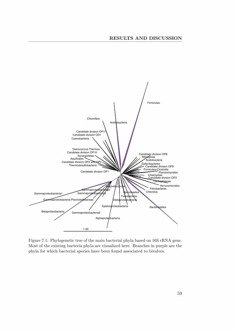
RESULTS AND DISCUSSION
Candidate division OP1
Thermodesulfobacteria
AquificalesCandidate division OP2 and OP5
SynergistetesCandidate division OP10
Deinococcus-Thermus
CyanobacteriaCandidate division OD1
Candidate division OP11
ChloroflexiActinobacteria
Firmicutes
Candidate division OP8Nitrospirae
AcidobacteriaDeferribacteres
Candidate division OP9Firmicutes-Clostridia
Planctomycetes
Candidate division OP3Chlamydiae
Lentisphaerae
VerrucomicrobiaFibrobacteres
Chlorobia
Bacteroidetes
Spirochaetes
FusobacteriaDeltaproteobacteria
Epsilonproteobacteria
Magnetococcus
Alphaproteobacteria
Gammaproteobacteria4Gammaproteobacteria3
Gammaproteobacteria2
Gammaproteobacteria-Piscirickettsiaceae
Betaproteobacteria
Gammaproteobacteria1
1.00
Figure 7.1: Phylogenetic tree of the main bacterial phyla based on 16S rRNA gene.Most of the existing bacteria phyla are visualized here. Branches in purple are thephyla for which bacterial species have been found associated to bivalves.
59

RESULTS AND DISCUSSION
bivalves, gastropods, echinoderms, ascidians, and fish. However, except for
the bathymodiolin mussels and a shallow water bivalve (the Pacific razor
clam Siliqua patula) none of these metazoa-associated bacteria have been
shown to occur inside nuclei. It has been suggested that intranuclear bacte-
ria may cause mass mortalities in bivalves but a causal relationship has never
been established. Because of their potentially lethal effect on bivalve pop-
ulations, I looked for the presence of intranuclear bacteria in economically
important and commercially available bivalve species, i.e. oysters (Cras-
sostrea gigas), razor clams (Siliqua patula and Ensis directus), blue mussels
(Mytilus edulis), manila clams (Venerupis philippinarum), and common cock-
les (Cerastoderma edule). Fluorescence in situ hybridization (FISH) revealed
the presence of intranuclear bacteria in all investigated bivalves except oys-
ters and blue mussels (see details in Manuscript II). A FISH probe targeting
all currently known intranuclear gammaproteobacteria was designed for fu-
ture high-throughput analyses of marine invertebrates. Furthermore, primers
were designed to quantify the abundance of intranuclear bacteria with real
time PCR. Preliminary tests with these primers showed massive amounts of
intranuclear bacteria in some bivalve species, raising the question if these
might significantly affect not only the health of the bivalves but possibly also
of the humans that eat them.
7.2 Diversity of bacteria associated with bivalves
A high diversity of bacterial phylotypes was found in our 16S rRNA sequence
analysis, some are recurrent bacteria that have shown up in previous bivalve
studies (Table 7.1). The representation of bacteria herein has a gammapro-
teobacteria dominance, as has been seen in the previous molecular studies of
bivalve-associated bacterial communities (Schulze et al. 2006, Cavallo et al.
2009). Bivalve sequences fell in clades with bacteria from organic-rich en-
vironments: oil spills, bone-falls, feces or invertebrate tissue. Most of them
belong to the Gammaproteobacteria group (Figure 7.2) but also there are
Alphaproteobacteria (Figure 7.3), Epsilonproteobacteria (Figure 7.3), Bac-
teroidetes (Figure 7.4), Actinobacteria and Spirochetes (Figure 7.5). It can
60

RESULTS AND DISCUSSION
be hypothesized that these bacteria are specialized on high-organic matter
habitats and that more analysis might show a host species-specific bacterial
community.
In the Epsilonproteobacteria group it was observed that there is a par-
ticular group that encloses hydrothermal vent sequences (7.3). Other two
sequences are from invertebrate tissue, Paralvinella palmiformis and B. plat-
ifrons. This last one is the closest relative to the sequence of B. childressi
analyzed herein. FISH analysis revealed that Epsilonproteobacteria are at-
tached to cilia-like structures of the mussel gill (see Fig. 2 in Manuscript
III). Other Epsilonproteobacteria have been described as invertebrate ec-
tosymbionts (e.g. in Rimicaris exoculata, which it is not surprising that the
diversity of this association is not limited to certain invertebrates (i.e. crus-
taceans) but it might extend widely in the whole invertebrate group.
Associated spirochaetes stand out because they seem to be a stable bac-
terial community in bivalve styles (Noguchi 1921, Bernard 1970, Paster et al.
1996, Prieur et al. 1990, Margulis and Fester 1991). In my FISH observations
(see Fig. 2 in Manuscript III) I see them well established in bivalve gill tissue.
Also, a spirochete sequence from DNA gills has been described in a Lucinoma
sp. cold-seep clam by Duperron et al. (2007). It might be worthwhile to do
a population genetics study to investigate the variability of these spirochetes
species within the different bivalves. It is not clear so far if spirochetes have
an ecological role or importance in the association with bivalves, but they
might be an ubiquitous microflora within the mollusks group (Prieur et al.
1990). Margulis et al. (1991) named as symbionts the studied spirochetes in
oysters. We might be able to study them as ubiquitous symbiotic bacteria
in bivalves.
61

RESULTS AND DISCUSSION
Vibrio sp. SOMBO17, cuttlelfish egg case, AJ936947Vibrio sp. W-10, DQ923444
clone 6, Ensis directus, SyltIsolate Tapes semidecussatusMytilus edulis, SyltListonella anguillarum, X71821
Crassostrea gigas., SyltVibrio sp. 24A, ANG gland of Sepia officinalis, AJ936940
Vibrio splendidus, Pecten maximus, AJ515226Mya sp., Crassostrea gigas., Mytilus edulis, SyltVibrio sp. 3d, AF388392Vibrio pomeroyi, AJ560649mucus bacterium 55, mucus, Oculina patagonica, AY654793Vibrio sp. A2, sea dragon, EF467288Vibrio sp. Mel 34, Crassostrea gigas, AJ582805Vibrio sp. P90, hydrocarbon polluted sediments, EU195934Vibrio sp. LMG 23856, Ruditapes philippinarum, EF599163Vibrio splendidus, AJ515230Vibrio lentus, AM162659Crassostrea sp., Ensis directus, Sylt
Mytilus edulis, Crassostrea sp., Ensis directus, SyltAntarctic bacterium PHNZ10C1, Antarctic surface seawater, DQ925852Aliivibrio logei, AY771721uncultured Vibrio sp., hindgut of Mudsuckers, DQ340191Aliivibrio wodanis, Y17575
Aliivibrio logei, AY292925Vibrio sp. 6(2006), diseased sporophyte, Laminaria japonica, DQ642809
Ensis directus clone, Syltclone 17, Crassostrea sp., Sylt
Psychromonas ossibalaenae, marine sediment adjacent to sperm whale carcasses, AB304808gamma proteobacterium endosymbiont of Siboglinum fiordicum, EU086771Psychromonas aquimarina, marine sediment adjacent to sperm whale carcasses, AB304805
Psychromonas japonica, marine sediment adjacent to sperm whale carcasses, AB304804
Psychromonas spp. cultivated
Bathymodiolus brooksi gill symbiont, (Chapopote Knoll, southern GoM)Shewanella affinis, AF500080Isolate Cerastoderma edule,SyltShewanella colwelliana, AB205577Shewanella sp. SOMBO46, cuttlelfish egg case, AJ936953
Alteromonadaceae bacterium T1, German Wadden Sea, AY177717uncultured bacterium, Japan Trench sediment , AB013824
clone 3, Crassostrea sp.., Syltuncultured bacterium, temperate estuarine mud, AY216447clone 37, Cerastoderma edule, Syltuncultured bacterium, Janssand intertidal sandy sediment, middle flat, EU707315
Alcanivorax sp. P75, hydrocarbon polluted sediments, EU195941uncultured bacterium, orange microbial mat on a grey whale carcass, Pacific Ocean, AY922230Alcanivorax borkumensis, Y12579Alcanivorax sp. DG813, AY258105Isolate 6 Tapes semidecussatus
Bathymodiolus thiotrophic group
Cycloclasticus spp.
Bathymodiolus methanotrophic group
Vestimentiferan symbionts group
NIX clade
Pseudomonas sp. 47, EU883662Pseudomonas stutzeri, EU531806
Pseudomonas putida, oil contaminated soil, EU849665Isolate 4 Tapes semidecussatusPseudomonas antarctica, soil, AM933495
Pseudomonas sp. Cam-1, AF098464Pseudomonas sp. Sag-1, AF098467
0.10
Figure 7.2: Gammaproteobacterial diversity in bivalves. The clones and isolatesobtained in this study are highlighted in green. Clones or isolates are shown andnamed after their host origin.62

RESULTS AND DISCUSSION
clone 133, Ensis, SyltRalstonia pickettii, lake sediment enriched with heavy metals, DQ997838Ralstonia sp. SA-3, soil, DQ854843
unc. alpha proteobacterium, grey whale bone, Pacific Ocean, AY922181alpha proteobacterium STF-07, North Sea oil reservoir, EU594271
clone 6, Crasosstrea sp., SyltTerasakiella pusilla, AB006768Terasakiella sp. UST061013-067, EF587999
uncultured organism, deep-sea octacoral, DQ395939Isolate, Tapes semidecussatusSulfitobacter litoralis, DQ097527Sulfitobacter sp. BR1, AY902210Sulfitobacter sp. S11-B-4, EU016167Sulfitobacter sp. BSw20064, Arctic seawater, EU365589Sulfitobacter sp. ARCTIC-P49, AY573043Sulfitobacter sp. B7, AY902203unidentified bacterium, AJ278782Sulfitobacter sp. HEL-77, water sample from 10 m depth, AJ534229Sulfitobacter sp. GAI-21, AF007257
clone B. childressi, nGoMuncultured Bathymodiolus platifrons gill symbiont, AB250697
uncultured bacterium, Indian Ocean hydrothermal vent, AY531588uncultured epsilon proteobacterium, Paralvinella palmiformis mucus secretions, AJ441206
uncultured epsilon proteobacterium, deep sea hydrothermal vent, AJ575999uncultured bacterium, iron mat, FJ535279
uncultured epsilon proteobacterium, microbial fuel cell fed with marine plankton, EU052242uncultured epsilon proteobacterium KTc1160, AF235116uncultured bacterium, Montastraea faveolata - diseased tissue, FJ203167
epsilon clone, Ensis directus, Crasosstrea sp., Syltepsilon clone, Crassostrea sp. Mexepsilon proteobacterium Oy-M7, adult oyster mantle, DQ357825
uncultured bacterium, Namibian upwelling system, EF645952uncultured bacterium, Zostera marina roots, EF029017epsilon clone, Mya sp., Syltclone 187, Crassostrea sp., France
uncultured bacterium, sediment from the Kings Bay, Svalbard, Arctic, EU050947Arcobacter sp. BSs20195, DQ514311uncultured bacterium, Pearl River Estuary sediments at 6cm depth, EF999357
uncultured bacterium, marine environment, AY548986unidentified epsilon proteobacterium, deepest cold-seep area of the Japan Trench, AB015256
uncultured epsilon proteobacterium, Riftia pachyptila's tube, AF449239uncultured bacterium, marine environment, AY548997clone 27, Crassostrea sp., Sylt
0.10
Vents
Figure 7.3: Alpha- and Epsilonproteobacterial diversity in bivalves. The clonesand isolates obtained in this study are highlighted in blue. Clones from Ensisdirectus, Crassostrea gigas, Tapes semidecussatus, B. childressi and Mya sp. 16SrRNA libraries. Hydrothermal vent group is highlighted also.
63
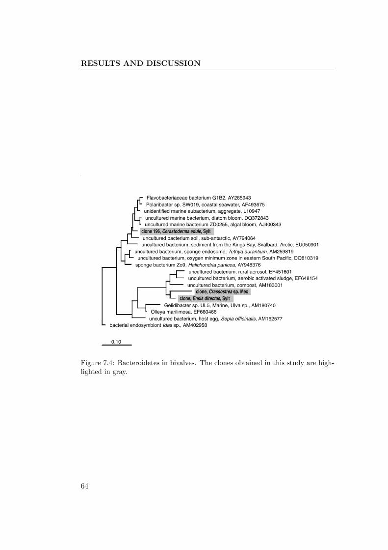
RESULTS AND DISCUSSION
Flavobacteriaceae bacterium G1B2, AY285943Polaribacter sp. SW019, coastal seawater, AF493675
unidentified marine eubacterium, aggregate, L10947uncultured marine bacterium, diatom bloom, DQ372843uncultured marine bacterium ZD0255, algal bloom, AJ400343
clone 196, Cerastoderma edule, Syltuncultured bacterium soil, sub-antarctic, AY794064uncultured bacterium, sediment from the Kings Bay, Svalbard, Arctic, EU050901
uncultured bacterium, sponge endosome, Tethya aurantium, AM259819uncultured bacterium, oxygen minimum zone in eastern South Pacific, DQ810319
sponge bacterium Zo9, Halichondria panicea, AY948376uncultured bacterium, rural aerosol, EF451601uncultured bacterium, aerobic activated sludge, EF648154uncultured bacterium, compost, AM183001
clone, Crassostrea sp. Mexclone, Ensis directus, Sylt
Gelidibacter sp. UL5, Marine, Ulva sp., AM180740Olleya marilimosa, EF660466
uncultured bacterium, host egg, Sepia officinalis, AM162577bacterial endosymbiont Idas sp., AM402958
0.10
Figure 7.4: Bacteroidetes in bivalves. The clones obtained in this study are high-lighted in gray.
64

RESULTS AND DISCUSSION
Spec
ies
Vibrio
Pseudom
onas
Achromob
acter-
Acineto
bacter
Shewan
ella sp
p.Enter
obact
eria
other
Gamma Micr
ococcu
s Spirochete
s Bactero
idetes Hyd
rocarb
on-
degrad
ers other
Alpha Epsilon
Chlamyd
ia Photo
bacteri
umPro
teolyt
ic bact
.Fer
mentat
ive
bact. Others
Ref
eren
ces
Oys
ters
Ost
rea
edulis
MN
oguch
i 1921
Cra
ssost
rea
gig
asCSCIS
CS
CIS
SIS
SIS
SIS
SC
CPr
ieur
1981,
Sugita
et a
l 1981,
Sch
ultze
et
al.
2006,
This
stu
dy
C.
virg
inic
aC 2
5%
C31%
MC
C26%
C 8
7%
Noguch
i 1921,
Murc
hel
ano
& B
row
n 1
968
C.
cucu
lata
CC
CC
CC
Nei
sser
ia,
Cory
nobac
terium
Raj
agopal
an &
Siv
alin
gan
1978
Muss
els
Myt
ilus
edulis
CSCS
SM
CC
Noguch
i 1921,
Prie
ur
1981,
this
stu
dy
Mer
cenar
ia m
erce
nar
ia
Myt
ilus
viridis
CC
CC
CC
Nei
sser
ia,
Cory
nobac
terium
Raj
agopal
an &
Siv
alin
gan
1978
Myt
ilus
coru
scus
CC
CC
Mora
xella
Sugita
et a
l 1981
Cla
ms
Cer
asto
der
ma
eduli
CS
Sth
is s
tudy
Tap
es (
Ven
erupis
) sp
p.
CIS
SC
ISCCS
ISCS
CS
MIS
ECS
CS
ISC
CSulfitobac
ter
Noguch
i 1921,
Prie
ur
1981,
Sugita
et a
l 1981,
Sch
ultze
et
al. 2
006,
This
stu
dy
Sili
qua
pat
ula
Ensi
s sp
p.
CS
SM
SS
SN
oguch
i 1921
Mya
are
nar
iaCCS
CC
SM
CS
Cory
nobac
terium
, Art
hro
bac
ter
Noguch
i 1921,
Cundel
l &
Yo
ung 1
975
Mac
tra
vener
iform
isC
CC
CM
Noguch
i 1921,
Sugita
et a
l 1981
Phac
oso
ma
japonic
um
CC
CSugita
et a
l 1981
Sca
phar
ca b
roughto
nii
CC
CSugita
et a
l 1981
Panopea
abru
pta
ISIS
ISIS
Sch
ultze
et
al.
2006
Tab
le7.1:Bacterialdiversity
studiesin
bivalves.Studieswherebacteria
havebeenobserved
microscopically(M
),cu
ltured
(C),isolated(I),ortheir16SrR
NA
sequenced(S).Somelettersare
repeatedbecause
more
thanonestudyhavecharacterized
thespecies.
Inredthesequencesandculturesfrom
this
study.
65
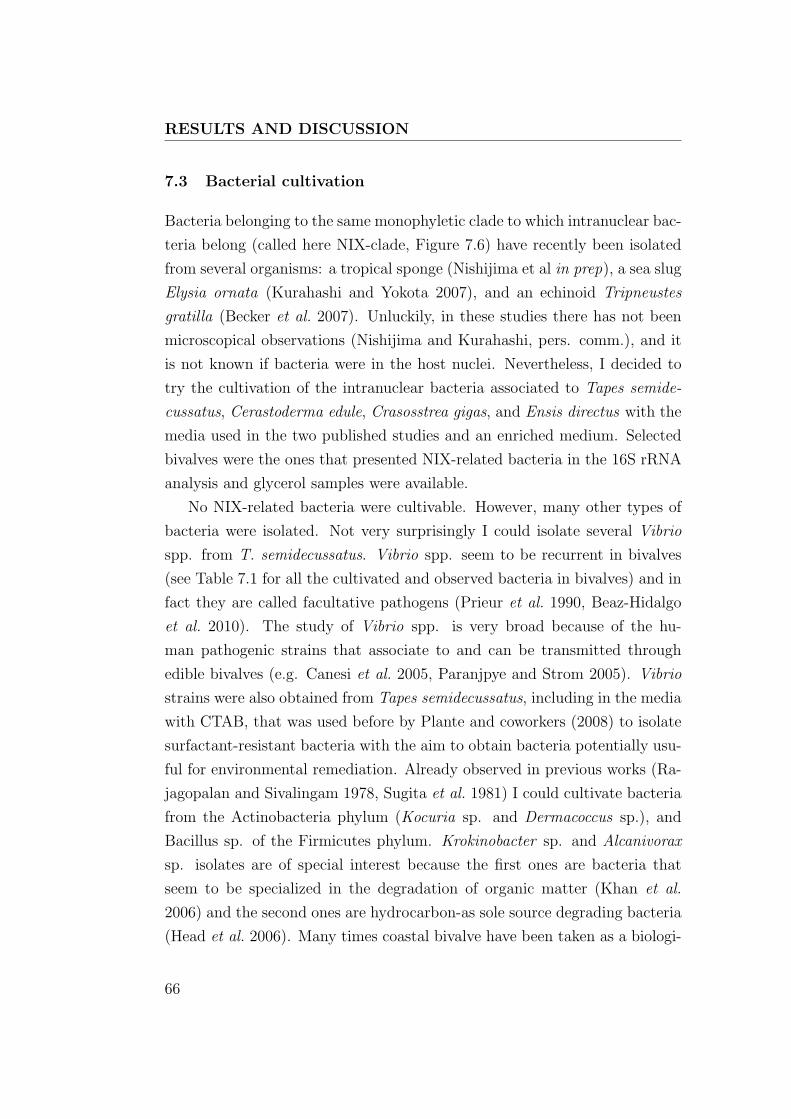
RESULTS AND DISCUSSION
7.3 Bacterial cultivation
Bacteria belonging to the same monophyletic clade to which intranuclear bac-
teria belong (called here NIX-clade, Figure 7.6) have recently been isolated
from several organisms: a tropical sponge (Nishijima et al in prep), a sea slug
Elysia ornata (Kurahashi and Yokota 2007), and an echinoid Tripneustes
gratilla (Becker et al. 2007). Unluckily, in these studies there has not been
microscopical observations (Nishijima and Kurahashi, pers. comm.), and it
is not known if bacteria were in the host nuclei. Nevertheless, I decided to
try the cultivation of the intranuclear bacteria associated to Tapes semide-
cussatus, Cerastoderma edule, Crasosstrea gigas, and Ensis directus with the
media used in the two published studies and an enriched medium. Selected
bivalves were the ones that presented NIX-related bacteria in the 16S rRNA
analysis and glycerol samples were available.
No NIX-related bacteria were cultivable. However, many other types of
bacteria were isolated. Not very surprisingly I could isolate several Vibrio
spp. from T. semidecussatus. Vibrio spp. seem to be recurrent in bivalves
(see Table 7.1 for all the cultivated and observed bacteria in bivalves) and in
fact they are called facultative pathogens (Prieur et al. 1990, Beaz-Hidalgo
et al. 2010). The study of Vibrio spp. is very broad because of the hu-
man pathogenic strains that associate to and can be transmitted through
edible bivalves (e.g. Canesi et al. 2005, Paranjpye and Strom 2005). Vibrio
strains were also obtained from Tapes semidecussatus, including in the media
with CTAB, that was used before by Plante and coworkers (2008) to isolate
surfactant-resistant bacteria with the aim to obtain bacteria potentially usu-
ful for environmental remediation. Already observed in previous works (Ra-
jagopalan and Sivalingam 1978, Sugita et al. 1981) I could cultivate bacteria
from the Actinobacteria phylum (Kocuria sp. and Dermacoccus sp.), and
Bacillus sp. of the Firmicutes phylum. Krokinobacter sp. and Alcanivorax
sp. isolates are of special interest because the first ones are bacteria that
seem to be specialized in the degradation of organic matter (Khan et al.
2006) and the second ones are hydrocarbon-as sole source degrading bacteria
(Head et al. 2006). Many times coastal bivalve have been taken as a biologi-
66

RESULTS AND DISCUSSION
wall-less spirochete, M87055clone, Crassostrea sp. Mex
Spirochaeta coccoides, host hindgut, Neotermes sp., AJ698092spirochete endosymbiont lucinid bivalve, AM236337
uncultured Spirochaeta sp., termite gut homogenate, Reticulitermes speratus, AB088873uncultured spirochete BHI80-158, Alvinella pompejana tubes, AJ431240uncultured spirochete sp. anaerobic lake hypersaline sediment, ASpC-2, AY995150uncultured spirochete hypersaline lake, AF454308clone, Ensis directus, Sylt
Borrelia sp. R57, Clethrionomys glareolus, AY626138Cristispira sp. CP1, U42638
uncultured organism deep-sea octacoral, DQ395607clones, Ensis directus, Cerastoderma edule., Sylt
Spironema culicis of mosquito Culex pipiens, AF166259uncultured spirochete, microbial mat, AY605160
uncultured bacterium, hypersaline microbial mat, DQ154857uncultured candidate, hypersaline microbial mat: Guerrero Negro, DQ329631
clone, Crassostrea sp., Syltuncultured spirochete, gut proctodeal, Nasutitermes sp., EF454267
uncultured spirochete, termite gut, Microcerotermes sp. 2, AB191843uncultured spirochete, biofilm at hydrothermal vent orifice, AM712343
Ilyobacter psychrophilus, AJ877255clone, Ensis directus, Syltuncultured bacterium, sediment from the Kings Bay, Svalbard, Arctic, EU050917Fusobacteria bacterium HAW-EB21, AY579753uncultured bacterium, Amsterdam mud volcano, deep Eastern Mediterranean, AY592371uncultured Fusobacterium sp., cold seep sediment, AB189363Fusobacteria bacterium Ko711, basalt, AF550592uncultured Fusobacteria bacterium, Paralvinella palmiformis mucus secretions, AJ441229uncultured bacterium, marine environment, AY548984uncultured prokaryote, hot fluid vent, AM268765
uncultured bacterium, anaerobic methanogenic UASB reactor, EF063619uncultured bacterium, full-scale anaerobic UASB bioreactor, AY426453
clone, Crassostrea sp. MexKocuria sp. HNR05, root, Chinese cabbage, EU373345
Isolate 1 Tapes semidecussatusDermacoccus sp. Ellin183, AF409025
Isolate 2 Tapes semidecussatusMicrococcus sp. 5, Carpodacus mexicanus, AY269868
0.10
Figure 7.5: Spirochaete and Fusobacteria diversity in bivalves. The clones andisolates obtained in this study are highlighted in violet. Clones or isolates areshown and named after their host origin.
67

RESULTS AND DISCUSSION
cal marker to assess pollution (e.g. Nishihama et al. 1998, Bresler et al. 1999,
Verlecar et al. 2006). The presence of Alcanivorax bacteria in clams tissue
could indicate a strong oil influence in North Sea beaches, and in fact this
can be an easy-to-evaluate biological marker for oil pollution: the presence
of Alcanivorax spp. in bivalves tissue.
7.4 Summary and Outlook
Bivalve gills provide to bacteria an ideal habitat, with protection from grazers
and constant fluid of nutrients. Bacteria might be complementing their host
nutrition or contributing to metabolite production. Degradation of bacteria
by bivalve enzymes has been observed and it seems that this degradation
provides dissolved compounds (Birkbeck and McHenery 1982, Amouroux
and Amouroux 1988, McHenery and Birkbeck 1985) and improves bivalve
nutrition (Delaunay et al. 1992). Bacteria could provide 5% to 10% carbon,
and 20% nitrogen from the bivalve requirements (Prieur et al. 1990). A stable
microbiota could be providing protection to bivalves thanks to competition
against other bacteria potentially pathogenic. Also, microbiota could be
secreting antimicrobial substances that have been observed to be commun in
bacteria isolated from bivalves (Zheng et al. 2005).
Ecological studies with molecular techniques are scarce and they could
help to disentangle the interaction patterns between the endemic microflora
and the invasive one, explaining the benefits and contrarieties that symbi-
otic or pathogenic bacteria bring along. It is important to understand the
distribution of pathogenic bacteria in the marine environments to predict po-
tential health concerns transmitted by seafood. Ecological parameters such
as nutrient availability, temperature, and salinity influence the presence and
persistence of bacteria. However, I suggest that bacteria like Vibrio, Pseu-
domonas, Spirochetes and Epsilonproteobacteria (see Table 7.1 for different
bacteria occurrence) present in bivalves are not only randomly there. It is
in part the result of the surrounding water community but it might also be
the result of a common evolution between host and bacteria that normally
associate to invertebrates or high organic matter content habitats. We would
68
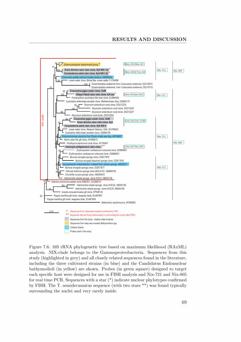
RESULTS AND DISCUSSION
Endonucleobacter bathymodioli group *
Ensis directus razor clam clone, Sylt NIX-I (4) *Cerastoderma edule clam clone, Sylt NIX-I (5) *
90
100
Tripneustes gratilla echinoid isolate bacteria, AM495252ocean water clone, Bohai Bay, ocean water, FJ154998
Oceanrickettsia ariakensis from Crassostrea ariakensis, DQ123914Oceanrickettsia ariakensis, from Crassostrea ariakensis, DQ118733
10098
81
Crassostrea gigas oyster clone, GoMSiliqua Patula razor clam clone, full seq*
Pomacanthus sexstriatus fish tract clone, EU884930
65
Cystodytes dellechiajei ascidian clone, Mediterranean Sea, DQ884170
100
Alcyonium antarcticum coral clone, DQ312235Alcyonium antarcticum coral clone, DQ312243Alcyonium antarcticum coral clone, DQ312237
83
Alcyonium antarcticum coral clone, DQ312244
10099
Crassostrea gigas oyster clone, GoM Ensis directus razor clam clone, Sylt
Cerastoderma edule clam clone, Sylt NIX-II
96
ocean water clone, Newport Harbour, USA, EU799933Cystodytes dellechiajei ascidian clone, DQ884169
83
99
Endozoicomonas elysicola from Elysia ornata sea slug, AB196667Salmo salar fish gill clone, AY494615
88
Pocillopora damicornis coral clone, AY700601Venerupis philippinarum clam clone **
84
Erythropodium caribaeorum octocoral clone, DQ889921Erythropodium caribaeorum octocoral clone, DQ889931Muricea elongata sponge clone, DQ917879
Muricea elongata bleached sponge clone, DQ917830
68
91
Spongiobacter nickelotolerans, isolated from marine sponge, AB205011Muricea elongata sponge clone, DQ917877
89
Petrosia ficiformis sponge clone MOLA 531, AM990755Chondrilla nucula sponge clone, AM259915
Halichondria okadia sponge clone HOC2, AB054136
84
Asterias amurensis seastar clone KMD001, EU599216
79
Halichondria okadia sponge clone HOC22, AB054156Halichondria okadia sponge clone HOC25, AB054159
Acesta excavata bivalve gill clone, EF508132
100
67
95
Ruppia maritima gill clone, seagrass beds, EU487857Ruppia maritima gill clone, seagrass beds, EU487858
63
Bdellovibrio bacteriovorus, AF084850
0.10
Bnix-643/Bnix-64
Bnix-643II/Tnix-64
Snix-64/Snix-643
Cnix-64/Cnix-1249
Tnix-64/Tnix-643
Sequences from this study - shallow water bivalves
Cultured strains
*
Probes used in this study
Nix-721
Nix-721
Nix-805
Nix-805
Nix-721
Nix-721
NIX
cla
de
Sequences from intranuclear bacteria confirmed by FISH
Sequences from deep-sea mussels Bathymodiolus spp.
Sequences that are found intranuclearly or sorrounding the nuclei (after FISH)**
Figure 7.6: 16S rRNA phylogenetic tree based on maximum likelihood (RAxML)analysis. NIX-clade belongs to the Gammaproteobacteria. Sequences from thisstudy (highlighted in grey) and all closely related sequences found in the literature,including the three cultivated strains (in blue) and the Candidatus Endonuclearbathymodioli (in yellow) are shown. Probes (in green square) designed to targeteach specific host were designed for use in FISH analysis and Nix-721 and Nix-805for real time PCR. Sequences with a star (*) indicate nuclear phylotypes confirmedby FISH. The T. semidecussatus sequence (with two stars **) was found typicallysurrounding the nuclei and very rarely inside.
69

RESULTS AND DISCUSSION
need more studies to be able to say that these bacteria are symbiotic, but
from a point of view of the broad symbiosis concept they are, as they appear
to be consistent in the bivalves microflora.
70


Part III
Manuscripts
72


Resulting manuscripts from this thesis work
and contributions:
Manuscript I: L. Raggi, F. Schubotz, K.-U. Hinrichs, J.M.
Petersen, J. Felden and N. Dubilier. Bacterial symbionts of
Bathymodiolus mussels and Escarpia tubeworms from Chapopote,
an asphalt seep in the southern Gulf of Mexico.
Manuscript in preparation.
L.R.: developed the concept together with N.D., did the 16S rRNA
sequencing and analyses, designed FISH probes and performed
the FISH experiments, helped with the carbon isotope analyses,
conceived and wrote the manuscript. F.S.: did the carbon isotope
analysis of lipids. K.U.H.: developed the carbon isotope analysis
method. J.M.P.: conceived and edited the manuscript with L.R.
J.F.: provided samples, and information about the geochemical
parameters from the Chapopote site. N.D.: developed the concept
with L.R., conceived and edited the manuscript.
Manuscript II: L.Raggi, D. Fink and N. Dubilier. An intranu-
clear bacterial parasite in shallow water bivalves.
Manuscript in preparation.
L.R.: developed the concept together with N.D., did the 16S rRNA
sequencing and analysis, designed FISH probes and performed the
FISH experiments, conceived and wrote the manuscript. D.F.:
performed the qPCR. N.D.: developed the concept with L.R., con-
ceived and edited the manuscript.
74

MANUSCRIPTS
Manuscript III: L.Raggi, and N. Dubilier. Minireview: Bacte-
rial diversity of shallow-water bivalves.
Manuscript in preparation.
L.R.: developed the concept, did the 16S rRNA sequencing and
analyses, performed cultivation experiments, conceived and wrote
the manuscript. N.D.: developed the concept with L.R., conceived
and edited the manuscript.
Manuscript IV: F.U. Zielinski, A. Pernthaler, S. Duperron,
L. Raggi, O. Giere, C. Borowski and N. Dubilier. (2009).
Widespread occurrence of an intranuclear parasite in bathymodi-
olin mussels. Environmental Microbiology 11(5):1150-67.
F.U.Z.: developed the concept, did the sampling, cloning, sequenc-
ing, and microscopy analyses. A.P.: performed some FISH ex-
periments. S.D.: provided support with sampling, phylogenetic
reconstruction, and probe design. L.R.: did 16S rRNA sequenc-
ing and FISH experiments on specimens from the Gulf of Mexico.
O.G.: helped with sampling and did the TEM. N.D.: developed
the concept, conceived and edited the manuscript.
75

Manuscript I:
Bacterial symbionts of Bathymodiolus
mussels and Escarpia tubeworms from
Chapopote, an asphalt seep in the southern
Gulf of Mexico.
Luciana Raggi, Florence Schubotz, Kai-Uwe Hinrichs,
Jill M. Petersen and Nicole Dubilier
In Preparation
76

Bacterial symbionts of Bathymodiolus mussels and Escarpia tubeworms
from Chapopote, an asphaltic seep in the southern Gulf of Mexico
L. Raggi1, F. Schubotz
2, K.-U. Hinrichs
2, J.M. Petersen
1, J. Felden
1, N. Dubilier
1
1 Max-Planck Institut for Marine Microbiology, Celsiusstr. 1, 28359, Bremen, Germany
2 MARUM, University of Bremen, Leobener Str., 28359 Bremen, Germany
Keywords: endosymbiosis, thiotrophic, methanotrophic, Bathymodiolus mussels, Escarpia
tubeworms, asphalt, cold seep
Running head: Chapopote Escarpia and Bathymodiolus symbioses
Corresponding author: Nicole Dubilier
Max Planck Institut for Marine Microbiology
Celsiusstr. 1
28359, Bremen
Germany
Phone: +49 421 2028 932
e-mail: [email protected]
This article is intended for publication in the journal:
Marine Ecology Progress Series
Chapopote Escarpia and Bathymodiolus symbioses
77

Abstract
Chemosynthetic life was recently discovered in the southern Gulf of Mexico (sGoM) where
lava-like flows of solidified asphalt cover a large area at 3000 m depth; with oil seeps and gas
hydrate deposits also present (MacDonald et al. 2004). Animals with chemosynthetic
symbionts such as vestimentiferan tubeworms, mussels, and clams colonize this site called
Chapopote. Based on morphological and molecular analyses (COI gene), two mussel species
are present at this site, Bathymodiolus heckerae and B. brooksi, and a single Escarpia
tubeworm species. Comparative 16S rRNA sequence analysis and FISH showed that all three
host species harbor intracellular sulfur-oxidizing symbionts that are highly similar or
identical to the symbionts found in the same host species from northern GoM (nGoM) sites.
The mussels also harbor methane-oxidizing symbionts, and these are identical to their
northern GoM conspecifics. Unexpectedly, we discovered a novel symbiont in B. heckerae
that is closely related to hydrocarbon degrading bacteria of the genus Cycloclasticus. We
found in B. heckerae the Methyl-toluene-phenol hydroxylase (MTPH) gene and stable carbon
isotope analyses of lipids indicative for heterotrophic bacteria were consistently heavier in B.
heckerae by 3‰ than in B. brooksi., indicating that the novel symbiont might use isotopically
heavy hydrocarbons from the asphalt seep as an energy and carbon source. The discovery of a
novel symbiont that may be able to metabolize hydrocarbons is particularly intriguing
because until now only methane and reduced sulfur compounds have been identified as
energy sources in chemosynthetic symbioses. The large amounts of hydrocarbons available at
Chapopote would provide these mussel symbioses with a rich source of nutrition.
Manuscript I
78

Introduction
The discovery of cold seeps was a surprising event showing great communities of big sized
organisms living at these recondite places (Paull et al. 1984). Cold seep ecosystems are some
of the most productive on Earth, and it was initially unclear how, with what it seemed to be
very little input of organic matter from photosynthesis, these new fauna could even survive
down there (Van Dover 2000). Association of these organisms with chemosynthetic bacteria
is the answer. More and more different sites and chemosynthetic-based habitats are been
discovered, as the underwater tools are each time more equipped to stay longer and deeper in
the sea floor. In April 2004 a natural asphalt (bitumen) deposit at 3000 m depth in the
Campeche Knolls, southern Gulf of Mexico (sGoM), was discovered and was named
Chapopote (MacDonald et al. 2004). Cold seeps with methane and hydrocarbon seepage are
widespread in the GoM as a result of its unique tectonics and geological history (Macgregor
1993, Bryant et al. 1991, Ewing 1991), however this is the first time that a classical cold-seep
fauna community was found coexisting with natural asphalt. The biology and microbiology
of the northern GoM seeps is well studied (e.g. Fisher 1993, Cavanaugh et al., 1987,
Cavanaugh 1993, Carney et al. 2006, Cordes et al. 2005, 2007, Duperron et al. 2007), in
contrast with the little studied southern GoM (Fig. 1). The presence of symbiotic bacteria in
Bathymodiolus mussels and in escarpid tubeworms is one of the intensively studied topics in
the nGoM. Three Bathymodiolus species in the northern Gulf of Mexico have been described;
B. childressi, with methanotrophic bacteria, B. heckerae and B. brooksi, with a dual
symbiosis of methanotrophic and thiotrophic bacteria. Also three different tubeworms genera
are recognized and all of them bear thiotrophic symbionts: Escarpia sp., Lamellibrachia sp.
and Sepiophila sp. The distribution of the different species varies along the GoM (Fig 1), in
the Louisiana Slope B. childressi, L. luymesi, and S. jonesi are common and abundant;
Atwater Canyon retain only B. brooksi and Missisipi Canyon only B. childressi. In the
Florida Escarpment B. heckerae, B. brooksi and E. laminata are found. The Alaminos
Canyon holds B. brooksi, B. childressi, and E. laminata. These species seem to be endemic of
the GoM except B. heckerae that is found also in the Blake Ridge diapir (Salerno et al. 2005).
All these species of both tubeworms and mussels get nutrients and benefits from their
Chapopote Escarpia and Bathymodiolus symbioses
79
chemosynthetic association using only sulfide, methane, or both as energy sources, no further
sources are currently known.
Lipid analysis of mussels in the northern Gulf of Mexico, that only contain methanotrophic
symbionts have been performed in former studies. It has been observed that depending on
whether methane has a biogenic or a thermogenic source, the isotopic values of the tissue
vary between -79 to -80 ‰ and -45 to -40 ‰, respectively (Jahnke et al. 1995, MacAvoy et
al. 2002). Methane values for the thermogenic methane in the northern GoM are between -44
and -46 ‰ and for biogenic methane -64 to -65 ‰ (Sassen et al. 1999). At the Chapopote
Knoll, the stable carbon isotope composition of methane is between -40 ‰ (in the asphalts) to
-60‰ (in the sediments), representing a mixture of thermogenic and biogenic methane
(Schubotz et al. subm.). In this study, we do genetic (phylogenetic and metabolic) and lipid
analyses focusing on the question of whether the asphalt in Chapopote site might be shaping
the community, in particular the bacterial symbiotic community of invertebrates, adding an
extra carbon source. This habitat is a novel setting because of this heavy oil called asphalt.
Asphalt has high amounts of asphaltenes that make it heavier than water. Thus, the oil stays
in the seafloor, while in other settings the oil leaks upward to the water surface. It creates
then an interface where hydrocarbons can be aerobically oxidized. Also, this is a system with
new substrate for the megafauna to settle, as there are not just carbonates but also solid
asphaltic formations.
Manuscript I
80

Material and methods
Specimen collection
Mussels and vestimentiferan tubeworms were collected at the base of Chapopote Knoll with
the ROV Quest aboard the RV Meteor during the M67/2 cruise (April 2005). Bathymodiolus
mussels were collected from a mussel bed (21°53.98´N; 93°26.12`W) at a water depth of
2923 m. Four mussels were recovered during dive 83; two were identified as species 1 and
the other two as species 2. The mussel gills were dissected immediately after recovery and
segments were frozen and stored at -20°C for DNA or lipid extraction. Other fragments were
fixed for FISH with 2% PFA and stored at 4°C in 0.5X PBS / 50% ethanol.
Tubeworms were collected in two different dives: dive 82 at 21°53.95´N; 93°26.23`W, 2918
m water depth, and dive 83 at 21°53.94´N; 93°26.25`W and water depth 2915 m. Three
tubeworms from dive 82 (Tbw 1, Tbw 2 and Tbw 3) and one from dive 83 (Tbw 4) were
extracted from their tube and different pieces were frozen for DNA extraction or fixed for
FISH.
DNA extraction and PCR amplification
DNA was extracted from frozen tissue according to Zhou et al. (1996) with the following
modifications. Briefly, 2 ml of extraction buffer and 20 µl proteinase K (20 mg/ml) were
added to approximately 100 mg of sample, and incubated for 1.5 hours at 37°C. Then 200 µL
of 20% SDS were added and incubated for 2 hr at 56°C. The liquid phase was recovered after
centrifugation at 14000 g for 20 min and cleaned once with 1 V
phenol/chloroform/isoamylalcohol (25:24:1) and a second time with
chloroform/isoamylalcohol (24:1). DNA was precipitated with 0.6 V isopropanol and
dissolved in Tris-EDTA buffer. The extracted DNA was used for both host and bacterial
symbiont analysis.
Host COI genes were amplified using 36 PCR cycles. For the mussels the primers LCO-
1560 and HCO-2148 (Jones et al. 2006) were used and for the tubeworms LCO1490 and
Chapopote Escarpia and Bathymodiolus symbioses
81

HCO2198 (Folmer et al. 1994). Bacterial 16S rRNA genes were amplified using 20 PCR
cycles with universal primers GM3 (8F) and GM4 (1492R) (Muyzer et al. 1995). Metabolic
marker genes were amplified using 23 PCR cycles. Primers aps1F and aps4R were used to
amplify aprA gene (Blazejak et al. 2006) and the pmoA gene was amplified with A189F and
MB661R primers (Costello & Lidstrom 1999). We tested all the ring-hydroxylating
monooxygenases primers used by Baldwin et al. (2003), and only the RMO-F and RMO-R
pair gave an amplification product of the expected size (see Table S2 for primer sequences
and annealing temperatures). The expected 500 bp product was cloned and sequenced. We
obtained sequences of 300 bp length which resulted to be a fragment of a
methane/toluene/phenol hydroxylase.
Tubeworm genetic analysis
COI PCR products of the four tubeworms were directly sequenced from the PCR product, in
both directions. Symbiont 16S rRNA and aprA genes of tubeworms 1 and 3 were cloned
using the TOPO-TA system (Invitrogen) and analyzed. Genes pmoA and MTPH could not be
amplified in the tubeworms.
Mussel genetic analysis
COI PCR products of the four mussels were directly sequenced in both directions. Symbiont
genes of each mussel (16S rRNA, aprA, pmoA and MTPH were cloned using the TOPO-TA
(Invitrogen) or pGEM-T Easy (Promega) cloning vectors. 16S rRNA clones were partially
sequenced with primer 907RC (Muyzer & Smalla 1998) and representative clones from each
host individual were chosen for full sequencing in both directions. Metabolic marker genes
were sequenced with the respective primers (both strands). aprA and pmoA genes were
analyzed in one individual of each species. MTPH was amplified from both individuals of B.
heckerae but the product of only one individual could be successfully cloned and sequenced.
Sequencing was performed using the BigDye terminator v3.1 Cycle Sequencing Kit along
with the Genetic Analyzer Abiprism 3130 (Applied Biosystems).
Manuscript I
82
Phylogenetic analysis
Sequences were analyzed with Sequencher (Genes Codes Corporation) and ARB (Ludwig et
al. 2004) sofwares. 16S rRNA Sequences were aligned within the Ref_96 SILVA database
(Pruesse et al. 2007). Databases for each metabolic marker gene were constructed with
publicly available sequences for each gene, and the alignment was produced with
CLUSTALW implemented in ARB (Ludwig et al. 2004). Phylogenetic trees were calculated
with the ARB software. All sequence comparisons are given as percentage sequence identity
(% similar nucleotides) after calculations of distance matrix. For tree reconstruction, only
long sequences (~1400 bp for 16S rRNA, ~650 bp for COI, ~1200 bp for aprA, ~450 bp for
pmoA) were used, except for the MTPH gene that we used the 300 bp sequence. Phylogenetic
trees of 16S rRNA gene sequences were calculated by neighbor joining, maximum parsimony
and maximum likelihood methods. As the differences in the resulting tree topographies were
not significant we only present the maximum likelihood results. Sequences with more than
99.7% identity (% identical nucleotides) were grouped and are shown as one single sequence.
To assess nodes robustness in the trees, 1000ML bootstrap replicates were run.
Probe design and testing
All probes used in this study were checked for mismatches against our sequences of interest
and the Silva 96 SSU Ref database (Pruesse et al. 2007). The Cycloclasticus probe (Cypu-
829) was originally used without formamide (Maruyama et al. 2003). We performed a
formamide series between 10% and 70%, and the probe hybridized between 10% and 40%
formamide. The probe NON338 (Amann et al. 1990) was used as a negative control and
EUB338 (Wallner et al. 1993) as a positive control. Two new probes were designed (using
the ARB Probe Design tool) to match the Escarpia symbiont, TbwT-643 and TbwT-139,
both of which were tested for FISH at 20% formamide. Since these probes gave low intensity
signals, we tested an HRP-labeled TbwT-643 probe. A 20% to 60% formamide series was
Chapopote Escarpia and Bathymodiolus symbioses
83

performed and signals were observed at all formamide concentrations (see probes and
formamide concentrations for all probes in Table 1S).
FISH and CARD-FISH
A piece of tubeworms and gills were dehydrated in an ethanol series and embedded in low-
melting temperature polyester wax (Steedman 1957). Wax cubes were cut into 5 or 6 µm
sections with an RM2165 microtome (Leica, Germany) and mounted on Superfrost-Plus
slides (Menzel-Gläser). Polyester wax was removed by washing three times in absolute
ethanol (5 min each), and sections were rehydrated in a 96%-80%-70% ethanol series.
Sections were permeabilized in Tris-HCl (20 mM, pH 8), proteinase K (0.05 mg ml-1 in Tris-
EDTA, pH 8, at 37°C), and washed in MilliQ water (5 min each). For in situ hybridizations
with fluorochrome- (FISH) or horseradish peroxidase (HRP)-labeled probes (CARD-FISH)
and subsequent staining with DAPI, sections were processed as described previously
(Duperron et al. 2007, Lösekann et al. 2008, Pernthaler et al. 2002).
Lipid biomarker/isotopic analysis
Lipids of freeze-dried and homogenized mussel gill and tubeworm tissue were extracted four
times with a modified Bligh and Dyer method described in Sturt et al. (2004). Briefly, in the
first two steps, dichloromethanol (DCM), methanol and a phosphate buffer (2:1:0.8) were
added to the soft tissue and cell lysis was initiated during microwave extraction (15 min at
70°C; Brand), for the last two steps the phosphate buffer was exchanged with 0.5 M
trichloroacetic acid. Total lipid extracts were collected after liquid-liquid extraction with
DCM and deionized Milli-Q water. Aliquots of the total lipid extracts were saponified into
free fatty acids and neutral lipids with aqueous 0.5M KOH in methanol (3 h at 80°C)
following the protocol of Elvert et al. (2003). Fatty acids and neutral lipids were derivatized
with Bis(trimethylsilyl)trifluoroacetamide (BSTFA), yielding trimethylsilyl (TMS) -
derivatives, before analysis by gas chromatography. Structural identification of compounds
was achieved using a GC-MSplus-DSQ system (Finnigan Trace). An injection standard
Manuscript I
84

(squalane) was added for quantification purposes prior to analysis on a Thermo-Finnigan
Trace GC coupled to a FID. Determination of compound specific stable carbon isotopic
compositions was performed on a gas chromatograph coupled to an isotopic ratio mass
spectrometer (GC-IRMS). Intact polar lipids (IPLs) were also analyzed with a HPLC-ESI-
MSn system as described previously (Sturt et al. 2004). All isotopic values are reported in the
delta notation (d13
C) and are relative to the Vienna PeeDee Belemnite Standard. The isotopic
compositions of the TMS-derivatives were corrected for the isotopic values of the methyl
groups attached during derivatisation (-47.2‰). The standard deviation of replicates and an
injection standard (hexatriacontan) was <1 ‰.
Statistics
The statistical analyses were performed with the SigmaStat software (version 3.5; Jandell
Scientific, San Rafael, CA). To determine differences in isotopic fatty acid values ANOVA
was performed. As the data were not normally distributed, comparisons were analyzed using
the nonparametric Kruskal-Wallis ANOVA on ranks with Dunn´s method as the post hoc
test.
Chapopote Escarpia and Bathymodiolus symbioses
85

Results
Host COI gene phylogeny
Cytochrome oxidase I (COI) gene sequences from the four tubeworms were >99.7%
identical, and shared >99.1% identity with the described species Escarpia laminata from the
northern Gulf of Mexico, E. spicata from the Guaymas and Santa Catalina Basins and with E.
southwardae from the Zaire Margin (McMullin et al. 2003, Black et al. 1997, Feldman et al.
1998, Andersen et al. 2004). Four almost identical haplotypes were found within our
sequences each with one synonymous substitution in three polymorphic sites. They fell
within a well supported clade of Escarpia sequences (98% bootstrap support) and this clade
grouped together with other seep vestimentiferans with 99% bootstrap support (Fig 2).
COI gene sequences of the mussels identified morphologically as Bathymodiolus sp. 1 were
99% identical and clustered with sequences from B. heckerae from the northern Gulf of
Mexico (>98.4% identity). Sequences of the two mussels identified as Bathymodiolus sp. 2
were 99.4% identical and clustered with B. brooksi sequences from the northern Gulf of
Mexico (>98.8% identity). From here on, we call B. heckerae to Bathymodiolus sp.1 and B.
brooksi to Bathymodiolus sp.2.
Tubeworm symbiont 16S rRNA phylogeny and in situ localization
The 9 full sequences obtained from the 16S rRNA clone libraries of both analyzed
tubeworms were identical between each other (a total of 209 partial sequences were analyzed
and also identical) and with the endosymbionts of Escarpia laminata and Lamellibrachia sp.
from the Northern GoM (Nelson & Fisher 2000, McMullin et al. 2003), and E. spicata and
Lamellibrachia barhami from the Guaymas Basin (Vrijenhoek et al. 2007). These sequences
together with the almost identical symbiont sequence of L. columna (99.9%) from Lau Basin,
and L. barhami from Middle Valley (Nelson & Fisher 2000, McMullin et al. 2003) formed a
well supported clade with a 100% value (Fig. 3b). The bacterial symbionts were clearly
observed in sections of the tubeworm trophosome tissue with the specific probe TbwT-643.
Manuscript I
86

They are present in highly abundant groups and patchily distributed in the trophosomal tissue
(Fig. 5a).
Bathymodiolus heckerae symbionts and in situ localization
The B. heckerae 16S rRNA clone library was dominated by sequences close related to
thiotrophic B. heckerae symbionts. Two different thiotrophic-related sequences were found
with a 96.3% identity between them and one of them was identical to a sequence from the
nGoM, and the second one had a 98.4% identity with the closest relative from the nGoM as
well. We also obtained one methanotroph-related phylotype, which matched exactly with
methanotrophic-related sequence from the nGoM (Fig. 3b). In addition to the above-
described bacteria a Cycloclasticus-related species was found in the B. heckerae data-set
which had a 97.9% identity with Cycloclasticus spirillensus and other cultivated
Cycloclasticus sp. (see Fig. 3b). All different bacteria were localized with fluorescence in situ
hybridization (FISH) in both individuals B. heckerae 1 and 2, including the Cycloclasticus-
related species (Fig. 4 and Fig. S2). This last one showed very low fluorescence intensity
with FISH therefore a CARD-FISH probe was used to have a more evident observation. The
different endosymbionts were observed with specific probes for thiotrophic- and
methanotrophic- Bathymodiolus symbiont (Table S1). The methanotrophic- and thiotrophic-
related endosymbionts were co-localizing in the bacteriocytes showing a higher abundance of
thiotrophic ones (4e-f, i-j). It is not clear in our observation if the biovolume of the thiotrophs
is also larger than the methanotrophs due to the small size of the thiotrophs and the big size of
the methanotrophs (Fig 4e). Cycloclasticus-related bacteria were co-localizing in B. heckerae
with the other symbionts and with a triple hybridization it was estimated that the
Cycloclasticus-related bacteria were making 6% of the total endosymbionts. These bacteria
were not detected in other Bathymodiolus tissues, not B. heckerae from nGoM, nor B.
childressi from the nGoM, neither B. brooksi from Chapopote.
Chapopote Escarpia and Bathymodiolus symbioses
87

Bathymodiolus brooksi symbionts and in situ localization
The B. brooksi 16S rRNA clone library was dominated by sequences close related to
thiotrophic symbionts of B. brooksi from the nGoM. A single thiotrophic- and one
methanotrophic-related phylotypes were obtained in this library (100% and 99.8% identities
to respective sequences from the nGoM). In addition a Psychromonas-related phylotype was
found and was most closely related to Psychromonas profunda (96.5% identity) and to a
whale-fall clone sequence (96.7% identity). All the different endosymbionts were localized in
situ with specific probes in both B. brooksi individuals. Thiotrophic- and methanotrophic-
related symbionts were present both inside bacteriocytes. Psychromonas-related bacteria
were present scarcely also within the mussel tissue; However, they do not seem to be in the
same focus plain as the endosymbionts (Fig. S1).
Metabolic marker genes
The aprA gene coding for the alpha subunit of APS reductase was amplified in B. heckerae,
B. brooksi and Escarpia sp. We analyzed 48 clones from B. heckerae 2 and as all the
sequences were identical we decided to analyze only 2 to 8 clones from each host species.
The sequences were identical between individuals for each species. The comparative
sequence analysis (Fig. 5a) grouped Bathymodiolus spp. aprA sequences with other
bathymodiolin spp. (72.5-93.9% identity). Escarpia sp. aprA grouped with other annelids
(gutless oligochaetes) and Astomonema nematode symbionts (82.5-87.8%). The pmoA gene
was only found in Bathymodiolus sp. No nucleotide differences were found within
individuals of each Bathymodiolus species. Comparative sequence analysis (Fig. 5b) showed
that B. heckerae and B. brooksi pmoA sequences are closely related to each other and to the
other Bathymodiolus spp. (90–100% identity). Methane-oxidazing free-living bacteria
sequences form a clade that is close-related (70-90%) to the Bathymodiolus group. The alpha
subunit of the MTPH gene could be amplified only from both B. heckerae individuals (Fig.
5c-d), suggesting that Cycloclasticus bacteria are only present in this species.
Manuscript I
88

Lipid analysis
The main fatty acids in all four investigated Escarpia species were 18:1ω7, 20:5, and 16:1ω7.
Other major fatty acids were 16:0, 18:2, 20:4, 20:2, 20:1, 22:2 and 18:0. 18:1ω7 and 16:1w7
are known biomarkers for sulfur oxidizing bacteria (Conway & Capuzzo 1991). Within all
samples 18:1ω7 and 16:1ω7 fatty acids are always most enriched and the longer and
unsaturated the fatty acids the more depleted they are (Fig. S4). This observation is consistent
with findings by Pond et al. (2002), who described a synthesis of elongated and desaturated
fatty acids from the bacterial starting product. The values of this study for the δ13
C fatty
acids of the Escarpia tubeworms range from -27‰ for the short chain fatty acids up to -43‰
for the longer or complex lipids, having no variation between individual compounds. Lower
values might be due to the fractionation during the chain elongation that takes place as the
two carbon acetyl groups are added to the carbon chain (Deniro & Epstein 1977, Monson &
Hayes 1982) or when the desaturating enzymes create monounsaturated acids from saturated
fatty acids (Monson & Hayes 1982, Abrajano et al. 1994). The stable carbon isotope
composition of the weighted average mean of all fatty acids is -31.2 ± 1.5 ‰. When plotting
mean values of any tubeworm fatty acid stable carbon isotopic composition against any other
tubeworm it can be observed that all values plot on the 1:1 slope (see example in Fig. 6c).
Three sterols dominated the neutral fraction, cholesterol, ergosta-dien-ol, and cholesta-dien-ol
(For a complete overview of compounds see table S3). Sterols have the most depleted values,
between -37.7 for cholesterol to -39.2‰ for ergosta-dien-ol.. The intact polar lipid
composition of the Escarpia species is very complex, among the most abundant head groups
are phosphatidylcholines (PC) and of phosphatidyl-ethanolamines (PE) as diacylglycerols
and plasmalogens. Also present are phosphatidyl-serines (PS) and glycosidic ceramides
(sphingolipids) between many other unknown.
The dominating fatty acids in both mussels were 16:1ω7 and 16:0. They also contained large
amounts of 18:0, 16:1ω7, 20:1ω7 and polyunsaturated fatty acids 18:3, 18:2, 20:3, 20:2 and
22:2. 16:1ω7, 16:1ω7 and 20:1ω7, biomarkers that have been used as indicators for
thiotrophy were detected in both mussel species. On the contrary, the methanotrophic lipid
16:1ω8 was only detected in B. brooksi , while diplopterol, which is also a marker for aerobic
Chapopote Escarpia and Bathymodiolus symbioses
89

methanotrophy (e.g. Hinrichs et al. 2003), was only observed in B. heckerae. Both mussels
contain also 4-methyl sterols, which to date have only been found in methanotrophic bacteria
(Jahnke et al. 1995, Schouten et al. 2000) and indeed they were always most depleted in 13
C
(-54.9 to -50.9‰). B. brooksi additionally contained moderate amounts of lanosterol, and
4,4,-dimethylcholesta-dienol. The weighted average stable carbon isotope composition of all
fatty acids for both B. heckerae species was -41.9‰ and -44.4‰ for the two B. brooksi
species (see Table S3 for complete list of fatty acid isotopic values). The bulk gill tissue of
both B. brooksi species is also approximately 2‰ lighter compared to both B. heckerae
species, consisting of -42.9 and -40.8‰ in average, respectively. And as general observation
the carbon isotopic values of the fatty acids compared to the bulk tissue have an average
offset of ca. 2.5 ‰ for all compounds (Fig. 6a). Compound specific stable carbon isotopes of
fatty acids cluster closely together but a general trend can be observed towards the more
unsaturated the fatty acids the more depleted values they have (e.g 18:3 and 20:3 being
approx. 3 ‰ lighter than the 18:1 and 20:1). When plotting the mean B. heckerae fatty acid
stable carbon isotopic composition against the mean B. brooksi fatty acids it can be observed
that all values plot below the 1:1 slope, indicating an average depletion of B. brooksi fatty
acids in comparison to B. heckerae (Fig. 6b). This is supported by ANOVA values with p =
<0.001 (H=19,099 with 3 degrees of freedom) and the Dunn´s method, showing significant
differences between species but not between individuals of the same species. The analysis of
intact polar lipids (IPLs) revealed the fatty acid combinations and head groups of the intact
membrane lipids. Main polar headgroups of both B. heckerae and B. brooksi were composed
PE, PC, phosphatidylphosphonoethanolamines (Phos-phono), and minor amounts of PS and
phosphatidylinositols (PI). Phosphatidyl-glycerols (PG) were only detected in B. heckerae. In
B. brooksi also minor amounts of the Sphingolipid ceramide-PE (PE-Cer) were detected
(Table S3).
Manuscript I
90

Discussion
Identity of tubeworms and mussels from the Chapopote asphalt seep
The chemosynthetic fauna present at Chapopote in the sGOM is reminiscent of cold seeps in
the nGOM, dominated by tubeworms and mussels. In the first description of the Chapopote
site by MacDonald et al. (2004), the tubeworms were identified morphologically as
Lamellibrachia sp., and the mussels as Bathymodiolus sp.. Our comparative pylogenetic
analysis of the COI genes of four tubeworms identified these as belonging to the genus
Escarpia. Analysis of the four mussels sampled identified these as belonging to two separate
species, one of which is closely related to B. heckerae from the nGOM, the other to B.
brooski from the nGOM. The phylogenetic resolution of the COI gene worked well for
bathymodiolin mussels, integrating the new species into a defined group, we could name the
two species B. heckerae and B. brooksi. However, as observed before (for rev. see McMullin
et al. 2003), the resolution of this gene is not sufficient for determining tubeworm species,
especially within the escarpids that have a very similar COI sequence. However it gives a
good definition of the tubeworm genera, in this case Escarpia. The phylogeny analysis of the
tubeworms including other molecular markers (as the ND4 mitochondrial gene) would be
needed to differentiate between Escarpia species. However, we can not let drop that
vestimentiferan tubeworms have a remarkable plasticity (Black et al. 1997) and therefore E.
laminata, E. southwardae and E. spicata could be the same species. To resolve this, a
population genetic study would be required.
Phylogeny and in situ distribution of previously described symbionts
The endosymbiotic bacteria of the Bathymodiolus mussels and Escarpia tubeworms
investigated in this study are, like the hosts themselves, closely related or even identical to
their counterparts in the nGOM. Although a clone library might not be representative of the
true abundance because of well-characterized method-biases (Reysenbach et al. 1992, Suzuki
& Giovannoni 1996, Acinas et al. 2005) our clone library analysis showed a dual symbiosis
Chapopote Escarpia and Bathymodiolus symbioses
91

in both mussel species, with a dominant abundance of sulfur-oxidizers. This was corroborated
with FISH observations (Fig. 4). The high abundance of thiotrophs contrasts with previous
studies of Bathymodiolus mussels from the nGOM. In all nGOM mussels investigated to
date, methanotrophic symbionts had a dominating abundance based on FISH and RNA slot
blot hybridizations (Cavanaugh 1993, Fisher et al. 1993, Duperron et al. 2007). Methane
concentrations were generally high at Chapopote, indicating that this was not the limiting
factor for the presence of methanotrophic symbionts (MacDonald et al. 2004). Unfortunately,
there are no geochemical data available for the site at which the mussels were sampled.
Methane and sulfide availability in the mussel habitat has been shown to directly influence
the relative abundance of thiotrophs and methanotrophs in the mussel gill tissue (Trask and
Van Dover 1999, Fiala-Medioni et al. 2002, Salerno et al. 2005, Duperron et al. 2007, Riou
et al. 2008). If methane is only delivered to the surface in short bursts during asphalt
eruptions, and shorter hydrocarbons are the first to diffuse out of the asphalt, then there may
not have been much methane available in the mussel habitat at the time of sampling. In these
areas, sulfide may be more abundant than methane, which would explain why thiotrophs are
relatively more abundant in the mussels at this site compared to the nGOM. At Chapopote, it
might be important that the hydrocarbons are trapped below the bituminous formations
because anaerobic hydrocarbon degradation (with sulfate) would support higher production
of sulfide (Boetius 2005). It will be important in future studies to carry out punctual
measurements of sulfide and methane concentrations in the different habitats to be able to
link them to the symbiotic microbial community.
Metabolic capabilities of previously described symbioses
Based on the phylogenetic relationship of the Escarpia tubeworm symbionts to the sulfur-
oxidizing symbionts of other tubeworms, we hypothesized that they are also sulfur-oxidizing
chemolithoautotrophs. To investigate this further, we analyzed stable carbon isotopes and
lipid profiles, and the presence of a key gene for sulfur oxidation, aprA, in the tubeworm
tissues. Based on our geochemical analyses on the Escarpia tubeworms and the lipid profile
showing 18:1w7 and 16:1w7 fatty acids presence in all four investigated individuals, we can
Manuscript I
92

conclude that they contain thiotrophic symbionts. If the tubeworms rely entirely on their
symbionts for their nutrition, we would expect the tubeworm tissue to have a carbon stable
isotopic composition of around -29‰, based on the fractionation of CO2 during
chemoautotrophy (Fang et al. 1993). The δ13C values of this study for the fatty acids of the
Escarpia tubeworms are indeed around this value, except for the lower values for the longer
fatty acids, which could be explained by host fractionation while synthesizing the longer fatty
acids. The compound-specific stable carbon isotope values of Escarpia sp. show no
significant differences between individuals (Fig. 6), indicating a similar nutritional strategy
for all tubeworm individuals investigated. We were able to amplify and sequence an aprA
gene from tubeworm trophosome tissue (Fig. 5a). Our aprA sequences clustered with those of
free-living sulfur-oxidizing bacteria and the other thiotrophic symbionts, suggesting that this
bacterial phylotype is capable of oxidizing sulfide. Before this study, there was only a single
published aprA sequence available from lamellibrachid or escarpid tubeworms, from a
tubeworm found at mud volcanoes in the Mediterranean (Duperron et al. 2009). The aprA
sequences from other thiotrophic symbionts from deep-sea chemosynthetic ecosystems such
as the clam and mussel symbionts fall into lineage I, as defined by Meyer & Kuever (2007).
In contrast, our sequence from the Chapopote Escarpia tubeworm symbiont falls into lineage
II, and clusters with sequences from symbionts of oligochaete worms found in shallow
marine sediments (Fig. 5).
According to our metabolic marker and lipid analyses, both mussel species contain pmoA,
aprA genes and lipids characteristic of thiotrophic (16:1w7 and 18:1w7) and methanotrophic
bacteria (16:1w8, 16:1w5, and diplopterol). The pmoA gene separates well B. heckerae from
B. brooksi bacterial phylotypes. Thus, the presence of the pmoA gene and the lipid profiles
suggest that symbionts in the mussels have the potential to oxidize methane and use it as a
source of energy and carbon, as was shown in previous studies for methanotrophy in
bathymodiolin mussels (Fisher et al. 1987, 1993, Nelson et al. 1995, Cavanaugh 1993). The
main isotopic values of both species are more enriched than typically observed for dual
symbiont bearing mussels (i.e. methanotrophic and thiotrophic symbionts). Nevertheless, this
is in direct correlation to the isotopic value of the carbon source in the Chapopote site. The
heavier isotopic values for the mussel’s lipids are explained by the combination of the
Chapopote Escarpia and Bathymodiolus symbioses
93

fractionation by methanotrophic bacteria that deplete by 10 - 20‰ (Barker & Fritz 1981), and
thiotrophic bacteria that provoke a depletion by 24.4‰ (Scott et al. 2004). The δ13
C of the
methane at Chapopote is between -41‰ and -70‰, and water column DIC has a δ13
C of
approximately 0‰ (Gruber et al. 1999). We could say then, that the isotopic values that we
got in this study could be the result of the uptake of the different carbon sources (CO2, CH4
and heavier hydrocarbons) in this complex symbiosis.
Novel symbionts in Bathymodiolus mussels, an adaptation?
Mussels in this novel environmental setting seem to have adapted well to this rare bituminous
settling surface after their normal association to carbonates. And it could be that
establishment of new symbioses helps this adaptation to happen. Independent of the host
needs, the presence of potential new bacterial symbionts seem to be related to a high organic
matter content in the environment, which in this case it does exist in Chapopote site and
would explain the presence of Psychromonas bacteria in B. brooksi tissue, and of
Cycloclasticus-related bacteria in B. heckerae. Psychromonas bacteria are heterotrophic
organisms frequently found in cold-water sediments. Only once, another Psychromonas-
related phylotype has been observed associated with an animal tissue, in the bones of a whale
fall (Goffredi et al. 2004). Cycloclasticus is a common organism that blooms in oil spills, and
it is commonly found and detected in water analysis (Kasai et al. 2002, Maruyama et al.
2003) . Here we suggest that the hydrocarbon load in the environment makes the presence of
this Cycloclasticus bacterium possible, as they have been found also in oily sediments from
shallow and deep waters. However, this is the first time to our knowledge that Cycloclasticus
sp. is observed as an intracellular bacterium as it has been shown by our FISH analysis. We
have showed here the presence of the gene for the MTPH enzyme in B. heckerae. We suggest
that the presence of this gene is due to the Cycloclasticus-related bacteria. The association
with the mussel host might provide this bacterium the advantage of being in an aerobic
habitat with available nutrients (hydrocarbons flowing through the gills) and for the mussel
this would provide a new nutrition source coming from the degradation of aromatic
compounds, including a detoxification of them. We are not aware of any specific lipid
Manuscript I
94

markers that have been found in the so far cultured Cycloclasticus spp. When analyzing the
lipid data we can observe that all lipids are generally heavier in B. heckerae than in B.
brooksi (Fig. 6). In fact, bulk tissue and compound specific stable carbon isotopes showed a
mean average enrichment in δ13
C for B. heckerae in comparison to B. brooksi. As we
discussed before it is clear that both mussels host a dual symbioses. Since the mussels co-
occur, it is highly likely that the symbionts of both utilize the same carbon sources. In
addition, the relative abundance of thiotrophs and methanotrophs is comparable for both
species, indicating that the relative contribution of thiotrophy and methanotrophy to the
nutrition of each mussel host is comparable. Based on these observations, we would expect
them to have similar lipid isotopic values. However, B. brooksi is consistently lighter than B.
heckerae, in bulk and compound-specific isotope analysis, and this is most likely explained
by the contribution of the additional hydrocarbon degrading symbiont to host nutrition.
Concluding remarks
The sGOM Chapopote Knoll fauna is similar to that at the West Florida Escarpment, nGoM,
having the presence of B. heckerae, B. brooksi and E. laminata. This gives an interesting
comparison between hosts and symbionts of both places. This study shows that the input of
hydrocarbons, derived probably from the asphalt, has directly influenced the diversity of
symbionts found in the local chemosynthetic mussels. The use of improved molecular
techniques and the discovery and investigation of novel environmental settings may reveal
that this phenomenon is more common than previously assumed. The unique environmental
conditions at Chapopote define not only the free-living microbial communities but also the
symbiotic ones.
Chapopote Escarpia and Bathymodiolus symbioses
95

Acknowledgements
We thank A. Boetius who endowed the mussel samples; and the officers, crew of the Meteor,
and pilots of the ROV Quest, for expert assistance. We thank H. Sahling for the map
composition and the fruitful discussion. This work was supported by the Max-Plank Society,
the International Max-Plank Research School for Marine Microbiology and the CONACyT-
DAAD scholarship.
References
Abrajano TA, Murphy DE, Fang J, Comet P, Brooks JM (1994) C-13 C-12 Ratios in Individual Fatty-Acids of
Marine Mytilids with and without Bacterial Symbionts. Organic Geochemistry 21:611-617
Acinas SG, Sarma-Rupavtarm R, Klepac-Ceraj V, Polz MF (2005) PCR-induced sequence artifacts and bias:
Insights from comparison of two 16S rRNA clone libraries constructed from the same sample. Applied
and Environmental Microbiology Appl Environ Microbiol 71:8966-8969
Amann RI, Krumholz L, Stahl DA (1990) Fluorescent-oligonucleotide probing of whole cells for determinative,
phylogenetic, and environmental studies in microbiology. Journal of Bacteriology 172:762
Andersen AC, Hourdez S, Marie B, Jollivet D, Lallier F, Sibuet M (2004) Escarpia southwardae sp. nov., a new
species of vestimentiferan tubeworm (Annelida, Siboglinidae) from West African cold seeps. Canadian
Journal of Zoology 82:980-999
Baldwin BR, Nakatsu CH, Nies L (2003) Detection and enumeration of aromatic oxygenase genes by multiplex
and real-time PCR. Appl Environ Microbiol 69:3350-3358
Barker JF, Fritz P (1981) Carbon isotope fractionation during microbial methane oxidation. 293:289-291
Black MB, Halanych KM, Maas PAY, Hoeh WR, Hashimoto J, Desbruyeres D, Lutz RA, Vrijenhoek RC (1997)
Molecular systematics of vestimentiferan tubeworms from hydrothermal vents and cold-water seeps.
Marine Biology 130:141-149
Blazejak A, Kuever J, Erseus C, Amann R, Dubilier N (2006) Phylogeny of 16S rRNA, ribulose 1,5-
bisphosphate carboxylase/oxygenase, and adenosine 5 '-phosphosulfate reductase genes from gamma-
and alphaproteobacterial symbionts in gutless marine worms (Oligochaeta) from Bermuda and the
Bahamas. Applied and Environmental Microbiology 72:5527-5536
Boetius A (2005) Microfauna-Macrofauna Interaction in the Seafloor: Lessons from the Tubeworm. PLoS
Biology 3
Bryant WR, Simmons GR, Grimm P (1991) The Morphology and Evolution of Basins on the Continental-Slope,
Northwest Gulf of Mexico. Aapg Bulletin-American Association of Petroleum Geologists 75:1517-
1518
Manuscript I
96
Carney SL, Formica MI, Divatia H, Nelson K, Fisher CR, Schaeffer SW (2006) Population structure of the
mussel "Bathymodiolus" childressi from Gulf of Mexico hydrocarbon seeps. Deep-Sea Research Part I-
Oceanographic Research Papers 53:1061-1072
Cavanaugh CM (1993) Methanotroph-invertebrate associations: Symbioses from deep-sea cold seeps and
hydrothermal vents. Trends in Microbial Ecology:227-230
Cavanaugh CM, Levering PR, Maki JS, Mitchell R, Lidstrom ME (1987) Symbiosis of Methylotrophic Bacteria
and Deep-Sea Mussels. Nature 325:346-348
Conway N, Capuzzo JM (1991) Incorporation and utilization of bacterial lipids in the Solemya-Velum
symbiosis. Marine Biology 108:277-291
Cordes EE, Carney SL, Hourdez S, Carney RS, Brooks JM, Fisher CR (2007) Cold seeps of the deep Gulf of
Mexico: Community structure and biogeographic comparisons to Atlantic equatorial belt seep
communities. Deep Sea Research Part I: Oceanographic Research Papers 54:637-653
Cordes EE, Hourdez S, Predmore BL, Redding ML, Fisher CR (2005) Succession of hydrocarbon seep
communities associated with the long-lived foundation species Lamellibrachia luymesi. Marine
Ecology-Progress Series 305:17-29
Costello AM, Lidstrom ME (1999) Molecular characterization of functional and phylogenetic genes from natural
populations of methanotrophs in lake sediments. Applied and Environmental Microbiology 65:5066-
5074
Deniro MJ, Epstein S (1977) Mechanism of carbon isotope fractionation associated with lipid-synthesis. Science
197:261-263
Duperron S, de Beer D, Zbinden M, Boetius A, Schipani V, Kahil N, Gaill F (2009) Molecular characterization
of bacteria associated with the trophosome and the tube of Lamellibrachia sp., a siboglinid annelid from
cold seeps in the eastern Mediterranean. Fems Microbiology Ecology 69:395-409
Duperron S, Sibuet M, MacGregor BJ, Kuypers MMM, Fisher CR, Dubilier N (2007) Diversity, relative
abundance and metabolic potential of bacterial endosymbionts in three Bathymodiolus mussel species
from cold seeps in the Gulf of Mexico. Environmental Microbiology 9:1423-1438
Elvert M, Boetius A, Knittel K, Jorgensen BB (2003) Characterization of specific membrane fatty acids as
chemotaxonomic markers for sulfate-reducing bacteria involved in anaerobic oxidation of methane.
Geomicrobiology Journal 20:403-419
Ewing TE (1991) Structural framework, Vol J. The geological Society of America, USA
Fang JS, Abrajano TA, Comet PA, Brooks JM, Sassen R, Macdonald IR (1993) Gulf of Mexico hydrocarbon
seep communities .11. Carbon isotopic fractionation during fatty-acid biosynthesis of seep organisms
and its implication for chemosynthetic processes. Chemical Geology 109:271-279
Feldman RA, Shank TM, Black MB, Baco AR, Smith CR, Vrijenhoek RC (1998) Vestimentiferan on a whale
fall. Biological Bulletin 194:116-119--
Fiala-Medioni A, McKiness ZP, Dando P, Boulegue J, Mariotti A, Alayse-Danet AM, Robinson JJ, Cavanaugh
CM (2002) Ultrastructural, biochemical, and immunological characterization of two populations of the
mytilid mussel Bathymodiolus azoricus from the Mid-Atlantic Ridge: evidence for a dual symbiosis.
Marine Biology
Chapopote Escarpia and Bathymodiolus symbioses
97

Mar Biol 141:1035-1043
Fiala-Medioni A, McKiness ZP, Dando P, Boulegue J, Mariotti A, Alayse-Danet AM, Robinson JJ, Cavanaugh
CM (2002) Ultrastructural, biochemical, and immunological characterization of two populations of the
mytilid mussel Bathymodiolus azoricus from the Mid-Atlantic Ridge: evidence for a dual symbiosis.
Marine Biology
Mar Biol 141:1035-1043
Fisher CR (1993) Oxidation of methane by deep-sea mytilids in the gulf-of-mexico. Biogeochemistry of Global
Change:606-618
879
Fisher CR, Childress JJ, Oremland RS, Bidigare RR (1987) The Importance of Methane and Thiosulfate in the
Metabolism of the Bacterial Symbionts of 2 Deep-Sea Mussels. Marine Biology 96:59-71--
Folmer O, Black M, Hoeh W, Lutz R, Vrijenhoek R (1994) DNA primers for amplification of mitochondrial
cytochrome c oxidase subunit I from diverse metazoan invertebrates. Mol Mar Biol Biotechnol 3:294-
299
Goffredi SK, Paull CK, Fulton-Bennett K, Hurtado LA, Vrijenhoek RC (2004) Unusual benthic fauna associated
with a whale fall in Monterey Canyon, California. Deep-Sea Research Part I-Oceanographic Research
Papers 51:1295-1306
Gruber N, Keeling CD, Bacastow RB (1999) Spatiotemporal patterns of carbon-13 in the global. Global
Biogeochemical Cycles 13:307-335
Hinrichs KU, Hmelo LR, Sylva SP (2003) Molecular fossil record of elevated methane levels in late pleistocene
coastal waters. Science 299:1214-1217
Jahnke LL, Summons RE, Dowling LM, Zahiralis KD (1995) Identification of Methanotrophic Lipid
Biomarkers in Cold-Seep Mussel Gills - Chemical and Isotopic Analysis. Applied and Environmental
Microbiology 61:576-582
Jones WJ, Won YJ, Maas PAY, Smith PJ, Lutz RA, Vrijenhoek RC (2006) Evolution of habitat use by deep-sea
mussels. Marine Biology
Mar Biol 148:841-851
Kasai Y, Kishira H, Harayama S (2002) Bacteria belonging to the genus Cycloclasticus play a primary role in
the degradation of aromatic hydrocarbons released in a marine environment. Appl Environ Microbiol
68:5625-5633
Loesekann T, Robador A, Niemann H, Knittel K, Boetius A, Dubilier N (2008) Endosymbioses between bacteria
and deep-sea siboglinid tubeworms from an Arctic cold seep (Haakon Mosby Mud Volcano, Barents
Sea). Environmental Microbiology 10:3237-3254
Ludwig W, Strunk O, Westram R, Richter L, Meier H, Yadhukumar, Buchner A, Lai T, Steppi S, Jobb G,
Forster W, Brettske I, Gerber S, Ginhart AW, Gross O, Grumann S, Hermann S, Jost R, Konig A, Liss
T, Lussmann R, May M, Nonhoff B, Reichel B, Strehlow R, Stamatakis A, Stuckmann N, Vilbig A,
Lenke M, Ludwig T, Bode A, Schleifer KH (2004) ARB: a software environment for sequence data.
Nucleic Acids Res 32:1363-1371
MacAvoy SE, Macko SA, Joye SB (2002) Fatty acid carbon isotope signatures in chemosynthetic mussels and
Manuscript I
98

tube worms from Gulf of Mexico hydrocarbon seep communities. Chemical Geology 185:1-8
MacDonald IR, Bohrmann G, Escobar E, Abegg F, Blanchon P, Blinova V, Bruckmann W, Drews M,
Eisenhauer A, Han X, Heeschen K, Meier F, Mortera C, Naehr T, Orcutt B, Bernard B, Brooks J, de
Farago M (2004) Asphalt volcanism and chemosynthetic life in the Campeche Knolls, Gulf of Mexico.
Science 304:999-1002
Maruyama A, Ishiwata H, Kitamura K, Sunamura M, Fujita T, Matsuo M, Higashihara T (2003) Dynamics of
microbial Populations and strong selection for Cycloclasticus pugetii following the Nakhodka oil spill.
Microbial Ecology 46:442-453--
McMullin ER, Hourdez S, Schaeffer SW, Fisher CR (2003) Phylogeny and biogeography of deep sea
vestimentiferan tubeworms and their bacterial symbionts. Symbiosis 34:1-41
Meyer B, Kuever J (2007) Molecular analysis of the distribution and phylogeny of dissimilatory adenosine-5'-
phosphosulfate reductase-encoding genes (aprBA) among sulfur-oxidizing prokaryotes. Microbiology
153:3478
Muyzer G, Smalla K (1998) Application of denaturing gradient gel electrophoresis (DGGE) and temperature
gradient gel electrophoresis (TGGE) in microbial ecology. Antonie Van Leeuwenhoek International
Journal of General and Molecular Microbiology 73:127-141
Muyzer G, Teske A, Wirsen CO, Jannasch HW (1995) Phylogenetic-Relationships of Thiomicrospira Species
and Their Identification in Deep-Sea Hydrothermal Vent Samples by Denaturing Gradient Gel-
Electrophoresis of 16s Rdna Fragments. Archives of Microbiology 164:165-172
Nelson D, Fisher C, Karl D (1995) The microbiology of deep-sea hydrothermal vents, Vol
Nelson K, Fisher C (2000) Absence of cospeciation in deep-sea vestimentiferan tube worms and their bacterial
endosymbionts. Symbiosis 28:1-15
Paull C, Hecker B, Commeau R, Freeman-Lynde R, Neumann C, Corso W, Golubic S, Hook J, Sikes E, Curray
J (1984) Biological communities at the Florida Escarpment resemble hydrothermal vent taxa. Science
226:965
Pernthaler A, Pernthaler J, Amann R (2002) Fluorescence in situ hybridization and catalyzed reporter deposition
for the identification of marine bacteria. Applied and Environmental Microbiology 68:3094-3101
Pond DW, Allen CE, Bell MV, Van Dover CL, Fallick AE, Dixon DR, Sargent JR (2002) Origins of long-chain
polyunsaturated fatty acids in the hydrothermal vent worms Ridgea piscesae and Protis hydrothermica.
Marine Ecology-Progress Series 225:219-226
Pruesse E, Quast C, Knittel K, Fuchs BM, Ludwig WG, Peplies J, Glockner FO (2007) SILVA: a comprehensive
online resource for quality checked and aligned ribosomal RNA sequence data compatible with ARB.
Nucleic Acids Research 35:7188-7196
Reysenbach AL, Giver LJ, Wickham GS, Pace NR (1992) Differential amplification of rRNA genes by
polymerase chain reaction. Applied and Environmental Microbiology 58:3417
Riou V, Halary S, Duperron S, Bouillon S, Elskens M, Bettencourt R, Santos RS, Dehairs F, Colaco A (2008)
Influence of CH4 and H2S availability on symbiont distribution, carbon assimilation and transfer in the
dual symbiotic vent mussel Bathymodiolus azoricus. Biogeosciences 5:1681-1691--
Salerno JL, Macko SA, Hallam SJ, Bright M, Won YJ, McKiness Z, Van Dover CL (2005) Characterization of
Chapopote Escarpia and Bathymodiolus symbioses
99

symbiont populations in life-history stages of mussels from chemosynthetic environments. Biological
Bulletin 208:145-155
Sassen R, Joye S, Sweet ST, DeFreitas DA, Milkov AV, MacDonald IR (1999) Thermogenic gas hydrates and
hydrocarbon gases in complex chemosynthetic communities, Gulf of Mexico continental slope. Organic
Geochemistry 30:485-497
Schouten S, Bowman JP, Rijpstra WIC, Damste JSS (2000) Sterols in a psychrophilic methanotroph,
Methylosphaera hansonii. Fems Microbiology Letters 186:193-195
Scott K, Lu X, Cavanaugh C, Liu J (2004) Optimal methods for estimating kinetic isotope effects from different
forms of the Rayleigh distillation equation 1. Geochimica et Cosmochimica Acta 68:433-442
Steedman HF (1957) Polyester Wax - New Ribboning Embedding Medium for Histology. Nature 179:1345-
1345
Sturt HF, Summons RE, Smith K, Elvert M, Hinrichs KU (2004) Intact polar membrane lipids in prokaryotes
and sediments deciphered by high-performance liquid chromatography/electrospray ionization
multistage mass spectrometry - new biomarkers for biogeochemistry and microbial ecology. Rapid
Communications in Mass Spectrometry 18:617-628
Suzuki MT, Giovannoni SJ (1996) Bias caused by template annealing in the amplification of mixtures of 16S
rRNA genes by PCR. Appl Environ Microbiol 62:625-630--
Trask JL, Van Dover CL (1999) Site-specific and ontogenetic variations in nutrition of mussels (Bathymodiolus
sp.) from the Lucky Strike hydrothermal vent field, Mid-Atlantic Ridge. Limnology and Oceanography
44:334-343--
Van Dover CL, Trask JL (2000) Diversity at deep-sea hydrothermal vent and intertidal mussel beds. Marine
Ecology-Progress Series 195:169-178
Vrijenhoek RC, Duhaime M, Jones WJ (2007) Subtype variation among bacterial endosymbionts of tubeworms
(Annelida: Siboglinidae) from the Gulf of California. The Biological Bulletin 212:180
Wallner G, Amann R, Beisker W (1993) Optimizing fluorescent in situ hybridization with rRNA-targeted
oligonucleotide probes for flow cytometric identification of microorganisms. Cytometry Part A 14:136-
143
Zhou J, Bruns M, Tiedje J (1996) DNA recovery from soils of diverse composition. Appl Environ Microbiol
62:316-322
Manuscript I
100

Tables and figures
Fig. 1 Gulf of Mexico Basin. The most studied sites are shown with their respective
Bathymodiolus and tube worm fauna.
Fig. 2a Phylogenetic affiliation of Escarpia tubeworms based on COI gene sequences.
Maximum-likelihood tree showing vestimentiferan tubeworm species from vent and seep
environments including the 5 individuals of this study (sequences highlighted in gray). Only
bootstrap values greater than 70 % are shown.
Fig. 2b Phylogenetic reconstruction of bacterial symbionts of vestimentiferan tubeworms
based on 16S rRNA gene sequences. Maximum-likelihood tree shows within the �-
proteobacteria phylum, thiotrophic symbionts of seep and vent vestimentiferans. Only one
phylotype was present in the two investigated tubeworms (in bold), and fell in group 1
(McMullin at al 2003) within Escarpia spicata, Escarpia laminata and Lamellibrachia spp.
Fig. 3a Phylogenetic affiliation of Bathymodiolus mussels based on COI gene sequences.
Maximum likelihood tree showing Bathymodiolus spp. from vent and seep environments
including the 4 individuals of this study (highlighted in gray). Only bootstrap values greater
than 70 % are shown.
Fig. 3b Phylogenetic reconstruction of bacterial symbionts of Bathymodiolus mussels based
on 16S rRNA gene sequences. Maximum-likelihood tree shows within the �γ-proteobacteria
phylum thiotrophic, Cycloclasticus-related, Psychromonas-related and methanotrophic
bacteria. The sequences from this study are shown in bold, Note that B. heckerae individuals
have two different thiotrophic phylotypes, one Cycloclasticus- and one methanotrophic-
related phylotypes. In our B. brooksi datasets we have found one thiotrophic-, one
Psychromonas, and one methanotrophic-related phylotypes.
Chapopote Escarpia and Bathymodiolus symbioses
101

Fig 4. Bathymodiolus mussels and Escarpia tubeworms from this study, and FISH images of
bacteriocytes in the mussel gill filaments and in the tubeworm trophosome. (a)
Bathymodiolus brooksi and B. heckerae mussels together with escarpid tubeworms settle in
the asphaltic sediment at Chapopote cold seep in the southern Gulf of Mexico. Each
metazoan species harbors its own specific bacterial phylotypes. (b) Escarpia tubeworms from
this study bear chemoautotrophic tubeworm symbionts. (c) Localization of the symbionts
(arrows) with a FISH specific probe through a tubeworm cross-section. (d-g) B. heckerae
mussel and respective FISH images: B. heckerae shell has an elongated shape (d); Its
filamentous gills house methanotrophic (pink) and thiotrophic (green) bacteria (e). Two
different chemoautotrophic bacterial phylotypes have been recognized. The host nuclei are in
blue, thiotrophs I in red, and thiotrophs II in yellow (f). A new hydrocarbon-degrader
symbiont in green, is co-existing with the methanotrophic bacteria in blue and the thiotrophs
in pink (g). (h-k) B. brooksi mussel and respective FISH images: the shape of the B. brooksi
(h) shell is more round and smaller than B. heckerae.. When analyzed with FISH B. brooksi
gill filaments (autofluorescence of the tissue is purple) house a methanotrophic bacterial
phylotyope in red, and a thiotrophic one in green (i). A detail of (i) shows host nuclei in blue,
methanotrophs in red and thiotrophs in green (j). A Psychromonas-related bacteria was found
to be associated with B. brooksi gill tissue (k). Scale bars: (c, i) = 50µm; (d, h) = 5 cm; (e, f,
g, k) = 5 µm; (j) = 10 µm.
Manuscript I
102

Fig 5. Phylogenetic reconstruction of bacterial symbionts based on metabolic marker genes.
Sequences highlighted in gray. (a) Maximum-likelihood tree based on the alpha subunit of
the APS reductase gene (aprA) sequences. (b) Maximum-likelihood tree based on the alpha
subunit of the particulate MMO gene (pmoA) sequences. We found this gene present only in
Bathymodiolus spp. The sequences of this study grouped with former Bathymodiolus
sequences. (c and d) Maximum-likelihood tree based on the phenol hydroxylase gene. We
could amplify the gene in B. heckerae and not in B.brooksi (see d). The sequence fall within
sequences related to high content hydrocarbon environments.
Fig 6. Stable carbon isotope measurements of lipids extracted from B. heckerae, B. brooksi,
and Escarpia sp. tubeworms tissue. (a) Carbon isotope values of lipids (circles and bars) and
bulk tissue (diamonds) from Bathymodiolus spp. and Escarpia sp. tissues. (b) B. heckerae
carbon isotope values plotted against B. brooksi values. (c) Escarpia tubeworms B and 4
carbon isotope values plotted against A1 and A2.
Table S1. Primers and probes. All the oligonucleotides used in this study are listed and their
anneling temperature or formamide concentration that were applied to them.
Table S2. Clone description of this study. In the case of the 16S rRNA gene the number of
partial sequences are shown and complete ones are in parenthesis. No data means weather we
did not obtained any sequence or we did not amplify from that individual. The dash meaning
we run the PCR but no product was obtained.
Table S3. Isotopic composition and quantity (mg of lipid per g of tissue) of fatty acids from
Escarpia tube worms
Table S4. Isotopic composition and quantity (mg of lipid per g of gill tissue) of fatty acids
from Bathymodiolus mussels.
Chapopote Escarpia and Bathymodiolus symbioses
103

Fig. S1 Cross-section of gill filament of B. brooksi. (a) Methanotrophic-related bacteria
(blue) and Psychromonas-related bacteria (orange). In gray the auto fluorescence of the
tissue. Psychromonas-related bacteria are not abundant and seem to be filamentous-like
bacteria. (Thiotrophic-related bacteria are not shown but they are similarly present as in B.
heckerae tissue). (b) Psychromonas-related bacteria in pink and DAPI in blue.
Fig. S2 Cross-section through gill filaments of B. heckerae. (a) Thiotrophic-related bacteria
T1 (in red), and T2 (in yellow). Scale bar in 40 µm. (b) A detail from a, scale bar 10 µm.
Manuscript I
104

100˚W 95˚W 90˚W 85˚W 80˚W
20˚N
25˚N
30˚N
USA
Mexico
YucatanPeninsula
Fig. 1. Gulf of Mexico Basin. The most studied sites are shown with their respective Bathymodiolus mussels and tubeworm fauna.
0 500
km
B. heckeraeB. brooksiE. laminata
Chapopote (this study)
B. heckeraeB. brooksiE. laminata
Florida Escarpment
B. brooksi“B”. childressiE. laminata
Alaminos Canyon
Atwater Canyon“B”. childressi L. luymesiS. jonesiEscarpia sp.
Louisiana Slope
B. brooksi
Mississipi Canyon“B“. childressi
Chapopote Escarpia and Bathymodiolus symbioses
105

Escarpia southwardae (Zaire Margin), AY326303Escarpia laminata (Alaminos Canyon, GoM), AY129128Escarpia laminata (Florida Escarpment, GoM), AY129131Escarpia spicata (Santa Catalina Basin whale fall), U84262Tubeworm 4 (Chapopote, GoM)Escarpia spicata (Guaymas seep), U74065Escarpia spicata (Guaymas vent), U74064Tubeworm 2 (Chapopote, GoM)Escarpia laminata, (Alaminos Canyon, GoM), AY129129Escarpia laminata, (West Florida Esc, GoM), U74063Escarpia laminata, (Alaminos Canyon, GoM), AY129130Tubeworm 3 (Chapopote, GoM)Tubeworm 1 (Chapopote, GoM)
Escarpia sp. (Louisiana Slope, GoM), AY129134
98
Seepiophila jonesi
90
100
Paraescarpia cf., echinospica (Nanaki Trough, Japan), D50594
Lamellibrachia spp.
100
100
Riftia pachyptila
Tevnia jerichonata
100
100
99
96
Oasisia alvinae
100
99
Ridgeia spp.
100
1000.10
Seepiophila jonesi endosymbiont (GB, nGoM), AY129092Lamellibrachia cf. luymesi endosymbiont (Green Canyon, nGoM), AY129100Lamellibrachia sp. endosymbiont (Bush Hill, nGoM), AY129110
unclassified escarpiid symbiont (GB, nGoM), AY129088Lamellibrachia barhami endosymbiont (Monterey Canyon, EP), AY129094Escarpia laminata endosymbiont (Atwater Canyon, nGoM), AY129102
Escarpia spicata endosymbiont (Whale fall, SCB), U77482Lamellibrachia columna endosymbiont (Lau Basin, WP), U77481
Lamellibrachia barhami endosymbiont (Guaymas seep), DQ232902Escarpia spicata endosymbiont (Guaymas seep), DQ232903Escarpia laminata tubeworms endosymbiont (Chapopote, sGoM)Lamellibrachia barhami endosymbiont (Middle Valley, NEP), AY129113Lamellibrachia sp. endosymbiont (Green Canyon, nGoM), U77479Escarpia laminata endosymbiont (Florida Escarpment, nGoM), AY129106
Vent group
Escarpia spicata endosymbiont, AF165908Escarpia spicata endosymbiont, AF165909Solemya terraeregina gill symbiont, U62131
Thyasira flexuosa gill symbiont, L01575 0.10
Group 3
Group 2
Group 1
FIG 2. Phylogenetic affiliation of Escarpia tubeworms and their bacterial symbionts. (a) Tree based on COI gene sequences. Maximum-likelihood tree showing vestimentiferan tubeworm species from vent and seep environments including the 4 individuals of this study (sequences highlighted in gray). Only bootstrap values greater than 70 % are shown. (b) Tree based on 16S rRNA gene sequences. Maximum-likelihood tree shows within the gamma-proteobacteria phylum, thiotrophic symbionts of seep and vent vestimentiferans. Only one phylotype was present in the two investigated tubeworms (in bold), and fell in group 1 (McMullin at al 2003) within Escarpia spicata, Escarpia laminata and Lamellibrachia spp.
(a) COI
(b) 16S rRNA
Manuscript I
106

98
100
78
82
86
76
98
100
99
Bathymodiolus heckerae, (Florida Escarpment, GoM), DQ513441Bathymodiolus heckerae, (South Florida, GoM), AY649794
Bathymodiolus heckerae, (Blake Ridge, Atl), AY649793Bathymodiolus sp1 Ind1, (Chapopote Knoll, GoM)Bathymodiolus sp1 Ind2, (Chapopote Knoll, GoM)
Bathymodiolus boomerang, (Barbados), DQ513449Bathymodiolus sp., (Nigeria), EF051242Bathymodiolus aff. boomerang, (Regab, Gulf of Congo), DQ513450
Bathymodiolus azoricus, (Lucky Strike, MAR), AB170061Bathymodiolus puteoserpentis, (Snake Pit, MAR), AB170062
Bathymodiolus brevior, (Lau Back Arc, WP), AY275544Bathymodiolus marisindicus, (Southern Central Indian Ridge), AB170042Bathymodiolus thermophilus, (Eastern Pacific Rise), AF456301
Bathymodiolus brooksi, (Florida Escarpment, GoM), AY649798Bathymodiolus brooksi, (Alaminos Canyon, GoM), AY649797Bathymodiolus sp2 Ind1, (Chapopote Knoll, GoM)Bathymodiolus sp2 Ind2, (Chapopote Knoll, GoM)
Bathymodiolus brooksi, (Alaminos Canyon, GoM), EF051244Bathymodiolus sp., (New Zeland), AB255739Bathymodiolus sp., (Manus, WP), AB101431
Bathymodiolus aduloides, (Japan margin), AB170054
B.childressi - japonicus group
Idas washingtonia, (New Zealand), AY275546Benthomodiolus lignicola, (New Zealand), AY275545
0.10
Vent
Seep
Seep
Vent
Seep-Vent
76
FIG 3. (a) Phylogenetic affiliation of Bathymodiolus mussels and their bacterial symbionts. (a) Tree based on COI gene sequences. Maximum likelihood tree showing Bathymodiolus spp. from vent and seep environments including the 4 individuals of this study (highlighted in gray). Only bootstrap values greater than 70 % are shown. (b) Phylogenetic reconstruction of bacterial symbionts of Bathymodiolus mussels based on 16S rRNA gene sequences. Maximum-likelihood tree shows within the Gammaproteo-bacteria phylum thiotrophic, Cycloclasticus-related, Psychromonas-related and methanotrophic bacteria. The phylotypes investigated in this study are shown in bold. Note that B. heckerae individuals have two different thiotrophic phylotypes, one Cycloclasticus-related and one methanotrophic phylotypes, and B. brooksi present only one thiotrophic, one Psychromonas-related, and one methanotrophic phylotypes.
(b)
(a)Thiotrophs
Methanotrophs
Psychromonas spp.
Cycloclasticus spp.
Psychromonas cultured strains
whale bone clone C3F9, marine environment, AY548975Bathymodiolus brooksi gill symbiont, (Chapopote Knoll, southern GoM)
B. azoricus and B. puteoserpentis thiotrophic symbionts
Bathymodiolus brooksi gill symbiont, (Chapopote Knoll, southern GoM)Bathymodiolus brooksi thiotroph, (Alaminos Canyon, northern GoM), AM236331Bathymodiolus thermophilus thioautotrophic gill symbiont, (vent, Galapagos Rift), DQ321716
Bathymodiolus sp. symbiont, (vent, Juan de Fuca), DQ077893
Mytilid associated bacteria
Bathymodiolus heckerae gill symbiont , (Chapopote Knoll, southern GoM)Bathymodiolus heckerae thiotroph, (WFE, northern GoM), AM236328Bathymodiolus marisindicus thiotrophic gill symbiont, (vent, Central Indian Ocean), DQ321715Bathymodiolus aff. brevior thioautotrophic gill symbiont (vent, Central Indian Ridge), DQ077891
uncultured bacterium, ridge flank crustal fluid, (crustal fluid), DQ513047Bathymodiolus heckeare gill symbiont, (Chapopote Knoll, southern GoM)
Bathymodiolus heckerae thiotroph, (WFE, northern GoM), AM236327Bathymodiolus sp. thiotroph, sea water, (seep, Gulf of Guinea), AJ745718
Cycloclasticus cultivated strains
dibenzofuran-degrading bacterium DBF-MAK, sea water, AB086228marine metagenome, AACY023983802uncultured bacterium, (Newport Harbor), EU799707
Bathymodiolus heckerae gill symbiont, (Chapopote Knoll, southern GoM)Bathymodiolus platifrons methanotroph, (vent, Okinawa Trough), AB036710Bathymodiolus childressi methanotroph, (cold seep, northern GoM), AM236329
Bathymodiolus japonicus methanotroph, (vent, Okinawa Through), AB036711Bathymodiolus heckerae gill symbiont, (Chapopote Knoll, southern GoM)Bathymodiolus heckerae methanotroph, (WFE, northern GoM), AM236325Bathymodiolus sp. methanotroph, sea water, AJ745717
Bathymodiolus azoricus methanotroph, (Menez Gwen, MAR), AM083967Bathymodiolus brooksi gill symbiont, (Chapopote Knoll, southern GoM)Bathymodiolus brooksi methanotroph, (cold seep, northern GoM), AM236330
methanotrophic endosymbiont of Idas sp., AM402955
B. azoricus and B. puteoserpentis methanotrophic symbionts
0.10
Chapopote Escarpia and Bathymodiolus symbioses
107

Manuscript I
108

Laxus oneistus symbiont, FJ573249Allochromatium vinosum, U84759
Olavius algarvensis symbiont 3 AB8871 , AM234051Olavius ilvae symbiont 3 AB19, AM234055
Bathymodiolus heckerae symbiont (Chapopote Knoll)Bathymodiolus brooksi symbiont (Chapopote Knoll)Bathymodiolus thermophilus, EF641960Bathymodiolus azoricus, EF641959Bathymodiolus brevior, EF641958
Candidatus Vesicomyosocius okutanii, AP009247Candidatus Ruthia, CP000488
Candidatus Pelagibacter ubique, CP000084Escarpia tubeworm symbiont (Chapopote Knoll)Astomonema symbiont, DQ890382Olavius algarvensis symbiont 1 AB8895, AM234050Olavius ilvae symbiont 1 APS41, AM234053Inanidrilus makropetalos gamma 1 symbiont, AM228901Inanidrilus leukodermatus gamma 1 symbiont, AM228902
Thiobacillus denitrificans, AY296750Lucinoma aff. kazani endosymbiont, AM236338Chlorobium tepidum, 821103..823079 AE006
SRB
0.20
Bathymodiolus symbiont, BAB56133B childressi endosymbiont, CAK55530
B. heckerae symbiont (Chapopote Knoll, GoM)B. childressi bacteria endosymbiont, CAJ85652B heckerae endosymbiont, CAJ85654B childressi endosymbiont, CAK55529
B brooksi endosymbiont, CAJ85650B. brooksi symbiont (Chapopote Knoll, GoM)
B. childressi endosymbiont, CAK55528B azoricus endosymbiont of, AAX48775
Methylosarcina fibrata, AAF04265Methylomicrobium pelagicum, AAC61804
uncultured methanotroph, AAG16920Methylobacter psychrophilus, AAX48776Methylomonas sp., ABD62312
Methylobacter sp., ABD622990.10
uncultured bacterium, Sewage sludge compost, AB286802uncultured bacterium, Sewage sludge compost, AB286772
uncultured bacterium, Hydrocarbon contaminated soil, DQ196344uncultured bacterium, Sewage sludge compost, AB286781
Bathymodiolus heckerae symbiont (Chapopote Knoll) uncultured bacterium, Sewage sludge compost, AB286756
uncultured microorganism, Lab-scale activated sludge degrading phenol, EF589029Burkholderia sp., Tomato rhizoplane, EF151008
Pseudomonas mendocina, Phenolic polluted river, AY875728Pseudomonas sp., Phenol-degrading bacterium, EF687004
Pseudomonas fluorescens, Phenolic polluted river, AY875742uncultured bacterium, Sewage sludge compost, AB286815Variovorax sp., Phenol-stimulated enrichment, AB051715
uncultured microorganism, Phenol-stimulated enrichment, AB051730uncultured bacterium, Sewage sludge compost, AB286796
Alicycliphilus sp., EF596778uncultured bacterium, Sewage sludge compost, AB286777
uncultured bacterium, Sewage sludge compost, AB286767
0.10 1.1 1.2 2.1 2.2
Fig. 5 Phylogenetic reconstruction of bacterial symbionts based on metabolic marker genes. The three sequences of this study are highlighted in gray. (a) Maximum-likelihood tree based on the alpha subunit of the APS reductase gene (aprA) sequences. (b) Maximum-likelihood tree based on the alpha subunit of the particulate MMO gene (pmoA) sequences. We found this gene present only in Bathymodiolus spp. The sequences of this study (in bold) grouped with former Bathymodiolus sequences. (c and d) Maximum-likelihood tree based on the phenol hydroxylase gene. We could amplify the gene in B. heckerae and not in B.brooksi (see d). The sequence of this study fall within sequences related to high content hydrocarbon environments.
a b
c d
Fig 4. Bathymodiolus mussels and Escarpia tubeworms from this study, and FISH images of bacteriocytes in the mussel gill filaments and in the tubeworm trophosome. (a) Bathymodiolus brooksi and B. heckerae mussels together with escarpid tubeworms settle in the asphaltic sediment at Chapopote cold seep in the southern Gulf of Mexico. Each metazoan species harbors its own specific bacterial phylotypes. (b) Escarpia tubeworms from this study bear chemoautotrophic tubeworm symbionts. (c) Localization of the symbionts (arrows) with a FISH specific probe through a tubeworm cross-section. (d-g) B. heckerae mussel and respective FISH images: B. heckerae shell has an elongated shape (d); Its filamentous gills house methanotrophic (pink) and thiotrophic (green) bacteria (e). Two different chemoautotrophic bacterial phylotypes have been recognized. The host nuclei are in blue, thiotrophs I in red, and thiotrophs II in yellow (f). A new hydrocarbon-degrader symbiont in green, is co-existing with the methanotrophic bacteria in blue and the thiotrophs in pink (g). (h-k) B. brooksi mussel and respective FISH images: the shape of the B. brooksi (h) shell is more round and smaller than B. heckerae.. When analyzed with FISH B. brooksi gill filaments (autofluorescence of the tissue is purple) house a methano-trophic bacterial phylotyope in red, and a thiotrophic one in green (i). A detail of (i) shows host nuclei in blue, methanotrophs in red and thiotrophs in green (j). A Psychromonas-related bacteria was found to be associated with B. brooksi gill tissue (k). Scale bars: (c, i) = 50μm; (d, h) = 5 cm; (e, f, g, k) = 5 μm; (j) = 10 μm.
Chapopote Escarpia and Bathymodiolus symbioses
109

δ13C
-70
-60
-50
-40
-30
-20
CnHx CH4
1 8:1w
1 6:1w
Squ a len
Cho leste ro
Ergo sta -dien -
Cho lesta-dien -
2 0:
1 8:
2 0:1w
2 2:
2 0:2 0:
1 6:1 8:
2 0:1w1
1 8:
1 6:1 8:
2 0:
2 0:1 8:
Fig. 6 Stable carbon isotope composition of lipids extracted from B. heckerae, B. brooksi, and Escarpia sp. tubeworms tissue. (a) Carbon isotope values of lipids (circles and bars) and bulk tissue (diamonds) from Bathymodiolus spp. and Escarpia sp. tissues. (b) B. heckerae carbon isotope values plotted against B. brooksi values. (c) Escarpia carbon isotope values tubeworms B and 4 plotted against A1 and A2.
B. hec
kera
e 1
B. hec
kera
e 2
B. bro
oksi
2
B. bro
oksi
1
Escar
pia
sp. 2
Escar
pia
sp. 1
Escar
pia
sp. 3
Escar
pia
sp. 4
Manuscript I
110

Chapopote Escarpia and Bathymodiolus symbioses
111

Manuscript I
112

Gene Primers Primer sequence (5´-3´)Anneling
temperatureRef.
GM3 (8F) AGAGTTTGATCATGGC Muyzer et al., 1995
GM4 (1492R)TACCTTGTTACGACTT Muyzer et al., 1998
907RC CCGTCAATTCCTTTGAGTTT
LCO-1560 ATRCTDATTCGWATTGA
HCO-2148 CCYCTAGGRTCATAAAAAGA
LCO-1490 GGTCAACAAATCATAAAGATATTG
HCO2198TAAACTTCAGGGTGACCAAAAAAT
CA
AprA-1-FW TGGCAGATCATGATYMAYGG
AprA-5-RV GCGCCAACYGGRCCRTA
A189F GGNGACTGGGACTTCTGG
MB661R CCGGMGCAACGTCYTTACC
RMO-F TCTCVAGCATYCAGACVGACG
RMO-R TTKTCGATGATBACRTCCCA
Species Probes Probe sequence (5´-3´)Formamide
% (46°C)
Psychromonasspp. Psychr-1453 GGTCATCGCCATCCCCGA 20 Eilers et al., 2000
This studyEscarpia sp.
thitrophs Tbw-139 TCCGAGTTGTCCCCCACTAC 20
This study
B. heckerae TII
Cycloclasticusspp.
Escarpia sp.
thitrophs Tbw-643 TCTACCACACTCTAGTCAGGCAG 20-60
Duperron et al.,
2005
Cypu-829 GGA AAC CCG CCC AAC AGT 20-50Maruyama et al.,
2003
Bathymodiolusspp.
methanotrophs
BangM-138 ACCAGGTTGTCCCCCACTAA 20
Duperron et al.,
2007
Bheck-193 AAGAGGGCTCCTTTT 20Duperron et al.,
2007
B. heckerae TI
and B. brooksi T Bthio-193 CGAAGATCCTCCACTTTA 20
55 Folmer et al., 1994
hydroxylase 53 Baldwin et al., 2003
pmoA 55 Holmes et al., 1995
Table 1S. Primers and probes. All the oligonucleotides used in this study are
listed and their anneling temperature or formamide concentration that were
applied to them.
16S rRNA 44
COI 55
aprA 54Meyer and Kuever
2007
Jones et al., 2006
Chapopote Escarpia and Bathymodiolus symbioses
113

Species SpecimenT1 T2 M C P aprA pmoA hydrox
1 119 (5) 56 (5) 5 (2) 3 (3) 6 42 3
2 180 (3) 3 (1) 47 94
1 156 (3) 21 (2) 3 (3) 4 94 x
2 111 (3) 63 (3) 4 89 x
A1 140 (5) 2 x -
B1 69 (4) 2 x -
Table 2S. Clone description of this study. In the case of the 16S rRNA gene the number of partial
sequences are shown and complete ones are in parenthesis. No data means we did not obtained any
sequence, dash that we did not try to amplify from that individual. The x means we run the PCR but
no product was obtained.
B. heckerae
B. brooksi
Escarpia sp.
Manuscript I
114

Compound mg lipid/g gill MEAN DEV mg lipid/g gill MEAN DEV mg lipid/g gill MEAN DEV mg lipid/g gill MEAN DEV
? 0,12 0,23 0,05
14:1 0,10 0,07 -44,2 0,6 0,17 -42,5 0,04 -43,6
14:0 0,14 -39,3 0,06 -41,2 1,0 0,21 -43,8 0,04 -42,2
15:0 0,14 0,03 0,16 -38,7 0,04 -38,0
16:1�8 0,00 10,69 -40,6 2,9 2,52 -49,3 4,4
16:1�7 6,21 -41,2 1,0 2,18 -41,4 0,2 23,63 -45,0 0,1 5,07 -45,1 0,8
16:1�5 0,69 0,08 -42,5 0,2 2,05 0,48 -45,0 0,7
16:2 0,32 -41,9 0,3 0,19 -41,6 0,7 0,08
16:2 0,54 0,02
16:0 1,97 -39,5 1,7 0,60 -41,6 0,4 4,87 -43,9 1,2 1,07 -44,3 0,6
17:1 ? 0,03 0,08 0,01
17:0 ? 0,06 0,05 0,01
17:0 0,16 0,27
17:0 0,04 0,06 -39,5 0,1
18:3 2,58 -42,1 2,3 0,79 -44,7 0,8 1,09 -45,1 0,8 0,26 -46,3 0,2
18:3 0,29 0,09 -44,2 0,0
18:2 0,21 -45,2 0,8 0,07 -45,0 1,4 0,24 -42,2 0,9 0,08 -43,9 0,4
18:2 0,19 -40,0 0,5 0,09 -39,4 0,9 0,33 -42,4 1,0 0,11 -44,2 0,2
18:1�11 0,14 -40,0 1,6 0,08 -41,3 0,8 0,36 -39,3 1,7 0,10 -43,8 0,6
18:1�9 0,12 -41,8 3,1 0,11 1,08 -39,8 1,9 0,24 -41,1 0,2
18:1�7 0,76 -40,5 1,0 0,32 -41,8 0,3 2,06 -41,9 0,44 -44,7 0,3
18:1�5 0,14 0,02 0,35 -42,9 0,11 -54,5 0,1
18:0 0,23 -36,9 1,5 0,15 -39,1 0,7 0,85 -42,5 0,8 0,20 -42,7 0,1
? 0,06 0,06 -42,5
? 0,24 -42,7 1,1 0,06
20:3 0,10 -42,5 0,6
20:3 1,32 -43,8 1,1 0,46 -43,8 0,3 0,05 -46,3 2,1 0,11 -46,7 0,6
20:2 0,51 -42,0 3,1 0,11 -44,0 1,3 0,78 -45,7 0,5 0,20 -43,1 2,0
20:2 1,75 -41,3 0,4 0,58 -42,0 0,5 1,70 -44,1 0,4 0,35 -44,4 0,4
20:2 0,32 -43,5 0,0 0,05 -45,4 0,3
20:1�13 0,50 -41,1 0,5 0,16 -40,9 0,3 0,59 -43,2 0,3 0,13 -44,4 1,1
20:1�9 0,36 -40,6 1,7 0,12 -44,1 2,6 0,80 -43,3 1,0 0,18 -42,7 0,2
20:1�7 0,95 -40,7 0,9 0,36 -41,0 0,5 3,11 -43,2 0,6 0,63 -43,4 0,0
22:2 0,19 -42,0 0,7
16:1 MAGE 0,18 -47,2 1,0 0,08 -46,4 1,1 0,11 0,03 -43,9 0,0
C16:1 MAGE 0,02 -43,9
16:0 MAGE 0,11 -37,8 0,03 -49,5 0,12 0,03 -45,2 1,2
? 0,70 -41,8 0,7 1,00 -44,9 0,4 0,19 -44,5 0,4
C17 MAGE 0,00
18:1 MAGE ? 0,04 0,01 0,01 0,00
18:1 MAGE ? 0,05 0,01 0,05 0,01
18:0 MAGE 0,05 0,02 0,14 0,05 -42,9 0,0
? 0,09 -43,8 0,14 0,02
C20:2 MAGE 0,13 -42,0 0,02 0,00 -53,5 0,5 0,16 -48,9 1,4
C20:1 MAGE 0,14 -40,9 0,04 -41,7 0,6 0,51 -43,8 1,7 0,17 -43,9 1,1
Cholesterol 0,08 -42,9 0,01 -35,1 0,5 0,05 0,03
4-Methyl-cholesta-N-en-3b-ol 0,13 -51,3 0,02 0,11 -57,0 0,6 0,12
4-Methyl-cholesta-N,N-dien-3b-ol 3,76 -50,8 0,3 0,83 -51,6 0,5 3,79 -53,2 1,7 3,37 -54,2 1,5
4-Methyl-cholesta-N,N,N-trien-3b-ol 0,12 0,03 0,13 0,12
? 0,06 0,06 0,16
Lanosterol ? 0,06 0,16 -52,8 0,4 0,45 -46,4 0,5
Stigmasterol? 0,03 0,14 -48,8 1,2 0,40 -43,8 0,3
Diplopterol 0,01
? 0,12 -42,4 0,1 0,03 0,10 -45,6 0,0
? 0,25 -43,0 0,3
? 0,19 -41,1 0,1 -41,8 0,5 0,01
? 0,06 -43,7 1,0 0,03 -43,7 0,7 0,004 0,02
16:1 FAME 0,11 -41,3 0,4
16:1 FAME 0,81 -45,0 0,0
16:0 FAME 0,33 -40,9 0,8
C18:3 FAME 0,16 -40,9 0,3
C18:2 FAME 0,02
C18:2 FAME 0,04
C18:1 FAME 0,06 -35,5 0,2
C18:1 FAME 0,13 -42,6 0,1
C18:0 FAME 0,05 -40,3 0,0
C20:3 FAME 0,08 -36,3 0,4
C20:3 FAME 0,01
C20:2 FAME 0,17 -43,3 0,3
C20:2 FAME 0,23 -43,0 0,6
C20:1 FAME 0,05 -39,9 0,5
C20:1 FAME 0,16 -40,8 0,7
C20:1 FAME 0,17 -41,7 0,7
Table 4S. Isotopic composition and quantity (mg of lipid per g of gill tissue) of fatty acids from Bathymodiolus mussels
B.heck Ind.1 B.heck Ind.2 B.brook Ind.1 B.brook Ind.2
Chapopote Escarpia and Bathymodiolus symbioses
115

head
Compound �13C (‰) mg/g �13C (‰) mg/g �13C (‰) mg/g �13C (‰) mg/g �13C (‰) mg/g �13C (‰) mg/g �13C (‰) mg/g �13C (‰)
14:0 FAs -38,0
16:1� FAs -28,8 4,93 -27,9 2,20 -29,1 2,19 -28,1 0,48 -28,2 -27,4 1,39 -26,83 -27,5
16:1� FAs 0,13 0,10 0,08 0,06 -31,6 0,10
16:0 FAs -36,5 3,39 -34,7 2,03 -35,7 1,74 -35,5 0,42 -34,0 -32,0 1,52 -31,85 -33,3
18:2 FAs -37,0 3,21 -36,1 1,60 -35,8 1,33 -36,4 0,28 -35,2 -33,2 1,30 -31,88 -36,0
18:2 FAs -37,0 1,03 0,51 0,65 0,04 0,24
18:1� FAs -28,8 14,55 -28,8 6,00 -28,7 5,82 -28,4 1,17 -28,8 -28,4 4,83 -27,11 -28,8
18:1� FAs 0,35 0,32 0,21 0,05 0,13
18:0 FAs -33,3 1,31 -36,6 0,76 -34,6 1,05 -34,9 0,08 -35,1 -30,7 0,25 -34,23
20:4 FAs -40,0 2,98 -37,3 1,72 -35,8 1,82 -36,5 0,43 -36,1 -34,9 2,36 -36,46 -36,8
20:5 FAs -38,1 4,77 -38,1 2,86 -36,2 2,20 -37,0 0,87 -36,6 -36,1 2,55 -37,33 -37,4
20:2 FAs -33,5 2,14 -34,2 1,03 -32,9 1,16 -31,7 0,26 -33,0 -31,8 0,73 -31,34 -34,1
20:1� FAs -39,1 2,71 -36,4 1,51 -34,4 1,86 -34,7 0,19 -34,8 -33,2 0,71 -32,31 -34,5
20:1� FAs -32,1 2,75 -31,5 1,48 -30,1 2,62 -29,0 0,11 -30,7 -30,5 0,32 -30,77 -32,1
22:2 FAs 1,33 -32,1 0,82 -30,6 0,95 -30,8 0,26 -33,6 -31,6 0,71
16:1 alcohol 0,17 -29,1
16:0 alcohol 0,19 -36,2 0,03 0,04 -36,2 0,01 -32,6 0,46 -34,7 0,04 -27,70 0,16 -33,1
18:1 alcohol 0,43 -30,2 0,04 0,11 -30,3 0,02 -29,3 0,91 -29,1 0,06 -33,01 0,32 -29,0
18:0 alcohol -42,0 0,45 -36,3 0,16 -36,4 0,19 -34,6 0,02 -35,2 0,18 -36,6 0,09 -32,26 0,28 -34,9
20:1 alcohol 0,51 -30,5 0,29 -30,8 0,21 -28,5 0,03 -32,6 0,14 -30,4 0,14 -29,42 0,21 -31,7
20:0 alcohol 0,01 0,10 -28,45
18:1 MAGE 0,01 -36,8
18:0 MAGE 0,18 -37,1 0,15 -35,6 0,14 -35,3 0,02 -36,1 0,09 -36,5 0,11 -31,80 0,07 -37,0
20:2 MAGE 0,05 -41,2 0,08 -41,6 0,01 -32,7
20:1 MAGE -24,7 0,22 -32,8 0,20 -32,3 0,16 -30,7 0,03 -32,4 0,08 -32,0 0,09 -37,24 0,09 -32,0
Cholesterol -41,8 5,05 -38,9 5,29 -36,6 2,75 -36,7 0,87 -38,7 0,51 -37,6 3,72 -35,48 0,81 -37,9
Cholesta-dien-ol -43,2 4,16 -40,9 3,81 -40,2 2,94 -39,7 0,53 -38,9 0,37 -38,3 1,93 -38,43 0,48 -36,1
Ergosta-dien-ol -42,6 2,24 -40,7 2,03 -38,7 1,52 -38,8 0,48 -40,2 0,33 -39,2 2,25 0,47 -38,1
Table 3S. Isotopic composition and quantity (mg of lipid per g of tissue) of fatty acids from Escarpia tube worms
Tube worm A1 Tube worm A2 Tube worm B Tube worm 4
body1 head bodybody head body head
Manuscript I
116

Manuscript II:
An intranuclear bacterial parasite in shallow
water bivalves
Luciana Raggi, Dennis Fink and Nicole Dubilier
In Preparation

An intranuclear bacterial parasite in shallow water bivalves
Luciana Raggi1, Dennis Fink
1, Nicole Dubilier
1
1Max Planck Institute for Marine Microbiology, Celsiussr. 1, 28359, Bremen, Germany
Keywords: Intranuclear bacteria, NIX, symbiosis, parasitic association, invertebrates
Running head: Intranuclear bacteria of bivalves.
Corresponding author: Nicole Dubilier
E-mail: [email protected]
Intended as a Brief report in Environmental Microbiology
Manuscript II
118

Abstract
Marine intranuclear bacteria have a potentially lethal effect on bivalve populations, thus
we set out to look for the presence of intranuclear bacteria in economically important and
commercially available bivalve species. These included oysters (Crassostrea gigas),
razor clams (Siliqua patula and Ensis directus), blue mussels (Mytilus edulis), manila
clams (Venerupis philippinarum), and common cockles (Cerastoderma edule). 16S rRNA
analysis and fluorescence in situ hybridization (FISH) revealed the presence of
intranuclear bacteria in all investigated bivalves except oysters and blue mussels. A FISH
probe targeting all currently known intranuclear Gammaproteobacteria was designed for
future high-throughput analyses of marine invertebrates. Furthermore, primers were
designed to quantify the abundance of intranuclear-related bacteria with real time PCR.
Preliminary tests showed massive amounts of intranuclear bacteria in some bivalve
species, raising the question as to whether these significantly affect not only the health of
the bivalves, but possibly also of the humans that eat them.
Introduction
Bacteria that invade eukaryotic nuclei are commonly found in protists, but have rarely
been observed in multicellular eukaryotes (Grandi et al. 1997; Arneodo et al. 2008).
Recently, we described intranuclear bacteria in deep-sea hydrothermal vent and cold seep
mussels of the genus Bathymodiolus (Zielinski et al. 2009). This bacterial parasite
appears to infect a nucleus as a single cell, replicate in massive numbers and eventually
cause the nucleus to burst, liberating thousands of bacteria with the possibility to infect
contiguous nuclei. Phylogenetic analyses showed that these bacteria belong to a
monophyletic clade of Gammaproteobacteria associated with marine animals as diverse
as sponges, corals, bivalves, gastropods, echinoderms, ascidians, and fish. However,
except for bathymodiolin mussels and a shallow water bivalve, the Pacific razor clam Siliqua patula (Kerk et al. 1992), none of these metazoa-associated bacteria have been
shown to occur inside nuclei. When this intranuclear bacterium was first described, it
was named “Nuclear Inclusion X” (NIX) (Elston, 1986), from here on we referrer to the
monophyletic group described in Zielinski et al. (2009) as the NIX clade. Intranuclear
non-Rickettsia-like bacteria have been observed by TEM in marine metazoans, such as
sponges (Vacelet 1970, Friedrich et al. 1999), Ruditapes (Tapes, Venerupis) decussatus
clams (Azevedo 1989), Siliqua patula razor clam (Elston 1986, Kerk et al. 1992) and
Intranuclear bacteria of bivalves
119

Bathymodiolus spp. mussels (Zielinski et al. 2009). With the exception of Kerk et al. and
Zielinski et al., the observations were not associated with molecular data. It has been
suggested that these intranuclear bacteria may cause massive mortality in bivalves, with
potential impact on hatchery economics (Elston 1986, Ayres et al. 2004). Within this
bacterial group there are cultivated bacteria, the Endozoicomonas sp. (Kurahashi and
Yokota 2007) and surfactant-resistant bacteria (Plante et al. 2008), all of which were
isolated from marine invertebrates. In the current study, we investigated the presence of
these intranuclear bacteria in coastal consumable bivalves with both culture-dependent
and -independent methods. In contrast with deep-sea bivalves, shallow-water bivalves
have a diverse and abundant microflora. Molecular studies of this diversity are limited
(e.g. Cavallo et al 2009), in part due to the fact that research on bacterial-bivalve
interactions is largely focused on known human pathogenic bacteria (e.g Vibrio spp.).
These studies have never reported the presence of bacteria related to the intranuclear
group. We report here that NIX bacteria belonging to the monophyletic group are
commonly present in bivalves and have a widespread distribution.
Material and Methods
Sampling site and processing Bivalves were collected in April 2008 from intertidal zones in Sylt, Germany. Ensis sp.
were collected in Königs Bay (Königshafen) and Mytilus edulis, Cerastoderma sp. and
Crassostrea gigas at Oddewatt. Collection of Ensis sp. clams is traditionally known to be
difficult due to their burying behavior. However, upon arrival to the beach, a small
community of around 200 clams was observed on the surface of the intertidal sediment.
Siliqua patula clams were collected from Kalaloch Beach on the Pacific coast of
Washington State, USA. Crassostrea gigas (France and Mexico), and Tapes semidecussatus (Italy) were obtained in local markets in Bremen and Mexico City. The
organisms were kept alive in seawater or on ice until further processing. The gills were
dissected and segments were place in 96% ethanol and stored at -10°C for DNA analysis.
Other fragments were fixed for FISH analysis with 1% PFA and stored at 4°C in 50%
ethanol/PBS, and a third fragment was stored in 6% glycerol for cultivation attempts.
Cultivation
Three different media were used in solid (with 1.5 % Agar) and liquid presentations:
Marine Medium 2216 (Difco), Minimum Medium with and without CTAB (0.1% yeast,
0.01% peptone, 1.5% agar, 100µm CTAB, dissolved in sea water), and WL Nutrient
Manuscript II
120

Medium (Difco) in 3.5% NaCl. No NIX were recovered, but many other bacteria were
(results of the cultivated bacteria will be presented in a separate report).
DNA extraction and PCR amplification DNA was extracted from frozen or ethanol fixed tissue following the Zhou et al. (1996)
protocol with small modifications. Briefly, 2 ml of extraction buffer and 20 µl proteinase
K (20 mg/ml) were added to approximately 100 mg of tissue, and incubated 1.5 hours.
200 µL of 20% SDS were added and incubated 2 hr at 56°C. The liquid phase was
recovered after centrifugation at 14000 g for 20 min and cleaned once with 1 V
phenol/chloroform/isoamilalcohol (25:24:1) and a second time with
chloroform/isoamylalcohol (24:1), precipitated with 0.6 V isopropanol and dissolved in
TE buffer. DNA was extracted from a variety of animals, but we report here only those
hosts for which a positive 16S rRNA sequence analysis confirmed the presence of NIX.
The extracted DNA was used for both host and bacterial symbiont analysis. Hosts COI
genes were amplified using 36 PCR cycles using the LCO1490 (5'-
GGTCAACAAATCATAAAGATATTG-3´) and HCO2198 (5´-
TAAACTTCAGGGTGACCAAAAAATCA-3´) primers (Folmer et al., 1994). Bacterial
16S rRNA genes were amplified using 20 PCR cycles with universal primers GM3 (8F)
and GM4 (1492R) (Muyzer et al., 1995).
Quantification of bacteria: real time PCR Primers for the amplification of a 100 bp fragment of the 16S rRNA were designed using
the probe design tool of ARB (Ludwig et al., 2004). The primers matched all sequences
in the NIX-clade, with no match to other sequences from bivalve-associated bacteria:
Nix-721F (5'-AGTGGCGAAGGCGACACT-3') and Nix-805R (5'-
GACATCGTTTACGGCGTGG-3'). Oligonucleotides were checked for their potential to
form secondary structures using the analysis tool NetPrimer
(http://www.premierbiosoft.com). For PCR conditions, the Eurogentech (Germany)
protocol was followed. The annealing temperature was 60ºC and 50 cycles were used.
Phylogenetic analysis COI products of the host invertebrates were directly sequenced (both strands). Bacterial
16S rRNA of each organism was cloned using the TOPO-TA system (Invitrogen) and
clones were partially sequenced with primer 907RC (Muyzer et al., 1998). Clone
sequences belonging to the NIX-clade from each host individual were chosen for full
Intranuclear bacteria of bivalves
121
sequencing in both directions. Sequence data were analyzed with Sequencher (Genes
Codes Corporation), 16S rRNA sequences were aligned with the ARB/SILVA aligner
(Pruesse et al. 2007), and phylogenetic trees were calculated with the ARB software
(Ludwig et al., 2004). All sequence comparisons are given as percentage sequence
identity (% similar nucleotides) after calculations of a Neighbor-Joining distance matrix.
For tree reconstruction, only long sequences (~1400 bp) were used. Maximum likelihood
phylogenetic tree was calculated using RAxML (GTRGAMMA distribution model, 100
bootstrap replicates).
FISH and CARD-FISH A gill piece from each specimen was dehydrated in an ethanol series and embedded in
low-melting polyester wax (Steedman, 1957). Wax cubes were sectioned with a Leica
microtome (5-6 µm) and mounted on Plus Frosted Slides (Menzel-Gläser). Polyester wax
was removed in three rinses in absolute ethanol (5 min each), and sections were
rehydrated in a short ethanol series (3 concentrations). For better penetration of probes,
sections were subsequently incubated in Tris-HCl (20 mM, pH 8), proteinase K (0.05 mg
ml-1 in Tris-EDTA, pH 8, at 37°C), and washed in MilliQ water (5 min each). For in situ hybridizations with mono- or horseradish peroxidase (HRP)-labeled probes (CARD-
FISH) and subsequent staining with DAPI, sections were processed as described
previously (Zielinski et al 2009, Duperron et al 2007). The probes used in this study were
designed based on the primer sequences. EUB338 probe was used as a positive control.
Probe NON338 was used as a negative control for background autofluorescence. All
hybridizations were performed using 20% formamide.
Manuscript II
122

Results and Discussion
16S rRNA sequence analysis In our 16S rRNA sequence analysis (Fig. 1), we obtained roughly 30 sequences from
each reported host (Table 1), with the exception of M. edulis, C. edule and T. semidecussatus, with 4, 61 and 51 clone sequences, respectively. We found sequences
belonging to the monophyletic group described in Zielinski et al. (2009), which we call
the NIX-clade, in five of the bivalve species analyzed: Ensis directus, Cerastoderma eduli, Crassostrea gigas, Siliqua patula, Tapes semidecussatus. The amount and quality
of DNA was limited, due to inefficient DNA extractions for some organisms. Two
different NIX phylotypes were found in some of the bivalves, however the phylotypes of
each host were nearly always different. Among the phylotypes found in this study (Figure
1), there are three cultivated strains isolated from a sponge (Nishijima et al. in prep), a
sea slug (Kurahashi and Yokota 2007) and an echinoid (Becker et al 2007). These
cultivated bacteria were not yet checked for in situ localization (personal communication
from researchers), which, should they be confirmed as intranuclear, could pave the way
towards physiological studies of intranuclear bacteria. All sequences in the NIX-clade
except two (from marine water) belong to animal-associated bacteria (Figure 1). This
suggests these bacteria are specialized at living within animal tissue and may get from
them nutrients, protection and surface for attachment, parameters potentially essential for
their life cycle. This study confirms previous findings that the NIX clade is a
monophyletic group (Zielinski, et al. 2009).
In situ localization and abundance
We could localize intranuclear and not intranuclear bacteria belonging to the NIX-clade
with specific probes (Table 2) in S. patula, E. directus, C. edule, and T. semidecussatus, which corresponded to only one of the phylotypes of each host (Fig. 2). We were not able
to localize the second phylotype that occurred in C. edule and E. directus, as the specific
probes (Cnix-643, Cnix-64, Cnix-1249 for C. edule, and Cnix-1249 for E. directus
phylotype) did not show an intranuclear signal, and any positive signal other than nuclear
could not be discern from background fluorescence. In the two different oysters, no
positive signals with any bacterial probe (i.e. Eub-338, Gamma, and Tnix-64) were
observed. In fact, the oyster tissue looked very clean (no bacteria or particles), which is
likely due to the normal and extensive depuration process that oysters are put through for
commercial use. In contrast, C. edule had a massive amount of particles and high
Intranuclear bacteria of bivalves
123

background, making it difficult to distinguish a positive signal outside nuclei. The second
phylotype may be present, but could not be observed under these conditions. S. patula and T. semidecussatus frequently had massive infections, as did C. edule, though to a
lesser degree (see Fig. 2-4). T. semidecussatus was unique in that NIX bacteria were
found normally surrounding the nucleus and rarely inside (Fig. 2). These FISH
observations suggest that NIX-bacteria are not exclusively intranuclear. However, as
observed in T. semidecussatus, they may need to live associated with nuclei, potentially
providing a mechanism for nucleus entry, easier than first thought.
The natural aggregation of NIX bacteria makes their quantification by FISH impossible.
As such, we sought to develop a qPCR (real time PCR) based assay for high-throughput
NIX-specific quantification, as described in the methods section. Preliminary results of
qPCR are consistent with FISH observations (Table 1). S. patula had the highest quantity
of NIXclade bacteria, as observed in both qPCR and in situ analyses (Fig 2X). According
to the NIX life cycle proposed by Zielinski et al. (2009), the bacterial infections observed
here are in the last developmental stage (Fig. 4). With the oligonucleotide probes
designed in this study, it would now be possible to corroborate the presence of
intranuclear-related bacteria in any of the organisms for which there is a published
sequence, such as in the fish Pomacanthus sextriatus, the sponges Petrosia ficiformis,
Chondrilla nucula, Muricea elongata, and the corals Halichondria okadia, Erythropodium caribaeorum.
Mass mortalities What we observed in Sylt, a group of ~200 clams lying on the intertidal sediment is a
low-scale mass mortalty. Some clams were dead and some did not perform their usual
burying when feeling contact. The cause of the dying Ensis clams in Sylt coast is unclear.
However, it is highly possible that the mortality was due to the presence of NIX-realted
bacterial parasite. Siliqua patula razor clams have been observed to experience mass
mortality related to a nuclear bacterial parasite (Elston 1986, Ayres et al. 2004) and it
would not be surprising that a species as closely related as E. directus can become
diseased by NIX bacteria as well. Typically, bacteria and parasites are persistent in
certain species and it is a delicate equilibrium, likely dependent on environmental factors
that keeps these organisms under control. An imbalance may cause certain parasites to
bloom and opportunistically damage their host. As such, a NIX disease ecology or
epidemiological study, requires a thorough assessment of environmental parameters,
which, with the use of the qPCR primers designed and tested here, could be correlated
with the emergence of the mass mortalities due to intracellular bacterial infections.
Manuscript II
124

Conclusions
It is clear that the distribution of these NIX-related bacteria is widespread, not only
biogeographically, as was suggested by Zielinski et al (2009), but also among
phylogenetically diverse host invertebrates. The persistent occurrence of these bacteria
suggests that they have both a parasitic stage and a non-parasitic host stage, some hosts
appear to support high numbers of bacteria without presenting any physical disease. Is the
symbiotic state of this bacteria-bivalve interaction in a host-commensal relationship
rather than host-parasitic? Perhaps the observed association is a transition between these
symbiotic concepts. The intranuclear presence of bacteria is an evolutionary process not
well understood, requiring further ecological and physiological studies. Furthermore, the
issue is of great importance due to the economical repercussions that mass mortality
could have for aquaculture, as well as the consequences the infections of these bivalves
have on human health.
Acknowledgements
We are grateful to R. Elston for providing the Siliqua patula samples and helpful
discussions. To M. Duhaime for her constructive revision. This work was supported by
the Max Planck Society, Germany. L.R. was supported by a CONACyT-DAAD
scholarship.
Intranuclear bacteria of bivalves
125

References
Altschul SF, Madden TL, Schaffer AA, Zhang JH, Zhang Z, et al. (1997) Gapped
BLAST and PSI-BLAST: a new generation of protein database search programs. Nucleic
Acids Research 25: 3389-3402
Ayres, D.L., Schumaker, E.J., and Elston, R.A. (2004) Recent mortality event of
Pacific razor clams, Siliqua patula, along the Pacific coast of Washington state associated
with record infection intensity of the gills by Nuclear Inclusion X (NIX). J Shellfish Res 23: 651.
Amann, R.I., Ludwig, W. and Schleifer, K.H. (1995) Phylogenetic identification and
in situ detection of individual microbial cells without cultivation. Microbiol. Rev. 59:
143-169
Cavallo, R. A., M. I. Acquaviva, and L. Stabili. (2009) Culturable heterotrophic
bacteria in seawater and Mytilus galloprovincialis from a Mediterranean area (Northern
Ionian Sea-Italy). Environmental Monitoring and Assessment 149:465-475
Duperron, S., Fiela-Medioni, A. Caprais, J-C-, Olu, K. and Sibuet, M. (2007)
Evidence for a chemoautotrophic symbiosis in a Mediterranean cold seep clam (Bivalvia:
Lucinidae): comparative sequence analysis of bacterial 16S rRNA, APS reductase and
RubisCO genes.
Elston, R.A. (1986) An Intranuclear pathogen [Nuclear Inclusion X (NIX)] Associated
with massive Mortalities of the Pacific Razor Clam, Siliqua patula. Journal 0f
Invertebrate Pahology: 47, 93-104
Elston, R.A. and Peacock, M.G. (1984) A Rickettsiales-like Infection in the pacific
Razor Clam Siliqua patula Journal of Invertebrate Pathology 44, 82-96
J. Gomez-Leon,1 L. Villamil,1 M. L. Lemos,2 B. Novoa,1 and A. Figueras (2005)
Isolation of Vibrio alginolyticus and Vibrio splendidus from Aquacultured Carpet Shell
Clam (Ruditapes decussatus) Larvae Associated with Mass Mortalities. Applied and
Environmental Microbiology, Jan. 2005, p. 98–104
Grandi, G., L. Guidi, and M. Chicca. (1997). Endonuclear bacterial symbionts in
termite species: An ultrastructural study. Journal of Submicroscopic Cytology and
Pathology 29:281-292
Kerk, D., Gee, A., Dewhirst, F.E., Drum, A.S. and Elston, R.A. (1992) Phylogenetic
Placement of “Nuclear Inclusion X (NIX)” into the Gamma Subclass of Proteobacteria on the Basis of 16S Ribosomal RNA Sequence Comparisons. System. Appl. Microbiol.
15, 191-196
Kurahashi, M. and Yokota, A. (2007) Endozoicimonas elysicola gen.nov., sp. nov., a γ-
Manuscript II
126

Proteobacterium isolated from the sea slug Elysa ornata. Systematic and applied
Microbiology 30 (2007) 202-206
Muyzer G, Teske A, Wirsen CO, Jannasch HW (1995) Phylogenetic relationships of
Thiomicrospira species and their identification in deep-sea hydrothermal vent samples by
denaturing gradient gel electrophoresis of 16S rDNA fragments. Archives of
Microbiology 164: 165-172
Pernthaler A, Pernthaler J, Amann R (2002) Fluorescence in situ hybridization and
catalyzed reporter deposition for the identification of marine bacteria. Applied and
Environmental Microbiology 68: 3094-3101
Steedman HF (1957) Polyester Wax - New Ribboning Embedding Medium for
Histology. Nature 179: 1345-1345
Sun, J. and Wu, X. (2004) Histology, Ultrastructure and morphogenesis of a rickettsia-
like organism causing disease in the oyster, Crassostrea ariakensis Gould. Journal of
Invertebrate Pathology 86 (2004): 77 – 86
Zhou JZ, Bruns MA, Tiedje JM (1996) DNA recovery from soils of diverse
composition. Applied and Environmental Microbiology 62: 316-322
Zielinski, F., Pernthaler, A. Duperron, S., Raggi, L., Giere, O., Borowski, C. and
Dubilier, N. (2009) Widespread occurrence of an intranuclear bacterial parasite in vent
and seep bathymodiolin mussels. Environmental Microbiology 11:1150-1167
Intranuclear bacteria of bivalves
127
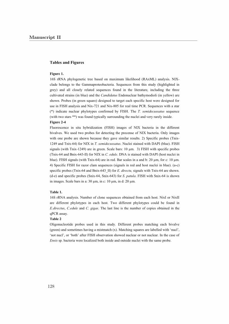
Tables and Figures
Figure 1.
16S rRNA phylogenetic tree based on maximum likelihood (RAxML) analysis. NIX-
clade belongs to the Gammaproteobacteria. Sequences from this study (highlighted in
grey) and all closely related sequences found in the literature, including the three
cultivated strains (in blue) and the Candidatus Endonuclear bathymodioli (in yellow) are
shown. Probes (in green square) designed to target each specific host were designed for
use in FISH analysis and Nix-721 and Nix-805 for real time PCR. Sequences with a star
(*) indicate nuclear phylotypes confirmed by FISH. The T. semidecussatus sequence
(with two stars **) was found typically surrounding the nuclei and very rarely inside.
Figure 2-4
Fluorescence in situ hybridization (FISH) images of NIX bacteria in the different
bivalves. We used two probes for detecting the precense of NIX bacteria. Only images
with one probe are shown because they gave similar results. 2) Specific probes (Tnix-
1249 and Tnix-64) for NIX in T. semidecussatus. Nuclei stained with DAPI (blue). FISH
signals (with Tnix-1249) are in green. Scale bars: 10 µm. 3) FISH with specific probes
(Tnix-64 and Bnix-643-II) for NIX in C. edule. DNA is stained with DAPI (host nuclei in
blue). FISH signals (with Tnix-64) are in red. Bar scales in a and b: 20 µm, for c: 10 µm.
4) Specific FISH for razor clam sequences (signals in red and host nuclei in blue). (a-c)
specific probes (Tnix-64 and Bnix-643_II) for E. directu, signals with Tnix-64 are shown.
(d-e) and specific probes (Snix-64, Snix-643) for S. patula. FISH with Snix-64 is shown
in images. Scale bars in a: 30 µm, in c: 10 µm, in d: 20 µm.
Table 1.
16S rRNA analysis. Number of clone sequences obtained from each host. NixI or NixII
are different phylotypes in each host. Two different phylotypes could be found in
E.directus, C.edule and C. gigas. The last line is the number of copies obtained in the
qPCR assay.
Table 2
Oligonucleotide probes used in this study. Different probes matching each bivalve
(green) and sometimes having a mistmatch (x). Matching squares are labelled with ‘nucl’,
‘not nucl’, or ‘both’ after FISH observation showed nuclear or not nuclear. In the case of
Ensis sp. bacteria were localized both inside and outside nuclei with the same probe.
Manuscript II
128

Intranuclear bacteria of bivalves
Endonucleobacter bathymodioli group *
Ensis directus razor clam clone, Sylt NIX-I (4) *Cerastoderma edule clam clone, Sylt NIX-I (5) *
90
100
Tripneustes gratilla echinoid isolate bacteria, AM495252ocean water clone, Bohai Bay, ocean water, FJ154998
Oceanrickettsia ariakensis from Crassostrea ariakensis, DQ123914Oceanrickettsia ariakensis, from Crassostrea ariakensis, DQ118733
10098
81
Crassostrea gigas oyster clone, GoMSiliqua Patula razor clam clone, full seq*
Pomacanthus sexstriatus fish tract clone, EU884930
65
Cystodytes dellechiajei ascidian clone, Mediterranean Sea, DQ884170
100
Alcyonium antarcticum coral clone, DQ312235Alcyonium antarcticum coral clone, DQ312243Alcyonium antarcticum coral clone, DQ312237
83
Alcyonium antarcticum coral clone, DQ312244
10099
Crassostrea gigas oyster clone, GoM Ensis directus razor clam clone, Sylt
Cerastoderma edule clam clone, Sylt NIX-II
96
ocean water clone, Newport Harbour, USA, EU799933Cystodytes dellechiajei ascidian clone, DQ884169
83
99
Endozoicomonas elysicola from Elysia ornata sea slug, AB196667Salmo salar fish gill clone, AY494615
88
Pocillopora damicornis coral clone, AY700601Venerupis philippinarum clam clone **
84
Erythropodium caribaeorum octocoral clone, DQ889921Erythropodium caribaeorum octocoral clone, DQ889931Muricea elongata sponge clone, DQ917879
Muricea elongata bleached sponge clone, DQ917830
68
91
Spongiobacter nickelotolerans, isolated from marine sponge, AB205011Muricea elongata sponge clone, DQ917877
89
Petrosia ficiformis sponge clone MOLA 531, AM990755Chondrilla nucula sponge clone, AM259915
Halichondria okadia sponge clone HOC2, AB054136
84
Asterias amurensis seastar clone KMD001, EU599216
79
Halichondria okadia sponge clone HOC22, AB054156Halichondria okadia sponge clone HOC25, AB054159
Acesta excavata bivalve gill clone, EF508132
100
67
95
Ruppia maritima gill clone, seagrass beds, EU487857Ruppia maritima gill clone, seagrass beds, EU487858
63
Bdellovibrio bacteriovorus, AF084850
0.10
Bnix-643/Bnix-64
Bnix-643II/Tnix-64
Snix-64/Snix-643
Cnix-64/Cnix-1249
Tnix-64/Tnix-643
Sequences from this study - shallow water bivalves
Cultured strains
*
Probes used in this study
Nix-721
Nix-721
Nix-805
Nix-805
Nix-721
Nix-721
NIX
cla
de
Sequences from intranuclear bacteria confirmed by FISH
Sequences from deep-sea mussels Bathymodiolus spp.
Sequences that are found intranuclearly or sorrounding the nuclei (after FISH)**
Fig. 1 16S rRNA phylogenetic tree based on maximum likelihood (RAxML) anal-ysis. NIX-clade belongs to the Gammaproteobacteria. Sequences from this study(highlighted in grey) and all closely related sequences found in the literature, in-cluding the three cultivated strains (in blue) and the Candidatus Endonuclearbathymodioli (in yellow) are shown. Probes (in green square) designed to targeteach specific host were designed for use in FISH analysis and Nix-721 and Nix-805for real time PCR. Sequences with a star (*) indicate nuclear phylotypes confirmedby FISH. The T. semidecussatus sequence (with two stars **) was found typicallysurrounding the nuclei and very rarely inside.
129
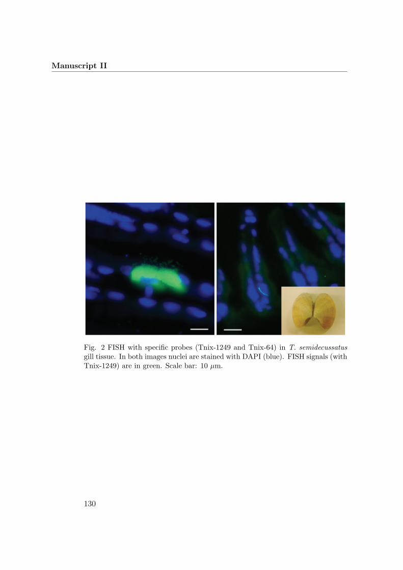
Manuscript II
Fig. 2 FISH with specific probes (Tnix-1249 and Tnix-64) in T. semidecussatusgill tissue. In both images nuclei are stained with DAPI (blue). FISH signals (withTnix-1249) are in green. Scale bar: 10 μm.
130

Intranuclear bacteria of bivalves
Fig. 3 FISH with specific probes (Tnix-64 and Bnix-643-II) in C. edule gill tissue.In a-c DNA is stained with DAPI (in blue), and FISH signals (with Tnix-64) arein red. Bar scales for a and b: 20 μm, for c: 10 μm. (a) image is without probeand a huge bacterial infection can be observed labeled with DAPI. Backgroundfluorescence of host tissue is observed in red.
131

Manuscript II
Fig. 4 Specific FISH for NIX-related sequences in razor clams (i.e. E.directus andS. patula gill tissue. Signals are in red and host nuclei labeled with DAPI in blue.(a-c) specific probes (Tnix-64 and Bnix-643-II) for E. directus, signals with Tnix-64 are shown. Background from tissue autofluorescence is seen in green. (d-e)specific probes (Snix-64, Snix-643) for S. patula. FISH with Snix-64 is shown inimages (red). Autofluorescence of tissue is blue. Scale bars in a: 30 μm, in c: 10μm, in d: 20 μm.
132

Intranuclear bacteria of bivalves
Table 1. 16S rRNA analysis. Number of clone sequences obtained from each host.NixI or NixII are different phylotypes in each host. Two different phylotypes couldbe found in E.directus, C.edule and C. gigas. The last line is the number of copiesobtained in the qPCR assay.
Table 2. Oligonucleotide probes used in this study. Different probes matchingeach bivalve (green) and sometimes having a mistmatch (x). Matching squares arelabelled with nucl, not nucl, or both after FISH observation showed nuclear or notnuclear. In the case of Ensis sp. bacteria were localized both inside and outsidenuclei with the same probe.
133

Manuscript III:
Minireview: Bacterial diversity of
shallow-water bivalves
Luciana Raggi-Hoyos, Nicole Dubilier
In Preparation

Minireview: Bacterial diversity in shallow-water bivalves
Luciana Raggi1, Nicole Dubilier1
1Max Planck Institute for Marine Microbiology, Celsiussr. 1, 28359,
Bremen, Germany
Keywords: bacteria-bivalve interaction, symbiosis, invertebrates
Running head: Bacterial diversity in bivalves
Corresponding author: Nicole Dubilier
E-mail: [email protected]
Bacteria diversity of bivalves
135

Abstract
Bacterial diversity in bivalves is a multidisciplinary topic, from biological
and basic research to economical interests, including aquaculture and
medicine. In this paper we review how these different disciplines have
influenced the few bivalve microbiota studies available. We present new
data from 16S rRNA analysis, including sequences from clone libraries and
isolates, and fluorescence in situ hybridization (FISH) from shallow-water
bivalves: Ensis directus, Cerastoderma edule, Crassostrea gigas, Siliqua
patula, Tapes semidecussatus, Mytilus edulis, and Mya arenaria, localizing
specific groups of bacteria associated with them. We also performed
cultivations from C. edule, and T. semidecussatus. The isolated bacteria
belong to bacteria phyla that are frequently found in organic rich
environments: oil spills, bone-falls, feces or invertebrate tissue. Most of
them belong to the Gammaproteobacteria group, and others to the
Alphaproteobacteria, Bacteroidetes, Actinobacteria and Spirochetes.
These associations might be occurring not only because of the surface
that bivalves provide to bacteria, but also because the association could
provide nutritional or protection benefits for both partners.
Manuscript III
136

Fig. 1 Phylogentic tree of the main bacterial phyla based on 16S rRNA gene. Almost all existing bacteria phyla
are visualized here. Branches in purple are the phyla for which bacterial species have been found associated to
bivalves.
Bacteria-bivalve associations
Bacteria-bivalve association studies have been enriched by the discovery
of intracellular bacterial symbionts of mussels and clams in the deep-sea.
However, most studies have been focused on species with intracellular
symbionts and there are scarce studies in the other associated bacteria.
Through its history, two topics have inspired the study of bacteria-bivalve
associations in shallow waters, the first one is the human pathogens that
edible bivalves can concentrate (Kueh and Chan 1984, Prieur et al. 1990,
Cabello et al 2005), and the second the economical importance that
Bacteria diversity of bivalves
137

Fig. 2 Mya arenaria transversal sections of gill tissue in left panels. FISH with specific
probes for spirochetes (Spiro-1400 – 5’-CTCGGGTGGTGTGACGGGCG–3’). In right
panels B. childressi, with spirochete specific probe (Spiro-1400) in top image, and in image
below with specific probe for epsilonproteobacteria (Epsy-682 – 5’
CGGATTTTACCCCTACAC-3’). Scale bars: 10 µm in left panels and bottom right image; 5
µm in top right image.
Manuscript III
138
healthy hatcheries have in aquaculture (Prieur 1987, Romero and Espejo
2001). In fact, because of these two reasons, already more than one
century ago the depuration processes (cleaning based on maximizing the
natural filtering activity of shellfish holding it in tanks of clean seawater)
in particular for oysters were implemented. The study of these
associations to use them as models for studying physiological interactions
between eukarya and bacteria, and the possibility to study evolutionary
pathways of symbiosis has seldom been the focus. With this last
intention, we synthesize all the available information about bacteria and
their association to shallow-water bivalves, and with molecular studies we
contribute to the understanding of bacterial diversity in bivalves. the
bacterial diversity of these living habitats. The first surprising thing that it
is noted when reviewing described bacteria associated with bivalves is
that their diversity is very broad and they cover almost all main branches
of the Bacteria kingdom (see Fig. 1).
Microflora diversity
Pathogenic, commensal and beneficial bacteria have all been described as
members of bivalve microflora. Most of the diversity studies performed
with bivalves are culture-dependent (Lovelace et al. 1968, Murchelano
and Brown 1968, Brisou 1962, Prieur 1981), meaning that the knowledge
that we have about bivalve microflora diversity is limited to the estimated
1% bacterial population that is cultivable (Kjelleberg et al. 1993) or even
less than 0.001% as described for oysters (Romero and Espejo 2001).
Occurrence of bacteria has been studied intensively in the bivalve
digestive tract (Prieur 1981, Minet et al. 1987) and in the crystalline style
(Bernard 1970, Paster et al. 1996, Prieur et al. 1990) Various bacterial
genera have been observed to be commonly
Bacteria diversity of bivalves
139

Table 1 Isolates from T. semidecussatus, C. edule and E. directus. Gill pieces
stored in glycerol were homogenized and cultivated in three different media.
Isolate Host Medium1 Identity2 Closest relative Acc.3
TI-1 T. semidecussatus HNM Kocuria sp. MOLA 658 AM990771
TI-2 T. semidecussatus HNM Dermacoccus sp. Ellin183 AF409025
TI-3 T. semidecussatus HNM Sulfitobacter sp. MOLA 8 AM990784
TI-4 T. semidecussatus HNM Uncultured bacterium clone PL-10B7 AY570580
TI-5 T. semidecussatus HNM Uncultured Vibrionaceae AY627367
TI-6 T. semidecussatus HNM Alcanivorax sp. PA23 EU647559
TI-7 T. semidecussatus HNM Pseudoalteromonas sp. BSs20060 EU433327
CI-1 C. edule HNM Shewanella sp. P117 EU195929
Vm1-1 T. semidecussatus MM+CTAB 1084/1121 (96%) FJ025776
Vm1-2 T. semidecussatus MM GQ149233
Os1 C. gigas MA AB198089
Vm1-3 T. semidecussatus MA AY292936
Vm1-4 T. semidecussatus MA DQ146980
Hs1-1 C. edule MA AB198089
Hs1-2 C. edule MA EU375181
E1-1 E.directus MA FN395284
E1-2 E.directus MA CP000903
Vm1-5 T. semidecussatus MA+CTAB FJ457601
Vm1-6 T. semidecussatus MA+CTAB FJ457601
1Media were used in solid (with 1.5 % Agar) and liquid presentations: MA - Marine
Medium 2216 (Difco), MM - Minimum Medium with and without CTAB (0.1% yeast,
0.01% peptone, 1.5% agar, 100µm CTAB, dissolved in sea water), and HNM - WL
Nutrient Medium (Difco) in 3.5% NaCl. 2This identity is with respect to closest relative of next column, it is shown first the number
of base pairs ration of the isolate sequence over the closest relative. 3Accesión numbers correspond to the published closest relatives.
associated to shallow-water bivalves: Vibrio, Pseudomonas, Spirochaetes,
Achromobacter, Flavobacteria, Micrococcus, Bacillus and also anaerobic
bacteria Bacteroidetes and Chlostridium.
Many studies have focused on Vibrio spp. since they are facultative
pathogens of humans and bivalves. Vibrio spp. are a regular component
Manuscript III
140

of the microflora and they might have an “important ecological niche”
(Prieur et al. 1990). Some have been related with the few described
bivalve diseases like bacillary necrosis or brown ring disease
(Grishkowsky and Liston 1974, Tubiash et al. 1970, Elston et al. 2008).
Recently, a review on the diversity and pathogenesis of Vibrio spp. was
made by Beaz-Hidalgo et al. (2010). With respect to bivalve lethality the
only two associated species are V. aestuarianus and V. splendidus. Other
species that seem to be commonly associated to bivalves are V.
alginolyticus and V. harveyi, and it is been observed that environmental
parameters such as salinity and temperature influence their diversity
(Pujalte et al 1999, Arias et al 1999, Beaz-Hidalgo et al 2010). Using
different media (High Nutrient Media, Minimum Media and Marine Agar,
with or without CTAB [Cetyl trimethylammonium bromide]), we have
been able to cultivate 18 different bacterial strains belonging to a variety
of bacterial phyla (Table 1). As expected, we obtained Vibrio strains from
Tapes semidecussatus, including in the media with CTAB, that was used
before by Plante and coworkers (2008) to isolate surfactant-resistant
bacteria with the aim of obtaining bacteria useful for environmental
remediation. As already observed in previous studies (Rajagopalan &
Sivalingan 1978, Sugita et al 1981) we could cultivate bacteria from the
Actinobacteria phylum (Kocuria sp. and Dermacoccus sp.), and Bacillus
sp. of the Firmicutes phylum. Krokinobacter sp. and Alcanivorax sp.
isolates are of special interest because the first ones are bacteria that
seem to be specialized in the degradation of organic matter (Khan et al.
2006) and the second are bacteria that use hydrocarbons as a sole carbon
and energy source (Head et al. 2006). Many times coastal bivalves have
been taken as a biological marker to assess pollution (e.g. Nishihama et
al 1998, Bresler et al 1999, Verlecar et al 2006). For example, the
presence of Alcanivorax bacteria in clam tissue indicates a strong oil
contamination in North Sea beaches (Brakstad and Lodeng 2005). Many
bacterial phylotypes were found in our 16S rRNA sequence analyses,
Bacteria diversity of bivalves
141

some are recurrent bacteria that have shown up in previous bivalve
studies (Table 2). Most of these sequences are from
gammaproteobacteria, as it has been seen in previous molecular studies
of bivalve-associated bacterial communities (Schulze et al. 2006, Cavallo
et al. 2009). We could detect groups of bacteria that seem to associate
with high-content organic matter: oil spills, bone-falls, feces or
invertebrate tissue. In addition to gammaproteobacteria there were also
Alphaproteobacteria, Bacteroidetes, Actinobacteria and Spirochetes. We
hypothesize that these bacteria are specialized in high-organic matter
habitats and that further analysis of more individuals and more species
will show a characteristic community perhaps host species-specific.
Associated spirochaetes stand out because they seem to be a stable
bacterial community in bivalves (Bernard et al. 1970, Paster et al. 1996,
Prieur 1990, Margulis and Fester 1991). Our FISH observations (Fig 2)
show them well established in bivalve gill tissue. It might be interesting to
study the variability of these spirochetes species within the different
bivalves. It is not clear so far if spirochetes have an ecological role or
importance in the association with bivalves, but they seem to be
ubiquitous microflora within the mollusk group (Prieur 1990) and perhaps
the whole invertebrate group, as other spirochetes have been found in
oligochaete worms (Ruehland et al. 2008). Margulis et al. (1991) named
as symbionts the studied spirochetes in oysters.
Do intracellular symbionts reduce microflora diversity?
Six families of bivalves have been found to be associated with intracellular
symbiotic bacteria: Vesicomyidae (Calyptogena , Vesicomya), Mytilidae
(Bathymodiolus, Idas), Solemyidae (Solemya, Acharax), Lucinidae
(Lucina, Codakia), Thyasiridae (Thyasira), and Teredinidae (Lyrodus). All
are deep-sea water organisms except for Solemya, Thyasira and lucinids.
It is remarkable that these organisms associate with such a restricted
Manuscript III
142

diversity of bacteria, perhaps due to the presence of the endosymbionts.
However it is still possible that diversity is in fact not reduced and we
have not been able to see it in cultures or in clone library analyses due to
the high abundance of the bacterial symbionts. In our 16S rRNA analysis
(sequencing and FISH) of B. childressi, we observed the presence of
different bacteria associated with the tissue apart from the well
characterized methanotrophic symbiont: an epsilon proteobacterium, a
Candidatus Endonuclear bathymodiolin and a spirochete phylotype. This
could suggest that the B. childressi recognition system is not as specific
as in the other Bathymodiolus spp. In fact B. childressi is one of the
lowest positioned bathymodiolin mussels in the COI phylogenetic analysis
except for the latest described B. sp. from Juan de Fuca (Duperron et al.
2009) and could have an immunological system more similar to the
shallow water bivalves. However, in-depth studies of other species might
reveal that the diversity of bathymodiolin mussels is much higher than
previously assumed.
Nutrition and protection
Bivalve gills provide an ideal habitat for bacteria, with protection from
grazers and constant flow of nutrients. Bacteria might be complementing
their host nutrition or contributing to metabolite production. Degradation
of bacteria by bivalve enzymes has been observed and it seems that this
degradation provide dissolved compounds (Birckbeck & McHenery 1982,
Amouroux & Amouroux 1988) and improves bivalve nutrition (Samain et
al. 1987). Bacteria could provide 5% to 10% of the carbon, and 20% of
the nitrogen from the bivalve requirements (Prieur et al 1990). A stable
microbiota could be providing protection to bivalves thanks to competition
against other, potentially pathogenic bacteria. Also, microbiota could be
secreting antimicrobial substances that have been observed to be
commun in bacteria isolated from bivalves (Zheng et al. 2005).
Bacteria diversity of bivalves
143

Table 2 Bacteria found in the different bivalves. Studies where bacteria have been observed microscopically (M), cultured (C), isolated (I), or
their 16S rRNA sequenced (S). Some letters are repeated because more than one study have characterized the species. In red the sequences
and cultures from this study.
Manuscript III
144

Summary and conclusions
Bacterial diversity in bivalves is a multidisciplinary topic, with from
researches in biological, medical, aquaculture and basic research.
Ecological studies with molecular techniques are scarce and they could
help to disentangle the interaction patterns between the endemic
microflora and the invasive ones, as well as the benefits that symbiotic or
the harm that pathogenic bacteria bring along. It is important to
understand the distribution of pathogenic bacteria in the marine
environments to predict potential health concerns transmitted by seafood.
Ecological parameters such as nutrient availability, temperature, and
salinity influence the presence and persistence of bacteria. However we
suggest that bacteria present in bivalves are not only randomly
associated with their hosts. It is in part the result of the surrounding
water community but also the result of a common evolution between host
and bacteria that normally associate with invertebrates or occur in
habitats with high organic matter content. We would need more studies to
be able to say that these symbiotic bacteria, consistently associated with
many diverse bivalves, are beneficial or harmful to their hosts.
Bibliography
Amouroux J, Amouroux J (1988) Comparative study of the carbon cycle in Venus
verrucosa fed on bacteria and phytoplankton. Marine Biology 97:339-347
Arias CR, Macian MC, Aznar R, Garay E, Pujalte MJ (1999) Low incidence of Vibrio
vulnificus among Vibrio isolates from sea water and shellfish of the western
Mediterranean coast. Journal of Applied Microbiology 86:125-134
Beaz-Hidalgo R, Balboa S, Romalde JL, Figueras MJ (2010) Diversity and
pathogenecity of Vibrio species in cultured bivalve molluscs. Environmental
Microbiology Reports 2:34-43
Bernard F (1970) Occurrence of the spirochaete genus Cristispira in western
Canadian marine bivalves. The Veliger 13:33-36
Birkbeck T, McHenery J (1982) Degradation of bacteria by Mytilus edulis. Marine
Bacteria diversity of bivalves
145

Biology 72:7-15
Brakstad O, Lødeng A (2005) Microbial diversity during biodegradation of crude oil in
seawater from the North Sea. Microbial ecology 49:94-103
Bresler V, Bissinger V, Abelson A, Dizer H, Sturm A, Kratke R, Fishelson L, Hansen
PD (1999) Marine molluscs and fish as biomarkers of pollution stress in littoral
regions of the Red Sea, Mediterranean Sea and North Sea. Helgoland Marine
Research 53:219-243
Brisou J (1968) La pollution microbienne, virale et parasitaire des eaux littorales et
ses conséquences pour la santé publique. Bulletin of the World Health
Organization 38:79
Cabello AE, Espejo RT, Romero J (2005) Tracing Vibrio parahaemolyticus in oysters
(Tiostrea chilensis) using a Green Fluorescent Protein tag. Journal of
Experimental Marine Biology and Ecology 327:157-166
Cavallo RA, Acquaviva MI, Stabili L (2009) Culturable heterotrophic bacteria in
seawater and Mytilus galloprovincialis from a Mediterranean area (Northern
Ionian Sea-Italy). Environmental Monitoring and Assessment 149:465-475
Elston RA, Hasegawa H, Humphrey KL, Polyak IK, Hase CC (2008) Re-emergence
of Vibrio tubiashii in bivalve shellfish aquaculture: severity, environmental
drivers, geographic extent and management. Diseases of aquatic organisms
82:119-134
Grischkowsky RS, Liston J (1974) Bacterial pathogenicity in laboratory-induced
mortality of the Pacific oyster (Crassostrea gigas, Thunberg). Proc Natl
Shellfish Assoc 64:82-91
Head IM, Jones DM, R\\"oling WFM (2006) Marine microorganisms make a meal of
oil. Nature Reviews Microbiology 4:173-182
Khan ST, Nakagawa Y, Harayama S (2006) Krokinobacter gen. nov., with three
novel species, in the family Flavobacteriaceae. International journal of
systematic and evolutionary microbiology 56:323
Kjelleberg S, Albertson N, Fl\\"ardh K, Holmquist L, Jouper-Jaan Å, Marouga R,
Östling J, Svenblad B, Weichart D (1993) How do non-differentiating bacteria
adapt to starvation? Antonie Van Leeuwenhoek 63:333--341
Kueh CSW, Chan K (1985) Bacteria in bivalve shellfish with special reference to the
oyster. Journal of Applied Microbiology 59:41-47
Lovelace T, Tubiash H, Colwell R (1968) Quantitative and qualitative commensal
bacterial flora of Crassostrea virginica in Chesapeake Bay. Proc Natl Shellfish
Assoc 58:82-87
Margulis L, Fester R (1991) Symbiosis as a source of evolutionary innovation:
speciation and morphogenesis, Vol. The MIT Press
Minet J, Barbosa T, Prieur D, Cormier M (1987) Mise en évidence du processus de
concentration des bactéries par la moule Mytilus edulis(L.)= The nature of the
Manuscript III
146

bacteria concentration process by the mussel Mytilus edulis (L.). Comptes
rendus de l'Académie des sciences Série 3, Sciences de la vie 305:351-354
Murchelano RA, Brown C (1968) Bacteriological study of the natural flora of the
Eastern oyster, Crassostrea virginica. Journal of Invertebrate Pathology
11:510-520
Nishihama Y, Horii T, Motoya S, Yamashita S (1998) Effect of oil pollution and its
recovery on the activity and taste of the bivalve Pseudocardium sachalinensis
caused by an oil spill accident off Tomakomai, Hokkaido. Scientific Reports of
Hokkaido Fisheries Experimental Station 53:17-24
Paster B, Pelletier D, Dewhirst F, Weisburg W, Fussing V, Poulsen L, Dannenberg S,
Schroeder I (1996) Phylogenetic position of the spirochetal genus Cristispira.
Applied and environmental microbiology 62:942
Plante CJ, Coe KM, Plante RG (2008) Isolation of surfactant-resistant bacteria from
natural, surfactant-rich marine habitats. Applied and environmental
microbiology 74:5093
Prieur D (1981) Experimental studies of trophic relationships between marine
bacteria and bivalve molluscs.
Prieur D, Nicolas JL, Plusquellec A, Vigneulle M (1990) Interactions between bivalve
mollusks and bacteria in the marine-environment. Oceanography and Marine
Biology 28:277-352--
Pujalte MJ, Ortigosa M, Macián MC, Garay E (1999) Aerobic and facultative
anaerobic heterotrophic bacteria associated to Mediterranean oysters and
seawater. International Microbiology 2:259
Rajagopalan K, Sivalingam P (1978) Bacterial flora of green mussel (Mytilus viridis
Linnaeus) and a naturally occurring rock oyster (Crassostrea cuculata).
Malaysian Applied Biology (Malaysia)
Romero J, Espejo R (2001) The prevalence of noncultivable bacteria in oysters
(Tiostrea chilensis, Philippi, 1845). Journal of Shellfish Research 20:1235-
1240
Ruehland C, Blazejak A, Lott C, Loy A, Erseus C, Dubilier N (2008) Multiple bacterial
symbionts in two species of co-occurring gutless oligochaete worms from
Mediterranean sea grass sediments. Environmental Microbiology 10:3404-
3416--
Schulze AD, Alabi AO, Tattersall-Sheldrake AR, Miller KM (2006) Bacterial diversity
in a marine hatchery: Balance between pathogenic and potentially probiotic
bacterial strains. Aquaculture 256:50-73
Sugita H, Tanaami H, Kobashi T, Deguchi Y (1981) Bacterial flora of coastal
bivalves. Nippon Suisan Gakkaishi 47:655-661
Verlecar X, Pereira N, Desai S, Jena K (2006) Marine pollution detection through
biomarkers in marine bivalves. Current science 91:1153-1157
Bacteria diversity of bivalves
147

Manuscript IV
Manuscript IV:
Widespread occurrence of an intranuclear
parasite in bathymodiolin mussels
Frank U. Zielinski, Annelie Pernthaler, Sebastien
Duperron, Luciana Raggi, Olav Giere, Christian
Borowski and Nicole Dubilier
Published in Environmental Microbiology
148

Widespread occurrence of an intranuclear bacterialparasite in vent and seep bathymodiolin mussels
Frank U. Zielinski,1,2 Annelie Pernthaler,3†
Sébastien Duperron,4 Luciana Raggi,1 Olav Giere,5
Christian Borowski1 and Nicole Dubilier1*1Max Planck Institute for Marine Microbiology, SymbiosisGroup, Celsiusstr. 1, 28359 Bremen, Germany.2Helmholtz Centre for Environmental Research – UFZ,Department of Environmental Microbiology, PermoserStr. 15, 04318 Leipzig, Germany.3California Institute of Technology, Division of Geologicaland Planetary Sciences, 1200 E California Blvd,Pasadena, CA 91125, USA.4Université Pierre et Marie Curie, Équipe Adaptation auxMilieux Extrêmes, 7 Quai St Bernard, 75005 Paris,France.5University of Hamburg, Biozentrum Grindel undZoologisches Museum, Martin-Luther-King Platz 3,20146 Hamburg, Germany.
Summary
Many parasitic bacteria live in the cytoplasm of multi-cellular animals, but only a few are known to regularlyinvade their nuclei. In this study, we describethe novel bacterial parasite “Candidatus Endonucleo-bacter bathymodioli” that invades the nuclei of deep-sea bathymodiolin mussels from hydrothermal ventsand cold seeps. Bathymodiolin mussels are wellknown for their symbiotic associations with sulfur-and methane-oxidizing bacteria. In contrast, the para-sitic bacteria of vent and seep animals have receivedlittle attention despite their potential importance fordeep-sea ecosystems. We first discovered the intra-nuclear parasite “Ca. E. bathymodioli” in Bathymodio-lus puteoserpentis from the Logatchev hydrothermalvent field on the Mid-Atlantic Ridge. Using primersand probes specific to “Ca. E. bathymodioli” we foundthis intranuclear parasite in at least six other bathy-modiolin species from vents and seeps around theworld. Fluorescence in situ hybridization and trans-mission electron microscopy analyses of the devel-
opmental cycle of “Ca. E. bathymodioli” showed thatthe infection of a nucleus begins with a single rod-shaped bacterium which grows to an unseptated fila-ment of up to 20 mm length and then dividesrepeatedly until the nucleus is filled with up to 80 000bacteria. The greatly swollen nucleus destroys itshost cell and the bacteria are released after thenuclear membrane bursts. Intriguingly, the only nucleithat were never infected by “Ca. E. bathymodioli” werethose of the gill bacteriocytes. These cells contain thesymbiotic sulfur- and methane-oxidizing bacteria,suggesting that the mussel symbionts can protecttheir host nuclei against the parasite. Phylogeneticanalyses showed that the “Ca. E. bathymodioli”belongs to a monophyletic clade of Gammaproteobac-teria associated with marine metazoans as diverse assponges, corals, bivalves, gastropods, echinoderms,ascidians and fish. We hypothesize that many of thesequences from this clade originated from intra-nuclear bacteria, and that these are widespread inmarine invertebrates.
Introduction
Bacteria inhabit every imaginable place on earth. With theevolution of the eukaryotes, numerous bacteria haveestablished a symbiotic or parasitic relationship insideeukaryotic cells. Most of these bacteria live in the cyto-plasm, but some have found their way into eukaryotic cellcompartments, including mitochondria (Chang and Mus-grave, 1970; Epis et al., 2008), chloroplasts (Wilcox,1986; Schmid, 2003a,b) and nuclei (Maillet and Folliot,1967; Grandi et al., 1997; Arneodo et al., 2008). Althougheukaryotic cell compartments have been investigated fordecades with light and electron microscopy, little is knownabout intracompartimental bacteria, particularly those thatlive in the nuclei of eukaryotes. Most commonly describedfrom protists (Fokin, 2004; Görtz, 2006), almost nothing isknown about intranuclear bacteria of metazoans. Rickett-sial Alphaproteobacteria occasionally invade nuclei oftheir arthropod or mammalian hosts but occur mainly inthe cytoplasm (Burgdorfer et al., 1968; Urakami et al.,1982; Pongponratn et al., 1998; Ogata et al., 2006). Apartfrom these facultative intranuclear Rickettsia, reports ofintranuclear bacteria that are morphologically or phy-logenetically distinct from the Rickettsiales are rare in
Received 29 July, 2008; accepted 11 November, 2008. *Forcorrespondence. E-mail [email protected]; Tel. (+49) 4212028 932; Fax (+49) 421 2028 580. †Present address: HelmholtzCentre for Environmental Research – UFZ, Department of Environ-mental Microbiology, Permoser Str. 15, 04318 Leipzig, Germany.
Environmental Microbiology (2009) doi:10.1111/j.1462-2920.2008.01847.x
© 2009 The AuthorsJournal compilation © 2009 Society for Applied Microbiology and Blackwell Publishing Ltd
Bathymodiolus intranuclear parasite
149

metazoans. Only a single 16S rRNA sequence is currentlyknown from an intranuclear bacterium that does notbelong to the Rickettsiales (Kerk et al., 1992). This Gam-maproteobacterium, called ‘Nuclear Inclusion X’ (NIX),causes mass mortalities in the Pacific razor clam Siliquapatula (Elston, 1986; Ayres et al., 2004). A few morpho-logical observations of non-Rickettsiales-like intranuclearbacteria have been described from the venerid clamRuditapes decussatus (Azevedo, 1989), and two marineAplysina sponges (formerly Verongia) (Vacelet, 1970;Friedrich et al., 1999).Deep-sea hydrothermal vents and cold seeps have
been studied extensively since their discovery25–30 years ago. Given the high diversity of metazoanscurrently described from vents and seeps (Sibuet andOlu-Le Roy, 2002; Wolff, 2005; Desbruyères et al.,2006a), remarkably little is known about their parasites(de Buron and Morand, 2004). To date about a dozenmetazoan macroparasites have been described (deBuron and Segonzac, 2006a,b). A few studies, using 18SrRNA-based molecular techniques, have suggested thatthere may be a high abundance of parasitic protist atvents (Atkins et al., 2000; Edgcomb et al., 2002; López-García et al., 2007), and the morphology of fungal, protist,bacterial and viral parasites has been described in moredetail in some vent and seep mussels, clams and limpets(Powell et al., 1999; Terlizzi et al., 2004; Ward et al., 2004;Mills et al., 2005; Van Dover et al., 2007). These studiessuggest the potential importance of parasites for vent andseep ecosystems, yet comprehensive investigations ofthe taxonomy, phylogeny, and life cycle of vent and seepparasites are still lacking.Mussels of the genus Bathymodiolus (Bivalvia: Mytil-
idae) occur worldwide at deep-sea hydrothermal ventsand cold seeps (Tarasov et al., 2005; DeChaine andCavanaugh, 2006; Génio et al., 2008). In the absence oflight and thus, photosynthetic carbon fixation, thesemussels depend on chemosynthetic bacterial symbiontsfor their nutrition (Stewart et al., 2005; Cavanaugh et al.,2006). These endosymbionts occur in the mussel’s gilltissue, in the cytoplasm of bacteriocytes that regularlyalternate with symbiont-free intercalary cells (Fiala-Médioni and Le Pennec, 1987; Distel et al., 1995).Bathymodiolin mussels can harbour two types ofchemosynthetic bacteria: a chemoautotrophic sulfur oxi-dizer, capable of fixing CO2 in the presence of sulfide orthiosulfate as energy sources, and a methane oxidizerthat uses methane as both a carbon and an energysource (Fisher et al., 1987; Nelson et al., 1995; Pimenovet al., 2002). Some mussel species harbour onlythiotrophic or only methanotrophic symbionts, while othermussel species harbour both types and thus live in a dualsymbiosis (DeChaine and Cavanaugh, 2006). The energysources for the bacterial symbionts, reduced sulfur com-
pounds and methane, are provided by the hydrothermalfluids at vents and through hydrocarbon seepage at coldseeps. Both the mussels and their chemosynthetic bacte-ria mutually benefit from their symbiosis: the musselfacilitates access to the reductants and oxidants thatare necessary for energy production (such as sulfide,methane and oxygen) by supplying its symbionts with aconstant fluid flow. In exchange, the bacterial symbiontsprovide carbon compounds that support the growth andmaintenance of host biomass (Stewart et al., 2005;Cavanaugh et al., 2006). In addition to their symbioticbacteria, some vent and seep mussels have recentlybeen described that are colonized by parasites such asviruses, Rickettsia- and Chlamydia-like bacteria, ciliates,fungi and trematodes, but beyond their morphologicaldescription, almost nothing is known about these para-sites (Powell et al., 1999; Ward et al., 2004; Van Doveret al., 2007).This study describes a novel bacterial parasite of
bathymodiolin mussels. Using comparative 16S rRNAsequence analysis, fluorescence in situ hybridization(FISH) and transmission electron microscopy (TEM), weshow that this parasite lives in the nuclei of mussels fromvents and seeps around the world. We describe the lifecycle of this intranuclear parasite through reconstructionof its developmental cycle from a solitary cell to the pro-liferation of up to 80 000 bacteria within a single greatlyenlarged nucleus. We propose the name “CandidatusEndonucleobacter bathymodioli” for this bacterium. Thegenus name ‘Endonucleobacter’ translates freely into‘bacterium living inside the nucleus’ and the species name‘bathymodioli’ refers to the host genus of vent and seepmussels, Bathymodiolus, in which we discovered thisparasite.
Results
Discovery of intranuclear bacteria in Bathymodiolusputeoserpentis
Three gammaproteobacterial 16S rRNA phylotypeswere found in gill tissues of B. puteoserpentis from theLogatchev hydrothermal vent field on the Mid-AtlanticRidge (Fig. 1A). In addition to the sequences alreadyknown from the sulfur- and methane-oxidizing symbionts(Duperron et al., 2006), a novel 16S rRNA sequence wasdiscovered (“Candidatus Endonucleobacter bathymodioli”in Fig. 1A). This novel sequence fell in a clade consistingof 16S rRNA sequences from bacteria associated withmarine animals including sponges, corals, a sea slug, anascidian, a sea urchin and a fish (93–97% identity,Table S1). This clade also included the sequence from the‘Nuclear Inclusion X’ parasite of the Pacific razor clamS. patula (Kerk et al., 1992). The monophyly of this clade
2 F. U. Zielinski et al.
© 2009 The AuthorsJournal compilation © 2009 Society for Applied Microbiology and Blackwell Publishing Ltd, Environmental Microbiology
Manuscript IV
150

was supported in both maximum likelihood and Bayesiananalyses (support values: 60 and 1.00). The cultivatedspecies within this clade were all heterotrophic: Endozo-icomonas elysicola, a strictly aerobic and mesophilic bac-terium from the gastrointestinal tract of the sea slug Elysiaornata (Kurahashi and Yokota, 2007), and three speciesfrom marine sponges, one rod-shaped (H425) and onespirillum-like (H262) bacterium (Sfanos et al., 2005), andSpongiobacter nickelotolerans (unpublished informationfrom GenBank). The closest relatives to the clade con-taining “Ca. E. bathymodioli” were Zooshikella gangwhen-sis, a chemoorganotrophic, aerobic and halophilic isolatefrom sediments of a Korean tidal flat (Yi et al., 2003) (91%identity to the “Ca. E. bathymodioli” sequence fromB. puteoserpentis) and two sequences from Rickettsia-like bacteria causing mass mortality in the oyster Cras-sostrea ariakensis (unpublished information in GenBankunder accession numbers DQ118733 und DQ123914).Fluorescence in situ hybridization analyses with probes
specific to the “Ca. E. bathymodioli” phylotype showedthat it originated from bacteria that occurred exclusively inthe nuclei of B. puteoserpentis cells (Fig. 2). Gill tissueswere most heavily colonized (Fig. 2A–D and F), but thebacteria were also observed in nuclei of the gut (Fig. 2E),digestive gland, labial palp, mantle and foot (data notshown). In the gill tissues, only nuclei of the symbiont-freeintercalary cells were infected, whereas intranuclear bac-teria were never observed in the bacteriocytes containingthe thiotrophic and methanotrophic symbionts (Fig. 2B–Dand F). Transmission electron microscopy analysis of gillfilaments confirmed that the bacteria were inside the hostnuclei (Fig. 3).
Widespread occurrence of “Ca. E. bathymodioli” indeep-sea mussels
Using PCR primers and FISH probes specific to the “Ca.E. bathymodioli” phylotype found in B. puteoserpentis, wesearched for these bacteria in other bathymodiolin hostsfrom hydrothermal vents and cold seeps in the AtlanticOcean, the Gulf of Mexico, and the Pacific Ocean (Fig. 4,Table 1, Table S2). We found “Ca. E. bathymodioli” in allhost species except B. aff. boomerang (Southeast Atlan-tic) and B. brevior (West Pacific). All “Ca. E. bathymodioli”sequences were very closely related to each other with98.8% identity between sequences on average and98.1% identity between the two most distant phylotypes(Fig. 1B). In most host species, a single 16S rRNA phy-lotype dominated the clone libraries. Less dominant phy-lotypes that differed by at most 2 bp from the dominantphylotypes were also found in several host species.These phylotypes often co-occurred in the same hostindividuals and were shared between individuals(Table S2). In B. sp. from Wideawake (Mid-Atlantic
Ridge), a single 16S rRNA clone was found with asequence that differed by 1.9% (26 bp) from all other “Ca.E. bathymodioli” phylotypes found in this species (called‘minor phylotype III’ in Table S2). This phylotype was mostclosely related to the sequence from B. aff. thermophilus(Pacific-Antarctic Ridge) (Fig. 1B). With the exception ofthis minor phylotype III, all other “Ca. E. bathymodioli”sequences fell into three clusters reflecting their geo-graphic origins from the Mid-Atlantic Ridge, the Pacific-Antarctic Ridge, and the Gulf of Mexico (Fig. 1B). Thecorrelation between genetic distances of the “Ca. E.bathymodioli” 16S rRNA phylotypes and geographical dis-tances of sample locations was statistically significantfor all phylotypes (P = 0.04 with minor phylotype III andP = 0.01 without minor phylotype III).
Developmental cycle of “Ca. E. bathymodioli”
Detailed FISH and TEM analyses of “Ca. E. bathymodioli”in B. puteoserpentis gill tissues revealed six distinctdevelopmental stages (Figs 2, 3 and 5). In Stage 1, asingle rod-shaped bacterium (1.8 ¥ 0.4 mm) is presentinside the nucleus (Fig. 2B and G). In Stage 2, thebacterium has grown to an unseptated filament of upto 18–20 mm length (Fig. 2H–J). In Stage 3, a looselywrapped filamentous coil is visible inside the host nucleus(Fig. 2M). We could not clearly discern if this coil consistsof several separate filaments or one long unseptatedfilament. However, on rare occasions we observed twofilaments of equal length (Fig. 2L) and filaments thatappeared to be in the process of longitudinal division insome nuclei (Fig. 2K), suggesting that the Stage 3 coilconsists of several filaments. The host nuclei in Stage 3have become more irregular in shape and chromatin isreduced to a thin layer along the nuclear membrane(Fig. 3A and D). Transverse binary fission of the filamen-tous coils leads to Stage 4 in which stacks of shorterfilaments of up to 8–10 mm length fill the host nucleus(Fig. 2E and N). At this stage, host nuclei are at least twoto three times the volume of uninfected nuclei. Repeatedtransverse binary fissions lead to the formation of numer-ous rods in Stage 5 with the nuclei further enlarged toabout five times the volume of uninfected nuclei. Chroma-tin is nearly completely reduced and barely visible alongsome parts of the nuclear membrane (Fig. 3E). In Stage6, the bacteria have reproduced massively forming around to oval aggregate of up to 30 mm in diameter that isstill surrounded by a membrane (Fig. 2P). At this stage,the infected nuclei are no longer visible within structurallyintact host cells, but occur extracellularly within the gillepithelium. At average bacterial sizes of 1.8 ¥ 0.4 mm,Stage 6 nuclei can contain between 10 000 and 80 000bacteria. These aggregates are eventually disrupted andthe bacteria released into the fluids surrounding the
Intranuclear bacteria in vent and seep mussels 3
© 2009 The AuthorsJournal compilation © 2009 Society for Applied Microbiology and Blackwell Publishing Ltd, Environmental Microbiology
Bathymodiolus intranuclear parasite
151

δ
α
γ
4 F. U. Zielinski et al.
© 2009 The AuthorsJournal compilation © 2009 Society for Applied Microbiology and Blackwell Publishing Ltd, Environmental Microbiology
Manuscript IV
152

mussel gills. Three-dimensional examples of the develop-mental Stages 2, 3, 4 and 6 can be viewed as supportinganimations (Videoes S1–S4). In addition to the detailedFISH and TEM analyses of the “Ca. E. bathymodioli”developmental stages in B. puteoserpentis, FISH analy-ses of B. azoricus, B. sp. (Wideawake), B. aff. thermophi-lus, B. brooksi, B. heckerae, and “B.” childressi gill tissuesshowed the presence of similar developmental stages inthese host species as well.
Discussion
Developmental cycle
The colonization of a nucleus requires several steps: theinfection of the host cell, passage through the cytoplasm,and penetration of the nuclear membrane. None of thesestages were observed in this study, presumably becausethese events occur on very short time scales and aretherefore only rarely visible. The first discernible infectionstage in our study was the presence of a single rod-shaped bacterium in the mussel nuclei that grows to anunseptated filament of about 20 mm and possibly longer(Stages 1 and 2 in Fig. 2G–J). This may be a character-istic feature of intranuclear bacteria. In the marine spongeAplysina, intranuclear bacteria can form filaments up to350 mm in length (Vacelet, 1970; Friedrich et al., 1999),and infectious forms of Holospora obtusa can reach20 mm in length in the macronucleus of the ciliate Para-mecium caudatum (Görtz, 2006). Unseptated filamentousgrowth has also been observed in Bdellovibrio bacterio-vorus, a deltaproteobacterial predator of Gram-negativebacteria (Angert, 2005; Dworkin, 2006). The transitionfrom Stage 2 to Stage 3 appears to take place throughlongitudinal fission (Fig. 2K). Longitudinal division is rareamong bacteria and has only been described in the sulfur-oxidizing symbionts of three marine host genera, thenematode Laxus sp. (Polz et al., 1992; 1994), the gutlessoligochaete Olavius (Giere and Krieger, 2001; Bright andGiere, 2005), and the sand-dwelling ciliates of the genusKentrophoros (Fauré-Fremiet, 1951; Raikov, 1971).
The shift from filamentous growth to massive reproduc-tion through transverse fission may be triggered by nutri-ent limitation. The chromatin of Stage 3 nuclei is greatlyreduced to a thin layer along the nuclear membrane(Fig. 3A and D). Given that this reduction does not appearto be physically induced because the bacteria do notcompletely fill out the nucleus at this stage, it is likely thatthe bacteria have used the chromatin for nutrition (seebelow). In B. bacteriovorus, transition to the multiplefission phase occurs when the cytoplasm is consumedand the host’s resources are exhausted (Horowitz et al.,1974; Angert, 2005; Lambert et al., 2006).Multiple rounds of transverse binary fission between
Stages 3 and 6 lead to massive swelling of the host nuclei.Greatly enlarged nuclei are typical for protists infectedwith intranuclear bacteria (Table S3) and have also beendescribed in the Pacific razor clam S. patula infected withthe intranuclear pathogen ‘NIX’ (Elston, 1986). It is intrigu-ing that in two of the host species shown to be infectedwith “Ca. E. bathymodioli” in this study (B. heckerae andB. puteoserpentis), previous light microscopical analysesindicated the presence of hypertrophied nuclei in sometissues (Ward et al., 2004). Transmission electron micros-copy and FISH analyses are needed to clarify if thesenuclear distortions were caused by viruses, as suggestedby Ward and colleagues (2004), or by intranuclear bacte-ria. Similarly, bacteria described as ‘Rickettsia-like’ and‘Chlamydia-like’ based on light microscopical analyses ofgill tissues of vent and seep mollusks (Powell et al., 1999;Terlizzi et al., 2004; Ward et al., 2004; Mills et al., 2005)might not be bacteria belonging to the Rickettsia andChlamydia but rather “Ca. E. bathymodioli” parasites.The completion of the “Ca. E. bathymodioli” cell cycle
requires the release of the infected nuclei from the hostcell. This most likely occurs through the destruction of thehost cell and rupture of the host cytoplasmic membrane.In ‘NIX’-infected S. patula, the host cells are ruptured bythe greatly enlarged nuclei, indicating a greater resiliencyof the nuclear over the cytoplasmic membrane (Elston,1986). When the “Ca. E. bathymodioli”-infected nucleibecome extracellular, they are still surrounded by a mem-
Fig. 1. 16S rRNA phylogenetic tree inferred from maximum likelihood and Bayesian analysis.A. The tree shows the “Candidatus Endonucleobacter bathymodioli” clade (grey trapezoid) together with its closest relatives. Colours ofsequences from marine invertebrates were assigned according to host phylogeny, with bacteria from bivalves shown in red, from sponges inblue, and from ascidians in purple (sea slug, sea urchin and fish bacteria are shown in black). Also included in the tree are (i) the closest freeliving relatives within the Oceanospirillales and Alteromonadales; (ii) the methane-oxidizing endosymbionts of bathymodiolin mussels (bluebox); (iii) the sulfur-oxidizing endosymbionts of bathymodiolin mussels (yellow box); and (iv) intranuclear bacteria belonging to the Rickettsialesfrom ciliates and ticks (purple box). Bdellovibrio bacteriovorus was used as an outgroup. The phylogenetic reconstruction is based primarily onnearly full sequences. Partial sequences (508–983 bp) are marked (°). Methane- and sulfur-oxidizing symbionts of bathymodiolin mussels arelabelled MOX and SOX respectively. Values at nodes represent maximum likelihood bootstrap values in percentage (first value) and posteriorprobabilities (second value). Scale bar represents 10% estimated base substitution. Only bootstrap values � 60 and posterior probabilities> 0.80 are shown. MAR, Mid-Atlantic Ridge; PAR, Pacific-Antarctic Ridge; GoM, Gulf of Mexico.B. Tree of “Ca. E. bathymodioli” sequences from vent and seep mussels (collection sites of mussels are shown). The “Ca. E. bathymodioli”sequences form a monophyletic clade.
Intranuclear bacteria in vent and seep mussels 5
© 2009 The AuthorsJournal compilation © 2009 Society for Applied Microbiology and Blackwell Publishing Ltd, Environmental Microbiology
Bathymodiolus intranuclear parasite
153

6 F. U. Zielinski et al.
© 2009 The AuthorsJournal compilation © 2009 Society for Applied Microbiology and Blackwell Publishing Ltd, Environmental Microbiology
Manuscript IV
154

brane. This membrane is most likely of nuclear origin,because it stained positively with 4,6-diamidino-2-phenylindole (DAPI), suggesting remnants of nuclearDNA along the inside of the membrane. Finally, the bac-teria must escape from the membrane-surrounded Stage6 nuclei, but how they do this remains unclear. Not unex-pectedly, we only very rarely saw single extracellular “Ca.E. bathymodioli”, as the washing procedure for the fixationof gill tissues would have removed most loosely attachedcells. Little is known about how other intranuclear bacteriaescape their hosts. In the ciliate Paramecium caudatum,infectious forms of the intranuclear bacterium Holosporaobtusa make use of the division apparatus of the hostnucleus to escape from the nucleus (Fokin, 2004; Görtz,2006). In the bacterial predator B. bacteriovorus, 15lipases have been identified that dissolve the outer mem-brane of its hosts and enable its release (Rendulic et al.,2004). We do not currently know if “Ca. E. bathymodioli”contribute actively to their release from the membrane-bound aggregate, for example, through the excretion oflipases, or if this process is passive, for example, throughmechanical disruption of the greatly swollen aggregratemembrane.
Chromatin as a nutritional source
The disappearance of chromatin during the developmentof “Ca. E. bathymodioli” suggests that the host DNA withits surrounding chromosomal proteins provides the nutri-tion for the growing and reproducing parasites. Chroma-tin reduction has also been observed in protists infectedwith intranuclear bacteria (Table S3). Clearly, DNA pro-vides a rich source of sugar, nitrogen and phosphorus.ATP for biomass synthesis could be acquired by break-ing down host DNA or host nucleotides could be used for
DNA synthesis. Recent studies have shown the impor-tance of extracellular DNA as a nutritional source forfree-living bacteria (Lennon, 2007; Pinchuk et al., 2008),but nothing is known about the metabolism of intra-nuclear bacteria and their genomes have not yet beensequenced. Although not an intranuclear bacterium butrather a bacterial parasite, B. bacteriovorus efficientlyconsumes its prey’s cellular contents including nucleicmaterial. It has been studied extensively (reviewed inJurkevitch, 2006) and its genome contains 20 differentdeoxyribonuclease genes for DNA hydrolysis (Rendulicet al., 2004). A further source of nutrition for “Ca. E.bathymodioli” may also be cytosolic substrates, particu-larly during later developmental stages in which theextreme enlargement of the nuclear membrane mightenable the leakage of substrates from the cytosol to thenucleus.A few bacteria that fall within the monophyletic clade to
which “Ca. E. bathymodioli” belongs have been cultivated:E. elysicola (isolated from a sea slug – Kurahashi andYokota, 2007) and three bacteria isolated from marinesponges, Spongiobacter nickelotolerans (unpublishedinformation from GenBank) and two unnamed speciescalled H262 and H425 (Sfanos et al., 2005). Cultivationinformation is only available for E. elysicola which is anaerobic, mesophilic heterotroph but details for growthsubstrates of E. elysicola were not described and it is notclear if this species can grow on DNA alone (Kurahashiand Yokota, 2007).
Host–parasite–symbiont interactions
Both our FISH and TEM analyses showed that onlysymbiont-free intercalary gill cells were infected by “Ca. E.bathymodioli” whereas the nuclei of bacteriocytes con-
Fig. 2. “Ca. E. bathymodioli” in various mussel tissues and developmental stages. (This figure was prepared as an RGB image and convertedto CMYK mode for print. The original RGB image is provided as Figure S1 in Supporting Information).A. Cross-section through a juvenile mussel showing the distribution of the intranuclear bacterium throughout the gill tissue. Intranuclearbacteria are shown in green and mussel tissue appears in orange. The morphology results from staining nuclei and bacterial endosymbioticDNA with DAPI which was assigned an orange colour. AB, ascending gill branch; DB, descending gill branch; ID, inner demibranch; M,mantle; OD, outer demibranch.B–D. Non-ciliated gill tissue with intranuclear bacterium in intercalary cells which alternate with bacteriocytes.E. Gut tissue. In images B–E intranuclear bacteria are shown in green and eukaryotic tissue is represented in yellow. Nuclei and bacterialendosymbiotic DNA in bacteriocytes appear in blue.F. Non-ciliated gill tissue with intranuclear bacteria; intranuclear bacteria appear in bright yellow, whereas eukaryotic tissue is represented by ayellowish to brownish colour. Chemoautotrophic and methanotrophic bacterial endosymbionts in bacteriocytes are shown in green and redrespectively. Nuclei were stained with DAPI and appear in blue.G–P. Developmental stages of “Ca. E. bathymodioli” in B. puteoserpentis gill tissues. The intranuclear bacterium appears in green, the nucleusin blue. Images H–M result from projection of a stack of several two-dimensional layers onto one single layer reflecting the overallthree-dimensional structure on a two-dimensional plane.G–J. Series showing growth from a single short rod to a single filament in Stages 1 and 2.K. Two overlapping filaments or filament in the process of longitudinal binary fission in transition from Stage 2 to Stage 3.L. Two separate filaments (Stages 2–3).M. Filamentous assembly consisting of either one single long coiled filament or several filaments (Stage 3).N. Stacks of shorter filaments (Stage 4) resulting from transverse fissions of coiled filaments.O. Long rods resulting from division of Stage 4 filaments.
Intranuclear bacteria in vent and seep mussels 7
© 2009 The AuthorsJournal compilation © 2009 Society for Applied Microbiology and Blackwell Publishing Ltd, Environmental Microbiology
Bathymodiolus intranuclear parasite
155

Fig. 3. Transmission electron microscopy images showing different stages in the development of “Ca. E. bathymodioli”.A. Oval elongated nucleus with intranuclear bacteria in longitudinal, transversal and cross-section most likely representing the filamentousassembly of Stage 3. Note the filamentous appearance of bacteria in this stage and that the filaments are unseptated.B. Uninfected nucleus (left) and infected nucleus (right). The infected nucleus represents a cross-section of either a twisted Stage 2 filamentor an early Stage 3 filamentous coil.C. Stage 4 nucleus showing a stack of shorter filaments in cross-section.D. Pear-shaped nucleus with intranuclear bacteria in longitudinal, transversal and cross-section most likely representing the multifilamentouscoil of Stage 3. The nucleus, normally at the basal end of the cell when uninfected, is now at the apical end.E. Swollen Stage 5 nucleus with bacteria in longitudinal and cross-section. Note the reduced length of each single bacterium as comparedwith the elongated form in Stage 3. Also notice the absence of chromatin except for narrow remnants along the nuclear membrane.F. Dividing filament (middle) showing transverse binary fission.G. Single bacterium showing electron dense particles distributed throughout the cell.H. One single bacterium in cross-section. Note the inner and outer membrane characteristic of Gram-negative bacteria.
8 F. U. Zielinski et al.
© 2009 The AuthorsJournal compilation © 2009 Society for Applied Microbiology and Blackwell Publishing Ltd, Environmental Microbiology
Manuscript IV
156
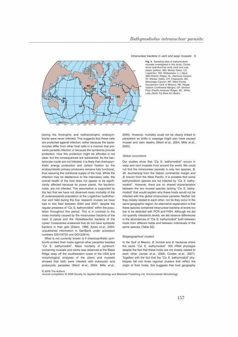
taining the thiotrophic and methanotrophic endosym-bionts were never infected. This suggests that these cellsare protected against infection, either because the bacte-riocytes differ from other host cells in a manner that pre-vents parasite infection or because the symbionts provideprotection. How this protection might be afforded is notclear, but the consequences are substantial. As the bac-teriocyte nuclei are not infected, it is likely that chemosyn-thetic energy production and carbon fixation by theendosymbiotic primary producers remains fully functional,thus assuring the nutritional supply of the host. While theinfection may be deleterious to the intercalary cells, theoverall health of the host does not appear to be signifi-cantly affected because its power plants, the bacterio-cytes, are not infected. This assumption is supported bythe fact that we have not observed mass mortality of theB. puteoserpentis population at the Logatchev hydrother-mal vent field during the four research cruises we havehad to this field between 2004 and 2007, despite theregular presence of “Ca. E. bathymodioli” within the popu-lation throughout this period. This is in contrast to themass mortality caused by the intranuclear bacteria of theclam S. patula and the Rickettsia-like bacteria of theoyster Crassostrea ariakensis that do not have symbioticbacteria in their gills (Elston, 1986; Ayres et al., 2004;unpublished information in GenBank under accessionnumbers DQ118733 und DQ123914).What is not currently known is if chemosynthetic sym-
bionts protect their hosts against other parasites besides“Ca. E. bathymodioli”. Mass mortality of symbiont-containing mussels and clams was observed at the BlakeRidge seep off the southeastern coast of the USA andmorphological analyses of the clams and musselsshowed that both were infested with eukaryotic andprokaryotic parasites (Ward et al., 2004; Mills et al.,
2005). However, mortality could not be clearly linked toparasitism as shifts in seepage might also have causedmussel and clam deaths (Ward et al., 2004; Mills et al.,2005).
Global occurrence
Our studies show that “Ca. E. bathymodioli” occurs inseep and vent mussels from around the world. We couldnot find the intranuclear parasite in only two species, B.aff. boomerang from the Gabon continental margin andB. brevior from the West Pacific. It is possible that somebathymodiolin species are not infected by “Ca. E. bathy-modioli”. However, there are no shared characteristicsbetween the two mussel species lacking “Ca. E. bathy-modioli” that would explain why these hosts would not beinfected with this global intranuclear parasite: Neither arethey closely related to each other, nor do they occur in thesame geographic region. An alternative explanation is thatthese species contained intranuclear bacteria at levels toolow to be detected with PCR and FISH. Although we didnot quantify infestation levels, we did observe differencesin the abundances of “Ca. E. bathymodioli” both betweenhosts from different fields and between individuals of thesame species (Table S2).
Biogeographical clusters
In the Gulf of Mexico, B. brooksi and B. heckerae sharethe same “Ca. E. bathymodioli” 16S rRNA phylotypedespite the fact that these hosts are not closely related toeach other (Jones et al., 2006; Cordes et al., 2007).Together with the fact that the “Ca. E. bathymodioli” phy-lotypes fall into three regional clusters that reflect theorigin of their hosts, this suggests that host geography
Fig. 4. Sampling sites of bathymodiolinmussels investigated in this study. Circlesshow hydrothermal vents (red) and coldseeps (yellow). MG, Menez Gwen; LO,Logatchev; WA, Wideawake; LI, Lilliput(Mid-Atlantic Ridge); AL, Alaminos Canyon;AT, Atwater Valley; CH, Chapopote; MC,Mississippi Canyon; WF, West FloridaEscarpment (Gulf of Mexico); RE, Regab(Gabon Continental Margin); GF, GermanFlats (Pacific-Antarctic Ridge); WL, WhiteLady (North Fiji Back-Arc Basin).
Intranuclear bacteria in vent and seep mussels 9
© 2009 The AuthorsJournal compilation © 2009 Society for Applied Microbiology and Blackwell Publishing Ltd, Environmental Microbiology
Bathymodiolus intranuclear parasite
157

Tab
le1.
Bathymodiolin
musselsinvestigated
inthisstudyandsamplingsites.
Bathymodiolus
species
Location
Cruisename
Recoverya
Month,year
Chief
scientist
Reference
Sam
pleID
Depth
Latitude
Longitude
B.azoricus
bMenez
Gwen
ATOS
Victor
Jun/Jul2001
P.M.Sarradin
Sarradinetal.(2001)
MG3,
5,10
850m
37°51′N
31°31′W
B.puteoserpentis
bLogatchev/Irina2
Hydromar
IQuest
Jan/Feb
2004
T.Kuhn
Kuhnetal.(2004)
35GTV-3
3016
m14°45.1629
′N44°58.7478
′W38ROV/6-5
3035
m56ROV/6-j6
3035
m66ROV/16-3
3053
mB.sp.b
Wideawake
Marsued
IIQuest
Apr
2005
K.Haase
Haase
etal.(2007)
109G
TV/A-3
2998
m4°48.64′S
12°22.36
′W125R
OV/1B-2
2987
m4°48.6293′S
12°22.3524
′W125R
OV/7-4
2986
m4°48.6275′S
12°22.3503
′W125R
OV/12-6
2987
m4°48.6231′S
12°22.3515
′WB.sp.b
Lilliput/M
ainLilliput
Marsued
IIQuest
Apr
2005
K.Haase
Haase
etal.(2005)
200R
OV/9-3
1496
m9°33.1783′S
13°11.8462
′WB.breviorb
White
Lady/LHOS
HyfifluxII
TV-grab
Aug/Sep
1998
P.Halbach
Halbach
etal.(1999)
39GTV-6
1976
m16°59.348′S
173°55.029
′E66GTV-12
2009
m16°59.449′S
173°54.940
′E99GTV-9
2000
m16°59.486′S
173°54.911
′EB.aff.thermophilusb
German
Flats(38°S)c
Foundation3
TV-grab
Jun/Jul2001
P.Stoffers
Stecher
etal.(2002);
Stoffers
etal.(2002)
30GTV-1-4
2212
m37°47.4673
′S110°54.8675′W
B.aff.boom
erangd
Gabon
Continental
Margin/Regab
Biozaire
2Victor
Nov
2001
M.Sibuet
Duperronetal.(2005);
Ondreas
etal.(2005)
Ang
M5
3150
m5°52.8134′S
9°37.9419′E
B.heckerae
dWestFlorida
Escarpm
ent
DeepSeeps
11/I
Alvin
Oct2003
C.R.Fisherand
B.Carney
–GoM
M6
3284
m26°02.00
′N84°55.03
′W
B.heckerae
dCam
peche
Knolls/Chapopote
MeteorM67/2
Quest
Apr
2006
G.Bohrm
ann
SpiessandBohrm
ann
(2006)
Sp1.1,Sp1.2
2915
m21°53.98
′N93°26.12
′W
B.brooksid
Sp2.1,Sp2.2
2915
mB.brooksid
Lousiana
Continental
Slope/Atwater
Valley
DeepSeeps
11/I
Alvin
Oct2003
C.R.Fisherand
B.Carney
–GoM
M24
1893
m27°34.10
′N88°29.8′W
B.brooksid
Lousiana
Continental
Slope/Alaminos
Canyon
GoM
M29
2226
m26°21.32
′N94°30.12
′W
“B.”childressid
GoM
M34
“B.”childressid
Lousiana
Continental
Slope/Mississipi
Canyon
Atlantis15/3
Alvin
May
2006
C.R.Fisher
–4178/mc853-2
1050
m28°07.40
′N89°08.20
′W
a.Sam
ples
wererecoveredusingtheremotely-operated
vehicles
Quest
(Marum
,University
ofBremen,Germany)
andVictor(Ifrem
er,France),themannedsubm
ersibleAlvin
(Woods
Hole
OceanographicInstitution,
USA),andaTV-controlledgrab
(Oktopus
GmbH
Kiel,Germany).
b.Species
from
hydrotherm
alvents.
c.Ventfieldnameaccordingto
Desbruyères
andcolleagues(2006b).
d.Species
from
coldseeps.
10 F. U. Zielinski et al.
© 2009 The AuthorsJournal compilation © 2009 Society for Applied Microbiology and Blackwell Publishing Ltd, Environmental Microbiology
Manuscript IV
158

and not cospeciation played a role in the establishment ofthese associations. This hypothesis is supported by ourstatistical analyses that showed a significant correlationbetween genetic and geographic distances. However, theinvestigation of more host species from different geo-graphic regions, particularly the West Pacific, is neededbefore this hypothesis can be substantiated.One mussel species, B. sp. from the Wideawake vent
field on the southern Mid-Atlantic Ridge harboured two“Ca. E. bathymodioli” phylotypes that differed from eachother by 1.9%. One falls into the Mid-Atlantic Ridgecluster whereas the other one falls in the Pacific-AntarcticRidge cluster (Fig. 1B). This indicates that although most“Ca. E. bathymodioli” bacteria cluster according to theirgeography, there may have been transoceanic crossing inthe past, in this case from vent sites on the Pacific-Antarctic Ridge to the southern Mid-Atlantic Ridge. His-torical dispersal across ocean basins has been suggestedfor some bathymodiolin host species (Jones et al., 2006;Olu-Le Roy et al., 2007), and there is no reason toassume that it would not have occurred in their intra-nuclear bacteria. Extensive comparative analyses of thephylogeny of bathymodiolin hosts, their symbionts andtheir intranuclear bacteria will provide an ideal data set for
a better understanding of historical dispersal patterns inthese deep–sea associations.
Conclusions
This study shows that intranuclear bacteria are wide-spread in bathymodiolin mussels from hydrothermal ventsand cold seeps. The 16S rRNA sequences of “Ca. E.bathymodioli” belong to a monophyletic clade that con-sists of sequences from bacteria associated with marinemetazoans as diverse as sponges, corals, sea slugs,clams, ascidians, sea urchins and fish. Only one of thesequences within this clade is known to originate from anintranuclear bacterium, the previously described ‘NIX’parasite from the razor clam S. patula (Elston, 1986; Kerket al., 1992; Ayres et al., 2004) but morphological descrip-tions of intranuclear bacteria exist for other animal hostswithin this clade, for example, from two species of Aply-sina sponges (Vacelet, 1970; Friedrich et al., 1999) andthe venerid clam Ruditapes decussatus (Azevedo, 1989)(Table 2). We postulate that many of the sequences withinthis clade originate from intranuclear bacteria, and thatthese parasites are widespread in marine animals, includ-ing clams and other shellfish consumed by humans.
Fig. 5. Proposed developmental cycle of “Candidatus Endonucleobacter bathymodioli”. The infection begins with a single rod-shapedbacterium inside the nucleus (Stage 1) that grows into an unseptated filament of up to 18–20 mm in length (Stage 2). Multiplication, possiblyby longitudinal fission, yields several such filaments that are loosely wrapped in a filamentous coil (Stage 3). These subsequently form stacksof shorter filaments of up to 8–10 mm in length (Stage 4). Repeated transverse binary fissions lead to numerous rod-shaped bacteria (Stage5). Further multiplication results in a voluminous membrane-surrounded bacterial aggregate containing up to 80 000 rod-shaped bacteria andthe destruction of the host cell (Stage 6). These aggregates are eventually disrupted and the bacteria released.
Intranuclear bacteria in vent and seep mussels 11
© 2009 The AuthorsJournal compilation © 2009 Society for Applied Microbiology and Blackwell Publishing Ltd, Environmental Microbiology
Bathymodiolus intranuclear parasite
159

Experimental procedures
Sampling sites and processing
Nine Bathymodiolus species from six deep-sea hydrothermalvent fields and five cold seep sites located in the Atlantic andPacific Ocean were investigated (Fig. 4, Table 1). One to fourindividuals of each species were examined (Table 1). Uponrecovery mussels were immediately transferred into chilledbottom sea water and processed as previously described(Duperron et al., 2005; 2006; 2007). Briefly, gill tissue wasfrozen and stored at -20°C for DNAextraction as well as fixedfor FISH and TEM investigations.
Cloning and sequencing “Ca. E. bathymodioli” fromB. puteoserpentis (Logatchev)
Genomic DNA was extracted from gill tissue according toZhou and colleagues (1996). PCR was performed using theuniversal bacterial primers GM3F and GM4R (Muyzer et al.,1995) and a high-quality Taq DNA polymerase (error rate2.7 ¥ 10-5; Eppendorf, Hamburg, Germany). Amplificationproducts were purified and ligated into pGEM-T Easy vectors(Promega). One Shot TOP10 competent E. coli cells (Invitro-gen) were subsequently transformed. A total of 1108 positivetransformants from four individuals (277 clones per indi-vidual) were picked by blue/white screening and grown over-night in V96 MicroWell Plates (Nunc, Wiesbaden, Germany)containing 200 ml Luria–Bertani/ampicillin broth per well. Atotal of 1009 clones were screened for the right sized insert(252 clones per individual) by PCR using the M13F/M13Rprimer pair (Yanisch-Perron et al., 1985) and 854 clones hadan insert with the expected size of approximately 1500 bp.384 clones with the right sized insert were partiallysequenced (96 clones per individual). PCR products werepurified in MultiScreen-HV plates (Millipore) using SephadexG50 Superfine resin (Amersham Biosciences) andsequenced using the BigDye Terminator v2.0 Cycle Sequenc-ing Kit along with the Genetic Analyzer Abiprism 3100(Applied Biosystems). The GM3F oligonucleotide (Muyzeret al., 1995) was used as sequencing primer. The resultingpartial sequences were analysed with BioEdit SequenceAlignment Editor version 7.0 (Hall, 1999) using the ClustalWimplementation (Thompson et al., 1994). Uniquely occurringsequences were ignored. Repetitive sequences weregrouped. Three clones per group and individual were fullysequenced by sequencing both the coding and non-codingDNA strands with the sequencing primers M13F, M13R(Yanisch-Perron et al., 1985), 534R (Muyzer et al., 1993),518F, 1099F and 1193R (Buchholz-Cleven et al., 1997).Cycle sequencing accessories, equipment and conditionswere as described for partial sequencing. Sequences wereassembled using Sequencher (Gene Codes Corporation,http://www.genecodes.com). The vectorial remnants and theprimer sites were discarded.
Cloning and sequencing “Ca. E. bathymodioli” fromother bathymodiolin mussels
Primers specifically targeting the 16S rRNA gene of “Ca. E.bathymodioli” (Logatchev) were designed using the reverseTa
ble
2.Intranuclear
bacteriadescribed
todate
inMetazoa
(excluding
facultativeintranuclear
Rickettsiae).
Metazoa
taxon
Hostspecies
Higher-rankingtaxon
Designationof
bacteria
inside
nuclei
Shape
Size(mm)
Phylogeny
Reference
Dem
ospongia
Porifera
Aplysinaaerophoba;
A.cavernicola
Intranuclear
bacteria
Filamentous
150–350;
atleast4.5
Gam
maa
Vacelet
(1970);Friedrichetal.(1999)
Bivalvia
Mollusca
Siliquapatula
‘Nuclear
InclusionX’(NIX)
Multilayered,mem
branerich,
complexlyfolded,partially
cleaved
25¥16
Gam
ma
Elston(1986);Kerketal.(1992)
Ruditapesdecussatus
Endonucleobiotic
bacteria
Sphericalto
ellipsoidal
ø1.3
ndAzevedo
(1989)
Bathymodiolusspp.
“Ca.
Endonucleobacter
bathym
odioli”
Rod-shapedto
filam
entous
1.8¥0.4;
upto
20Gam
ma
Thisstudy
a.Phylogeny
basedon
FISHwith
generalgam
maproteobacterialprobe,no.16SrRNAsequence
available.
nd,notdeterm
ined.
12 F. U. Zielinski et al.
© 2009 The AuthorsJournal compilation © 2009 Society for Applied Microbiology and Blackwell Publishing Ltd, Environmental Microbiology
Manuscript IV
160
and complementary sequence of probe Bnix64 as a forwardprimer (bnix-64F: AGCGGTAACAGGTCTAGC) and thesequence of probe Bnix1249 as a reverse primer (bnix-1267R: GCAGCTTCGCGACCGTCT) resulting in a 1203 bpamplification product (for probe design see below). A combi-nation of the bnix-64F primer together with the universalbacterial reverse primer GM4R was eventually chosen tocover 1409 bp of the 16S rRNA gene. Steps for cloning,sequencing and analysing intranuclear bacteria belonging to“Ca. E. bathymodioli” were as described above for B. puteo-serpentis with the exception that only 16 transformants perindividual were picked and screened for the right sized insert,and the positive clones fully sequenced. The annealing tem-peratures for the bnix-64F/bnix-1267R and bnix-64F/GM4Rprimer pairs were 56°C and 45°C respectively. Sequencingwas performed using the BigDye Terminator v3.1 CycleSequencing Kit along with the Genetic Analyzer Abiprism3130 (Applied Biosystems).
Accession numbers
The 16S rRNA gene sequences belonging to “CandidatusEndonucleobacter bathymodioli” have been registered at theEMBL database (Kulikova et al., 2007) under accessionnumbers FM162182 to FM162195, and FM244838(Table S2).
Phylogenetic reconstruction
Sequences were analysed using ARB (Ludwig et al., 2004)and compared with the NCBI nucleotide database usingnucleotide BLAST (McGinnis and Madden, 2004), the RDP-Xdatabase using the Sequence Match tool (Cole et al., 2007),and the Silva rRNA database using the SINA webaligner(Pruesse et al., 2007). Highly similar sequences wereincluded in the analysis and aligned using ClustalX. Positionsdisplaying more than 25% gaps as well as positions ambigu-ously aligned were removed from the analysis. The finalalignment comprised 1416 positions. Phylogenetic analyseswere performed using Bayesian as well as maximum likeli-hood analysis. The former was run using MrBayes 3 (v3.1.2)(Ronquist and Huelsenbeck, 2003) under a General TimeReversible model along with Gamma-distributed rates of evo-lution and a proportion of invariant sites. Analyses were per-formed for 2 000 000 generations using four parallel MonteCarlo Markov chains. Sample trees were taken every 1000generations. Posterior probabilities calculated over 5000 besttrees were used as support values for nodes in the tree. Themaximum likelihood analysis was performed using PHYLIP
based on 100 jumble replicates. To assess the robustness ofnodes, 1000 ML bootstrap replicates were run. The alignmentused for the phylogenetic analysis can be obtained from theEMBL database (accession number: Align_001264).
Statistical analyses
To test the hypothesis that genetic distances between “Ca. E.bathymodioli” 16S rRNA sequences were correlated withtheir geographical distances, a Mantel test was performedusing the program R-package (Casgrain and Legendre,
2008). Geographical distances were estimated using GoogleEarth v.4 based on minimum oceanic distances betweensample locations.
Design of probes targeting “Ca. E. bathymodioli”
Based on the 16S rRNA full sequence (1468 bp) of “Ca. E.bathymodioli” from B. puteoserpentis (Logatchev) threeprobes were designed using the probe design tool of ARB(Ludwig et al., 2004). To verify their specificity, probes Bnix64(GCTAGACCTGTTACCGCT), Bnix643 (CCGTACTCTAGCCACCCA) and Bnix1249 (GCAGCTTCGCGACCGTCT)were checked against the 16S dataset of the RibosomalDatabase Project X using the online Probe Match tool (Coleet al., 2007). The most recent probe match against theRDP-X dataset 10.4 (October 2008) revealed that probeBnix64 targeted also 12 gammaproteobacterial sequencesbelonging to the Oceanospirillales and Alteromonadales.Probe Bnix643 targeted also 13 cyanobacterial sequences,five unclassified gammaproteobacterial sequences and onedeltaproteobacterial sequence. However, probe Bnix1249was confirmed to specifically target “Ca. E. bathymodioli”(Logatchev). With the amplification of more “Ca. E. bathymo-dioli” phylotypes from other bathymodiolin species it becameevident that the probes Bnix1249 and Bnix643 had one mis-match each to some of the other intranuclear phylotypes(Table S2). All three probes are deposited in the oligonucle-otide probe database ‘probeBase’ (Loy et al., 2007) underaccession numbers pB-01516 to pB-01518.All probes were fluorescently labelled (biomers.net, Ulm,
Germany). Specific hybridization conditions for all threeprobes were determined by varying the formamide concen-tration in the hybridization buffer (Pernthaler et al., 2002). Allprobes hybridized equally well with the target organism at35% formamide concentration. Probe EUB338 (Amann et al.,1990) covering most bacteria was used as a positive controland the NON338 probe (Wallner et al., 1993) as a control forbackground autofluorescence.
Fluorescence in situ hybridization
Subsamples of B. puteoserpentis tissues such as gill, gut,digestive gland, labial palps, mantle and foot were fixed in 1¥phosphate buffered saline (PBS: 137 mM NaCl, 2.7 mM KCl,10 mM Na2HPO4, 2 mM KH2PO4) containing 2% paraformal-dehyde at 4°C for 9–18 h. Samples were washed three timesby placing them in fresh 1¥ PBS for 10 min each time andsubsequently transferred into cold PBS/ethanol solution con-taining 1¥ PBS and pure ethanol in equal parts. Sampleswere kept at 4°C on board the research vessel, air-freightedback to the laboratory at 4°C and finally stored at -20°C.Fixed specimens were embedded in Steedman’s Wax(Steedman, 1957) and sectioned with a microtome into10 mm thick sections. The sections were placed onto Super-Frost slides (Fisher Scientific), dewaxed in three successivebaths of absolute ethanol for 5 min each and air dried. Sec-tions were then covered with 200 ml of hybridization buffer(Pernthaler et al., 2002) containing fluorescently labelled oli-gonucleotide probes (5 ng ml-1 final concentration), coveredwith a glass coverslip and hybridized at 46°C for 10 min in a
Intranuclear bacteria in vent and seep mussels 13
© 2009 The AuthorsJournal compilation © 2009 Society for Applied Microbiology and Blackwell Publishing Ltd, Environmental Microbiology
Bathymodiolus intranuclear parasite
161

histological microwave oven (Microwave Research andAppli-cations, Laurel, MD, USA) with the power output set to 20%.The slides were rinsed in 1¥ PBS, MQwater, and absoluteethanol for 1 min each. Single hybridizations targeting spe-cifically “Ca. E. bathymodioli” (Logatchev) were performedusing probe Bnix1249 labelled with Cy3. For visualization ofall bacteria in B. puteoserpentis, Cy3-labelled EUB338 wasused (Amann et al., 1990). Double hybridizations targetingeukaryotic 18S rRNA were performed using Cy5-labelledEUK516 (Amann et al., 1990). Triple hybridizations targetingthe chemoautotrophic endosymbiont, the methanotrophicendosymbiont and the eukaryotic 18S rRNA were performedusing the probes BMARt-193 (Cy3), BMARm-845 (Cy5)(Duperron et al., 2006) and EUK516 (Fluos). For quadruplehybridizations targeting additionally “Ca. E. bathymodioli”(Logatchev) Fluos-labelled Bnix1249 was used.
Deconvolution (restoration) microscopy
The air dried slides were embedded in a DAPI-amendedmountant and evaluated on a DeltaVision RT RestorationMicroscopy System (Applied Precision, Issaquah, WA, USA)using an Olympus IX71 (Olympus, Center Valley, PA, USA)equipped with appropriate filter sets for Cy3, Cy5, DAPI andfluorescein. For image capture and deconvolution the Soft-Worx image analysis software was used (Applied Precision,Issaquah, WA, USA). Images were further processed andanalysed using the Imaris software package (http://www.bitplane.com) that was also used to assign colours tothe different wavelengths.
Transmission electron microscopy
Gill filaments of B. puteoserpentis from Logatchev were fixedin a modified Trump’s fixative (McDowell and Trump, 1976)(0.05 M sodium cacodylate solution containing 2% glutaral-dehyde and 2% paraformaldehyde, pH 7.3) and storedtherein for several months. The tissues were dehydrated inan ethanol series and embedded in the acrylic resin LR White(Sigma). Ultrathin sections were stained with uranyl acetateand lead citrate, and examined with a Zeiss EM 109-S2.Transmission electron microscopy was performed on fiveadult specimens. Fifty-seven gill filaments were investigatedin total.
Estimation of “Ca. E. bathymodioli” numbers in Stage 6aggregates
The total number of “Ca. E. bathymodioli” cells in a Stage 6aggregate was estimated by dividing the volume of an aggre-gate by the volume of a single rod-shaped bacterial cell. Thevolume of aggregates was calculated by assuming that these
were spheres V d=( )16
3p . The diameter d of Stage 6 bac-
terial aggregates was determined from several FISH and fourTEM images which ranged between 16 and 32 mm. Thevolume of single rod-shaped bacterial cells was calculated byassuming that these were cylinders capped at both ends
by hemispheres spherocylinder,V d h d= +( )⎡⎣⎢
⎤⎦⎥
12
12
13
2p . The
length h and diameter (width) d was determined for severalintranuclear cells using TEM images (average size1.8 ¥ 0.4 mm, it follows that h = 1.4 mm, d = 0.4 mm).
Acknowledgements
We are grateful to the chief scientists and the captains andcrews of the involved research vessels (Table 1) as well asthe chief engineers and crews of the ROV’s Quest 4000 andVictor 6000 and the manned submersible Alvin for providingexcellent support to obtain deep-sea hydrothermal vent andcold seep samples. We appreciate our laboratory assistantsSilke Wetzel and Sabine Gaude and their indispensableskills. We are grateful to Victoria Orphan for letting us use herDeltaVision RT Restoration Microscopy System along withthe SoftWorx deconvolution and image analysis software aswell as the Imaris image analysis software package. Theauthor (F.U.Z.) is particularly thankful to Victoria Orphan forhosting him in her lab at the California Institute of Technologyduring parts of this study. We thank Shana Goffredi and threeanonymous reviewers for helpful comments and suggestionsfor the improvement of this manuscript. This work wassupported by grants from the Priority Program 1144 ofthe German Science Foundation (DFG). This is publicationnumber 24 of the Priority Program 1144 ‘From Mantle toOcean: Energy-, Material- and Life cycles at Spreading Axes’of the DFG.
References
Amann, R.I., Binder, B.J., Olson, R.J., Chisholm, S.W.,Devereux, R., and Stahl, D.A. (1990) Combination of 16SrRNA-targeted oligonucleotide probes with flow-cytometryfor analyzing mixed microbial populations. Appl EnvironMicrobiol 56: 1919–1925.
Angert, E.R. (2005) Alternatives to binary fission in bacteria.Nat Rev Microbiol 3: 214–224.
Arneodo, J.D., Bressan, A., Lherminier, J., Michel, J., andBoudon-Padieu, E. (2008) Ultrastructural detection of anunusual intranuclear bacterium in Pentastiridius leporinus(Hemiptera: Cixiidae). J Invertebr Pathol 97: 310–313.
Atkins, M.S., Teske, A.P., and Anderson, O.R. (2000) Asurvey of flagellate diversity at four deep-sea hydrothermalvents in the Eastern Pacific Ocean using structural andmolecular approaches. J Eukaryot Microbiol 47: 400–411.
Ayres, D.L., Schumaker, E.J., and Elston, R.A. (2004) Recentmortality event of Pacific razor clams, Siliqua patula, alongthe Pacific coast of Washington state associated withrecord infection intensity of the gills by Nuclear Inclusion X(NIX). J Shellfish Res 23: 651.
Azevedo, C. (1989) Fine structure of endonucleobiotic bac-teria in the gill epithelium of Ruditapes decussatus. MarBiol 100: 339–341.
Bright, M., and Giere, O. (2005) Microbial symbiosis in Anne-lida. Symbiosis 38: 1–45.
Buchholz-Cleven, B.E.E., Rattunde, B., and Straub, K.L.(1997) Screening for genetic diversity of isolates of anaero-bic Fe(II)-oxidizing bacteria using DGGE and whole-cellhybridization. Syst Appl Microbiol 20: 301–309.
Burgdorfer, W., Anacker, R.L., Bird, R.G., and Bertram, D.S.
14 F. U. Zielinski et al.
© 2009 The AuthorsJournal compilation © 2009 Society for Applied Microbiology and Blackwell Publishing Ltd, Environmental Microbiology
Manuscript IV
162

(1968) Intranuclear growth of Rickettsia rickettsii.J Bacteriol 96: 1415–1418.
de Buron, I., and Morand, S. (2004) Deep-sea hydrothermalvent parasites: why do we not find more? Parasitology 128:1–6.
de Buron, I., and Segonzac, M. (2006a) Hydrothermal ventparasites. Denisia 18: 29–30. Supplement & Corrigendum,November 2006, URL: http://www.noc.soton.ac.uk/chess/docs/handbook_corrigendum.pdf.
de Buron, I., and Segonzac, M. (2006b) Hydrothermal ventparasites. Denisia 18: 29–30.
Casgrain, P., and Legendre, P. (2008) The R Package forMultivariate and Spatial Analysis. Montréal, Canada: Uni-versité de Montréal.
Cavanaugh, C.M., McKiness, Z.P., Newton, I.L.G., andStewart, F.J. (2006) Marine chemosynthetic symbioses. InThe Prokaryotes. Dworkin, M., Falkow, S., Rosenberg, E.,Schleifer, K.H., and Stackebrandt, E. (eds). Berlin,Germany: Springer-Verlag, pp. 473–507.
Chang, K.P., and Musgrave, A.J. (1970) Ultrastructure ofrickettsia-like microorganisms in midgut of a plant bug,Stenotus binotatus Jak. (Heteroptera: Miridae). Can JMicrobiol 16: 621–622.
Cole, J.R., Chai, B., Farris, R.J., Wang, Q., Kulam-Syed-Mohideen, A.S., McGarrell, D.M., et al. (2007) The riboso-mal database project (RDP-II): introducing myRDP spaceand quality controlled public data. Nucleic Acids Res 35:D169–D172.
Cordes, E.E., Carney, S.L., Hourdez, S., Carney, R.S.,Brooks, J.M., and Fisher, C.R. (2007) Cold seeps of thedeep Gulf of Mexico: community structure and biogeo-graphic comparisons to Atlantic equatorial belt seep com-munities. Deep-Sea Res I 54: 637–653.
DeChaine, E.G., and Cavanaugh, C.M. (2006) Symbioses ofmethanotrophs and deep-sea mussels (Mytilidae: Bathy-modiolinae). In Molecular Basis of Symbiosis. Overmann,J. (ed.). Berlin, Germany: Springer-Verlag, pp. 227–249.
Desbruyères, D., Segonzac, M., and Bright, M. (eds) (2006a)Handbook of deep-sea hydrothermal vent fauna. Denisia18: 544. Supplement & Corrigendum, December 2006,URL: http://www.noc.soton.ac.uk/chess/docs/handbook_corrigendum.pdf.
Desbruyères, D., Segonzac, M., and Bright, M. (eds) (2006b)Handbook of deep-sea hydrothermal vent fauna. Denisia18: 544.
Distel, D.L., Lee, H.K.W., and Cavanaugh, C.M. (1995) Intra-cellular coexistence of methanotrophic and thioautotrophicbacteria in a hydrothermal vent mussel. Proc Natl Acad SciUSA 92: 9598–9602.
Duperron, S., Nadalig, T., Caprais, J.C., Sibuet, M., Fiala-Medioni, A., Amann, R., and Dubilier, N. (2005) Dual sym-biosis in a Bathymodiolus sp. mussel from a methane seepon the Gabon continental margin (southeast Atlantic): 16SrRNA phylogeny and distribution of the symbionts in gills.Appl Environ Microbiol 71: 1694–1700.
Duperron, S., Bergin, C., Zielinski, F., Blazejak, A., Perntha-ler, A., McKiness, Z.P., et al. (2006) A dual symbiosisshared by two mussel species, Bathymodiolus azoricusand Bathymodiolus puteoserpentis (Bivalvia: Mytilidae),from hydrothermal vents along the northern Mid-AtlanticRidge. Environ Microbiol 8: 1441–1447.
Duperron, S., Sibuet, M., MacGregor, B.J., Kuypers, M.M.M.,Fisher, C.R., and Dubilier, N. (2007) Diversity, relativeabundance and metabolic potential of bacterial endosym-bionts in three Bathymodiolus mussel species from coldseeps in the Gulf of Mexico. Environ Microbiol 9: 1423–1438.
Dworkin, M. (2006) Prokaryotic life cycles. In The Prokary-otes. Dworkin, M., Falkow, S., Rosenberg, E., Schleifer,K.H., and Stackebrandt, E. (eds). Berlin, Germany:Springer-Verlag, pp. 140–166.
Edgcomb, V.P., Kysela, D.T., Teske, A., Gomez, A.D., andSogin, M.L. (2002) Benthic eukaryotic diversity in theGuaymas Basin hydrothermal vent environment. Proc NatlAcad Sci USA 99: 7658–7662.
Elston, R.A. (1986) An intranuclear pathogen [Nuclear Inclu-sion X (NIX)] associated with massive mortalities of thePacific razor clam, Siliqua patula. J Invertebr Pathol 47:93–104.
Epis, S., Sassera, D., Beninati, T., Lo, N., Beati, L., Piesman,J., et al. (2008) Midichloria mitochondrii is widespread inhard ticks (Ixodidae) and resides in the mitochondria ofphylogenetically diverse species. Parasitology 135: 485–494.
Fauré-Fremiet, E. (1951) The marine sand-dwelling ciliates ofCape Cod. Biol Bull 100: 59–70.
Fiala-Médioni, A., and Le Pennec, M. (1987) Trophic struc-tural adaptations in relation to the bacterial association ofbivalve mollusks from hydrothermal vents and subductionzones. Symbiosis 4: 63–74.
Fisher, C.R., Childress, J.J., Oremland, R.S., and Bidigare,R.R. (1987) The importance of methane and thiosulfate inthe metabolism of the bacterial symbionts of two deep-seamussels. Mar Biol 96: 59–71.
Fokin, S.I. (2004) Bacterial endocytobionts of Ciliophora andtheir interactions with the host cell. Int Rev Cytol 236:181–249.
Friedrich, A.B., Merkert, H., Fendert, T., Hacker, J., Proksch,P., and Hentschel, U. (1999) Microbial diversity in themarine sponge Aplysina cavernicola (formerly Verongiacavernicola) analyzed by fluorescence in situ hybridization(FISH). Mar Biol 134: 461–470.
Génio, L., Johnson, S.B., Vrijenhoek, R.C., Cunha, M.R.,Tyler, P.A., Kiel, S., and Little, C.T.S. (2008) New record of“Bathymodiolus” mauritanicus Cosel 2002 from the Gulf ofCadiz (NE Atlantic) mud volcanoes. J Shellfish Res 27:53–61.
Giere, O., and Krieger, J. (2001) A triple bacterial endosym-biosis in a gutless oligochaete (Annelida): ultrastructuraland immunocytochemical evidence. Invertebr Biol 120:41–49.
Görtz, H.-D. (2006) Symbiotic associations between ciliatesand prokaryotes. In The Prokaryotes. Dworkin, M.,Falkow, S., Rosenberg, E., Schleifer, K.H., and Stack-ebrandt, E. (eds). Berlin, Germany: Springer-Verlag, pp.364–402.
Grandi, G., Guidi, L., and Chicca, M. (1997) Endonuclearbacterial symbionts in two termite species: an ultrastruc-tural study. J Submicrosc Cytol Pathol 29: 281–292.
Haase, K., Flies, C., Fretzdorff, S., Giere, O., Houk, A.,Koschinsky, A., et al. (2005) Marsüd 2. In Meteor Berichte05. Mid-Atlantic Expedition 2005. Cruise No. 64, Leg 1:
Intranuclear bacteria in vent and seep mussels 15
© 2009 The AuthorsJournal compilation © 2009 Society for Applied Microbiology and Blackwell Publishing Ltd, Environmental Microbiology
Bathymodiolus intranuclear parasite
163

Leitstelle Meteor. Institut für Meereskunde der UniversitätHamburg, pp. 1–59. http://www.ifm-geomar.de/fileadmin/ifm-geomar/fb4/fb4_fe2/spetersen/fahrtbericht_M64_1.pdf
Haase, K.M., Petersen, S., Koschinsky, A., Seifert, R.,Devey, C.W., Keir, R., et al. (2007) Young volcanism andrelated hydrothermal activity at 5°S on the slow-spreadingsouthern Mid-Atlantic Ridge. Geochem Geophys Geosyst8: Q11002, doi: 11010.11029/12006GC001509.
Halbach, P., Koschinsky, A., Seifert, R., Giere, O., Kuhn, T.,Armstrong, R., et al. (1999) Diffuse hydrothermal fluidactivity, biological communities, and mineral formation inthe North Fiji Basin (SW Pacific): preliminary results of theR/V Sonne Cruise SO-134. InterRidge News 8: 38–44.
Hall, T.A. (1999) BioEdit: a user-friendly biological sequencealignment editor and analysis program for Windows 95/98/NT. Nucleic Acids Symp Ser 41: 95–98.
Horowitz, A.T., Kessel, M., and Shilo, M. (1974) Growth cycleof predacious bdellovibrios in a host-free extract systemand some properties of the host extract. J Bacteriol 117:270–282.
Jones, W.J., Won, Y.J., Maas, P.A.Y., Smith, P.J., Lutz, R.A.,and Vrijenhoek, R.C. (2006) Evolution of habitat use bydeep-sea mussels. Mar Biol 148: 841–851.
Jurkevitch, E. (2006) The genus Bdellovibrio. In The Prokary-otes. Dworkin, M., Falkow, S., Rosenberg, E., Schleifer,K.H., and Stackebrandt, E. (eds). Berlin, Germany:Springer-Verlag, pp. 12–30.
Kerk, D., Gee, A., Dewhirst, F.E., Drum, A.S., and Elston,R.A. (1992) Phylogenetic placement of “Nuclear InclusionX (NIX)” into the gamma subclass of proteobacteria on thebasis of 16S ribosomal RNA sequence comparisons. SystAppl Microbiol 15: 191–196.
Kuhn, T., Alexander, B., Augustin, N., Birgel, D., Borowski,C., de Carvalho, L., et al. (2004) The Logatchev hydrother-mal field – revisited: preliminary results of the R/VMETEOR Cruise HYDROMAR I (M60/3). InterRidge News13: 1–4.
Kulikova, T., Akhtar, R., Aldebert, P., Althorpe, N., Andersson,M., Baldwin, A., et al. (2007) EMBL nucleotide sequencedatabase in 2006. Nucleic Acids Res 35: D16–D20.
Kurahashi, M., and Yokota, A. (2007) Endozoicomonas elysi-cola gen nov., sp. nov., a g-proteobacterium isolated fromthe sea slug Elysia ornata. Syst Appl Microbiol 30: 202–206.
Lambert, C., Morehouse, K.A., Chang, C.Y., and Sockett,R.E. (2006) Bdellovibrio: growth and development duringthe predatory cycle. Curr Opin Microbiol 9: 639–644.
Lennon, J.T. (2007) Diversity and metabolism of marine bac-teria cultivated on dissolved DNA. Appl Environ Microbiol73: 2799–2805.
López-García, P., Vereshchaka, A., and Moreira, D. (2007)Eukaryotic diversity associated with carbonates and fluid–seawater interface in Lost City hydrothermal field. EnvironMicrobiol 9: 546–554.
Loy, A., Maixner, F., Wagner, M., and Horn, M. (2007) probe-Base – an online resource for rRNA-targeted oligonucle-otide probes: new features 2007. Nucleic Acids Res 35:D800–D804.
Ludwig, W., Strunk, O., Westram, R., Richter, L., Meier, H.,Yadhukumar, et al. (2004) ARB: a software environment forsequence data. Nucleic Acids Res 32: 1363–1371.
McDowell, E.M., and Trump, B.F. (1976) Histologic fixativessuitable for diagnostic light and electron microscopy. ArchPathol Lab Med 100: 405–414.
McGinnis, S., and Madden, T.L. (2004) BLAST: at the core ofa powerful and diverse set of sequence analysis tools.Nucleic Acids Res 32: W20–W25.
Maillet, P.L., and Folliot, R. (1967) Nouvelles observationssur le transport de microorganismes intranucléairesappelés particules Phi par les spermatozoides chez desinsectes Homoptères. C R Hebd Seances Acad Sci D 264:965–968.
Mills, A.M., Ward, M.E., Heyl, T.P., and Van Dover, C.L.(2005) Parasitism as a potential contributor to massiveclam mortality at the Blake Ridge Diapir methane-hydrateseep. J Mar Biol Assoc UK 85: 1489–1497.
Muyzer, G., Dewaal, E.C., and Uitterlinden, A.G. (1993) Pro-filing of complex microbial populations by denaturing gra-dient gel electrophoresis analysis of polymerase chainreaction-amplified genes coding for 16S rRNA. ApplEnviron Microbiol 59: 695–700.
Muyzer, G., Teske, A., Wirsen, C.O., and Jannasch, H.W.(1995) Phylogenetic relationships of Thiomicrospiraspecies and their identification in deep-sea hydrothermalvent samples by denaturing gradient gel electrophoresis of16S rDNA fragments. Arch Microbiol 164: 165–172.
Nelson, D.C., Hagen, K.D., and Edwards, D.B. (1995) The gillsymbiont of the hydrothermal vent mussel Bathymodiolusthermophilus is a psychrophilic, chemoautotrophic, sulfurbacterium. Mar Biol 121: 487–495.
Ogata, H., La Scola, B., Audic, S., Renesto, P., Blanc, G.,Robert, C., et al. (2006) Genome sequence of Rickettsiabellii illuminates the role of amoebae in gene exchangesbetween intracellular pathogens. Plos Genet 2: 733–744.
Olu-Le Roy, K., von Cosel, R., Hourdez, S., Carney, S.L., andJollivet, D. (2007) Amphi-Atlantic cold-seep Bathymodiolusspecies complexes across the equatorial belt. Deep-SeaRes I 54: 1890–1911.
Ondreas, H., Olu, K., Fouquet, Y., Charlou, J.L., Gay, A.,Dennielou, B., et al. (2005) ROV study of a giant pockmarkon the Gabon continental margin. Geo-Mar Lett 25: 281–292.
Pernthaler, A., Pernthaler, J., and Amann, R. (2002) Fluores-cence in situ hybridization and catalyzed reporter deposi-tion for the identification of marine bacteria. Appl EnvironMicrobiol 68: 3094–3101.
Pimenov, N.V., Kalyuzhnaya, M.G., Khmelenina, V.N.,Mityushina, L.L., and Trotsenko, Y.A. (2002) Utilization ofmethane and carbon dioxide by symbiotrophic bacteria ingills of Mytilidae (Bathymodiolus) from the Rainbow andLogachev hydrothermal fields on the Mid-Atlantic Ridge.Microbiology 71: 587–594.
Pinchuk, G.E., Ammons, C., Culley, D.E., Li, S.M.W.,McLean, J.S., Romine, M.F., et al. (2008) Utilization ofDNA as a sole source of phosphorus, carbon, and energyby Shewanella spp. Ecological and physiological implica-tions for dissimilatory metal reduction. Appl Environ Micro-biol 74: 1198–1208.
Polz, M.F., Felbeck, H., Novak, R., Nebelsick, M., and Ott,J.A. (1992) Chemoautotrophic, sulfur-oxidizing symbioticbacteria on marine nematodes – morphological and bio-chemical characterization. Microb Ecol 24: 313–329.
16 F. U. Zielinski et al.
© 2009 The AuthorsJournal compilation © 2009 Society for Applied Microbiology and Blackwell Publishing Ltd, Environmental Microbiology
Manuscript IV
164

Polz, M.F., Distel, D.L., Zarda, B., Amann, R., Felbeck, H.,Ott, J.A., and Cavanaugh, C.M. (1994) Phylogenetic analy-sis of a highly specific association between ectosymbiotic,sulfur-oxidizing bacteria and a marine nematode. ApplEnviron Microbiol 60: 4461–4467.
Pongponratn, E., Maneerat, Y., Chaisri, U., Wilairatana, P.,Punpoowong, B., Viriyavejakul, P., and Riganti, M. (1998)Electron-microscopic examination of Rickettsia tsutsuga-mushi infected human liver. Trop Med Int Health 3: 242–248.
Powell, E.N., Barber, R.D., Kennicutt, M.C., and Ford, S.E.(1999) Influence of parasitism in controlling the health,reproduction and PAH body burden of petroleum seepmussels. Deep-Sea Res I 46: 2053–2078.
Pruesse, E., Quast, C., Knittel, K., Fuchs, B.M., Ludwig, W.,Peplies, J., and Glockner, F.O. (2007) SILVA: a compre-hensive online resource for quality checked and alignedribosomal RNA sequence data compatible with ARB.Nucleic Acids Res 35: 7188–7196.
Raikov, I.B. (1971) Bactéries épizoique et mode de nutritiondu cilié psammophile Kentrophoros fistulosum Fauré-Fremiet (étude au microscope électronique). Protistologica7: 365–378.
Rendulic, S., Jagtap, P., Rosinus, A., Eppinger, M., Baar, C.,Lanz, C., et al. (2004) A predator unmasked: life cycle ofBdellovibrio bacteriovorus from a genomic perspective.Science 303: 689–692.
Ronquist, F., and Huelsenbeck, J.P. (2003) MrBayes 3:Bayesian phylogenetic inference under mixed models. Bio-informatics 19: 1572–1574.
Sarradin, P.M., Desbruyères, D., Dixon, D., Almeida, A.,Briand, P., Caprais, J.C., et al. (2001) ATOS cruise R/VL’Atalante, ROV Victor, June 22nd–July 21st 2001. Inter-Ridge News 10: 18–20.
Schmid, A.M.M. (2003a) Endobacteria in the diatomPinnularia (Bacillariophyceae). I. “Scattered ct-nucleoids”explained: DAPI-DNA complexes stem from exoplastidialbacteria boring into the chloroplasts. J Phycol 39: 122–138.
Schmid, A.M.M. (2003b) Endobacteria in the diatom Pinnu-laria (Bacillariophyceae). II. Host cell cycle-dependenttranslocation and transient chloroplast scars. J Phycol 39:139–153.
Sfanos, K., Harmody, D., Dang, P., Ledger, A., Pomponi, S.,McCarthy, P., and Lopez, J. (2005) Amolecular systematicsurvey of cultured microbial associates of deep-watermarine invertebrates. Syst Appl Microbiol 28: 242–264.
Sibuet, M., and Olu-Le Roy, K. (2002) Cold seep communi-ties on continental margins: structure and quantitative dis-tribution relative to geological and fluid venting patterns. InOcean Margin Systems. Wefer, G., Billett, D., Hebbeln, D.,Jørgensen, B.B., Schlüter, M., and Van Weering, T. (eds).Berlin, Germany: Springer-Verlag, pp. 235–251.
Spiess, V., and Bohrmann, G. (2006) Meteor cruise M67/2 –short cruise report. [WWW document]. URL http://www.ifm.zmaw.de/fileadmin/files/leitstelle/meteor/M67/M67-2-SCR.pdf
Stecher, J., Turkay, M., and Borowski, C. (2002) Faunalassemblages on the Pacific-Antarctic Ridge near the Foun-dation Seamount Chain (37 degrees 30′S, 110 degrees30′W). Cah Biol Mar 43: 271–274.
Steedman, H.F. (1957) Polyester wax – new ribboningembedding medium for histology. Nature 179: 1345.
Stewart, F.J., Newton, I.L.G., and Cavanaugh, C.M. (2005)Chemosynthetic endosymbioses: adaptations to oxic–anoxic interfaces. Trends Microbiol 13: 439–448.
Stoffers, P., Worthington, T., Hekinian, R., Petersen, S.,Hannington, M., Türkay, M., and Party, S.S.S. (2002)Silicic volcanism and hydrothermal activity documented atthe Pacific-Antarctic Ridge. EOS Trans Am Geophys Union83: 301–304.
Tarasov, V.G., Gebruk, A.V., Mironov, A.N., and Moskalev,L.I. (2005) Deep-sea and shallow-water hydrothermal ventcommunities: two different phenomena? Chem Geol 224:5–39.
Terlizzi, C.M., Ward, M.E., and Van Dover, C.L. (2004) Obser-vations on parasitism in deep-sea hydrothermal vent andseep limpets. Dis Aquat Org 62: 17–26.
Thompson, J.D., Higgins, D.G., and Gibson, T.J. (1994)Clustal W: improving the sensitivity of progressive multiplesequence alignment through sequence weighting, position-specific gap penalties and weight matrix choice. NucleicAcids Res 22: 4673–4680.
Urakami, H., Tsuruhara, T., and Tamura, A. (1982) Intra-nuclear Rickettsia tsutsugamushi in cultured mouse fibro-blasts (L-cells). Microbiol Immunol 26: 445–447.
Vacelet, J. (1970) Description de cellules a bactéries intranu-cléaires chez des éponges Verongia. J Microsc 9: 333–346.
Van Dover, C.L., Ward, M.E., Scott, J.L., Underdown, J.,Anderson, B., Gustafson, C., et al. (2007) A fungal epi-zootic in mussels at a deep-sea hydrothermal vent. MarEcol 28: 54–62.
Wallner, G., Amann, R., and Beisker, W. (1993) Optimizingfluorescent in situ hybridization with rRNA-targetedoligonucleotide probes for flow cytometric identificationof microorganisms. Cytometry 14: 136–143.
Ward, M.E., Shields, J.D., and Van Dover, C.L. (2004) Para-sitism in species of Bathymodiolus (Bivalvia: Mytilidae)mussels from deep-sea seep and hydrothermal vents. DisAquat Org 62: 1–16.
Wilcox, L.W. (1986) Prokaryotic endosymbionts in the chlo-roplast stroma of the dinoflagellateWoloszynskia pascheri.Protoplasma 135: 71–79.
Wolff, T. (2005) Composition and endemism of the deep-seahydrothermal vent fauna. Cah Biol Mar 46: 97–104.
Yanisch-Perron, C., Vieira, J., and Messing, J. (1985)Improved M13 phage cloning vectors and host strains:nucleotide sequences of the M13mp18 and pUC19vectors. Gene 33: 103–119.
Yi, H., Chang, Y.H., Oh, H.W., Bae, K.S., and Chun, J. (2003)Zooshikella ganghwensis gen nov., sp nov., isolated fromtidal flat sediments. Int J Syst Evol Microbiol 53: 1013–1018.
Zhou, J.Z., Bruns, M.A., and Tiedje, J.M. (1996) DNA recov-ery from soils of diverse composition. Appl Environ Micro-biol 62: 316–322.
Supporting information
Additional Supporting Information may be found in the onlineversion of this article:
Intranuclear bacteria in vent and seep mussels 17
© 2009 The AuthorsJournal compilation © 2009 Society for Applied Microbiology and Blackwell Publishing Ltd, Environmental Microbiology
Bathymodiolus intranuclear parasite
165

Fig. S1. Figure 2 of main document in original RGB mode(for figure caption, see Fig. 2 in main document).Table S1. Closest relatives of “Candidatus Endonucleo-bacter bathymodioli” (Logatchev).Table S2. Nucleotide differences of dominant and minor“Ca. E. bathymodioli” 16S rRNA sequences.Table S3. Intranuclear bacteria described from protists.Videos. Animations show stacks of 2D images created bymoving the focal plane through the z-axis of 10 mm thick
sections at 0.2 mm intervals. S1: developmental stage 2; S2:developmental stage 3; S3: developmental stage 4; S4:developmental stage 6.
Please note: Wiley-Blackwell are not responsible for thecontent or functionality of any supporting materials suppliedby the authors. Any queries (other than missing material)should be directed to the corresponding author for thearticle.
18 F. U. Zielinski et al.
© 2009 The AuthorsJournal compilation © 2009 Society for Applied Microbiology and Blackwell Publishing Ltd, Environmental Microbiology
Manuscript IV
166


Part IV
Concluding remarks
168

Chapter 8
General Summary, Conclusions and Outlook
8.1 Symbiont diversity in Chapopote
My marine microbiology studies started after the discovery of the Chapopote
Seep in the Gulf of Mexico. I finished my Bachelor studies analyzing the mi-
crobial diversity of sediment samples coming from this unusual site. It was
quite interesting to see that microbial communities were different in two dif-
ferent but nearby spots within the site. It seemed that the presence/absence
of oil in the samples was determining the microbial diversity profile. I was
very excited when I was allowed to analyze the Bathymodiolus spp. com-
ing from this same site as part of my PhD thesis work. These mussels had
been studied all around the world and in particular in the northern Gulf
of Mexico (GOM). Then, it was very interesting to get to characterize this
Bathymodiolus-bacteria symbiosis in this new habitat. If the free-living bac-
terial community of two very close spots was already very different, I thought
with more reasons the symbionts from two very distant sites on each side of
the Gulf of Mexico would differ greatly. My knowledge about symbiosis was
very limited at that time and I never took into account the very fine system
that these associations are. Symbiosis is such a specific systems that when
I analyzed the mussels from the southern GoM, mussels and their bacteria
were exactly the same (based on standard COI and 16S rRNA analysis) as the
ones from the north. 1000 miles of separation was meaning nothing for the
symbiosis profile. However, two bacterial phylotypes, different to the classi-
cal symbionts, seemed to be present after the 16S rRNA sequence analysis.
169

CONCLUDING REMARKS
They were indeed present after localization by FISH. The most surprising
one was the phylotype related to cultivated Cycloclasticus sp. bacteria, be-
cause this bacterium seemed to cheat the host-symbiont recognition system
and could co-habit with the chemosynthetic symbionts. Whether this new
symbiosis described with detail in the body of this thesis is a stable associa-
tion, we do not know. I have analyzed the two B. heckerae organisms where
Cycloclasticus phylotype was found, but we would need to have a better
sample collection to be able to do statistical analysis about the distribution
of this new association. The re-visit of Chapopote site is essential to do a
further characterization of this symbiosis. Experiments in situ could be then
performed, as the injection of labeled hydrocarbons to site and the collection
of the mussels to analyze C incorporation. Alternatively, incubation of fresh
gills on board with the labeled hydrocarbons could be done. However, if this
is not performed in a pressurized chamber and with all the abiotic factors
controlled, the metabolic mechanisms might be very far from the original
ones. It might also be interesting to try cultivation of bacteria from homog-
enized gills as not being an obligate symbiont might make easier to growth
it on culture. Actually, the current knowledge of the Bathymodiolin mussels
about transmission tells us that transmission in this system is horizontal (or
environmental) and bacterial symbionts should have a free-living stage. With
the right conditions the symbiotic bacteria should be cultivable. However,
cultivation is not an easy task and therefore cultivation-independent methods
are an alternative. Isolation or enrichment of symbionts with physical mech-
anisms are in development and this is opening the doors to have complete
genomes to analyze genomic evolution and biochemical pathways important
in the description of symbiotic interactions. But for performing any of these
experiments that I mentioned here above, more Bathymodiolus mussels from
Chapopote would be needed.
8.2 The S and P concept
The S concept is applicable to many of the bacteria associated with the
bivalves studied in this thesis, while the thiotrophic and methanotrophic en-
170

CONCLUDING REMARKS
dosymbionts of Bathymodiolus mussels are clearly P-symbionts. S-symbionts
from this study could be Cycloclasticus-related phylotype, the NIX phylo-
types and possibly all bacteria described in shallow-water mussels. Of course
more physiological characterization to determine whether these bacteria are
mutualists, commensalists, or parasites is still needed. Nonetheless, this
study is one of the first molecular characterizations about bacterial diversity
in bivalves. It is clear that bacteria like NIX-bacteria are S-symbionts as they
are not always present in the organism, the host does not need them obligato-
rily and the bacteria have a parasitic behavior. For Cycloclasticus-bacterium
we have a different set up as this bacterium might be becoming a P-symbiont.
However, population ecology and physiology studies to analyse the specific
recurrence and activity of this phylotype would be needed to describe this
species as a P-symbiont. The shallow-water bivalves of this study seem to
be associated only to S-symbionts, however, it is still an early hypothesis
because not many studies are at hand. After this study and the previous
ones there are some non-pathogenic bacteria that seem to be present regu-
larly: Vibrio spp., Shewanella spp. and spirochetes. Vibrio species present
in bivalves are most of the time non-pathogenic. I suggest they could be
protecting pathogenic ones of infecting the host. Having then a mutualis-
tic role as in the symbiosis of squid-Vibrio, where Vibrio has a function of
protection. In this case, the Vibrio function would be through competition
and not through luminescence. Shewanella are marine bacteria that have
the ability to chelate metals, they might then be protecting bivalves from
the accumulation of them. Spirochetes seem to be also well adapted to bi-
valves tissue. They are frequently present in bivalves and with a particular
distribution. However the role of any of the non-chemosynthetic bacteria in
bivalves has not been studied in a process-oriented way and there is the lack
of information not just in the whole physiological direction but also in the
pure phylogenetic characterization and biological distribution. I think the
cultivation of bacteria associated to bivalves should be further established to
then be able to do experiments with sterile bivalves (as treated with antibi-
otics) and not just observe the effect on the bivalves but study the active
proteins and sugars to unveil the interaction processes.
171

CONCLUDING REMARKS
8.3 Conclusions
This PhD thesis contributes to the understanding of the diversity of chemosyn-
thetic and non-chemosynthetic bivalve symbioses. Bivalves are a worthy
model to study symbiosis: 1) they have a simpler immunological system than
vertebrates, 2) many of the species are large organisms which gives more area
and biological material to work with, 3) they are vectors for transmission of
bacterial and virus diseases to humans. And finally, 4) in the phylogenetic
evolution of bivalves we can observe the different physiological behaviours
with every different association: from extracellular heterotrophic to the in-
tracellular chemosynthetic ones. Thus it is always important to analyze both
bivalve and symbiotic bacteria phylogenies to be able to characterize the evo-
lution of bivalve symbioses. Bivalve communication system might be similar
within the whole group, then non-chemosynthetic bivalves could give us an-
swers about the communication between bacteria and their bivalve hosts in
a chemosynthetic symbiosis.
172

173

Bibliography
Amann, R., Ludwig, W., and Schleifer, K. (1995). Phylogenetic identifica-
tion and in situ detection of individual microbial-cells without cultivation.
Microbiological Reviews , 59(1), 143–169.
Amouroux, J. and Amouroux, J. (1988). Comparative study of the carbon
cycle in Venus verrucosa fed on bacteria and phytoplankton. Marine Bi-
ology , 97(3), 339–347.
Andersson, A., Mackenzie, F., and Lerman, A. (2005). Coastal ocean and
carbonate systems in the high co2 world of the anthropocene. American
Journal of Science, 305(9), 875.
Armstrong, E., Yan, L., Boyd, K., Wright, P., and Burgess, J. (2001).
The symbiotic role of marine microbes on living surfaces. Hydrobiologia,
461(1), 37–40.
Barry, J., Buck, K., Kochevar, R., Nelson, D., Fujiwara, Y., Goffredi, S., and
Hashimoto, J. (2002). Methane-based symbiosis in a mussel, Bathymodio-
lus platifrons, from cold seeps in Sagami Bay, Japan. Invertebrate Biology ,
121(1), 47–54.
Baumann, P. (2005). Biology bacteriocyte-associated endosymbionts of plant
sap-sucking insects. Annu Rev Microbiol , 59, 155–89.
Beaz-Hidalgo, R., Balboa, S., Romalde, J., and Figueras, M. (2010). Di-
versity and pathogenecity of vibrio species in cultured bivalve molluscs.
Environmental Microbiology Reports , 2(1), 34–43.
174

BIBLIOGRAPHY
Becker, P., Gillan, D., and Eeckhaut, I. (2007). Microbiological study of the
body wall lesions of the echinoid Tripneustes gratilla. Diseases of aquatic
organisms , 77(1), 73.
Bergquist, D., Fleckenstein, C., Szalai, E., Knisel, J., and Fisher, C. (2004).
Environment drives physiological variability in the cold seep mussel Bathy-
modiolus childressi. Limnology and Oceanography , 49(3), 706–715.
Bernard, F. (1970). Occurrence of the spirochaete genus Cristispira in west-
ern canadian marine bivalves. The Veliger , 13, 33–36.
Birkbeck, T. and McHenery, J. (1982). Degradation of bacteria by Mytilus
edulis. Marine Biology , 72(1), 7–15.
Birkbeck, T., McHenery, J., and Nottage, A. (1987). Inhibition of filtration
in bivalves by marines vibrios. Aquaculture, 67(1), 247–248.
Black, M., Halanych, K., Maas, P., Hoeh, W., Hashimoto, J., Desbruyeres,
D., Lutz, R., and Vrijenhoek, R. (1997). Molecular systematics of ves-
timentiferan tubeworms from hydrothermal vents and cold-water seeps.
Marine Biology , 130(2), 141–149.
Blazejak, A., Kuever, J., Erseus, C., Amann, R., and Dubilier, N. (2006).
Phylogeny of 16S rRNA, ribulose 1,5-bisphosphate carboxylase/oxygenase,
and adenosine 5 ’-phosphosulfate reductase genes from gamma- and al-
phaproteobacterial symbionts in gutless marine worms (Oligochaeta) from
Bermuda and the Bahamas. Applied and Environmental Microbiology ,
72(8), 5527–5536.
Bourtzis, K. (2008). Wolbachia-based technologies for insect pest population
control. Transgenesis and the Management of Vector-Borne Disease, 627,
104–113.
Bourtzis, K. and Miller, T. (2003). Insect symbiosis . CRC Press, Boca Raton,
FL.
Bowman, J. (2006). The methanotrophs–the families methylococcaceae and
methylocystaceae. The prokaryotes , pages 266–289.
175

BIBLIOGRAPHY
Bresler, V., Bissinger, V., Abelson, A., Dizer, H., Sturm, A., Kratke, R.,
Fishelson, L., and Hansen, P. (1999). Marine molluscs and fish as biomark-
ers of pollution stress in littoral regions of the Red Sea, Mediterranean Sea
and North Sea. Helgoland Marine Research, 53(3), 219–243.
Bright, M. and Bulgheresi, S. (2010). A complex journey: transmission of
microbial symbionts. Nature Reviews Microbiology , 8(3), 218–230.
Bright, M. and Sorgo, A. (2003). Ultrastructural reinvestigation of the
trophosome in adults of Riftia pachyptila (Annelida, Siboglinidae). In-
vertebrate Biology , 122(4), 347–368.
Brusca, R. and Brusca, G. (1990). Invertebrates . Sinauer Associates, Inc,
Sunderland, MA.
Buchner, P. (1965). Endosymbiosis of animals with plant microorganisms .
Interscience New York.
Burgess, D. (1999). Pharmacodynamic principles of antimicrobial therapy in
the prevention of resistance. Chest , 115(suppl 1), 19S.
Canesi, L., Gallo, G., Gavioli, M., and Pruzzo, C. (2002). Bacteria-hemocyte
interactions and phagocytosis in marine bivalves. Microscopy Research and
Technique, 57(6), 469–476.
Canesi, L., Betti, M., Ciacci, C., Lorusso, L., Gallo, G., and Pruzzo, C.
(2005). Interactions between Mytilus haemocytes and different strains of
Escherichia coli and Vibrio cholerae o1 el tor: role of kinase-mediated
signalling. Cellular Microbiology , 7(5), 667–674.
Carney, R. (1994). Consideration of the oasis analogy for chemosynthetic
communities at gulf of mexico hydrocarbon vents. Geo-Marine Letters ,
14(2-3), 149–159.
Carter, R. (1988). Coastal environments: an introduction to the physical,
ecological and cultural systems of coastlines . Academic Press.
176

BIBLIOGRAPHY
Cary, S. and Giovannoni, S. (1993). Transovarial inheritance of endosym-
biotic bacteria in clams inhabiting deep-sea hydrothermal vents and cold
seeps. Proceedings of the National Academy of Sciences , 90(12), 5695.
Cavallo, R., Acquaviva, M., and Stabili, L. (2009). Culturable heterotrophic
bacteria in seawater and Mytilus galloprovincialis from a mediterranean
area (northern ionian sea-italy). Environmental Monitoring and Assess-
ment , 149(1-4), 465–475.
Cavanaugh, C. (1985). Symbioses of chemoautotrophic bacteria and marine
invertebrates from hydrothermal vents and reducing sediments. Bulletin
of the Biological Society of Washington, (6), 373–388.
Cavanaugh, C. (1993). Methanotroph-invertebrate associations: symbioses
from deep-sea cold seeps and hydrothermal vents. Trends in Microbial
Ecology , pages 227–230.
Cavanaugh, C., Gardiner, S., Jones, M., Jannasch, H., and Waterbury,
J. (1981). Prokaryotic cells in the hydrothermal vent tube worm Rif-
tia pachyptila Jones : possible chemoautotrophic symbionts. Science,
213(4505), 340–342.
Cavanaugh, C., Levering, P., Maki, J., Mitchell, R., and Lidstrom, M.
(1987). Symbiosis of methylotrophic bacteria and deep-sea mussels. Na-
ture, 325(6102), 346–348.
Cavanaugh, C., McKiness, Z., Newton, I., and Stewart, F. (2006). Marine
chemosynthetic symbioses. The prokaryotes , 1, 475–507.
Chen, D., Montllor, C., and Purcell, A. (2000). Fitness effects of two facul-
tative endosymbiotic bacteria on the pea aphid, Acyrthosiphon pisum, and
the blue alfalfa aphid, a. kondoi. Entomologia experimentalis et applicata,
95(3), 315–323.
Childress, J., Fisher, C., Favuzzi, J., Arp, A., and Oros, D. (1993). The role
of a zinc-based, serum-borne sulphide-binding component in the uptake
177

BIBLIOGRAPHY
and transport of dissolved sulphide by the chemoautotrophic symbiont-
containing clam Calyptogena elongata. Journal of Experimental Biology ,
179(1), 131.
Cordes, E., Hourdez, S., Predmore, B., Redding, M., and Fisher, C. (2005).
Succession of hydrocarbon seep communities associated with the long-lived
foundation species Lamellibrachia luymesi. Marine Ecology-Progress Se-
ries , 305, 17–29.
Cordes, E., Carney, S., Hourdez, S., Carney, R., Brooks, J., and Fisher, C.
(2007). Cold seeps of the deep Gulf of Mexico: community structure and
biogeographic comparisons to atlantic equatorial belt seep communities.
Deep Sea Research Part I: Oceanographic Research Papers , 54(4), 637–
653.
de Bary, A. (1878). Ueber symbiose. Tageblatt der 51 Versammlung
Deutscher Naturforscher und Aerzte in Cassel , 51, 121–126.
DeChaine, E. and Cavanaugh, C. (2005). Symbioses of methanotrophs and
deep-sea mussels (Mytilidae: Bathymodiolinae). Molecular Basis of Sym-
biosis . Springer-Verlag, Berlin, Germany.
DeChaine, E., Bates, A., Shank, T., and Cavanaugh, C. (2006). Off-axis sym-
biosis found: characterization and biogeography of bacterial symbionts of
Bathymodiolus mussels from lost city hydrothermal vents. Environmental
microbiology , 8(11), 1902–1912.
Delaunay, F., Marty, Y., Moal, J., and Samain, J. (1992). Growth and lipid
class composition of Pecten maximus (L.) larvae grown under hatchery
conditions. Journal of Experimental Marine Biology and Ecology , 163(2),
209–219.
Demaneche, S., Meyer, C., Micoud, J., Louwagie, M., Willison, J., and Jouan-
neau, Y. (2004). Identification and functional analysis of two aromatic-
ring-hydroxylating dioxygenases from a sphingomonas strain that degrades
various polycyclic aromatic hydrocarbons. Applied and Environmental Mi-
crobiology , 70(11), 6714–6725.
178

BIBLIOGRAPHY
Di Meo, C., Wilbur, A., Holben, W., Feldman, R., Vrijenhoek, R., and
Cary, S. (2000). Genetic variation among endosymbionts of widely dis-
tributed vestimentiferan tubeworms. Applied and Environmental Microbi-
ology , 66(2), 651–658.
Distel, D. and Cavanaugh, C. (1994). Independent phylogenetic origins of
methanotrophic and chemoautotrophic bacterial endosymbioses in marine
bivalves. Journal of Bacteriology J. Bacteriol., 176(7), 1932–1938.
Distel, D., Lee, H., and Cavanaugh, C. (1995). Intracellular coexistence
of methanotrophic and thioautotrophic bacteria in a hydrothermal vent
mussel. Proceedings of the National Academy of Sciences U. S. A., 92(21),
9598–9602.
Distel, D., Baco, A., Chuang, E., Morrill, W., Cavanaugh, C., and Smith, C.
(2000). Marine ecology: Do mussels take wooden steps to deep-sea vents?
Nature, 403(6771), 725–726.
Distel, D., Morrill, W., MacLaren-Toussaint, N., Franks, D., and Waterbury,
J. (2002). Teredinibacter turnerae gen. nov., sp nov., a dinitrogen-fixing,
cellulolytic, endosymbiotic gamma-proteobacterium isolated from the gills
of wood-boring molluscs (bivalvia : Teredinidae). International Journal of
Systematic and Evolutionary Microbiology , 52, 2261–2269.
Dubilier, N., Giere, O., and Grieshaber, M. (1995). Morphological and eco-
physiological adaptations of the marine oligochaete Tubificoides Benedii to
sulfidic sediments. American Zoologist , 35(2), 163–173.
Dubilier, N., Windoffer, R., and Giere, O. (1998). Ultrastructure and stable
carbon isotope composition of the hydrothermal vent mussels Bathymodi-
olus brevior and B. sp. affinis brevior from the North Fiji Basin, western
Pacific. Marine Ecology Progress Series , 165, 187–193.
Dubilier, N., Amann, R., Erseus, C., Muyzer, G., Park, S., Giere, O., and
Cavanaugh, C. (1999). Phylogenetic diversity of bacterial endosymbionts
in the gutless marine oligochete Olavius loisae (annelida). Marine Ecology-
Progress Series , 178, 271–280.
179
BIBLIOGRAPHY
Dubilier, N., Mulders, C., Ferdelman, T., de Beer, D., Pernthaler, A.,
Klein, M., Wagner, M., Erseus, C., Thiermann, F., Krieger, J., Giere, O.,
and Amann, R. (2001). Endosymbiotic sulphate-reducing and sulphide-
oxidizing bacteria in an oligochaete worm. Nature, 411(6835), 298–302.
Dubilier, N., Blazejak, A., and R
”uhland, C. (2006). Symbioses between bacteria and gutless marine
oligochaetes. Molecular basis of symbiosis , pages 251–275.
Dubilier, N., Bergin, C., and Lott, C. (2008). Symbiotic diversity in marine
animals: the art of harnessing chemosynthesis. Nature Reviews Microbiol-
ogy , 6(10), 725–740.
Duperron, S., Nadalig, T., Caprais, J., Sibuet, M., Fiala-Medioni, A.,
Amann, R., and Dubilier, N. (2005). Dual symbiosis in a Bathymodiolus
sp. mussel from a methane seep on the gabon continental margin (south-
east Atlantic): 16S rRNA phylogeny and distribution of the symbionts in
gills. Applied and Environmental Microbiology , 71(4), 1694–1700.
Duperron, S., Sibuet, M., MacGregor, B., Kuypers, M., Fisher, C., and
Dubilier, N. (2007). Diversity, relative abundance and metabolic potential
of bacterial endosymbionts in three Bathymodiolus mussel species from
cold seeps in the Gulf of Mexico. Environmental Microbiology , 9(6), 1423–
1438.
Duperron, S., Lorion, J., Samadi, S., Gros, O., and Gaill, F. (2009).
Symbioses between deep-sea mussels (mytilidae: Bathymodiolinae) and
chemosynthetic bacteria: diversity, function and evolution. Comptes Ren-
dus Biologies , 332(2-3), 298–310.
Dyksterhouse, S., Gray, J., Herwig, R., Lara, J., and Staley, J. (1995). Cyclo-
clasticus pugetii gen. nov., sp. nov., an aromatic hydrocarbon-degrading
bacterium from marine sediments. International Journal of Systematic and
Evolutionary Microbiology , 45(1), 116.
180

BIBLIOGRAPHY
Elsaied, H., Kaneko, R., and Naganuma, T. (2006). Molecular characteriza-
tion of a deep-sea methanotrophic mussel symbiont that carries a rubisco
gene. Marine Biotechnology , 8(5), 511–520.
Ewing, T. (1991). The geological Society of America, USA.
Fang, J., Abrajano, T., Comet, P., Brooks, J., Sassen, R., and Macdonald,
I. (1993). Gulf of Mexico hydrocarbon seep communities. 11. carbon iso-
topic fractionation during fatty-acid biosynthesis of seep organisms and
its implication for chemosynthetic processes. Chemical Geology , 109(1-4),
271–279.
Fiala-Medioni, A., Metivier, C., Herry, A., and Pennec, M. (1986). Ultra-
structure of the gill of the hydrothermal-vent mytilid bathymodiolus sp.
Marine Biology , 92(1), 65–72.
Fiala-Medioni, A., Michalski, J., Jolles, J., Alonso, C., and Montreuil, J.
(1994). Lysosomic and lysozyme activities in the gill of bivalves from deep
hydrothermal vents. Comptes rendus de l’Academie des sciences. Serie 3,
Sciences de la vie, 317(3), 239–244.
Fiala-Medioni, A., McKiness, Z., Dando, P., Boulegue, J., Mariotti, A.,
Alayse-Danet, A., Robinson, J., and Cavanaugh, C. (2002). Ultrastruc-
tural, biochemical, and immunological characterization of two populations
of the mytilid mussel Bathymodiolus azoricus from the Mid-Atlantic Ridge:
evidence for a dual symbiosis. Marine Biology Mar. Biol., 141(6), 1035–
1043.
Fisher, C. (1990). Chemoautotrophic and methanotrophic symbioses in ma-
rine invertebrates. Rev. Aquat. Sci , 2(399.436).
Fisher, C. (1993). Oxidation of methane by deep-sea mytilids in the Gulf of
Mexico. Biogeochemistry of Global Change, pages 606–618.
Fisher, C. and Childress, J. (1992). Organic carbon transfer from methan-
otrophic symbionts to the host hydrocarbon-seep mussel. Symbiosis , 12(3),
221–235.
181
BIBLIOGRAPHY
Fisher, C., Childress, J., Oremland, R., and Bidigare, R. (1987). The im-
portance of methane and thiosulfate in the metabolism of the bacterial
symbionts of 2 deep-sea mussels. Marine Biology , 96(1), 59–71–.
Franck, Z., Leize, E., Oros, D., Hourdez, S., Van Dorsselaer, A., and Chil-
dress, J. (2000). Haemoglobin structure and biochemical characteristics
of the sulphide-binding component from the deep-sea clam Calyptogena
magnifica. Cah. Biol. Mar , 41, 413–423.
Frank, A. (1877). Uber die biologischen Verhaltnissen des Thallus Uber die
biologischen Verhaltnissen des Thallus Uber die biologischen Verhaltnissen
des Thallus eineger Krustenflechten. Beitr Biol Pflanzen, 2, 123–200.
Friedrich, A., Merkert, H., Fendert, T., Hacker, J., Proksch, P., and
Hentschel, U. (1999). Microbial diversity in the marine sponge Aplysina
cavernicola (formerly Verongia cavernicola) analyzed by fluorescence in
situ hybridization (FISH). Marine Biology , 134(3), 461–470.
Fujiwara, Y., Takai, K., Uematsu, K., Tsuchida, S., Hunt, J., and Hashimoto,
J. (2000). Phylogenetic characterization of endosymbionts in three hy-
drothermal vent mussels: influence on host distributions. Marine Ecology
Progress Series , 208, 147–155.
Gaston, K. (1994). Species diversity in ecological communities: historical
and geographical perspectives. Biodiversity Letters , 2(4), 125.
Gestal, C., Costa, M., Figueras, A., and Novoa, B. (2007). Analysis of differ-
entially expressed genes in response to bacterial stimulation in hemocytes
of the carpet-shell clam Ruditapes decussatus : identification of new an-
timicrobial peptides. Gene, 406(1-2), 134–143.
Giovannoni, S., DeLong, E., Olsen, G., and Pace, N. (1988). Phylogenetic
group-specific oligodeoxynucleotide probes for identification of single mi-
crobial cells. Journal of Bacteriology , 170(2), 720.
Goffredi, S., Paull, C., Fulton-Bennett, K., Hurtado, L., and Vrijenhoek, R.
(2004). Unusual benthic fauna associated with a whale fall in Monterey
182

BIBLIOGRAPHY
Canyon, California. Deep-Sea Research Part I-Oceanographic Research
Papers , 51(10), 1295–1306.
Gortz, H. (1983). Endonuclear symbionts in ciliates. Intracellular Symbiosis ,
page 145.
Gortz, H. (2006). Symbiotic associations between ciliates and prokaryotes.
The prokaryotes , pages 364–402.
Graf, J. and Ruby, E. (1998). Host-derived amino acids support the pro-
liferation of symbiotic bacteria. Proceedings of the National Academy of
Sciences , 95(4), 1818–1822.
Grill, B. and Zuschin, M. (2001). Modern shallow-to deep-water bivalve death
assemblages in the red sea–ecology and biogeography. Palaeogeography,
Palaeoclimatology, Palaeoecology , 168(1-2), 75–96.
Gustafson, R., Turner, R., Lutz, R., and Vrijenhoek, R. (1998). A new
genus and five new species of mussels (Bivalvia, Mytilidae) from deep-sea
sulfide/hydrocarbon seeps in the Gulf of Mexico. Malacologia-Philadelphia,
40, 63–112.
Halanych, K., Feldman, R., and Vrijenhoek, R. (2001). Molecular evidence
that Sclerolinum brattstromi is closely related to vestimentiferans, not to
frenulate pogonophorans (Siboglinidae, Annelida). The Biological Bulletin,
201(1), 65.
Harada, M., Yoshida, T., Kuwahara, H., Shimamura, S., Takaki, Y., Kato,
C., Miwa, T., Miyake, H., and Maruyama, T. (2009). Expression of genes
for sulfur oxidation in the intracellular chemoautotrophic symbiont of the
deep-sea bivalve Calyptogena okutanii. Extremophiles , 13(6), 895–903.
Hashimoto, J. and Okutani, T. (1994). Four new mytilid mussels associ-
ated with deep-sea chemosynthetic communities around Japan. Japanese
Journal of Malacology (Japan).
Head, I., Jones, D., and Roling, W. (2006). Marine microorganisms make a
meal of oil. Nature Reviews Microbiology , 4(3), 173–182.
183

BIBLIOGRAPHY
Hentschel, U., Steinert, M., and Hacker, J. (2000). Common molecular mech-
anisms of symbiosis and pathogenesis. Trends in microbiology , 8(5), 226–
231.
Hertig, M. and Wolbach, S. (1924). Studies on rickettsia-like microorganisms
in insects. J Med Res , 44(3), 329–374 7.
Hurtado, L. A., Mateos, M., Lutz, R. A., and Vrijenhoek, R. C. (2003).
Coupling of bacterial endosymbiont and host mitochondrial genomes in the
hydrothermal vent clam Calyptogena magnifica. Appl Environ Microbiol ,
69(4), 2058–2064.
International Aphid Genomics Consortium (2010). Genome sequence of the
pea aphid Acyrthosiphon pisum. PLoS Biol , 8(2), e1000313.
Ivanova, E., Nicolau, D., Yumoto, N., Taguchi, T., Okamoto, K., Tatsu, Y.,
Yoshikawa, S., and Ivanova, E. (1998). Impact of conditions of cultiva-
tion and adsorption on antimicrobial activity of marine bacteria. Marine
Biology , 130(3), 545–551.
Ivanova, E., Flavier, S., and Christen, R. (2004). Phylogenetic relation-
ships among marine Alteromonas-like proteobacteria: emended description
of the family Alteromonadaceae and proposal of Pseudoalteromonadaceae
fam. nov., Colwelliaceae fam. nov., Shewanellaceae fam. nov., Moritel-
laceae fam. nov., Ferrimonadaceae fam. nov., Idiomarinaceae fam. nov.
and Psychromonadaceae fam. nov. International journal of systematic and
evolutionary microbiology , 54(5), 1773.
Iwasaki, H., Kyuno, A., Shintaku, M., Fujita, Y., Fujiwara, Y., Fujikura, K.,
Hashimoto, J., Martins, L., Gebruk, A., and Miyazaki, J. (2006). Evolu-
tionary relationships of deep-sea mussels inferred by mitochondrial DNA
sequences. Marine Biology , 149(5), 1111–1122.
Jahnke, L., Summons, R., Dowling, L., and Zahiralis, K. (1995). Identifica-
tion of methanotrophic lipid biomarkers in cold-seep mussel gills - chemi-
cal and isotopic analysis. Applied and Environmental Microbiology , 61(2),
576–582.
184

BIBLIOGRAPHY
Jones, B. and Nishiguchi, M. (2004). Counterillumination in the hawaiian
bobtail squid, Euprymna scolopes berry (mollusca: Cephalopoda). Marine
Biology , 144(6), 1151–1155.
Jones, W., Won, Y., Maas, P., Smith, P., Lutz, R., and Vrijenhoek, R. (2006).
Evolution of habitat use by deep-sea mussels. Marine Biology Mar. Biol.,
148(4), 841–851.
Kasai, Y., Kishira, H., and Harayama, S. (2002). Bacteria belonging to the
genus Cycloclasticus play a primary role in the degradation of aromatic hy-
drocarbons released in a marine environment. Applied and Environmental
Microbiology , 68(11), 5625–33.
Kasai, Y., Shindo, K., Harayama, S., and Misawa, N. (2003). Molecular char-
acterization and substrate preference of a polycyclic aromatic hydrocarbon
dioxygenase from cycloclasticus sp strain a5. Applied and Environmental
Microbiology , 69(11), 6688–6697.
Khan, S., Nakagawa, Y., and Harayama, S. (2006). Krokinobacter gen. nov.,
with three novel species, in the family Flavobacteriaceae. International
journal of systematic and evolutionary microbiology , 56(2), 323.
Koga, R., Tsuchida, T., and Fukatsu, T. (2003). Changing partners in an ob-
ligate symbiosis: a facultative endosymbiont can compensate for loss of the
essential endosymbiont Buchnera in an aphid. Proc Biol Sci , 270(1533),
2543–50.
Kurahashi, M. and Yokota, A. (2007). Endozoicomonas elysicola gen. nov.,
sp nov., a gamma-proteobacterium isolated from the sea slug Elysia ornata.
Systematic and Applied Microbiology , 30(3), 202–206–.
Kuwahara, H., Yoshida, T., Takaki, Y., Shimamura, S., Nishi, S., Harada,
M., Matsuyama, K., Takishita, K., Kawato, M., Uematsu, K., Fujiwara,
Y., Sato, T., Kato, C., Kitagawa, M., Kato, I., and Maruyama, T. (2007).
Reduced genome of the thioautotrophic intracellular symbiont in a deep-
sea clam, Calyptogena okutanii. Curr Biol , 17(10), 881–886.
185
BIBLIOGRAPHY
Lane, D., Pace, B., Olsen, G., Stahl, D., Sogin, M., and Pace, N. (1985).
Rapid determination of 16S ribosomal RNA sequences for phylogenetic
analyses. Proc Natl Acad Sci U S A, 82(20), 6955–9.
Lanoil, B., Sassen, R., La Duc, M., Sweet, S., and Nealson, K. (2001). Bac-
teria and archaea physically associated with Gulf of Mexico gas hydrates.
Applied and Environmental Microbiology , 67(11), 5143.
Lee, K. and Ruby, E. (1994). Effect of the squid host on the abundance and
distribution of symbiotic Vibrio fischeri in nature. Applied and environ-
mental microbiology , 60(5), 1565.
Lemos, M., Toranzo, A., and Barja, J. (1985). Antibiotic activity of epiphytic
bacteria isolated from intertidal seaweeds. Microbial Ecology , 11(2), 149–
163.
MacAvoy, S., Macko, S., and Joye, S. (2002). Fatty acid carbon isotope
signatures in chemosynthetic mussels and tube worms from Gulf of Mexico
hydrocarbon seep communities. Chemical Geology , 185(1-2), 1–8.
MacDonald, I., Bohrmann, G., Escobar, E., Abegg, F., Blanchon, P., Blinova,
V., Bruckmann, W., Drews, M., Eisenhauer, A., Han, X., Heeschen, K.,
Meier, F., Mortera, C., Naehr, T., Orcutt, B., Bernard, B., Brooks, J.,
and de Farago, M. (2004). Asphalt volcanism and chemosynthetic life in
the Campeche Knolls, Gulf of Mexico. Science, 304(5673), 999–1002.
Madigan, M. and Martinko, J. (2009). Brock biology of microorganisms. 12 .
Upper Saddle River, NJ, USA: Pearson Prentice Hall.
Mandel, M., Wollenberg, M., Stabb, E., Visick, K., and Ruby, E. (2009). A
single regulatory gene is sufficient to alter bacterial host range. Nature,
458(7235), 215–218.
Margulis, L. and Fester, R. (1991). Symbiosis as a source of evolutionary
innovation: speciation and morphogenesis . The MIT Press.
186

BIBLIOGRAPHY
Maruyama, A., Ishiwata, H., Kitamura, K., Sunamura, M., Fujita, T., Mat-
suo, M., and Higashihara, T. (2003). Dynamics of microbial populations
and strong selection for Cycloclasticus pugetii following the Nakhodka oil
spill. Microbial Ecology , 46(4), 442–453.
May, R. (1992). How many species inhabit the earth. Scientific American,
267(4), 18–24.
McDonald, I., Bodrossy, L., Chen, Y., and Murrell, J. (2008). Molecular
ecology techniques for the study of aerobic methanotrophs. Applied and
environmental microbiology , 74(5), 1305.
McFall-Ngai, M. and Kimbell, M. (2001). The Euprymna scolopes-Vibrio
fischeri symbiosis: from genes to demes. American Zoologist , 41(6), 1519–
1519.
McFall-Ngai, M. and Ruby, E. (2000). Developmental biology in marine
invertebrate symbioses. Current Opinion in Microbiology Curr. Opin. Mi-
crobiol., 3(6), 603–607.
McFall-Ngai, M., Nyholm, S., and Castillo, M. (2010). The role of the im-
mune system in the initiation and persistence of the Euprymna scolopes-
Vibrio fischeri symbiosis. Seminars in Immunology , 22(1), 48–53.
McHenery, J. and Birkbeck, T. (1985). Uptake and processing of cultured
microorganisms by bivalves. Journal of Experimental Marine Biology and
Ecology , 90(2), 145–163.
McHugh, D. (2000). Molecular phylogeny of the annelida. Canadian Journal
of Zoology , 78(11), 1873–1884.
McKiness, Z., McMullin, E., Fisher, C., and Cavanaugh, C. (2005). A new
bathymodioline mussel symbiosis at the Juan de Fuca hydrothermal vents.
Marine Biology Mar. Biol., 148(1), 109–116.
McMullin, E., Hourdez, S., Schaeffer, S., and Fisher, C. (2003). Phylogeny
and biogeography of deep sea vestimentiferan tubeworms and their bacte-
rial symbionts. Symbiosis , 34(1), 1–41–.
187

BIBLIOGRAPHY
Mercot, H. and Poinsot, D. (2009). Infection by Wolbachia: from passengers
to residents. Comptes Rendus Biologies , 332(2-3), 284–297.
Min, K. and Benzer, S. (1997). Wolbachia, normally a symbiont of
Drosophila, can be virulent, causing degeneration and early death. Pro-
ceedings of the National Academy of Sciences , 94(20), 10792.
Miyazaki, J., Shintaku, M., Kyuno, A., Fujiwara, Y., Hashimoto, J., and
Iwasaki, H. (2004). Phylogenetic relationships of deep-sea mussels of the
genus Bathymodiolus (Bivalvia: Mytilidae). Marine Biology , 144(3), 527–
535.
Miyazaki, J., de Oliveira Martins, L., Fujita, Y., Matsumoto, H., and Fuji-
wara, Y. (2010). Evolutionary process of deep-sea Bathymodiolus mussels.
Montllor, C., Maxmen, A., and Purcell, A. (2002). Facultative bacterial
endosymbionts benefit pea aphids Acyrthosiphon pisum under heat stress.
Ecological Entomology , 27(2), 189–195.
Nehring, R. (1991). Oil and gas resources . The geological Society of America,
USA.
Nelson, D., Fisher, C., and Karl, D. (1995). The microbiology of deep-sea
hydrothermal vents . Boca Raton, FL: CRC.
Nercessian, O., Bienvenu, N., Moreira, D., Prieur, D., and Jeanthon, C.
(2005). Diversity of functional genes of methanogens, methanotrophs and
sulfate reducers in deep-sea hydrothermal environments. Environmental
microbiology , 7(1), 118–132.
Newton, I., Woyke, T., Auchtung, T., Dilly, G., Dutton, R., Fisher, M.,
Fontanez, K., Lau, E., Stewart, F., Richardson, P., Barry, K., Saunders, E.,
Detter, J., Wu, D., Eisen, J., and Cavanaugh, C. (2007). The Calyptogena
magnifica chemoautotrophic symbiont genome. Science, 315(5814), 998–
1000.
188

BIBLIOGRAPHY
Nishihama, Y., Horii, T., Motoya, S., and Yamashita, S. (1998). Effect
of oil pollution and its recovery on the activity and taste of the bivalve
Pseudocardium sachalinensis caused by an oil spill accident off Tomakomai,
Hokkaido. Scientific Reports of Hokkaido Fisheries Experimental Station,
53, 17–24.
Noguchi, H. (1921). Cristispira in north american shellfish. a note on a
spirillum found in oysters. The Journal of experimental medicine, 34(3),
295.
Nussbaumer, A., Fisher, C., and Bright, M. (2006). Horizontal endosymbiont
transmission in hydrothermal vent tubeworms. Nature, 441(7091), 345–
348.
Nyholm, S. and McFall-Ngai, M. (2004). The winnowing: establishing the
squid–Vibrio symbiosis. Nature Reviews Microbiology , 2(8), 632–642.
Nyholm, S., Stewart, J., Ruby, E., and McFall-Ngai, M. (2009). Recogni-
tion between symbiotic Vibrio fischeri and the haemocytes of Euprymna
scolopes. Environmental Microbiology , 11(2), 483–493.
Oliver, K., Russell, J., Moran, N., and Hunter, M. (2003). Facultative bacte-
rial symbionts in aphids confer resistance to parasitic wasps. Proceedings of
the National Academy of Sciences of the United States of America, 100(4),
1803.
Oliver, K., Moran, N., and Hunter, M. (2005). Variation in resistance to
parasitism in aphids is due to symbionts not host genotype. Proceedings
of the National Academy of Sciences of the United States of America,
102(36), 12795.
Orcutt, B., Boetius, A., Elvert, M., Samarkin, V., and Joye, S. (2005). Molec-
ular biogeochemistry of sulfate reduction, methanogenesis and the anaer-
obic oxidation of methane at Gulf of Mexico cold seeps. Geochimica Et
Cosmochimica Acta, 69(23), 5633–5633.
189

BIBLIOGRAPHY
Paranjpye, R. and Strom, M. (2005). A Vibrio vulnificus type IV pilin con-
tributes to biofilm formation, adherence to epithelial cells, and virulence.
Infection and immunity , 73(3), 1411.
Paster, B., Pelletier, D., Dewhirst, F., Weisburg, W., Fussing, V., Poulsen,
L., Dannenberg, S., and Schroeder, I. (1996). Phylogenetic position of the
spirochetal genus Cristispira. Applied and environmental microbiology ,
62(3), 942.
Petersen, J. and Dubilier, N. (2009). Methanotrophic symbioses in marine
invertebrates. Environmental Microbiology Reports , 1(5), 319–335.
Petersen, J., Ramette, A., Lott, C., Cambon-Bonavita, M., Zbinden, M., and
Dubilier, N. (2010). Dual symbiosis of the vent shrimp Rimicaris exoculata
with filamentous gamma-and epsilonproteobacteria at four Mid-Atlantic
Ridge hydrothermal vent fields. Environmental microbiology , 12(8), 2204–
2218.
Pond, D., Bell, M., Dixon, D., Fallick, A., Segonzac, M., and Sargent, J.
(1998). Stable-carbon-isotope composition of fatty acids in hydrother-
mal vent mussels containing methanotrophic and thiotrophic bacterial en-
dosymbionts. Applied and Environmental Microbiology , 64(1), 370–375–.
Prieur, D., Nicolas, J., Plusquellec, A., and Vigneulle, M. (1990). Interac-
tions between bivalve mollusks and bacteria in the marine-environment.
Oceanography and Marine Biology , 28, 277–352–.
Prince, R. (2005). The microbiology of marine oil spill bioremediation.
Petroleum Microbiology , pages 317–335.
Radjasa, O. and Sabdono, A. (2009). Bacterial symbionts of reef’s inverte-
brates as a sustainable source of marine natural products. Current Re-
search in Bacteriology , 2(1), 7–13.
Rajagopalan, K. and Sivalingam, P. (1978). Bacterial flora of green mussel
(Mytilus viridis Linnaeus) and a naturally occurring rock oyster (Cras-
sostrea cuculata). Malaysian Applied Biology (Malaysia).
190

BIBLIOGRAPHY
Read, C. (1970). Parasitism and symbiology: an introductory text . Ronald
Press, New York.
Rinkevich, B. and Muller, W. (1996). Invertebrate immunology . Springer
Verlag.
Rouse, G., Goffredi, S., and Vrijenhoek, R. (2004). Osedax : bone-eating
marine worms with dwarf males. Science, 305(5684), 668.
Rousset, V., Pleijel, F., Rouse, G., Erseus, C., and Siddall, M. (2007). A
molecular phylogeny of annelids. Cladistics , 23(1), 41–63.
Ruby, E., Urbanowski, M., Campbell, J., Dunn, A., Faini, M., Gunsalus, R.,
Lostroh, P., Lupp, C., McCann, J., Millikan, D., Schaefer, A., Stabb, E.,
Stevens, A., Visick, K., Whistler, C., and Greenberg, E. (2005). Com-
plete genome sequence of Vibrio fischeri : A symbiotic bacterium with
pathogenic congeners. Proceedings of the National Academy of Sciences of
the United States of America, 102(8), 3004–3009.
Ruehland, C., Blazejak, A., Lott, C., Loy, A., Erseus, C., and Dubilier, N.
(2008). Multiple bacterial symbionts in two species of co-occurring gutless
oligochaete worms from mediterranean sea grass sediments. Environmental
Microbiology , 10(12), 3404–3416–.
Salerno, J., Macko, S., Hallam, S., Bright, M., Won, Y., McKiness, Z., and
Van Dover, C. (2005). Characterization of symbiont populations in life-
history stages of mussels from chemosynthetic environments. Biological
Bulletin, 208(2), 145–155.
Sassen, R., Joye, S., Sweet, S., DeFreitas, D., Milkov, A., and MacDonald,
I. (1999). Thermogenic gas hydrates and hydrocarbon gases in complex
chemosynthetic communities, Gulf of Mexico continental slope. Organic
Geochemistry , 30(7), 485–497.
Sassen, R., Roberts, H., Carney, R., Milkov, A., DeFreitas, D., Lanoil, B.,
and Zhang, C. (2004). Free hydrocarbon gas, gas hydrate, and authi-
genic minerals in chemosynthetic communities of the northern Gulf of Mex-
191

BIBLIOGRAPHY
ico continental slope: relation to microbial processes. Chemical Geology ,
205(3-4), 195–217.
Scarborough, C., Ferrari, J., and Godfray, H. (2005). Aphid protected from
pathogen by endosymbiont. Science, 310(5755), 1781.
Schulze, A., Alabi, A., Tattersall-Sheldrake, A., and Miller, K. (2006). Bac-
terial diversity in a marine hatchery: Balance between pathogenic and
potentially probiotic bacterial strains. Aquaculture, 256(1-4), 50–73.
Seewald, J. (2003). Organic-inorganic interactions in petroleum-producing
sedimentary basins. Nature, 426(6964), 327–333.
Segonzac, M., De Saint-Laurent, M., and Casanova, B. (1993). L’enigme du
comportement trophique des crevettes Alvinocarididae des sites hydrother-
maux de la dorsale medio-atlantique.
Shigenobu, S., Watanabe, H., Hattori, M., Sakaki, Y., and Ishikawa, H.
(2000). Genome sequence of the endocellular bacterial symbiont of aphids
Buchnera sp. APS. Nature, 407(6800), 81–86.
Siozios, S., Sapountzis, P., Ioannidis, P., and Bourtzis, K. (2008). Wolbachia
symbiosis and insect immune response. Insect Science, 15(1), 89–100.
Smith, E., Scott, K., Nix, E., Korte, C., and Fisher, C. (2000). Growth and
condition of seep mussels Bathymodiolus childressi at a Gulf of Mexico
brine pool. Ecology , 81(9), 2392–2403.
Southward, E. (2009). Gill symbionts in thyasirids and other bivalve molluscs.
Journal of the Marine Biological Association of the UK , 66(04), 889–914.
Stanley, S. (1970). Relation of shell form to life habits of the Bivalvia (Mol-
lusca). Geological Society of America.
Stouthamer, R., Breeuwer, J., and Hurst, G. (1999). Wolbachia pipientis :
microbial manipulator of arthropod reproduction. Annual Reviews in Mi-
crobiology , 53(1), 71–102.
192

BIBLIOGRAPHY
Streams, M., Fisher, C., and Fiala-Medioni, A. (1997). Methanotrophic sym-
biont location and fate of carbon incorporated from methane in a hydro-
carbon seep mussel. Marine Biology , 129(3), 465–476.
Sugita, H., Tanaami, H., Kobashi, T., and Deguchi, Y. (1981). Bacterial
flora of coastal bivalves. Nippon Suisan Gakkaishi , 47, 655–661.
Theisen, A., Ali, M., Radajewski, S., Dumont, M., Dunfield, P., McDon-
ald, I., Dedysh, S., Miguez, C., and Murrell, J. (2005). Regulation of
methane oxidation in the facultative methanotroph Methylocella silvestris
bl2. Molecular microbiology , 58(3), 682–692.
Trask, J. and Van Dover, C. (1999). Site-specific and ontogenetic variations in
nutrition of mussels (Bathymodiolus sp.) from the Lucky Strike hydrother-
mal vent field, Mid-Atlantic Ridge. Limnology and Oceanography , 44(2),
334–343–.
Tsuchida, T., Koga, R., and Fukatsu, T. (2004). Host plant specialization
governed by facultative symbiont. Science, 303(5666), 1989.
Usher, K. (2008). The ecology and phylogeny of cyanobacterial symbionts in
sponges. Marine Ecology , 29(2), 178–192.
Vacelet, J. and Donadey, C. (1977). Electron-microscope study of associa-
tion between some sponges and bacteria. Journal of Experimental Marine
Biology and Ecology , 30(3), 301–314.
Van Dover, C. and Trask, J. (2000). Diversity at deep-sea hydrothermal vent
and intertidal mussel beds. Marine Ecology-Progress Series , 195, 169–178.
Verlecar, X., Pereira, N., Desai, S., and Jena, K. (2006). Marine pollution
detection through biomarkers in marine bivalves. Current science, 91(9),
1153–1157.
von Cosel, R. and Olu, K. (1998). Gigantism in Mytilidae. A new Bathymodi-
olus from cold seep areas on the Barbados accretionary prism. Comptes
Rendus de l’Academie des Sciences-Series III-Sciences de la Vie, 321(8),
655–663.
193

BIBLIOGRAPHY
Vrijenhoek, R., Duhaime, M., and Jones, W. (2007). Subtype variation
among bacterial endosymbionts of tubeworms (Annelida: Siboglinidae)
from the Gulf of California. The Biological Bulletin, 212(3), 180.
Won, Y., Jones, W., and Vrijenhoek, R. (2008). Absence of cospeciation
between deep-sea Mytilids and their thiotrophic endosymbionts. Journal
of Shellfish Research, 27(1), 129–138.
Woyke, T., Teeling, H., Ivanova, N., Huntemann, M., Richter, M., Gloeckner,
F., Boffelli, D., Anderson, I., Barry, K., Shapiro, H., et al. (2006). Sym-
biosis insights through metagenomic analysis of a microbial consortium.
Nature, 443(7114), 950–955.
Xu, Y., Nogi, Y., Kato, C., Liang, Z., Ruger, H., De Kegel, D., and Glans-
dorff, N. (2003). Psychromonas profunda sp. nov., a psychropiezophilic
bacterium from deep Atlantic sediments. International journal of system-
atic and evolutionary microbiology , 53(2), 527.
Zhao, K. and Lerche, I. (1993). Dynamical, thermal and hydrocarbon ma-
turity impacts of emplaced salt sheets in sedimentary sequences. mesozoic
and early cenozoic development of the Gulf of Mexico and Caribbean re-
gion. A context for hydrocarbon exploration. pages 123–133.
Zheng, L., Han, X., Chen, H., Lin, W., and Yan, X. (2005). Marine bac-
teria associated with marine macroorganisms: the potential antimicrobial
resources. Annals of microbiology , 55(2), 119–124.
Zielinski, F., Pernthaler, A., Duperron, S., Raggi, L., Giere, O., Borowski,
C., and Dubilier, N. (2009). Widespread occurrence of an intranuclear
bacterial parasite in vent and seep bathymodiolin mussels. Environmental
Microbiology , 11(5), 1150–1167.
194

195

Glossary
Bacteriocyte specialized cell of an eukaryotic organism that bears symbi-
otic bacteria.
Ectosymbiont inhabiting outside the organism.
Endosymbiont inhabiting inside the host cell (endocellularly).
Endogenous that live inside the organism, but not necessarily endocellu-
larly.
P-symbiont the primary and most abundant symbiont in a host organism.
Generally they are vertically transmitted endosymbionts.
S-symbiont the secondary and less abundant symbiont in a host organism.
Generally they are horizontally transmitted and their presence is facultative.
Vertical transmission symbionts are transmitted from the parents to the
offspring.
Horizontal transmission symbionts are taken from the environment whether
direct or indirect contact with host related organism.
Facultative symbionts their presence is not obligatory for host survival.
Chemosynthesis - Chemosynthetic organisms convert one or more carbon
molecules (usually carbon dioxide or methane) and nutrients into organic
matter using methane (methanotrophs) or inorganic molecules such as hy-
drogen sulphide (thiotrophs) as a source of energy, rather than sunlight, as
in photosynthesis.
Thiotrophy Thiotrophic organisms or sulphur oxidizers (also called chemoau-
totrophic) use reduced sulphur compounds (e.g. hydrogen sulphide) as elec-
tron donors and fix CO2 to generate their organic matter.
Methanotrophy - is a special case of methylotrophy, using single-carbon
compounds that are more reduced than carbon dioxide, as a carbon and
energy source.
Autotroph - organism able to synthesize organic compounds from CO2.
Heterotroph organism with a nutrition that is not based in CO2 but in
the uptake of organic compounds.
196

197

Acknowledgements
And finally the moment to say Thanks!:
To my reviewers Prof. Dr. Uli Fischer and Dr. Nicole Dubilier, and the
members of my thesis committee in the beginning and in the end: Dr. Antje
Boetius, Dr. Heiko Sahling, Dr. Kai-Uwe Hinrichs, Dr. Frank Zielinski
and Prof. Dr. Barbara Reinhold for their valuable comments and fruitful
discussions.
To Nicole, for your guidance, shared knowledge and contagious enthusiasm.
Thanks for letting me join your group. It has been a great experience!
To all the people I worked with and helped me all the time: Nicole, Jill,
Silke, Frank, Florence, Janine, Dennis, Paola, Cecilia, Caroline V, Stefan,
Rebecca, Betty...
To Heiko, who gave me help whenever I needed it, almost since the first day
I arrived to Bremen.
To Elva and Luisa who seeded the first desire on me to come to Bremen.
To all symbiosis group, Molecular Ecology, and MPI people. In particular
to Christiane, Karl-Heinz, Rudy, Jens, Bernhard, and Katrin for the warm
hosting in this cold country.
To our librarian Bernd Stickfort for the many articles that he searched for
me.
198
To my marmic friends: Alexandra, Angela, Angelique, Astrid, Ilaria, Joanna,
Melissa, Paola, Sandra, Daniel, Ivo, Lars, Mohammad, Elmar, Sonja, Dennis
and Karina for being always there.
Thanks to all our coupled friends that shared lot of nice couple moments:
Karina and Sven, Mohammad and Siham (plus children), Ceci y Fer, Tania
y Tona, Andrew and Joe, Mina and Danny, Alban and Diana, Claudia and
Sven, Renzo and Petra, Marina and Frank, Manuel and Sybil, Claudia and
Falk, Astrid and Simon, Ivo and Jessi.
To my capoeira Mestre and friends: Flavio-Grilo, Martin-Quase, Andres-Los
Andes, Stefan-Aranha, Stefanie-Basurinha, Ole-Koala, Eli-Magrinha, Ula-
Brigadeiro, Esther-Fofinha, Maren-Barriguda, Gunter-Marinheiro, Matthias-
Caoleo, Dennis-Baixista, Heinrich-Girafa, Andreas...and all the others be-
cause without their support, joy, and energy this wouldn’t have been possible.
To Ximena for being always there and keep me updated about friends and
life in Mexico.
To my Pablo, because his immense patience, comprehension, and love to me
and my work, have made of this a happy life.
To my parents for the infinite trust on me, and the unconditional support.
To all the people I am not mentioning here but always gave me a hand or a
smile.
Erklarung
Gem §6 (5) Nr. 1 - 3 Promotionsordnung erklare ich hiermit, dass ich die
Arbeit mit dem Titel:
Bacterial-invertebrate symbioses: from an asphalt cold seep to
shallow waters
1. ohne unerlaubte fremde Hilfe angefertigt habe,
2. keine anderen, als die von mir angegebenen Quellen und Hilfsmittel
benutzt habe,
3. die den benutztenWerken wrotlich oder inhaltlich entnommenen Stellen
als solche kenntlich gemacht habe.
Ebenfalls erklare ich hiermit, dass es sich bei den von mir abgegebenen Ar-
beiten um drei identische Exemplare handelt.
Luciana Raggi Hoyos
Bremen,