bacterial toxins induce sustained mrna expression of the
TRANSCRIPT
INFECTION AND IMMUNITY, Dec. 2009, p. 5583–5592 Vol. 77, No. 120019-9567/09/$12.00 doi:10.1128/IAI.00121-09Copyright © 2009, American Society for Microbiology. All Rights Reserved.
Bacterial Toxins Induce Sustained mRNA Expression of the SilencingTranscription Factor klf2 via Inactivation of RhoA and Rhophilin 1�
Kristina Dach, Josip Zovko, Michael Hogardt, Isabel Koch, Katrin van Erp,Jurgen Heesemann, and Reinhard Hoffmann*
Max von Pettenkofer-Institut, Department of Bacteriology, Pettenkoferstr. 9A, 80336 Munich, Germany
Received 2 February 2009/Returned for modification 21 March 2009/Accepted 17 September 2009
Yersiniae bearing the Yersinia virulence plasmid pYV impact the transcriptome of J774A.1 macrophage-likecells in two distinct ways: (i) by suppressing, in a Yersinia outer protein P (YopP)-dependent manner, theinduction of inflammatory response genes and (ii) by mRNA induction of the silencing transcription factor klf2.Here we show that klf2 induction by Yersinia enterocolitica occurs in several cell lines of macrophage andsquamous and upper gastrointestinal epithelial origin as well as in bone marrow-derived dendritic cells.Several strains of Pseudomonas aeruginosa and Staphylococcus aureus are equally effective as Y. enterocolitica ininducing klf2 expression. Screening of mutant strains or incubation with recombinant toxins identified therho-inactivating toxins YopT from Yersinia spp., ExoS from Pseudomonas aeruginosa, EDIN-B from Staphylo-coccus aureus, and C3bot from Clostridium botulinum as bacterial inducers of klf2 mRNA. klf2 mRNA inductionby these toxins does not require de novo protein synthesis. Serum response factor or actin depolymerizationdoes not seem to be involved in regulating klf2 expression in response to bacterial infection. Instead, shorthairpin RNA-mediated inactivation of RhoA and its effector rhophilin 1 is sufficient to induce long-term klf2expression. Thus, bacteria exploit the RhoA-rhophilin signaling cascade to mediate sustained expression of theimmunosuppressive transcription factor klf2.
The genus Yersinia comprises three species that arepathogenic to humans and rodents: Yersinia pestis, the eti-ologic agent of plague, causes systemic and life-threateningdisease; and Yersinia enterocolitica and Yersinia pseudotuber-culosis are enteropathogens which cause gastrointestinaldiseases, including mesenterial lymphadenitis, and, rarely,systemic infections. Yersinia is endowed with a unique ca-pacity to withstand the host attack by injecting antihosteffector proteins (Yersinia outer proteins [Yops]) into pro-fessional phagocytes via a type III protein secretion system(TTSS) (11) Both the TTSS and Yops are encoded by a70-kb virulence plasmid (pYV) that is common to all patho-genic Yersinia spp. The six established effector proteins in-terfere with distinct signaling pathways, resulting in paraly-sis of phagocyte function. In particular, there are threedifferent effector proteins that interfere with signaling fromsmall GTPases. The protein kinase YopO (YpkA in Y.pseudotuberculosis) physically interacts with RhoA andRac-1 (7) (although the functional relevance of this inter-action has not been shown to date) and inhibits G�q signal-ing (42). YopE is a GTPase activating protein (GAP) forRho, Rac, and Cdc42, accelerating GTP hydrolysis and con-verting these proteins into the inactive, GDP-bound form(68). YopT, a cysteine protease, inactivates RhoA by cleav-age adjacent to a prenylated cysteine located near the car-boxy terminus, resulting in membrane release and cytoplas-mic redistribution of RhoA (60). However, some studies
also showed some effect of YopT on Rac and Cdc42 inbiochemical assays performed in vitro (61), but after infec-tion of living cells, YopT seems to act mainly on RhoA (2).
Recent large-scale gene expression studies identified a num-ber of genes induced by the action of Yersinia Yops in macro-phages (24, 54) and epithelial cells (8). We and others (43)have identified the transcription factor klf2 as one gene withparticularly pronounced induction in host cells in response to,for example, infection with Yersinia enterocolitica (24), Pseudo-monas aeruginosa (44), or Staphylococcus aureus (40). How-ever, in most of these cases, the causative bacterial toxins havenot been identified, with the exception of Clostridium botuli-num C3 toxin (58).
KLF2 (formerly termed LKLF, for lung Kruppel-like factor)belongs to the KLF zinc finger family of transcription factors.KLF family members, such as KLF6, play a role in manycellular processes, including apoptosis, proliferation, differen-tiation, and development. KLF6, a tumor suppressor, has beenshown to be induced by bacterial toxins (43). KLF2 has beenidentified by virtue of its homology with Eklf and is expressedprimarily in the adult lung and, to a much lower extent, in thespleen (5). klf2�/� mice die between 11.5 and 13.5 dayspostconception, from severe hemorrhage (69) due to abnormaltunica media formation (32). However, experiments employingchimeric mice provided evidence that Klf2 is essential for nor-mal lung development (70). It was shown recently that Klf2 isupregulated upon maturation of single positive T cells and thatexpression of klf2 is required to program the quiescent state ofsingle positive T cells. klf2�/� T cells have a spontaneouslyactivated phenotype and are rapidly eliminated by Fas ligand-induced apoptosis (32). Consistently, inducible expression ofklf2 in Jurkat T cells is sufficient to induce a quiescent pheno-type characterized by reduced proliferation, reduced protein
* Corresponding author. Mailing address: Institute for Medical Mi-crobiology, Immunology and Hygiene, Technical University Munich,Trogerstr. 30, 81675 Munich, Germany. Phone: 49-89-4140-4155. Fax:49-89-4140-4933. E-mail: [email protected].
� Published ahead of print on 28 September 2009.
5583
Dow
nloa
ded
from
http
s://j
ourn
als.
asm
.org
/jour
nal/i
ai o
n 23
Jan
uary
202
2 by
45.
227.
32.1
64.

synthesis, and decreased surface expression of activation mark-ers (10). Moreover, in endothelial cells, klf2 is induced inresponse to shear stress (14) and inhibits expression of celladhesion molecules in response to proinflammatory cytokines(59). Together, these data show that KLF2 has broadly inac-tivating functions in an ontogenetically diverse collection ofcell types.
Here we further investigate bacterial mechanisms to inducemRNA expression of klf2 in host cells. Specifically, we asked ifthe ability to induce klf2 mRNA is specific for macrophage-likecells, if it is specific to Yersinia, which bacterial proteins arecapable of inducing klf2 mRNA, and how exactly bacterialtoxins mediate sustained klf2 expression.
MATERIALS AND METHODS
Bacteria and mutants. Cell lines and bacteria employed are listed in Tables 1and 2. For generation of P. aeruginosa exoS mutants, the exoS gene of strainPAO1 was subcloned into pCR2.1-Topo (Invitrogen), leading to the plasmidpTexoS (30). The � fragment from plasmid pHP45-�, conferring resistanceto streptomycin and spectinomycin (48), was ligated into the unique HincIIrestriction site of exoS. The disrupted exoS gene was cloned into the mobi-lizable suicide vector pEX18Ap, carrying the counterselectable sacB marker(23). The resulting plasmid, pEXexoS::�, was conjugated into strains P.aeruginosa PAO1 and PAK by triparental mating with Escherichia co-li(pRK2013). Transconjugants were selected on Luria-Bertani (LB) agarplates containing 500 �g/ml carbenicillin. Sucrose-resistant exoS mutantstrains with a carbenicillin-sensitive and streptomycin-resistant phenotypewere selected on Pseudomonas isolation agar containing 5% sucrose. PAO1and PAK exoS mutants were confirmed by Southern hybridization (data notshown) and Western blotting using polyclonal ExoS antibody. Recombinantsix-His-tagged EDIN-B toxin was a gift from M. Aepfelbacher (University of
TABLE 1. Cell lines used in this study
Cell line Tissue of origin Source (strain) orreference
J774A.1 Mouse macrophages ATCC TIB-67P388D1 Mouse macrophages ATCC CCL-46RAW264.7 Mouse macrophages ATCC TIB-71ANA-1 Mouse macrophages 12U-937 Human histiocytic lymphoma ATCC CRL-1593.2Jurkat Human T-cell acute lymphoblastic
leukemia56
Daudi Human B-cell lymphoma ATCC CCL-213HeLa Human cervix adenocarcinoma ATCC CCL-2AGS Human gastric adenocarcinoma ATCC CRL-1739MKN-28 Human gastric adenocarcinoma 28
TABLE 2. Bacterial strains used in this study
Strain Description Source or reference
Yersinia strainsWA-C Plasmidless derivative of WA(pYV) 21WA(pYV) Y. enterocolitica O:8 218081 Y. enterocolitica O:8 63WA(pTTS, pYopT) WA-C derivative translocating YopT only 66WA(pTTS, pYopT C139S) WA-C derivative translocating a catalytically inactive YopT 35, 60WA(pTTS, pYopE) WA-C derivative translocating YopE only 66WA(pTTS, pYopO) WA-C derivative translocating YopO only 66WA(pYV�YopT) YopT-deficient WA(pYV) derivative 67534 Y. pseudotuberculosis clinical isolate Max von Pettenkofer Institute
clinical strain collection591 Y. pseudotuberculosis clinical isolate Max von Pettenkofer Institute
clinical strain collection601 Y. pseudotuberculosis clinical isolate Max von Pettenkofer Institute
clinical strain collection686 Y. pseudotuberculosis clinical isolate Max von Pettenkofer Institute
clinical strain collection
Other strainsEnteropathogenic E. coli E2348/96 19Enterohemorrhagic E. coli O157:EDL933 19Enterohemorrhagic E. coli 413/89-1 74Citrobacter rodentium 17Citrobacter freundii ATCC 29219Listeria monocytogenes EGD ATCC BAA-697Helicobacter pylori P12 55Helicobacter pylori TIGR 65Campylobacter jejuni C31 Clinical isolate R. Haas, Munich, GermanyCampylobacter jejuni C63 20Campylobacter jejuni C64 20Hafnia alvei 10790 29Shigella flexneri M90T 53Pseudomonas aeruginosa PAO1 64Pseudomonas aeruginosa PAK 16Pseudomonas aeruginosa PA103 16PAO1�pcrD TTSS-deficient derivative of PAO1 26PAO1�exoS exoS-deficient derivative of PAO1 This studyPAK�pcrD TTSS-deficient derivative of PAK 26PAK�exoS exoS-deficient derivative of PAK This studyStaphylococcus aureus 57 Clinical isolate M. Aepfelbacher, Hamburg,
GermanyStaphylococcus aureus Newman ATCC 25904Salmonella enterica serovar Typhimurium SL1344 25
5584 DACH ET AL. INFECT. IMMUN.
Dow
nloa
ded
from
http
s://j
ourn
als.
asm
.org
/jour
nal/i
ai o
n 23
Jan
uary
202
2 by
45.
227.
32.1
64.

Hamburg, Germany), and recombinant C. botulinum C3 toxin was a gift fromStefan Linder (Ludwig Maximilians University, Munich, Germany). Recom-binant six-His-tagged SycE protein was a gift from G. Wilharm (University ofMunich, Germany).
Cell culture and bacterial infection. Dendritic cells (DCs) were generatedfrom bone marrows of C57BL/6 mice as described previously (37). Cell lineswere cultured in Dulbecco’s modified Eagle’s medium with 10% fetal calf serum.Unless noted otherwise, stationary-phase overnight bacterial cultures were di-luted 1:10 in fresh LB medium and grown to an optical density at 600 nm of 0.5to 0.6 (about 6 � 108 CFU/ml). Cells were infected at multiplicities of infectionof 100:1 and 10:1. Campylobacter jejuni strains were grown on Columbia bloodagar plates (Becton Dickinson) in 5% CO2 and restreaked every 48 h. Forinfections, bacteria were harvested from agar plates and diluted in Dulbecco’smodified Eagle’s medium. Helicobacter pylori strains were grown on GC agarplates (Doenitz Prolab, Augsburg, Germany) supplemented with 8% horse se-rum, a complex vitamin mixture, vancomycin (10 mg/liter), trimethoprim (5mg/liter), and nystatin (1 mg/liter) and were incubated for 1 to 2 days in amicroaerobic atmosphere (85% N2, 10% CO2, 5% O2) at 37°C. Stationary-phaseYersinia cultures grown overnight at 27°C were diluted 1:10 in fresh LB mediumand shifted to 37°C for 2 h, and infections were performed as described above.Recombinant EDIN-B and SycE were applied at 60 �g/ml overnight, and re-combinant C. botulinum C3 toxin was applied at 25 �g/ml overnight. In someexperiments, cells were incubated for 2 hours to overnight with 1 to 10 �M Rhokinase inhibitor H1152 (Calbiochem).
Generation of lentivirus and transduction of AGS cells. The BLOCK-iT len-tiviral RNA interference (RNAi) expression system (Invitrogen) was used ac-cording to the manufacturer’s specifications. Briefly, short hairpin RNA(shRNA) oligonucleotides were selected with BLOCK-iT RNAi Designer soft-ware from Invitrogen. To avoid sequence homology with other genes, BLASTanalysis (http://www.ncbi.nlm.nih.gov/BLAST/) was performed. The followingtarget mRNA sequences were used to design shRNAs (starting positions arenoted in parentheses): rhoA (687), GCCGGTGAAACCTGAAGAAGG; rhoB(841), GCATCCAAGCCTACGACTACC; rhoC (630), GCCTCCAGGTCCGCAAGAACA; rhpn1 (384), GCTGATCTCAGTGCACTTTGG; cit (685), GCGTTCATCTGATGGGATACG; rok2 (2228), GCAGCTGGAATCTAACAATAG; and ktn1 (1390), GCAGATGAAGTTTCAGCAAGT. A random-sequence shRNA was used as a control. Oligonucleotides were annealed andthen cloned into the entry vector pENTR/U6. TOP10 competent E. coli cellswere transformed, and colonies were selected on LB plates containing 50 �g/mlkanamycin. After sequence verification, the U6 RNAi cassette was transferred byLR recombination to the pLENTI6/BLOCK-iT-DEST plasmid. A lentiviralstock was produced by cotransfection of 293FT cells with the resulting expressionconstruct and ViraPower packaging mix, using the FuGene 6 transfection re-agent (Roche, Mannheim, Germany). At 48 h posttransfection, viral superna-tants were harvested and filtered. AGS cells were infected with lentivirusescontaining the packaged shRNAs. Gene transfer was detected using a lentiviralvector containing the gfp marker gene. Stable cells were generated by selectionwith 10 �g/ml blasticidin for 10 days.
Real-time reverse transcription-PCR. Total cellular RNA was isolated withTrizol RNA isolation reagent (Invitrogen) according to the manufacturer’srecommendations. After random-hexamer-primed first-strand cDNA synthe-sis (Superscript II; Invitrogen), real-time PCR was performed in an ABIPrism 7000 sequence detection system (Applied Biosystems) (with detectionof hprt, klf2, rhoA, rhoB, rhoC, rok2, rhpn1, cit, and ktn1). Amplification wasdone for 40 cycles, using an initial denaturation at 95°C for 10 min followedby cycles of 95°C for 15 s and 60°C for 1 min. Primers and fluorescent probesfor murine klf2 and hprt were described previously (24). Human klf2 and hprtPCR assay kits were purchased from Applied Biosystems (Assays-on-De-mand). A Universal ProbeLibrary (Roche, Mannheim, Germany) was used incombination with the Universal ProbeLibrary assay design center to designspecific primers and probes for rhoA, rhoB, rhoC, rhpn1, rok, cit, and ktn1.Gene expression levels were recorded relative to the hprt housekeepingcontrol as follows: E � 2��CT(E � gene expression value; �CT � differencein crossing points between threshold cycles for hprt and the gene of interest).All PCR experiments were performed at least four times, and standarddeviations were calculated and displayed as error bars. For graphical display,the maximum or Y. enterocolitica-elicited klf2 mRNA expression value inevery graph was given an arbitrary value of 10, and the remaining values andstandard deviations were scaled accordingly, graphwise. Statistical analysiswas performed with Student’s t test, as implemented in Microsoft Excel,considering P values of �0.05 statistically significant.
RESULTS
Yersinia enterocolitica, Yersinia pseudotuberculosis, Pseudomo-nas aeruginosa, and Staphylococcus aureus induce klf2 mRNA inmacrophages. To confirm and extend our previous analyses(24), we first evaluated whether bacteria other than Yersiniaenterocolitica were able to induce klf2 mRNA expression inJ774A.1 cells (Fig. 1). We first examined a panel of clinical Y.pseudotuberculosis isolates, which demonstrated that thesestrains are also able to induce klf2 mRNA expression in hostcells (Fig. 1A). Next, we screened a panel of phylogeneticallydiverse bacterial strains, many of whom are known to possesssystems for delivery of bacterial proteins into the cytoplasm ofhost cells (Fig. 1B). In addition to Yersinia, Pseudomonasaeruginosa strains PAO1 and PAK, but not strain PA103, andStaphylococcus aureus strain 57, but not strain Newman, in-duced klf2 mRNA. Salmonella enterica serovar Typhimuriumstrain SL1344 induced only low levels of klf2 mRNA inJ774A.1 cells. No significant klf2 induction could be demon-strated for strains of enterohemorrhagic Escherichia coli, en-teropathogenic Escherichia coli, Citrobacter spp., Listeriamonocytogenes, Helicobacter pylori, Campylobacter jejuni,Hafnia alvei, or Shigella flexneri. Similar results were obtainedwith multiplicities of infection of 10:1 and 100:1 and after 2 and4 h of infection (not shown).
YopT from Y. enterocolitica, ExoS from P. aeruginosa,EDIN-B from S. aureus, and C3 toxin from C. botulinum induceklf2 mRNA expression. To identify the bacterial protein re-sponsible for klf2 induction, we screened several mutant strainsof Y. enterocolitica and P. aeruginosa for klf2 induction in hostcells. As displayed in Fig. 2A, translocation of YopT aloneinduced klf2 mRNA expression, while the yopT deletion mu-tant WA(pYV�YopT) did not induce significant klf2 mRNAlevels in J774A.1 cells. A catalytically inactive YopT C139Smutant or strains secreting only YopE or YopO proteins werenot able to induce klf2 mRNA expression.
For P. aeruginosa, strains PAO1 and PAK, but not strainPA103, induced klf2 mRNA in J774A.1 cells. klf2 inductionwas dependent on the TTSS, as pcrD mutant strainsPAO1�pcrD and PAK�pcrD, which harbor an impairedTTSS, were not able to induce klf2 mRNA anymore (Fig. 2B).Together, these findings suggest ExoS, a type III secreted toxinproduced by strains PAO1 and PAK, but not by strain PA103,as a likely candidate mediating klf2 induction. Consistently,exoS deletion mutants PAO1�exoS and PAK�exoS were nolonger able to induce klf2 mRNA (Fig. 2B).
For S. aureus, strain 57 (a virulent clinical isolate [13])induced high levels of klf2 mRNA in J774A.1 cells, while theS. aureus reference strain Newman did not. One importantdifference is that strain 57, but not Newman, producesEDIN-B, a C3-like ADP-ribosyltransferase which modifiesRho GTPases (M. Aepfelbacher, unpublished observa-tions). We incubated J774A.1 cells overnight with purifiedrecombinant EDIN-B, and this treatment indeed resulted inthe induction of high levels of klf2 mRNA compared totreatment with an irrelevant protein (the Y. enterocoliticaYopE chaperone SycE) (Fig. 2C). This finding could beconfirmed by using the prototypical Rho-specific ADP ribo-syltransferase, C3 toxin from C. botulinum (4), which alsoinduced high levels of klf2 mRNA in host cells (Fig. 2C).
VOL. 77, 2009 klf2 INDUCTION VIA RHO AND RHOPHILIN 5585
Dow
nloa
ded
from
http
s://j
ourn
als.
asm
.org
/jour
nal/i
ai o
n 23
Jan
uary
202
2 by
45.
227.
32.1
64.

FIG. 1. klf2 mRNA induction in J774A.1 cells by different gram-positive and gram-negative bacteria (detailed in Table 2). The Y. enterocolitica-elicited klf2 level was scaled to an arbitrary value of 10. Levels of klf2 mRNA were recorded relative to the HPRT housekeeping control. Errorbars represent 1 standard deviation from the mean. Mock, uninfected cells. Horizontal brackets or asterisks indicate statistically significant (t test;P � 0.05) differences in klf2 mRNA expression levels. (A) klf2 induction by different clinical isolates of Y. pseudotuberculosis. (B) klf2 inductionby a collection of phylogenetically diverse bacteria.
5586 DACH ET AL. INFECT. IMMUN.
Dow
nloa
ded
from
http
s://j
ourn
als.
asm
.org
/jour
nal/i
ai o
n 23
Jan
uary
202
2 by
45.
227.
32.1
64.

YopT induces klf2 mRNA in cell lines of macrophage andepithelial origin. We next wondered whether induction of klf2mRNA upon bacterial infection could occur in cells other thanJ774A.1 cells. We tested a panel of mouse and human cell linesof macrophage, lymphocyte, and epithelial origin for klf2 in-duction upon infection with Y. enterocolitica (Fig. 3). klf2mRNA was induced in macrophage-like (J774A.1, RAW264.7,P388D.1, and Ana-1), squamous epithelial (HeLa), and gastricepithelial (AGS and MKN-28) cells by strain WA(pTTS,pYopT). We also found klf2 mRNA expression induced inbone marrow-derived DCs (Fig. 3). Thus, YopT is the onlyeffector Yop required to induce klf2 in macrophage/DC andepithelial cell lines, although other bacterial components mayalso contribute. In contrast, B (Daudi) or T (Jurkat) lympho-cytes or histiocytes (U937) did not induce klf2 mRNA in re-sponse to Y. enterocolitica (Fig. 3). It was shown earlier, how-ever, that Yops are effectively translocated into these cells (9,
73). We concluded that YopT-mediated induction of klf2mRNA expression takes place mainly in macrophages, DCs,and epithelial cells.
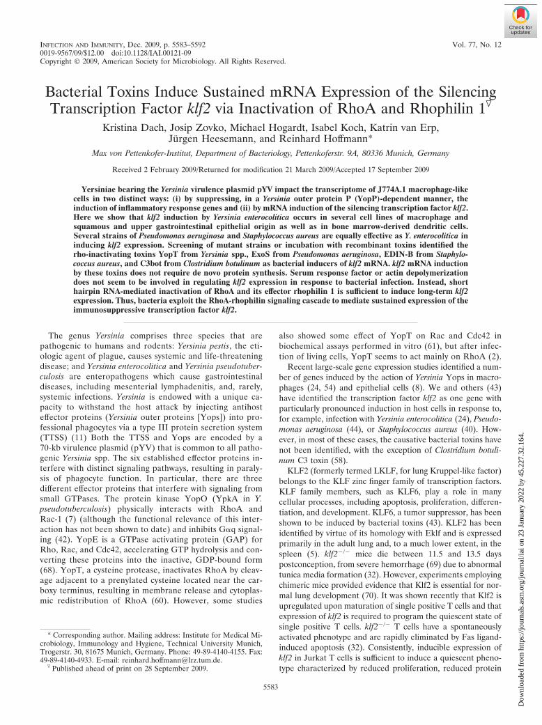
shRNA-mediated knockdown of RhoA induces klf2 mRNAexpression. As stated above, all bacterial toxins identified toinduce klf2 mRNA expression interfere with small GTPases ofthe rho family, with the C3 toxins interacting specifically withRhoA, RhoB, and RhoC (4). To identify the Rho proteinresponsible for klf2 induction, we generated stable shRNA-expressing cell lines for each of the Rho proteins individually.As shown in Fig. 4A to C, rhoA, rhoB, and rhoC shRNA-expressing cells showed substantial downregulation of the cor-responding mRNA levels relative to those in control shRNA-expressing cells. However, rhoB and rhoC shRNAs also hadmodest effects on rhoA mRNA expression levels: rhoB shRNAresulted in a 62% reduction of the rhoA expression level, andrhoC shRNA resulted in a 68% reduction. This problem oc-
FIG. 2. Identification of bacterial klf2-inducing proteins. (A) klf2 mRNA expression in J774A.1 cells after infection with different Y. entero-colitica mutant strains (detailed in Table 2). The graphical display is as described in the legend to Fig. 1. *, statistically significant (t test; P � 0.05)induction of klf2 mRNA compared to infection with strain WA-C. (B) klf2 mRNA levels in J774A.1 cells after infection with different P. aeruginosawild-type and mutant strains (see Table 2). The graphical display is as described in the legend to Fig. 1. Horizontal brackets indicate statisticallysignificant (t test; P � 0.05) differences in klf2 mRNA expression levels. (C) klf2 induction by recombinant EDIN-B or C. botulinum C3 toxin inJ774A.1 cells. The graphical display is as described in the legend to Fig. 1. sycE, irrelevant protein used as negative control.
VOL. 77, 2009 klf2 INDUCTION VIA RHO AND RHOPHILIN 5587
Dow
nloa
ded
from
http
s://j
ourn
als.
asm
.org
/jour
nal/i
ai o
n 23
Jan
uary
202
2 by
45.
227.
32.1
64.

curred with several different shRNA constructs. As displayedin Fig. 4D, however, klf2 mRNA expression was highest inrhoA shRNA-transduced cells, while rhoB shRNA- and rhoCshRNA-transduced cells expressed lower levels of klf2 mRNA.This suggests that rhoA is involved in the regulation of klf2mRNA expression levels.
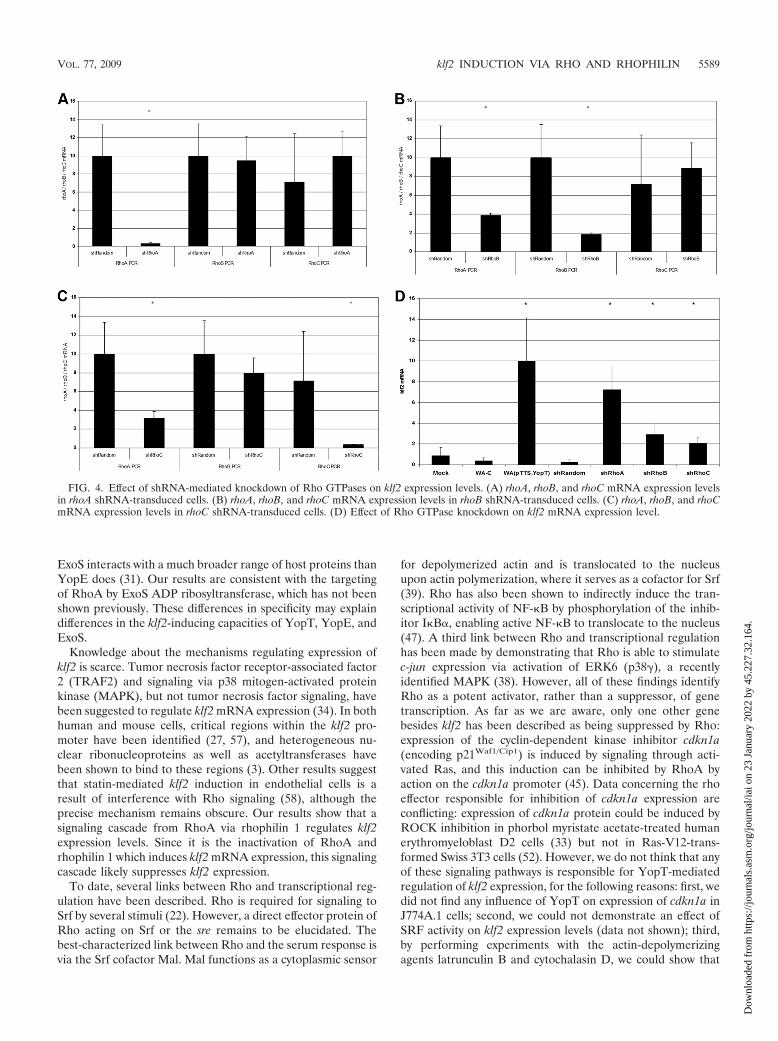
The Rho effector protein rhophilin 1 is involved in klf2mRNA induction. Rho proteins exert their cellular function bybinding, in their active GTP-bound state, to Rho effector pro-teins. To better characterize the RhoA-dependent signalingcascade regulating klf2 expression, we first investigated the roleof the well-characterized effector protein Rho kinase. Treat-ment of J774A.1 cells with different concentrations of the spe-cific, cell-permeating inhibitor H1152 for different times, how-ever, did not result in klf2 induction (Fig. 5A). Similar resultswere obtained with the less specific inhibitor H1077, which alsoinhibits the Rho effector proteins PRK1, PRK2, and MSK1(not shown). Thus, Rho kinase seems not to be involved inregulation of klf2 expression levels.
We next investigated the role of several other Rho effectorproteins by shRNA-mediated knockdown. shRNAs specific forcitron, rho kinase 2, kinectin 1, and rhophilin 1 all mediatedeffective knockdowns of their respective target mRNAs, asmeasured by real-time reverse transcription-PCR (not shown).Only rhophilin 1 shRNA-transduced cells, however, inducedsubstantial levels of klf2 mRNA (Fig. 5B). Importantly, thespecificity of the Rhpn-1 small interfering RNA was controlledby extensive manual BLAST searches, which demonstrated nooccurrence of the small interfering RNA sequence in any othermRNA transcript (data not shown). Thus, we suggest that a
signaling axis via RhoA and rhophilin 1 regulates klf2 mRNAlevels in response to bacterial infection.
DISCUSSION
Recent large-scale gene expression studies identified a num-ber of genes induced by the action of Yersinia Yops in macro-phages (24, 54) and epithelial cells (8). We have identified thetranscription factor klf2 as one gene with particularly pro-nounced induction in J774A.1 cells in response to infectionwith virulent, Yop-translocating yersiniae. Given the immuno-suppressive action of klf2 in a variety of cell types, it seemsplausible that klf2 induction constitutes a novel immunosup-pressive strategy of bacteria.
In the case of infection with Yersinia, the Rho-inactivatingcysteine protease YopT mediates sustained klf2 expression inhost cells. In the absence of YopT, klf2 is only transientlyexpressed. Yersinia possesses a second Rho-GTPase-inactivat-ing protein, YopE, which acts as a GAP for Rho, Rac, andCdc42 in vitro (68). However, after infection of living cells withYopE-translocating yersiniae, YopE seems to interact with amuch smaller range of Rho proteins: it acts primarily on Racrather than on Rho or Cdc42 in vivo, and it inhibits Cdc42-mediated Rac activation induced by bradykinin but not directactivation of Rac by sphingosine-1-phosphate (6). Consis-tently, YopT and YopE have been shown to differently affectthe cytoskeleton and phagocytic capacity of DCs (1). Com-pared to P. aeruginosa ExoS, it should be noted that YersiniaExoS contains an additional ADP ribosyltransferase domaininactivating small GTPases (49) and that the GAP domain of
FIG. 3. klf2 mRNA levels induced by wild-type Y. enterocolitica and YopT mutants in cell lines derived from different human or mouse tissues.The graphical display is as described in the legend to Fig. 1. Note the logarithmic scale. *, statistically significant (t test; P � 0.05) induction ofklf2 mRNA compared to infection of the respective cell line with strain WA-C. DC, mouse bone marrow-derived DCs.
5588 DACH ET AL. INFECT. IMMUN.
Dow
nloa
ded
from
http
s://j
ourn
als.
asm
.org
/jour
nal/i
ai o
n 23
Jan
uary
202
2 by
45.
227.
32.1
64.
ExoS interacts with a much broader range of host proteins thanYopE does (31). Our results are consistent with the targetingof RhoA by ExoS ADP ribosyltransferase, which has not beenshown previously. These differences in specificity may explaindifferences in the klf2-inducing capacities of YopT, YopE, andExoS.
Knowledge about the mechanisms regulating expression ofklf2 is scarce. Tumor necrosis factor receptor-associated factor2 (TRAF2) and signaling via p38 mitogen-activated proteinkinase (MAPK), but not tumor necrosis factor signaling, havebeen suggested to regulate klf2 mRNA expression (34). In bothhuman and mouse cells, critical regions within the klf2 pro-moter have been identified (27, 57), and heterogeneous nu-clear ribonucleoproteins as well as acetyltransferases havebeen shown to bind to these regions (3). Other results suggestthat statin-mediated klf2 induction in endothelial cells is aresult of interference with Rho signaling (58), although theprecise mechanism remains obscure. Our results show that asignaling cascade from RhoA via rhophilin 1 regulates klf2expression levels. Since it is the inactivation of RhoA andrhophilin 1 which induces klf2 mRNA expression, this signalingcascade likely suppresses klf2 expression.
To date, several links between Rho and transcriptional reg-ulation have been described. Rho is required for signaling toSrf by several stimuli (22). However, a direct effector protein ofRho acting on Srf or the sre remains to be elucidated. Thebest-characterized link between Rho and the serum response isvia the Srf cofactor Mal. Mal functions as a cytoplasmic sensor
for depolymerized actin and is translocated to the nucleusupon actin polymerization, where it serves as a cofactor for Srf(39). Rho has also been shown to indirectly induce the tran-scriptional activity of NF-�B by phosphorylation of the inhib-itor I�B�, enabling active NF-�B to translocate to the nucleus(47). A third link between Rho and transcriptional regulationhas been made by demonstrating that Rho is able to stimulatec-jun expression via activation of ERK6 (p38), a recentlyidentified MAPK (38). However, all of these findings identifyRho as a potent activator, rather than a suppressor, of genetranscription. As far as we are aware, only one other genebesides klf2 has been described as being suppressed by Rho:expression of the cyclin-dependent kinase inhibitor cdkn1a(encoding p21Waf1/Cip1) is induced by signaling through acti-vated Ras, and this induction can be inhibited by RhoA byaction on the cdkn1a promoter (45). Data concerning the rhoeffector responsible for inhibition of cdkn1a expression areconflicting: expression of cdkn1a protein could be induced byROCK inhibition in phorbol myristate acetate-treated humanerythromyeloblast D2 cells (33) but not in Ras-V12-trans-formed Swiss 3T3 cells (52). However, we do not think that anyof these signaling pathways is responsible for YopT-mediatedregulation of klf2 expression, for the following reasons: first, wedid not find any influence of YopT on expression of cdkn1a inJ774A.1 cells; second, we could not demonstrate an effect ofSRF activity on klf2 expression levels (data not shown); third,by performing experiments with the actin-depolymerizingagents latrunculin B and cytochalasin D, we could show that
FIG. 4. Effect of shRNA-mediated knockdown of Rho GTPases on klf2 expression levels. (A) rhoA, rhoB, and rhoC mRNA expression levelsin rhoA shRNA-transduced cells. (B) rhoA, rhoB, and rhoC mRNA expression levels in rhoB shRNA-transduced cells. (C) rhoA, rhoB, and rhoCmRNA expression levels in rhoC shRNA-transduced cells. (D) Effect of Rho GTPase knockdown on klf2 mRNA expression level.
VOL. 77, 2009 klf2 INDUCTION VIA RHO AND RHOPHILIN 5589
Dow
nloa
ded
from
http
s://j
ourn
als.
asm
.org
/jour
nal/i
ai o
n 23
Jan
uary
202
2 by
45.
227.
32.1
64.
the actin polymerization status is not involved in regulating klf2expression (data not shown); and fourth, we found rhophilin 1rather than ROCK to be involved in klf2 regulation.
Rhophilin 1 was first described in 1996, when it was detectedin a yeast two-hybrid screen with Rho as bait. It interactsstrongly with GTP-bound RhoA, less with RhoB, and littlewith RhoC (71). Rhpn1 is expressed in various tissues (41) andis highly expressed in the mouse testis, where it interacts withropporin (18). Other putative binding partners have been iden-tified by yeast two-hybrid screens. Among these are Trim37,Krt15, Cnksr1, Efemp2, and Ndp52 (51). Rhpn1 contains sev-eral protein-protein interaction motifs, such as HR1, a centralBRO1 domain, and a C-terminal PDZ domain (46). The HR1domain or Rho binding domain was first described as part ofprotein kinase PRK1, which binds RhoA (15). It is found in arange of signaling proteins, including rhotekin and PRK2, andis required for GTPase binding (50, 71). The exact molecularfunctions of BRO1 domains are not known. They are requiredfor cargo protein deubiquitination and play a role in endosome
metabolism (36). PDZ domains are protein interaction do-mains that are often found in multidomain scaffolding proteinsthat organize intracellular signaling at particular subcellularlocations (62). Taken together, the presence of different pro-tein interaction motifs suggest that rhophilin 1 may serve as asignaling protein. Thus, the following sequence of eventsseems likely in YopT-modified regulation of klf2 expression.YopT cleaves RhoA from its geranylgeranylated membraneanchor, which must not necessarily inactivate RhoA (72) butcould also change the subcellular localization of RhoA suffi-ciently to inhibit effector protein binding. However, the exactsubcellular localization of rhophilin 1 in macrophages is un-clear, so it remains to be determined whether YopT-mediatedRhoA inactivation or compartmentalization is responsible forinhibiting the RhoA-rhophilin 1 interaction. How exactlyrhophilin 1 then regulates klf2 mRNA expression remains tobe determined.
We described earlier that klf2 is also induced transiently (�1h after infection) by Yop-devoid yersiniae. Translocation of
FIG. 5. Impact of Rho effector proteins on klf2 expression level. (A) klf2 mRNA levels in J774A.1 cells treated with the ROCK inhibitor H1152for the indicated times (O/N, overnight) at the indicated concentrations. Similar results were obtained with the inhibitor H1077. (B) Impact ofindicated Rho effector protein knockdown by shRNA on klf2 expression level.
5590 DACH ET AL. INFECT. IMMUN.
Dow
nloa
ded
from
http
s://j
ourn
als.
asm
.org
/jour
nal/i
ai o
n 23
Jan
uary
202
2 by
45.
227.
32.1
64.

YopT results in a sustained (�2 h) expression of klf2 mRNA(24). Together with the data presented here, this suggests thefollowing model of klf2 regulation in the context of bacterialinfection (Fig. 6). Immediately after bacterial contact, hostcells induce klf2 by an as yet uncharacterized signaling cascade,but MAPK or NF-�B signaling may be involved. This earlyinduction of klf2 may be viewed as a physiological regulatory“loop” to prevent overwhelming inflammatory activation of thehost cell. Two hours after infection, signaling via RhoA andrhophilin 1 suppresses klf2 expression. Bacteria mediate long-term expression of klf2 by suppressing this inhibitory action ofRhoA via Rho-inactivating enzymes. As noted above, this mayconstitute a novel immunosuppressive strategy. Future studieswill identify the relevance of klf2 induction for infections invivo as well as the precise role of rhophilin 1 in suppression oftranscriptional responses to bacterial infection.
ACKNOWLEDGMENT
This work was supported by the German Federal Ministry of Edu-cation and Research under the auspices of the National GenomeResearch Network, NGFN 2 (grant no. IE-S31T10).
REFERENCES
1. Adkins, I., M. Koberle, S. Grobner, E. Bohn, I. B. Autenrieth, and S. Borg-mann. 2007. Yersinia outer proteins E, H, P, and T differentially target thecytoskeleton and inhibit phagocytic capacity of dendritic cells. Int. J. Med.Microbiol. 297:235–244.
2. Aepfelbacher, M., C. Trasak, G. Wilharm, A. Wiedemann, K. Trulzsch, K.Krauss, P. Gierschik, and J. Heesemann. 2003. Characterization of YopTeffects on Rho GTPases in Yersinia enterocolitica-infected cells. J. Biol.Chem. 278:33217–33223.
3. Ahmad, N., and J. B. Lingrel. 2005. Kruppel-like factor 2 transcriptionalregulation involves heterogeneous nuclear ribonucleoproteins and acetyl-transferases. Biochemistry 44:6276–6285.
4. Aktories, K., and J. T. Barbieri. 2005. Bacterial cytotoxins: targeting eukary-otic switches. Nat. Rev. Microbiol. 3:397–410.
5. Anderson, K. P., C. B. Kern, S. C. Crable, and J. B. Lingrel. 1995. Isolationof a gene encoding a functional zinc finger protein homologous to erythroidKruppel-like factor: identification of a new multigene family. Mol. Cell. Biol.15:5957–5965.
6. Andor, A., K. Trulzsch, M. Essler, A. Roggenkamp, A. Wiedemann, J. Heese-mann, and M. Aepfelbacher. 2001. YopE of Yersinia, a GAP for RhoGTPases, selectively modulates Rac-dependent actin structures in endothe-lial cells. Cell. Microbiol. 3:301–310.
7. Barz, C., T. N. Abahji, K. Trulzsch, and J. Heesemann. 2000. The YersiniaSer/Thr protein kinase YpkA/YopO directly interacts with the smallGTPases RhoA and Rac-1. FEBS Lett. 482:139–143.
8. Bohn, E., S. Muller, J. Lauber, R. Geffers, N. Speer, C. Spieth, J. Krejci, B.Manncke, J. Buer, A. Zell, and I. B. Autenrieth. 2004. Gene expressionpatterns of epithelial cells modulated by pathogenicity factors of Yersiniaenterocolitica. Cell. Microbiol. 6:129–141.
9. Boyd, A. P., N. Grosdent, S. Totemeyer, C. Geuijen, S. Bleves, M. Iriarte, I.Lambermont, J. N. Octave, and G. R. Cornelis. 2000. Yersinia enterocoliticacan deliver Yop proteins into a wide range of cell types: development of adelivery system for heterologous proteins. Eur. J. Cell Biol. 79:659–671.
10. Buckley, A. F., C. T. Kuo, and J. M. Leiden. 2001. Transcription factor LKLFis sufficient to program T cell quiescence via a c-Myc-dependent pathway.Nat. Immunol. 2:698–704.
11. Cornelis, G. R. 2002. The Yersinia Ysc-Yop ‘type III’ weaponry. Nat. Rev.Mol. Cell Biol. 3:742–752.
12. Cox, G. W., B. J. Mathieson, L. Gandino, E. Blasi, D. Radzioch, and L.Varesio. 1989. Heterogeneity of hematopoietic cells immortalized by v-myc/v-raf recombinant retrovirus infection of bone marrow or fetal liver. J. Natl.Cancer Inst. 81:1492–1496.
13. Czech, A., T. Yamaguchi, L. Bader, S. Linder, K. Kaminski, M. Sugai, andM. Aepfelbacher. 2001. Prevalence of Rho-inactivating epidermal cell dif-ferentiation inhibitor toxins in clinical Staphylococcus aureus isolates. J. In-fect. Dis. 184:785–788.
14. Dekker, R. J., S. van Soest, R. D. Fontijn, S. Salamanca, P. G. de Groot, E.VanBavel, H. Pannekoek, and A. J. Horrevoets. 2002. Prolonged fluid shearstress induces a distinct set of endothelial cell genes, most specifically lungKruppel-like factor (KLF2). Blood 100:1689–1698.
15. Flynn, P., H. Mellor, R. Palmer, G. Panayotou, and P. J. Parker. 1998.Multiple interactions of PRK1 with RhoA. Functional assignment of the Hr1repeat motif. J. Biol. Chem. 273:2698–2705.
16. Frank, D. W., G. Nair, and H. P. Schweizer. 1994. Construction and char-acterization of chromosomal insertional mutations of the Pseudomonasaeruginosa exoenzyme S trans-regulatory locus. Infect. Immun. 62:554–563.
17. Frankel, G., A. D. Phillips, M. Novakova, H. Field, D. C. Candy, D. B.Schauer, G. Douce, and G. Dougan. 1996. Intimin from enteropathogenicEscherichia coli restores murine virulence to a Citrobacter rodentium eaeAmutant: induction of an immunoglobulin A response to intimin and EspB.Infect. Immun. 64:5315–5325.
18. Fujita, A., K. Nakamura, T. Kato, N. Watanabe, T. Ishizaki, K. Kimura, A.Mizoguchi, and S. Narumiya. 2000. Ropporin, a sperm-specific binding pro-tein of rhophilin, that is localized in the fibrous sheath of sperm flagella.J. Cell Sci. 113:103–112.
19. Gansheroff, L. J., M. R. Wachtel, and A. D. O’Brien. 1999. Decreasedadherence of enterohemorrhagic Escherichia coli to HEp-2 cells in the pres-ence of antibodies that recognize the C-terminal region of intimin. Infect.Immun. 67:6409–6417.
20. Gebert, B., W. Fischer, E. Weiss, R. Hoffmann, and R. Haas. 2003. Helico-bacter pylori vacuolating cytotoxin inhibits T lymphocyte activation. Science301:1099–1102.
21. Heesemann, J., and R. Laufs. 1983. Construction of a mobilizable Yersiniaenterocolitica virulence plasmid. J. Bacteriol. 155:761–767.
22. Hill, C. S., J. Wynne, and R. Treisman. 1995. The Rho family GTPasesRhoA, Rac1, and CDC42Hs regulate transcriptional activation by SRF. Cell81:1159–1170.
23. Hoang, T. T., R. R. Karkhoff-Schweizer, A. J. Kutchma, and H. P. Schweizer.1998. A broad-host-range Flp-FRT recombination system for site-specificexcision of chromosomally-located DNA sequences: application for isolationof unmarked Pseudomonas aeruginosa mutants. Gene 212:77–86.
24. Hoffmann, R., K. Van Erp, K. Trulzsch, and J. Heesemann. 2004. Transcrip-tional responses of murine macrophages to infection with Yersinia entero-colitica. Cell. Microbiol. 6:377–390.
25. Hoiseth, S. K., and B. A. Stocker. 1981. Aromatic-dependent Salmonellatyphimurium are non-virulent and effective as live vaccines. Nature 291:238–239.
26. Hornef, M. W., A. Roggenkamp, A. M. Geiger, M. Hogardt, C. A. Jacobi, andJ. Heesemann. 2000. Triggering the ExoS regulon of Pseudomonas aerugi-nosa: a GFP-reporter analysis of exoenzyme (Exo) S, ExoT and ExoU syn-thesis. Microb. Pathog. 29:329–343.
27. Huddleson, J. P., S. Srinivasan, N. Ahmad, and J. B. Lingrel. 2004. Fluidshear stress induces endothelial KLF2 gene expression through a definedpromoter region. Biol. Chem. 385:723–729.
28. Imanishi, K., K. Yamaguchi, M. Suzuki, S. Honda, N. Yanaihara, and K.Abe. 1989. Production of transforming growth factor-alpha in human tumourcell lines. Br. J. Cancer 59:761–765.
29. Janda, J. M., S. L. Abbott, and M. J. Albert. 1999. Prototypal diarrheagenicstrains of Hafnia alvei are actually members of the genus Escherichia. J. Clin.Microbiol. 37:2399–2401.
30. Kaufman, M. R., J. Jia, L. Zeng, U. Ha, M. Chow, and S. Jin. 2000. Pseudo-monas aeruginosa mediated apoptosis requires the ADP-ribosylating activityof exoS. Microbiology 146:2531–2541.
31. Krall, R., J. Sun, K. J. Pederson, and J. T. Barbieri. 2002. In vivo RhoGTPase-activating protein activity of Pseudomonas aeruginosa cytotoxinExoS. Infect. Immun. 70:360–367.
32. Kuo, C. T., M. L. Veselits, K. P. Barton, M. M. Lu, C. Clendenin, and J. M.
FIG. 6. Proposed model of klf2 regulation in the context of bacte-rial infection. Immediately after bacterial contact, host cells induce klf2by an as yet uncharacterized signaling cascade; however, MAPK orNF-�B signaling may be involved. Two hours after infection, signalingvia RhoA and rhophilin 1 suppresses klf2 expression. Bacteria mediatelong-term expression of klf2 by suppressing this inhibitory action ofRhoA via Rho-inactivating enzymes.
VOL. 77, 2009 klf2 INDUCTION VIA RHO AND RHOPHILIN 5591
Dow
nloa
ded
from
http
s://j
ourn
als.
asm
.org
/jour
nal/i
ai o
n 23
Jan
uary
202
2 by
45.
227.
32.1
64.

Leiden. 1997. The LKLF transcription factor is required for normal tunicamedia formation and blood vessel stabilization during murine embryogene-sis. Genes Dev. 11:2996–3006.
33. Lai, J. M., S. Wu, D. Y. Huang, and Z. F. Chang. 2002. Cytosolic retentionof phosphorylated extracellular signal-regulated kinase and a Rho-associatedkinase-mediated signal impair expression of p21(Cip1/Waf1) in phorbol 12-myristate-13-acetate-induced apoptotic cells. Mol. Cell. Biol. 22:7581–7592.
34. Lin, Y., J. Ryan, J. Lewis, M. A. Wani, J. B. Lingrel, and Z. G. Liu. 2003.TRAF2 exerts its antiapoptotic effect by regulating the expression of Krup-pel-like factor LKLF. Mol. Cell. Biol. 23:5849–5856.
35. Locher, M., B. Lehnert, K. Krauss, J. Heesemann, M. Groll, and G. Wil-harm. 2005. Crystal structure of the Yersinia enterocolitica type III secretionchaperone SycT. J. Biol. Chem. 280:31149–31155.
36. Luhtala, N., and G. Odorizzi. 2004. Bro1 coordinates deubiquitination in themultivesicular body pathway by recruiting Doa4 to endosomes. J. Cell Biol.166:717–729.
37. Lutz, M. B., N. Kukutsch, A. L. Ogilvie, S. Rossner, F. Koch, N. Romani, andG. Schuler. 1999. An advanced culture method for generating large quanti-ties of highly pure dendritic cells from mouse bone marrow. J. Immunol.Methods 223:77–92.
38. Marinissen, M. J., M. Chiariello, and J. S. Gutkind. 2001. Regulation ofgene expression by the small GTPase Rho through the ERK6 (p38 gamma)MAP kinase pathway. Genes Dev. 15:535–553.
39. Miralles, F., G. Posern, A. I. Zaromytidou, and R. Treisman. 2003. Actindynamics control SRF activity by regulation of its coactivator MAL. Cell113:329–342.
40. Moreilhon, C., D. Gras, C. Hologne, O. Bajolet, F. Cottrez, V. Magnone, M.Merten, H. Groux, E. Puchelle, and P. Barbry. 2005. Live Staphylococcusaureus and bacterial soluble factors induce different transcriptional re-sponses in human airway cells. Physiol. Genomics 20:244–255.
41. Nakamura, K., A. Fujita, T. Murata, G. Watanabe, C. Mori, J. Fujita, N.Watanabe, T. Ishizaki, O. Yoshida, and S. Narumiya. 1999. Rhophilin, asmall GTPase Rho-binding protein, is abundantly expressed in the mousetestis and localized in the principal piece of the sperm tail. FEBS Lett.445:9–13.
42. Navarro, L., A. Koller, R. Nordfelth, H. Wolf-Watz, S. Taylor, and J. E.Dixon. 2007. Identification of a molecular target for the Yersinia proteinkinase A. Mol. Cell 26:465–477.
43. O’Grady, E., H. Mulcahy, C. Adams, J. P. Morrissey, and F. O’Gara. 2007.Manipulation of host Kruppel-like factor (KLF) function by exotoxins fromdiverse bacterial pathogens. Nat. Rev. Microbiol. 5:337–341.
44. O’Grady, E. P., H. Mulcahy, J. O’Callaghan, C. Adams, and F. O’Gara. 2006.Pseudomonas aeruginosa infection of airway epithelial cells modulates ex-pression of Kruppel-like factors 2 and 6 via RsmA-mediated regulation oftype III exoenzymes S and Y. Infect. Immun. 74:5893–5902.
45. Olson, M. F., H. F. Paterson, and C. J. Marshall. 1998. Signals from Ras andRho GTPases interact to regulate expression of p21Waf1/Cip1. Nature 394:295–299.
46. Peck, J. W., M. Oberst, K. B. Bouker, E. Bowden, and P. D. Burbelo. 2002.The RhoA-binding protein, rhophilin-2, regulates actin cytoskeleton organi-zation. J. Biol. Chem. 277:43924–43932.
47. Perona, R., S. Montaner, L. Saniger, I. Sanchez-Perez, R. Bravo, and J. C.Lacal. 1997. Activation of the nuclear factor-kappaB by Rho, CDC42, andRac-1 proteins. Genes Dev. 11:463–475.
48. Prentki, P., and H. M. Krisch. 1984. In vitro insertional mutagenesis with aselectable DNA fragment. Gene 29:303–313.
49. Radke, J., K. J. Pederson, and J. T. Barbieri. 1999. Pseudomonas aeruginosaexoenzyme S is a biglutamic acid ADP-ribosyltransferase. Infect. Immun.67:1508–1510.
50. Reid, T., T. Furuyashiki, T. Ishizaki, G. Watanabe, N. Watanabe, K. Fuji-sawa, N. Morii, P. Madaule, and S. Narumiya. 1996. Rhotekin, a newputative target for Rho bearing homology to a serine/threonine kinase, PKN,and rhophilin in the rho-binding domain. J. Biol. Chem. 271:13556–13560.
51. Rual, J. F., K. Venkatesan, T. Hao, T. Hirozane-Kishikawa, A. Dricot, N. Li,G. F. Berriz, F. D. Gibbons, M. Dreze, N. Ayivi-Guedehoussou, N. Klitgord,C. Simon, M. Boxem, S. Milstein, J. Rosenberg, D. S. Goldberg, L. V. Zhang,S. L. Wong, G. Franklin, S. Li, J. S. Albala, J. Lim, C. Fraughton, E.Llamosas, S. Cevik, C. Bex, P. Lamesch, R. S. Sikorski, J. Vandenhaute,H. Y. Zoghbi, A. Smolyar, S. Bosak, R. Sequerra, L. Doucette-Stamm, M. E.Cusick, D. E. Hill, F. P. Roth, and M. Vidal. 2005. Towards a proteome-scalemap of the human protein-protein interaction network. Nature 437:1173–1178.
52. Sahai, E., M. F. Olson, and C. J. Marshall. 2001. Cross-talk between Ras andRho signaling pathways in transformation favours proliferation and in-creased motility. EMBO J. 20:755–766.
53. Sansonetti, P. J., D. J. Kopecko, and S. B. Formal. 1982. Involvement of aplasmid in the invasive ability of Shigella flexneri. Infect. Immun. 35:852–860.
54. Sauvonnet, N., B. Pradet-Balade, J. A. Garcia-Sanz, and G. R. Cornelis.
2002. Regulation of mRNA expression in macrophages after Yersinia en-terocolitica infection. Role of different Yop effectors. J. Biol. Chem. 277:25133–25142.
55. Schmitt, W., and R. Haas. 1994. Genetic analysis of the Helicobacter pylorivacuolating cytotoxin: structural similarities with the IgA protease type ofexported protein. Mol. Microbiol. 12:307–319.
56. Schneider, U., H. U. Schwenk, and G. Bornkamm. 1977. Characterization ofEBV-genome negative “null” and “T” cell lines derived from children withacute lymphoblastic leukemia and leukemic transformed non-Hodgkin lym-phoma. Int. J. Cancer 19:621–626.
57. Schrick, J. J., M. J. Hughes, K. P. Anderson, M. L. Croyle, and J. B. Lingrel.1999. Characterization of the lung Kruppel-like transcription factor gene andupstream regulatory elements. Gene 236:185–195.
58. Sen-Banerjee, S., S. Mir, Z. Lin, A. Hamik, G. B. Atkins, H. Das, P. Banerjee,A. Kumar, and M. K. Jain. 2005. Kruppel-like factor 2 as a novel mediatorof statin effects in endothelial cells. Circulation 112:720–726.
59. SenBanerjee, S., Z. Lin, G. B. Atkins, D. M. Greif, R. M. Rao, A. Kumar,M. W. Feinberg, Z. Chen, D. I. Simon, F. W. Luscinskas, T. M. Michel, M. A.Gimbrone, Jr., G. Garcia-Cardena, and M. K. Jain. 2004. KLF2 is a noveltranscriptional regulator of endothelial proinflammatory activation. J. Exp.Med. 10:10.
60. Shao, F., P. M. Merritt, Z. Bao, R. W. Innes, and J. E. Dixon. 2002. AYersinia effector and a Pseudomonas avirulence protein define a family ofcysteine proteases functioning in bacterial pathogenesis. Cell 109:575–588.
61. Shao, F., P. O. Vacratsis, Z. Bao, K. E. Bowers, C. A. Fierke, and J. E. Dixon.2003. Biochemical characterization of the Yersinia YopT protease: cleavagesite and recognition elements in Rho GTPases. Proc. Natl. Acad. Sci. USA100:904–909.
62. Sheng, M., and C. Sala. 2001. PDZ domains and the organization of su-pramolecular complexes. Annu. Rev. Neurosci. 24:1–29.
63. Snellings, N. J., M. Popek, and L. E. Lindler. 2001. Complete DNA se-quence of Yersinia enterocolitica serotype 0:8 low-calcium-response plasmidreveals a new virulence plasmid-associated replicon. Infect. Immun. 69:4627–4638.
64. Stover, C. K., X. Q. Pham, A. L. Erwin, S. D. Mizoguchi, P. Warrener, M. J.Hickey, F. S. Brinkman, W. O. Hufnagle, D. J. Kowalik, M. Lagrou, R. L.Garber, L. Goltry, E. Tolentino, S. Westbrock-Wadman, Y. Yuan, L. L. Brody,S. N. Coulter, K. R. Folger, A. Kas, K. Larbig, R. Lim, K. Smith, D. Spencer,G. K. Wong, Z. Wu, I. T. Paulsen, J. Reizer, M. H. Saier, R. E. Hancock, S. Lory,and M. V. Olson. 2000. Complete genome sequence of Pseudomonas aerugi-nosa PAO1, an opportunistic pathogen. Nature 406:959–964.
65. Tomb, J. F., O. White, A. R. Kerlavage, R. A. Clayton, G. G. Sutton, R. D.Fleischmann, K. A. Ketchum, H. P. Klenk, S. Gill, B. A. Dougherty, K.Nelson, J. Quackenbush, L. Zhou, E. F. Kirkness, S. Peterson, B. Loftus, D.Richardson, R. Dodson, H. G. Khalak, A. Glodek, K. McKenney, L. M.Fitzegerald, N. Lee, M. D. Adams, J. C. Venter, et al. 1997. The completegenome sequence of the gastric pathogen Helicobacter pylori. Nature 388:539–547.
66. Trulzsch, K., A. Roggenkamp, M. Aepfelbacher, G. Wilharm, K. Ruckde-schel, and J. Heesemann. 2003. Analysis of chaperone-dependent Yop se-cretion/translocation and effector function using a mini-virulence plasmid ofYersinia enterocolitica. Int. J. Med. Microbiol. 293:167–177.
67. Trulzsch, K., T. Sporleder, E. I. Igwe, H. Russmann, and J. Heesemann.2004. Contribution of the major secreted Yops of Yersinia enterocolitica O:8to pathogenicity in the mouse infection model. Infect. Immun. 72:5227–5234.
68. Von Pawel-Rammingen, U., M. V. Telepnev, G. Schmidt, K. Aktories, H.Wolf-Watz, and R. Rosqvist. 2000. GAP activity of the Yersinia YopE cyto-toxin specifically targets the Rho pathway: a mechanism for disruption ofactin microfilament structure. Mol. Microbiol. 36:737–748.
69. Wani, M. A., R. T. Means, Jr., and J. B. Lingrel. 1998. Loss of LKLF functionresults in embryonic lethality in mice. Transgenic Res. 7:229–238.
70. Wani, M. A., S. E. Wert, and J. B. Lingrel. 1999. Lung Kruppel-like factor,a zinc finger transcription factor, is essential for normal lung development.J. Biol. Chem. 274:21180–21185.
71. Watanabe, G., Y. Saito, P. Madaule, T. Ishizaki, K. Fujisawa, N. Morii, H.Mukai, Y. Ono, A. Kakizuka, and S. Narumiya. 1996. Protein kinase N(PKN) and PKN-related protein rhophilin as targets of small GTPase Rho.Science 271:645–648.
72. Wong, K. W., and R. R. Isberg. 2005. Yersinia pseudotuberculosis spatiallycontrols activation and misregulation of host cell Rac1. PLoS Pathog. 1:e16.
73. Yao, T., J. Mecsas, J. I. Healy, S. Falkow, and Y. Chien. 1999. Suppressionof T and B lymphocyte activation by a Yersinia pseudotuberculosis virulencefactor, yopH. J. Exp. Med. 190:1343–1350.
74. Zhu, C., T. S. Agin, S. J. Elliott, L. A. Johnson, T. E. Thate, J. B. Kaper, andE. C. Boedeker. 2001. Complete nucleotide sequence and analysis of thelocus of enterocyte effacement from rabbit diarrheagenic Escherichia coliRDEC-1. Infect. Immun. 69:2107–2115.
Editor: J. B. Bliska
5592 DACH ET AL. INFECT. IMMUN.
Dow
nloa
ded
from
http
s://j
ourn
als.
asm
.org
/jour
nal/i
ai o
n 23
Jan
uary
202
2 by
45.
227.
32.1
64.