bacteroides fragilis enterotoxin induces human -defensin-2 ... · quantify mrna molecules, standard...
TRANSCRIPT

INFECTION AND IMMUNITY, May 2010, p. 2024–2033 Vol. 78, No. 50019-9567/10/$12.00 doi:10.1128/IAI.00118-10Copyright © 2010, American Society for Microbiology. All Rights Reserved.
Bacteroides fragilis Enterotoxin Induces Human �-Defensin-2Expression in Intestinal Epithelial Cells via a Mitogen-Activated
Protein Kinase/I�B Kinase/NF-�B-Dependent Pathway�
Young Mee Yoon,1† Jin Young Lee,1† Doyoung Yoo,1 Young-Suk Sim,1 Young-Jeon Kim,2
Yu-Kyoung Oh,3 Ju Seop Kang,4 Sunil Kim,5 Joo Sung Kim,6 and Jung Mogg Kim1*Department of Microbiology, Hanyang University College of Medicine, Seoul, South Korea1; Department of
Biotechnology, Joongbu University, Choongnam, South Korea2; College of Pharmacy, Seoul National University, Seoul,South Korea3; Department of Pharmacology, Hanyang University College of Medicine, Seoul, South Korea4;
Department of Biomedical Engineering, Hanyang University, Seoul, South Korea5; and Department ofInternal Medicine and Liver Research Institute, Seoul National University College of
Medicine, Seoul, South Korea6
Received 4 February 2010/Returned for modification 4 March 2010/Accepted 5 March 2010
Enterotoxigenic Bacteroides fragilis (ETBF) produces an approximately 20-kDa heat-labile enterotoxin (BFT)that plays an essential role in mucosal inflammation. Although spontaneous disappearance of ETBF infectionis common, little information is available on regulated expression of antibacterial factors in response to BFTstimulation. This study investigates the role of BFT in human �-defensin 2 (hBD-2) induction from intestinalepithelial cells. Stimulation of HT-29 and Caco-2 intestinal epithelial cell lines with BFT resulted in theinduction of hBD-2. Activation of a reporter gene for hBD-2 was dependent on the presence of NF-�B bindingsites. In contrast, suppression of AP-1 did not affect hBD-2 expression in BFT-stimulated cells. Inhibition ofp38 mitogen-activated protein kinase (MAPK) using SB203580 and small interfering RNA (siRNA) transfec-tion resulted in a significant reduction in BFT-induced I�B kinase (IKK)/NF-�B activation and hBD-2expression. Our results suggest that a pathway including p38 MAPK, IKK, and NF-�B activation is requiredfor hBD-2 induction in intestinal epithelial cells exposed to BFT, and may be involved in the host defensefollowing infection with ETBF.
Enterotoxigenic Bacteroides fragilis (ETBF) strains havebeen identified during an investigation of diarrheal illness inanimals and young children (27, 28, 36, 47). Recently, ETBFinfection has been reported to be associated with inflammatorybowel diseases (IBD) (2, 32, 37) and colorectal cancer (39, 46).B. fragilis enterotoxin (BFT), an approximately 20-kDa heat-labile metalloprotease, is regarded as a virulence factor for thediseases. Although ETBF strains are proposed to be entericpathogens, all human studies of ETBF infection have demon-strated that between �4% and 30% of infected individualsasymptomatically colonize with ETBF and that infection isself-limited (33, 36). These reports suggest that antibacterialfactors induced by ETBF infection may be upregulated in theinfected area of the intestine and regulate enteric inflamma-tion.
To prevent intestinal infection, the luminal flora and patho-gens are controlled by epithelially derived antimicrobial pep-tides that are constitutively expressed or inducible (29). Forexample, defensins, cathelicidin LL-37, lysozyme, phospho-lipase A, and proteins with bactericidal properties such asubiquicidin, ribosomal proteins, and eosinophilic proteins havebeen reported (4, 11, 29, 38).
Defensins can act as endogenous antimicrobials by mem-brane permeabilization, activation of cell wall lytic enzymes,and disruption of membrane-bound multienzyme complexesand intracellular events (10, 29, 34). Human defensins arecationic, amphipathic peptides of 3.5 to �6 kDa that are char-acterized by three intramolecular disulfide bonds. They are sub-divided into �- and �-defensins. Four different neutrophil �-de-fensins have been reported: human neutrophil protein 1 (HNP-1)to HNP-4. In addition, two human �-defensins (HD-5 and HD-6)are constitutively expressed in Paneth cells of the small intestine(6). The epithelial human �-defensin (hBD-1) is constitutive inthe intestinal tract, whereas epithelial hBD-2 is induced by cyto-kines or in response to inflammatory stimuli such as bacterialinfection (29, 35, 42). Recently, human �-defensins have beenreported to inhibit the activity of Clostridium difficile toxin B (9).In light of these reports, it is possible that defensins may affectinflammatory responses due to ETBF-derived BFT and contrib-ute to host defense. However, little is known about the regulatedexpression of �-defensins in response to BFT stimulation.
Activation of mitogen-activated protein kinase (MAPK), nu-clear factor �B (NF-�B), or activator protein 1 (AP-1) isknown to be important for the induction of hBD-2 in severalcell lines (24, 30, 35, 40, 41, 43, 44). Although these signalingmolecules are reported to be upregulated in intestinal epithe-lial cells exposed to BFT (12, 14, 19, 20, 21, 23, 45), there is noevidence of BFT-induced MAPK and NF-�B activation lead-ing to hBD-2 expression. In the studies reported here, weinvestigated the regulation of hBD-2 induction in response toBFT stimulation and found that stimulation with BFT en-
* Corresponding author. Mailing address: Department of Microbi-ology, Hanyang University College of Medicine, 17 Haengdang-dong,Sungdong-gu, Seoul 133-791, Korea. Phone: 82-2-2220-0645. Fax: 82-2-2282-0645. E-mail: [email protected].
† Y. M. Yoon and J. Y. Lee contributed equally to this work.� Published ahead of print on 15 March 2010.
2024
on March 6, 2019 by guest
http://iai.asm.org/
Dow
nloaded from

hances hBD-2 expression in intestinal epithelial cells throughactivation of the MAPK/I�B kinase (IKK)/NF-�B pathway.
MATERIALS AND METHODS
Reagents. Lipopolysaccharide (LPS)-free fetal bovine serum (FBS), antibiot-ics, L-glutamine, and Trizol were obtained from GIBCO BRL (Gaithersburg,MD). Dulbecco’s modified Eagle’s medium (DMEM) was purchased by SigmaChemical Co. (St. Louis, MO). Antibodies against IKK-�, IKK-�, phospho-IKK-�/�, pan-extracellular signal-regulated kinase 1/2 (pan-ERK1/2 [p44/p42]), phos-pho-ERK1/2, pan-Jun N-terminal kinase (pan-JNK [p54/p46]), phospho-JNK,pan-p38, phospho-p38, and actin were acquired from Cell Signaling Technology,Inc. (Beverly, MA). Goat anti-rabbit and anti-mouse secondary antibodies con-jugated to horseradish peroxidase were purchased from Transduction Labora-tories (Lexington, KY). Antibodies against p50, p52, p65, c-Rel, and RelB wereobtained from Santa Cruz Biotechnology (Santa Cruz, CA). PD98059,SB203580, SP600125, and Bay 11-7085 were acquired from Calbiochem (LaJolla, CA).
Purification of BFT and cell culture conditions. BFT was purified from theculture supernatants of a highly toxigenic strain of ETBF as described previously(12, 14, 19–21, 23). The purity of the BFT preparations was confirmed by sodium
dodecyl sulfate-polyacrylamide gel electrophoresis (SDS-PAGE). Typical prep-arations of BFT contained 0.5 to 1.2 mg of protein per milliliter as measured bythe bicinchoninic acid (BCA) protein assay. The buffers used in the purificationwere prepared using LPS-free water (Baxter Healthcare Corp., Deerfield, IL).The activity of LPS in BFT solutions (1 mg/ml) was less than 1 endotoxin unit/ml(quantitative chromogenic Limulus amebocyte lysate; BioWhittaker, Walkers-ville, MD). BFT was frozen in aliquots at �80°C immediately after purification.The human colon adenocarcinoma cell line HT-29 (ATCC HTB 38) and Caco-2human ileocecal epithelial cell line (ATCC HTB 37) were grown in DMEM with10% FBS and 2 mM glutamine. Cells were seeded at 0.5 � 106 to 2 � 106 cellsper well onto six-well plates and allowed to attach overnight. After 12 h of serumstarvation, cells were incubated with BFT for the indicated period.
Quantitative RT-PCR and ELISA. Cells were treated with BFT, after whichtotal cellular RNA was extracted using Trizol. Reverse transcription (RT)-PCRamplification was performed as described previously (23). The primers andexpected PCR product sizes were as follows (1, 30): hBD-1, 5�-CTCTGTCAGCTCAGCCTC-3� (sense) and 5�-CTTGCAGCACTTGGCCTTCCC-3� (anti-sense), 272 bp; hBD-2, 5�-CCAGCCATCAGCCATGAGGGT-3� (sense) and5�-GGAGCCCTTTCTGAATCCGCA-3� (antisense), 254 bp; and human �-ac-tin, 5�-TGACGGGGTCACCCACACTGTGCCCATCTA-3� (sense) and 5�-CTAGAAGCATTGCGGTGGACGATGGAGGG-3� (antisense), 661 bp. To
FIG. 1. Time course of hBD mRNA expression in HT-29 and Caco-2 cells after treatment with BFT. HT-29 (A) and Caco-2 cells (B) weretreated with BFT (300 ng/ml) for the indicated periods. Levels of hBD-1, hBD-2, and �-actin mRNA were analyzed by quantitative RT-PCR usingeach standard RNA. The values are expressed as means � SD of five different experiments.
VOL. 78, 2010 hBD-2 INDUCTION BY B. FRAGILIS ENTEROTOXIN 2025
on March 6, 2019 by guest
http://iai.asm.org/
Dow
nloaded from

quantify mRNA molecules, standard RNAs for hBD-1 and hBD-2 weregenerated by an in vitro transcription using T7 RNA polymerase, as describedpreviously (7, 16). Standard RNA for human �-actin was kindly provided byMartin F. Kagnoff of the University of California, San Diego. The sizes ofPCR products generated from standard RNAs for hBD-1, hBD-2, and human�-actin are 368 bp, 371 bp, 319 bp, and 520 bp, respectively. mRNA levels of5 � 103 mRNA molecules/g of total RNA were considered positive; al-though lower levels could be detected and quantified, they were consideredunlikely to be biologically meaningful, as they would reflect, on average, lessthan one mRNA transcript/20 cells (13, 22).
The amounts of hBD-2 in culture supernatants were measured by a commer-cially available enzyme-linked immunosorbent assay (ELISA; Phoenix Pharma-ceuticals, Belmont, CA) according to the manufacturer’s instructions. One ex-periment was performed in triplicate wells. This experiment was repeated morethan 3 times. The detection limit of the ELISA kit was 10 pg/ml.
EMSA. Cells were harvested and nuclear extracts prepared as described pre-viously (21). Concentrations of protein in the extracts were determined by theBradford assay (Bio-Rad, Hercules, CA). The electrophoretic mobility shift assay(EMSA) was performed according to the manufacturer’s instructions (Promega,Madison, WI). In brief, 5 g of nuclear extract was incubated for 30 min at roomtemperature with a -32P-labeled oligonucleotide probe (5�-AGTTGAGGGGACTTTCCCAGGC-3� for the NF-�B binding site; 5�-CGCTTGATGACTCAGCCGGAA-3� for the AP-1 binding site). After incubation, both bound DNA andfree DNA were resolved on 5% polyacrylamide gels, as described previously (14,21). A supershift assay was used to identify the specific members of the NF-�Bfamily activated by BFT stimulation. EMSA was performed as described above,except that rabbit antibodies (1 g/reaction) against NF-�B proteins p50, p52,p65, c-Rel, and RelB were added during the binding reaction period (21).
Plasmids, transfection, and luciferase assays. To analyze hBD-2 promoteractivity, wild-type (hBD-2-2338-luc) and mutant plasmids (NF-�B-mutant-1�2�3-luc, AP-1-mutant-luc, and AP-1�NF-�B-mutant-luc) were kindly do-nated by Jurgen Harder, Clinical Research Unit, Department of Dermatology,University Hospital Kiel, Kiel, Germany (44). The reporter plasmid containingAP-1-luciferase was purchased from BD Sciences (Franklin Lakes, NJ) (14). p2xNF-�B-, p�-actin- and pRSV-�-galactosidase transcriptional reporters werekindly provided by Martin F. Kagnoff of the University of California, San Diego(8). Cells in six-well dishes were transfected with 1.5 g of plasmid DNA usingFuGENE 6 transfection reagent (Roche, Mannheim, Germany). The transfectedcells were incubated for 24 h at 37°C in a 5% CO2 incubator and were thentreated with BFT for the indicated time. Luciferase activity was determined inaccordance with the manufacturer’s instructions (Tropix, Inc., Bedford, MA).Light release was quantitated for 10 s using a luminometer (MicroLumat Plus,Berthold GmbH & Co. KG, Bad Wildbad, Germany), as previously described(25).
TAM-67 is a dominant-negative c-Jun superrepressor that lacks the transac-tivation domain of c-Jun and is a potent inhibitor of AP-1-mediated transacti-vation (3, 26). TAM-67 dimerizes with c-Jun or c-Fos family members and bindsDNA, resulting in the inhibition of wild-type c-Jun and c-Fos function. TheTAM-67 used in the present study was a gift from Andreas von Knethen of theUniversity of Erlangen, Erlangen, Germany. Small interfering RNAs (siRNAs)against IKK-�, the NF-�B p65 subunit, and p38 were designed as describedpreviously (17, 25). The siRNAs were synthesized by Qiagen (Valencia, CA).The negative (nonsilencing) control siRNA was also purchased from Qiagen.Transfection of dominant-negative superrepressor or siRNA into cells was per-formed as described previously (14). Cells were cultured in 6-well plates to 50 to80% confluence, and the cells were then transfected with the dominant-negativesuperrepressor, siRNA, or nonsilencing siRNA using FuGENE 6 (Roche) as atransfection reagent. Briefly, 1 g of siRNA or 1.5 g plasmid DNA was dilutedin serum-free medium to produce a final volume of 100 l, to which 3 l ofFuGENE 6 was added, and the mixture was incubated for 15 min at roomtemperature. The transfection mixture was added to the respective wells, eachcontaining 300 l of medium (10% FBS content). Transfected cells were incu-bated for 48 h prior to the assay.
A retroviral system containing a mammalian expression vector encoding ahemagglutinin (HA) epitope-tagged mutant I�B� (I�B�-AA) with substitutionsof serine for alanine at positions 32 and 36 was used to block NF-�B activationas described previously (15).
Immunoblots. Cells were washed with ice-cold PBS and lysed in 0.5 ml/welllysis buffer (150 mM NaCl, 20 mM Tris pH 7.5, 0.1% Triton X-100, 1 mMphenylmethylsulfonyl fluoride [PMSF], and 10 g/ml aprotinin). Fifteen to 50 gof protein per lane was size fractionated on a 6% polyacrylamide minigel (Mini-Protein II; Bio-Rad) and electrophoretically transferred to a nitrocellulose mem-brane (0.1-m pore size). The immunoreactive proteins to which the primary
antibodies had bound were visualized using goat anti-rabbit or anti-mouse sec-ondary antibodies conjugated to horseradish peroxidase, followed by enhancedchemiluminescence (ECL system; Amersham Life Science, Buckinghamshire,England) and exposure to X-ray film.
In vitro kinase assay. An HTScan IKK-� kinase assay kit was purchased fromCell Signaling Technology. This kit contains the GST-IKK-� kinase protein, abiotinylated peptide substrate, and a phosphoserine antibody for detection of thephosphorylated form of the substrate peptide. TransAM NF-�B and TransAMAP-1 ELISA kits were obtained from Active Motif (Carlsbad, CA). Each assaywas performed according to the manufacturer’s instructions (12, 18, 25).
Statistical analyses. Data are presented as the mean � standard deviation(SD) for quantitative RT-PCR and the mean � standard error of the mean(SEM) for ELISA, luciferase assays, and kinase assays. Wilcoxon’s rank sum testwas used for statistical analysis. P values of �0.05 were considered statisticallysignificant.
RESULTS
BFT induces hBD-2 expression in intestinal epithelial cells.Stimulation with BFT increased the expression of hBD-2mRNA as assessed by quantitative RT-PCR. Increased hBD-2mRNA expression in HT-29 cells was first noted at 2 h afterstimulation, peaked at 6 h poststimulation, and decreased tobaseline thereafter (Fig. 1A). Similar increases in hBD-2 tran-scripts were observed following BFT stimulation of one addi-
FIG. 2. Expression of hBD-2 transcripts and proteins in intestinalepithelial cells stimulated with BFT. (A) HT-29 cells were treated withthe indicated concentrations of BFT for 6 h. mRNA expression ofhBD-2 (closed circles) and �-actin (open circles) was analyzed byquantitative RT-PCR using each standard RNA. The values are ex-pressed as means � SD of three different experiments. (B) HT-29 andCaco-2 cells were treated with BFT (300 ng/ml) for 24 h. The concen-tration of hBD-2 protein in culture supernatants was determined byELISA (mean � SEM; n 5). *, P � 0.05 versus untreated controls.
2026 YOON ET AL. INFECT. IMMUN.
on March 6, 2019 by guest
http://iai.asm.org/
Dow
nloaded from

tional human intestinal epithelial cell line, Caco-2 (Fig. 1B). Incontrast to hBD-2 expression, expression of hBD-1 mRNA wasconstitutive and not affected by BFT in either cell line. hBD-1transcript levels in each cell line ranged from �105 to �106
transcripts/g cellular RNA. The magnitude of hBD-2 mRNAexpression was dependent on the concentration of stimulatedBFT (Fig. 2A). To confirm that the expressed hBD-2 tran-scripts are linked to protein synthesis, we measured the pro-duction of the hBD-2 protein in culture supernatants. Asshown in Fig. 2B, stimulation of HT-29 and Caco-2 cells withBFT resulted in the increased hBD-2 release.
Binding of NF-�B to the hBD-2 promoter is required for theinduction of the hBD-2 gene in BFT-stimulated intestinal ep-ithelial cells. Since promoters for hBD-2 gene induction con-tain binding sites for NF-�B and AP-1, we asked whetherhBD-2 induction might correlate with NF-�B and AP-1 bind-ing sites in HT-29 and Caco-2 cells stimulated with BFT. In thisexperiment, we used four different hBD-2 promoter constructs
that were kindly donated by Jurgen Harder, Clinical Re-search Unit, Department of Dermatology, University Hos-pital Kiel, Kiel, Germany (Fig. 3A) (44). BFT stimulationled to a significant increase in luciferase transcription fromthe hBD-2 promoter. In this system, mutation of all threeNF-�B sites significantly decreased BFT-induced hBD-2promoter activation. However, mutation of the AP-1 bind-ing site did not result in any significant changes in hBD-2promoter activation (Fig. 3B).
AP-1 is not involved in the induction of the hBD-2 gene inBFT-stimulated cells. Because we found that mutation of theAP-1 binding site in a reporter plasmid did not affect hBDpromoter activity (Fig. 3B), we reevaluated whether AP-1 sig-naling could not regulate the induction of hBD-2 in BFT-stimulated intestinal epithelial cells. In a preliminary study,stimulation of Caco-2 cells with BFT did not increase AP-1signals (data not shown); however, BFT increased the DNAbinding activity of AP-1 in HT-29 cells (Fig. 4A). Transfection
FIG. 3. Activation of the hBD-2 promoter in HT-29 and Caco-2 cells after stimulation with BFT. (A) The hBD-2 promoter constructs.Nucleotide positions are marked relative to the hBD-2 transcription start. Three NF-�B sites and one AP-1 site in the hBD-2 promoter (bp �2338to �1) linked to the luciferase gene were mutated in different combinations. (Adapted from reference 44.) (B) HT-29 or Caco-2 cells weretransfected with the wild-type (�2338-luc) or several mutated hBD-2-promoter-luciferase plasmids for 24 h. After transfection, cells werestimulated with BFT (300 ng/ml) for another 6 h. Data are expressed as mean fold induction � SEM of luciferase activity relative to unstimulatedcontrols (n 5). The mean fold induction of �-actin reporter gene activity relative to that of unstimulated controls remained relatively constantthroughout each experiment.
VOL. 78, 2010 hBD-2 INDUCTION BY B. FRAGILIS ENTEROTOXIN 2027
on March 6, 2019 by guest
http://iai.asm.org/
Dow
nloaded from

with a dominant-negative c-Jun superrepressor was used forsuppression of AP-1 activity in HT-29 cells. As shown in Fig.4B, transfection with the c-Jun superrepressor did not reducehBD-2 promoter activation induced by BFT stimulation, al-though the c-Jun superrepressor almost completely suppressedAP-1 activity in HT-29 cells stimulated with BFT. Consistentwith this, the levels (mean � SD transcripts/g of totalRNA) of hBD-2 mRNA induced by BFT did not changewhen the AP-1 signal was blocked: unstimulated control,�1.0 � 103; BFT, (8.9 � 6.1) � 105; BFT plus dominant-negative c-Jun, (7.7 � 5.8) � 105; and BFT plus controlplasmid, (10.5 � 5.3) � 105 (n 3). These results indicatethat AP-1 is not involved in the induction of hBD-2 inintestinal epithelial cells stimulated with BFT.
Suppression of NF-�B activity attenuates hBD-2 expressionin HT-29 cells stimulated with BFT. We next reevaluatedwhether BFT-induced NF-�B activation is associated withhBD-2 expression. For these experiments, HT-29 cells weretransfected with retrovirus containing dominant-negative I�B�(retrovirus-I�B�-AA). The transfected cells were then stimu-
lated with BFT for 1 h, and the ability of NF-�B to bind toDNA was assessed by EMSA. Transfection with retrovirus-I�B�-AA completely blocked NF-�B binding in BFT-stimu-lated HT-29 cells; however, the control retrovirus containing agreen fluorescent protein (GFP) plasmid (retrovirus-GFP) didnot reduce NF-�B binding (Fig. 5A). The cells transfected withretrovirus-I�B�-AA were stimulated with BFT for 24 h, andthe level of hBD-2 production was determined by ELISA.Transfection with retrovirus-I�B�-AA resulted in significantinhibition of hBD-2 secretion (Fig. 5B).
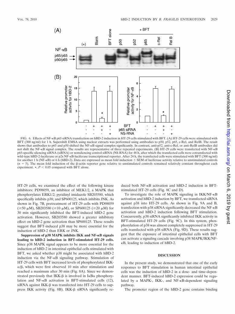
Since activation of p65/p50 heterodimeric NF-�B in re-sponse to BFT stimulation was observed (Fig. 6A), we per-formed another experiment using p65 siRNA to suppress NF-�B. In this experiment, blocking of NF-�B with p65 siRNAsignificantly decreased BFT-induced expression of hBD-2 (Fig.6B). However, the nonsilencing control siRNA (NS-RNA) hadno significant effect. These results demonstrate a direct con-nection between NF-�B-dependent signaling and hBD-2 in-duction.
MAPK is associated with hBD-2 induction in BFT-stimu-lated HT-29 cells. BFT strongly activated the phosphorylationof ERK1/2, p38, and JNK in HT-29 cells. Activation of allthree MAPK signaling molecules was first noted 5 min afterstimulation (Fig. 7A). To evaluate the relationship betweenMAPK activation and hBD-2 induction in BFT-stimulated
FIG. 4. Relationship between AP-1 signaling and hBD-2 expres-sion in BFT-stimulated HT-29 cells (A) HT-29 cells were stimulatedwith BFT (300 ng/ml) for the indicated periods of time. AP-1 activitywas assessed by EMSA. The results are representative of three re-peated experiments. (B) HT-29 cells were transfected with the pAP-1-or wild-type hBD-2-luciferase transcriptional reporter, together withthe dominant-negative c-Jun superrepressor, as indicated. Then, 48 hlater, the cells were stimulated with BFT (300 ng/ml) for another 1 h(AP-1) or 6 h (hBD-2), after which luciferase assays were performed.Data are expressed as mean fold induction in luciferase activity relativeto unstimulated controls � SEM (n 5). The mean fold induction ofthe �-actin reporter gene relative to unstimulated controls remainedrelatively constant throughout each experiment. *, P � 0.05 comparedwith BFT alone.
FIG. 5. Inhibition of NF-�B suppresses hBD-2 mRNA expressionin HT-29 cells stimulated with BFT. (A) HT-29 cells were transfectedwith either retrovirus containing I�B�-superrepressor (I�B�-AA) orcontrol virus (GFP). At 48 h after transfection, the cells were stimu-lated with BFT (300 ng/ml) for 1 h. NF-�B binding activity was assayedby EMSA. � represents the positive control in which HT-29 cells weretreated with tumor necrosis factor alpha (TNF-�) (20 ng/ml); � rep-resents the negative control. The results are representative of threerepeated experiments. (B) Cells were treated with BFT (300 ng/ml) for24 h. The concentration of hBD-2 protein in culture supernatants wasdetermined by ELISA (mean � SEM; n 5).
2028 YOON ET AL. INFECT. IMMUN.
on March 6, 2019 by guest
http://iai.asm.org/
Dow
nloaded from
HT-29 cells, we examined the effect of the following kinaseinhibitors: PD98059, an inhibitor of MEK1/2, a MAPK thatphosphorylates ERK1/2; pyridinyl imidazole SB203580, whichspecifically inhibits p38; and SP600125, which inhibits JNK. Asshown in Fig. 7B, pretreatment of HT-29 cells with PD98059(�50 M), SB203580 (�10 M), or SP600125 (�20 M) for30 min significantly inhibited the BFT-induced hBD-2 geneactivation. However, SB203580 showed a greater inhibitoryeffect on hBD-2 gene activation than SP600125. These resultssuggest that BFT-induced p38 may be more essential for theinduction of hBD-2 than ERK or JNK.
Suppression of p38 MAPK inhibits IKK and NF-�B signalsleading to hBD-2 induction in BFT-stimulated HT-29 cells.Since p38 MAPK signal appears to be more essential for theinduction of hBD-2 in intestinal epithelial cells stimulated withBFT, we asked whether p38 might be associated with hBD-2induction via the NF-�B signaling pathway. Stimulation ofHT-29 cells with BFT increased levels of phosphorylated IKK-�/�, which were first observed 10 min after stimulation andreached a maximum after 30 min (Fig. 8A). Since we demon-strated previously that IKK-� is involved in I�B� phosphory-lation and NF-�B activation in BFT-stimulated cells (12),siRNA against IKK-� was transfected into HT-29 cells to sup-press IKK activity (Fig. 8B). IKK-� siRNA significantly re-
duced both NF-�B activation and hBD-2 induction in BFT-stimulated HT-29 cells (Fig. 8C and D).
To investigate the role of MAPK signaling in IKK/NF-�Bactivation and hBD-2 induction by BFT, we transfected siRNAagainst p38 into HT-29 cells. As shown in Fig. 9A and B,transfection with p38 siRNA significantly decreased the NF-�Bactivation and hBD-2 induction following BFT stimulation.Concurrently, p38 siRNA significantly inhibited IKK activity inBFT-stimulated HT-29 cells (Fig. 9C). In this system, phos-phorylation of p38 was almost completely suppressed in HT-29cells transfected with p38 siRNA (Fig. 9D). These results sug-gest that the exposure of intestinal epithelial cells with BFTcan activate a signaling cascade involving p38 MAPK/IKK/NF-�B, leading to induction of hBD-2.
DISCUSSION
In the present study, we demonstrated that one of the earlyresponses to BFT stimulation in human intestinal epithelialcells was the induction of hBD-2 in a dose- and time-depen-dent manner. BFT-induced hBD-2 expression could be regu-lated by a MAPK-, IKK-, and NF-�B-dependent signalingpathway.
The promoter region of the hBD-2 gene contains binding
FIG. 6. Effects of NF-�B p65 siRNA transfection on hBD-2 induction in HT-29 cells stimulated with BFT. (A) HT-29 cells were stimulated withBFT (300 ng/ml) for 1 h. Supershift EMSA using nuclear extracts was performed using antibodies to p50, p52, p65, c-Rel, and RelB. The resultshows that antibodies to p65 and p50 shifted the NF-�B signal complex significantly. In contrast, anti-p52, anti-c-Rel, or anti-RelB antibodies didnot shift the NF-�B signal complex. The results are representative of three repeated experiments. (B) HT-29 cells were transfected with NF-�Bp65-specific silencing siRNA (siRNA) or nonsilencing control siRNA (NS RNA) for 48 h, after which the transfected cells were cotransfected withwild-type hBD-2-luciferase or p2x NF-�B-luciferase transcriptional reporter. After 24 h, the transfected cells were stimulated with BFT (300 ng/ml)for another 1 h (NF-�B) or 6 h (hBD-2). Data are expressed as mean fold induction � SEM of luciferase activity relative to unstimulated controls(n 5). The mean fold induction of the �-actin reporter gene relative to unstimulated controls remained relatively constant throughout eachexperiment. *, P � 0.05 compared with BFT alone.
VOL. 78, 2010 hBD-2 INDUCTION BY B. FRAGILIS ENTEROTOXIN 2029
on March 6, 2019 by guest
http://iai.asm.org/
Dow
nloaded from

sites for both transcription factors NF-�B and AP-1. Thus, theinduction of hBD-2 by cytokines or various bacteria such asSalmonella spp., pathogenic Escherichia coli, and Helicobacterpylori requires the activation of NF-�B and/or AP-1 and theirbinding to the promoter region (30, 40, 41, 43, 44). Since BFTstimulation activates NF-�B and AP-1 signaling in intestinalepithelial cells (14, 23), it is possible that BFT-mediated in-duction of hBD-2 may be associated with the binding of NF-�Band/or AP-1 to the hBD-2 promoter. The present studyshowed that mutation of NF-�B sites significantly reducedBFT-mediated hBD-2 promoter activation. The importance ofNF-�B in the signal transduction pathway leading to hBD-2gene induction by BFT was confirmed by the observation thatthe transfection of HT-29 cells with retrovirus-I�B�-AA or p65siRNA significantly downregulated the hBD-2 induction byBFT stimulation.
In contrast, a reporter with an AP-1 binding site mutationdid not show any significant change in hBD-2 promoter acti-vation in BFT-stimulated cells. The lack of involvement of
FIG. 8. Suppression of IKK activity by siRNA in HT-29 intestinalepithelial cells stimulated with BFT. (A) HT-29 cells were stimulated withBFT (300 ng/ml) for the indicated periods. Phosphorylation and proteinexpression of IKK-�, IKK-�, and actin were assessed by immunoblotanalysis. The results are representative of three repeated experiments.(B) HT-29 cells were transfected with siRNA against IKK-� for 48 h. Thetransfected cells were stimulated with BFT (300 ng/ml) for 1 h. IKKkinase activity was measured using the HTScan IKK-� kinase assay kit.Data are expressed as mean fold induction � SEM of kinase activityrelative to untreated controls (n 5). (C and D) The siRNA-transfectedcells were then cotransfected with p2x NF-�B- or wild-type hBD-2-lucif-erase reporter for another 24 h. BFT (300 ng/ml) was added to cotrans-fected cells for 1 h (NF-�B) (C) or 6 h (hBD-2) (D). Data are expressedas mean fold induction � SEM of luciferase activity relative to untreatedcontrols (n 5). The mean fold induction of the �-actin reporter generelative to untreated controls remained relatively constant throughouteach experiment. Asterisks indicate values of BFT plus siRNA that aresignificantly different from those of BFT alone (P � 0.05).
FIG. 7. BFT activates MAPKs in HT-29 cells. (A) HT-29 cells werestimulated with BFT (300 ng/ml) for the indicated periods of time.ERK1/2, p38, and JNK activities were measured by immunoblot anal-ysis. Results are representative of five independent experiments.(B) HT-29 cells were transfected with the wild-type hBD-2-luciferasetranscriptional reporter for 24 h. The transfected cells were preincu-bated with PD98059 (open circles), SB203580 (open squares), orSP600125 (filled circles) for 30 min and then stimulated with BFT (300ng/ml) for another 6 h. Data are expressed as the mean fold inductionin luciferase activity relative to unstimulated controls � SEM (n 5).*, P � 0.05 compared with BFT alone.
2030 YOON ET AL. INFECT. IMMUN.
on March 6, 2019 by guest
http://iai.asm.org/
Dow
nloaded from

AP-1 in BFT-induced hBD-2 expression was further confirmedby transfecting HT-29 cells with a dominant-negative c-Junsuperrepressor. Considering that AP-1 activation is linked toexpression of chemokine interleukin-8 (IL-8) and monocytechemoattractant protein 1 (MCP-1) in intestinal epithelial cellsexposed to BFT and may be involved in the development ofenteritis (14), AP-1 activation seems to be primarily involved inthe BFT-induced mucosal inflammation rather than hBD-2induction.
Our findings are different from those of a study in whichhBD-2 induction in human gingival epithelial cells infectedwith the periodontal bacterium Fusobacterium nucleatum wasnot blocked by NF-�B inhibitors but was mainly regulated byAP-1 (24). Both NF-�B and AP-1 are required for full hBD-2promoter activation upon stimulation of keratinocytes withPseudomonas aeruginosa (44) and of Caco-2 intestinal epithe-lial cells with pathogenic E. coli (43). Therefore, NF-�B-de-pendent and AP-1-independent hBD-2 induction seems to beunique to BFT-stimulated intestinal epithelial cells.
MAPK activation is known to be an important event under-lying hBD-2 induction. Although BFT activates MAPK andNF-�B signaling in intestinal epithelial cells (12, 14, 19, 20, 21,23, 45), the relationship between NF-�B and MAPK signalingin intestinal epithelial cells stimulated with BFT is unclear. Togain insight into BFT-induced signaling pathways involved inhBD-2 induction, we attempted to determine whether NF-�Band MAPK signaling might cooperate to induce hBD-2 expres-sion in intestinal epithelial cells. In the present study, the p38inhibitor SB203580 showed a greater inhibitory effect onhBD-2 induction than the ERK inhibitor PD98059 or the JNKinhibitor SP600125. These results suggest that p38 MAPK ismore essential for hBD-2 induction in BFT-stimulated intes-tinal epithelial cells than ERK or JNK. Since several reportshave demonstrated that p38 MAPK may be associated withIKK-dependent NF-�B activation (5, 25, 31), we assessed theeffects of p38 activation on IKK and NF-�B signaling in BFT-stimulated cells. Our study showed that suppression of p38activity in BFT-stimulated HT-29 cells significantly reducedphospho-IKK-�/� activity, NF-�B activity, and hBD-2 expres-sion, suggesting that the activated p38 molecule may act up-stream of IKK and NF-�B in BFT-induced hBD-2 expression.
In summary, we have demonstrated that exposure of intes-tinal epithelial cells to BFT results in the activation of a sig-naling cascade involving p38 MAPK, IKK, NF-�B, and subse-quent hBD-2 induction in intestinal epithelial cells. Based onthese findings, we propose that the induction of hBD-2 seemsto enhance the innate epithelial defense against ETBF infec-tion. If dysregulation of this cascade occurs, it may lead toinsufficient expression of hBD-2 in intestine, thereby increas-ing the chances of ETBF-associated diseases such as colitis,IBD, and colon tumorigenesis.
ACKNOWLEDGMENTS
We thank Jurgen Harder for providing hBD-2-luciferase plasmids,Martin F. Kagnoff for providing p�-actin- and pRSV-�-galactosidase-luciferase plasmids and standard �-actin RNA, Andreas von Knethenfor providing the dominant-negative c-Jun plasmid, and Han Jin Leefor expert technical help.
This work was supported by a National Research Foundation ofKorea (NRF) grant funded by the government of Korea (MEST)(MRC program no. 2009-0091463) and by the Basic Science Research
FIG. 9. Transfection with siRNA against p38 inhibits the activationof IKK and NF-�B and the expression of hBD-2 in HT-29 cells stim-ulated with BFT. (A and B) The siRNA-transfected cells were cotrans-fected with p2x NF-�B- or wild-type hBD-2-luciferase reporter foranother 24 h. BFT (300 ng/ml) was added to cotransfected cells for 1 h(NF-�B) (A) or 6 h (hBD-2) (B). Data are expressed as mean foldinduction � SEM of luciferase activity relative to untreated controls(n 5). The mean fold induction of the �-actin reporter gene relativeto untreated controls remained relatively constant throughout eachexperiment. Asterisks indicate values of BFT plus siRNA that aresignificantly different from those of BFT alone (P � 0.05). (C) BFT(300 ng/ml) was added to the siRNA-transfected HT-29 cells for theindicated period. IKK kinase activity was measured using an HTScanIKK-� kinase assay kit. Data are expressed as mean fold induction �SEM of kinase activity relative to untreated controls (n 5). Asterisksindicate values of BFT plus siRNA that are significantly different fromthose of BFT alone (P � 0.05). (D) The siRNA-transfected cells werecombined with BFT (300 ng/ml) for 30 min. Cell lysates were analyzedby immunoblotting with the indicated antibodies. Results shown arerepresentative of three independent experiments. Lanes: 1, unstimu-lated control in nontransfected cells; 2, BFT in nontransfected cells; 3,BFT in p38 siRNA-transfected cells; 4, BFT in NS-RNA-transfectedcells.
VOL. 78, 2010 hBD-2 INDUCTION BY B. FRAGILIS ENTEROTOXIN 2031
on March 6, 2019 by guest
http://iai.asm.org/
Dow
nloaded from

Program through the NRF funded by the Ministry of Education, Sci-ence and Technology (R11-2008-044-01004-0).
None of the authors of this study has any financial or commercialconflicts of interest.
REFERENCES
1. Bals, R., X. Wang, R. L. Meegalla, S. Wattler, D. J. Weiner, M. C. Nehls, andJ. M. Wilson. 1999. Mouse beta-defensin 3 is an inducible antimicrobialpeptide expressed in the epithelia of multiple organs. Infect. Immun. 67:3542–3547.
2. Basset, C., J. Holton, A. Bazeos, D. Vaira, and S. Bloom. 2004. Are Helico-bacter species and enterotoxigenic Bacteroides fragilis involved in inflamma-tory bowel disease? Dig. Dis. Sci. 49:1425–1432.
3. Brown, P. H., R. Alani, L. H. Preis, E. Szabo, and M. J. Birrer. 1993.Suppression of oncogene-induced transformation by a deletion mutant ofc-jun. Oncogene 8:877–886.
4. Cash, H. L., C. V. Whitham, C. L. Behrendt, and L. V. Hooper. 2006.Symbiotic bacteria direct expression of an intestinal bactericidal lectin. Sci-ence 313:1126–1130.
5. Chio, C. C., Y. H. Chang, Y. W. Hsu, K. H. Chi, and W. W. Lin. 2004.PKA-dependent activation of PKC, p38 MAPK and IKK in macrophage:implication in the induction of inducible nitric oxide synthase and interleu-kin-6 by dibutyryl cAMP. Cell. Signal. 16:565–575.
6. De Smet, K., and R. Contreras. 2005. Human antimicrobial peptides: de-fensins, cathelicidins and histatins. Biotechnol. Lett. 27:1337–1347.
7. Eckmann, L., J. Fierer, and M. F. Kagnoff. 1996. Genetically resistant (Ityr)and susceptible (Itys) congenic mouse strains show similar cytokine re-sponses following infection with Salmonella dublin. J. Immunol. 156:2894–2900.
8. Elewaut, D., J. A. DiDonato, J. M. Kim, F. Truong, L. Eckmann, and M. F.Kagnoff. 1999. NF-kappa B is a central regulator of the intestinal epithelialcell innate immune response induced by infection with enteroinvasive bac-teria. J. Immunol. 163:1457–1466.
9. Giesemann, T., G. Guttenberg, and K. Aktories. 2008. Human alpha-de-fensins inhibit Clostridium difficile toxin B. Gastroenterology 134:2049–2058.
10. Hale, J. D., and R. E. Hancock. 2007. Alternative mechanisms of action ofcationic antimicrobial peptides on bacteria. Expert Rev. Anti Infect. Ther.5:951–959.
11. Howell, S. J., D. Wilk, S. P. Yadav, and C. L. Bevins. 2003. Antimicrobialpolypeptides of the human colonic epithelium. Peptides 24:1763–1770.
12. Kim, J. M., D. H. Lee, J. S. Kim, J. Y. Lee, H. G. Park, Y. J. Kim, Y. K. Oh,H. C. Jung, and S. Kim. 2009. 5,7-Dihydroxy-3,4,6-trimethoxyflavone inhibitsthe inflammatory effects induced by Bacteroides fragilis enterotoxin via dis-sociating the complex of heat shock protein 90 and I�B� and I�B kinase-in intestinal epithelial cell culture. Clin. Exp. Immunol. 155:541–551.
13. Kim, J. M., H. C. Jung, D. J. Jin, K. I. Im, I. S. Song, and C. Y. Kim. 1997.Cytokine genes are downregulated when attachment of Entamoeba histo-lytica to HT-29 colon epithelial cells is prevented. Scand. J. Immunol. 45:613–617.
14. Kim, J. M., H. Y. Jung, J. Y. Lee, J. Youn, C. H. Lee, and K. H. Kim. 2005.Mitogen-activated protein kinase and activator protein-1 dependent signalsare essential for Bacteroides fragilis enterotoxin-induced enteritis. Eur. J. Im-munol. 35:2648–2657.
15. Kim, J. M., J. S. Kim, H. C. Jung, Y. K. Oh, H. Y. Chung, C. H. Lee, and I. S.Song. 2003. Helicobacter pylori infection activates NF-kappaB signaling path-way to induce iNOS and protect human gastric epithelial cells from apop-tosis. Am. J. Physiol. Gastrointest. Liver Physiol. 285:G1171–G1180.
16. Kim, J. M., J. S. Kim, J. Y. Lee, Y. J. Kim, H. J. Youn, I. Y. Kim, Y. J. Chee,Y. K. Oh, N. Kim, H. C. Jung, and I. S. Song. 2007. Vacuolating cytotoxin inHelicobacter pylori water-soluble proteins upregulates chemokine expressionin human eosinophils via Ca2� influx, mitochondrial reactive oxygen inter-mediates, and NF-�B activation. Infect. Immun. 75:3373–3381.
17. Kim, J. M., J. S. Kim, Y. J. Kim, Y. K. Oh, I. Y. Kim, Y. J. Chee, J. S. Han,and H. C. Jung. 2008. Conjugated linoleic acids produced by Lactobacillusdissociates IKK-gamma and Hsp90 complex in Helicobacter pylori-infectedgastric epithelial cells. Lab. Invest. 88:541–552.
18. Kim, J. M., J. W. Kang, M. Y. Cha, D. Yoo, N. Kim, I. K. Kim, J. Ku, S. Kim,H. S. Ma, H. C. Jung, I. S. Song, and J. S. Kim. 1 March 2010, posting date.Novel guggulsterone derivative GG-52 inhibits NF-�B signaling in intestinalepithelial cells and attenuates acute murine colitis. Lab. Invest. [Epub aheadof print.]
19. Kim, J. M., J. Y. Lee, and Y. J. Kim. 2008. Inhibition of apoptosis inBacteroides fragilis enterotoxin-stimulated intestinal epithelial cells throughthe induction of c-IAP-2. Eur. J. Immunol. 38:2190–2199.
20. Kim, J. M., J. Y. Lee, Y. M. Yoon, Y. K. Oh, J. S. Kang, Y. J. Kim, and K. H.Kim. 2006. Bacteroides fragilis enterotoxin induces cyclooxygenase-2 andfluid secretion in intestinal epithelial cells through NF-�B activation. Eur.J. Immunol. 36:2446–2456.
21. Kim, J. M., S. J. Cho, Y. K. Oh, H. Y. Jung, Y. J. Kim, and N. Kim. 2002.Nuclear factor-kappa B activation pathway in intestinal epithelial cells is a
major regulator of chemokine gene expression and neutrophil migrationinduced by Bacteroides fragilis enterotoxin. Clin. Exp. Immunol. 130:59–66.
22. Kim, J. M., Y. J. Kim, and Y. J. Cho. 2000. Synergy of Bacteroides fragilis andEscherichia coli in the induction of KC gene expression in mouse peritonealtissues. Scand. J. Infect. Dis. 32:643–649.
23. Kim, J. M., Y. K. Oh, Y. J. Kim, H. B. Oh, and Y. J. Cho. 2001. Polarizedsecretion of CXC chemokines by human intestinal epithelial cells in responseto Bacteroides fragilis enterotoxin: NF-kappaB plays a major role in theregulation of IL-8 expression. Clin. Exp. Immunol. 123:421–427.
24. Krisanaprakornkit, S., J. R. Kimball, and B. A. Dale. 2002. Regulation ofhuman beta-defensin-2 in gingival epithelial cells: the involvement of mito-gen-activated protein kinase pathways, but not the NF-kappaB transcriptionfactor family. J. Immunol. 168:316–324.
25. Lee, J. Y., H. Kim, M. Y. Cha, H. G. Park, Y. J. Kim, I. Y. Kim, and J. M.Kim. 2009. Clostridium difficile toxin A promotes dendritic cell maturationand chemokine CXCL2 expression through p38, IKK, and the NF-kappaBsignaling pathway. J. Mol. Med. 87:169–180.
26. Lee, J. Y., H. R. Park, Y. K. Oh, Y. J. Kim, J. Youn, J. S. Han, and J. M. Kim.2007. Effects of transcription factor activator protein-1 on interleukin-8expression and enteritis in response to Clostridium difficile toxin A. J. Mol.Med. 85:1393–1404.
27. Myers, L. L., and D. S. Shoop. 1987. Association of enterotoxigenic Bac-teroides fragilis with diarrheal disease in young pigs. Am. J. Vet. Res. 48:774–775.
28. Niyogi, S. K., P. Dutta, U. Mitra, and D. K. Pal. 1997. Association ofenterotoxigenic Bacteroides fragilis with childhood diarrhoea. Indian J. Med.Res. 105:167–169.
29. Nuding, S., L. T. Zabel, C. Enders, E. Porter, K. Fellermann, J. Wehkamp,H. A. Mueller, and E. F. Stange. 2009. Antibacterial activity of humandefensins on anaerobic intestinal bacterial species: a major role of HBD-3.Microbes Infect. 11:384–393.
30. O’Neil, D. A., E. M. Porter, D. Elewaut, G. M. Anderson, L. Eckmann, T.Ganz, and M. F. Kagnoff. 1999. Expression and regulation of the humanbeta-defensins hBD-1 and hBD-2 in intestinal epithelium. J. Immunol. 163:6718–6724.
31. Park, K. J., R. B. Gaynor, and Y. T. Kwak. 2003. Heat shock protein 27association with the I�B kinase complex regulates tumor necrosis factoralpha-induced NF-�B activation. J. Biol. Chem. 278:35272–35278.
32. Prindiville, T. P., R. A. Sheikh, S. H. Cohen, Y. J. Tang, M. C. Cantrell, andJ. Silva, Jr. 2000. Bacteroides fragilis enterotoxin gene sequences in patientswith inflammatory bowel disease. Emerg. Infect. Dis. 6:171–174.
33. Rhee, K. J., S. Wu, X. Wu, D. L. Huso, B. Karim, A. A. Franco, S. Rabizadeh,J. E. Golub, L. E. Mathews, J. Shin, R. B. Sartor, D. Golenbock, A. R.Hamad, C. M. Gan, F. Housseau, and C. L. Sears. 2009. Induction ofpersistent colitis by a human commensal, enterotoxigenic Bacteroides fragilis,in wild-type C57BL/6 mice. Infect. Immun. 77:1708–1718.
34. Sahl, H. G., U. Pag, S. Bonness, S. Wagner, N. Antcheva, and A. Toss. 2005.Mammalian defensins: structures and mechanism of antibiotic activity.J. Leukoc. Biol. 77:466–475.
35. Schneider, J. J., A. Unholzer, M. Schaller, M. Schafer-Korting, and H. C.Korting. 2005. Human defensins. J. Mol. Med. 83:587–595.
36. Sears, C. L. 2009. Enterotoxigenic Bacteroides fragilis: a rogue among sym-biotes. Clin. Microbiol. Rev. 22:349–369.
37. Sears, C. L., S. Islam, A. Saha, M. Arjumand, N. H. Alam, A. S. Faruque,M. A. Salam, J. Shin, D. Hecht, A. Weintraub, R. B. Sack, and F. Qadri.2008. Association of enterotoxigenic Bacteroides fragilis infection with in-flammatory diarrhea. Clin. Infect. Dis. 47:797–803.
38. Tollin, M., P. Bergman, T. Svenberg, H. Jornvall, G. H. Gudmundsson, andB. Agerberth. 2003. Antimicrobial peptides in the first line defence of humancolon mucosa. Peptides 24:523–530.
39. Toprak, N. U., A. Yagci, B. M. Gulluoglu, M. L. Akin, P. Demirkalem, T.Celenk, and G. A. Soyletir. 2006. Possible role of Bacteroides fragilis entero-toxin in the aetiology of colorectal cancer. Clin. Microbiol. Infect. 12:782–786.
40. Wada, A., K. Ogushi, T. Kimura, H. Hojo, N. Mori, S. Suzuki, A. Kumatori,M. Se, Y. Nakahara, M. Nakamura, J. Moss, and T. Hirayama. 2001. Heli-cobacter pylori-mediated transcriptional regulation of the human beta-defen-sin 2 gene requires NF-kappaB. Cell. Microbiol. 3:115–123.
41. Wada, A., N. Mori, K. Oishi, H. Hojo, Y. Nakahara, Y. Hamanaka, M.Nagashima, I. Sekine, K. Ogushi, T. Niidome, T. Nagatake, J. Moss, and T.Hirayama. 1999. Induction of human beta-defensin-2 mRNA expression byHelicobacter pylori in human gastric cell line MKN45 cells on cag pathoge-nicity island. Biochem. Biophys. Res. Commun. 263:770–774.
42. Wehkamp, J., J. Schauber, and E. F. Stange. 2007. Defensins and cathelici-dins in gastrointestinal infections. Curr. Opin. Gastroenterol. 23:32–38.
43. Wehkamp, J., J. Harder, K. Wehkamp, B. Wehkamp-von Meissner, M.Schlee, C. Enders, U. Sonnenborn, S. Nuding, S. Bengmark, K. Fellermann,J. M. Schroder, and E. F. Stange. 2004. NF-kappaB- and AP-1-mediatedinduction of human beta defensin-2 in intestinal epithelial cells by Esche-richia coli Nissle 1917: a novel effect of a probiotic bacterium. Infect. Immun.72:5750–5758.
44. Wehkamp, K., L. Schwichtenberg, J. M. Schroder, and J. Harder. 2006.
2032 YOON ET AL. INFECT. IMMUN.
on March 6, 2019 by guest
http://iai.asm.org/
Dow
nloaded from

Pseudomonas aeruginosa- and IL-1�-mediated induction of human beta-defensin-2 in keratinocytes is controlled by NF-kappaB and AP-1. J. Invest.Dermatol. 126:121–127.
45. Wu, S., J. Powell, N. Mathioudakis, S. Kane, E. Fernandez, and C. L. Sears.2004. Bacteroides fragilis enterotoxin induces intestinal epithelial cell secre-tion of interleukin-8 through mitogen-activated protein kinases and a ty-rosine kinase-regulated nuclear factor-�B pathway. Infect. Immun. 72:5832–5839.
46. Wu, S., K. J. Rhee, E. Albesiano, S. Rabizadeh, X. Wu, H. R. Yen, D. L.Huso, F. L. Brancati, E. Wick, F. McAllister, F. Housseau, D. M. Pardoll,and C. L. Sears. 2009. A human colonic commensal promotes colontumorigenesis via activation of T helper type 17 T cell responses. Nat.Med. 15:1016–1022.
47. Zhang, G., B. Svenungsson, A. Karnell, and A. Weintraub. 1999. Prevalenceof enterotoxigenic Bacteroides fragilis in adult patients with diarrhea andhealthy controls. Clin. Infect. Dis. 29:590–594.
Editor: S. R. Blanke
VOL. 78, 2010 hBD-2 INDUCTION BY B. FRAGILIS ENTEROTOXIN 2033
on March 6, 2019 by guest
http://iai.asm.org/
Dow
nloaded from