based registry study. acute stroke patients.a cross
TRANSCRIPT

Page 1/25
Adjustment of oral diet based on �exibleendoscopic evaluation of swallowing (FEES) inacute stroke patients.A cross-sectional hospital-based registry study.Tobias Braun ( [email protected] )
Universitaetsklinikum Gießen und Marburg - Standort Gießen https://orcid.org/0000-0003-1738-2773Martin Juenemann
Universitaetsklinikum Giessen and Marburg, Standort Giessen; Department of NeurologyMaxime Viard
Universitaetsklinikum Giessen and Marburg, Standort Giessen; Department of NeurologyMarco Meyer
Department of Neurology/Geriartrics, Diakonie Klinikum Jung-Stilling SiegenIris Reuter
Universitaetsklinikum Giessen and Marburg, Standort Giessen; Department of NeurologyMario Prosiegel
Lecturer at Faculty of Languages and Literatures, Department I, Ludwig-Maximilians-University (LMU),MunichManfred Kaps
Universitaetsklinikum Giessen und Marburg, Standort Giessen; Department of NeurologyChristian Tanislav
Department of Neurology/Geriatrics, Diakonie Klinikum Jung-Stilling Siegen
Research article
Keywords: Stroke, FEES, Dysphagia, Aspiration, Management of Dysphagia, Deglutition, DeglutitionDisorders
Posted Date: April 23rd, 2019
DOI: https://doi.org/10.21203/rs.2.9300/v1
License: This work is licensed under a Creative Commons Attribution 4.0 International License. Read Full License

Page 2/25
Version of Record: A version of this preprint was published on November 12th, 2019. See the publishedversion at https://doi.org/10.1186/s12883-019-1499-8.

Page 3/25
AbstractBackground Diagnosing dysphagia in acute stroke patients is crucial, as this comorbidity determines theoutcome; we therefore investigated the impact of �exible nasolaryngeal endoscopy (FEES) in acute strokepatients. Methods The FEES investigation as performed in acute stroke patients treated at a largeuniversity hospital, allocated as a standard procedure for all patients suspected of dysphagia. Wecorrelated our �ndings with baseline data, disability status, pneumonia, duration of hospitalisation,necessity for mechanical ventilation and treatment on the intensive care unit. Results We investigated152 patients (173 FEES). The median age was 73; 94 were male. Ischemic stroke was diagnosed in 125patients (82.2%); 27 (17.8%) suffered intracerebral haemorrhage. Dysphagia was diagnosed in 72.4% ofthe patients, and was associated with higher stroke severity on admission (median NIHSS 11 [IQR 6-17]vs. 7 [4-12], p=.013; median mRS 5 [IQR 4-5] vs. 4 [IQR 3-5], p=.012). Short-term mortality was higheramong patients diagnosed with dysphagia (7.2% vs. 0%, p=.107). FEES examinations revealed that only30.9% of the patients had an adequate oral diet. A change of oral diet was associated with a betteroutcome at discharge (mRS; p=.006), less need of mechanical ventilation (p=.028), shorter period ofhospitalisation (p=.044), and lower rates of pneumonia (p=.007) and mortality (p=.011). Discussion Dueto the inability of clinical assessments to detect silent aspiration, FEES might be better suited to identifystroke patients at risk and may contribute to a better functional outcome and lower rates of pneumoniaand mortality. Our �ndings also point to a low awareness of dysphagia, even in a specialised strokecentre. FEES in acute stroke patients helps to adequately adjust the oral diet for the vast majority ofstroke patients (69.1%), potentially avoiding severe complications.
BackgroundDysphagia occurs in the course of many neurological diseases and frequently determines the outcome [1]with stroke being the most common cause. Up to 80% of stroke patients suffer from dysphagia,depending on the choice of diagnostics tests used (screening tests, comprehensive swallowingassessment by SLT and instrumented methods, such as VFS or FEES) [2]. Pneumonia due to dysphagiais the leading cause of death in stroke patients and may cause fever [3]; hyperthermia is known to beassociated with a worse functional outcome in stroke [4]. Penetration and aspiration increase the risk forpneumonia up to 11.5-fold in stroke patients [2]. Another factor associated with a worse outcome is newor pre-existing malnutrition, which can be caused by dysphagia [5]. Thus, one of the positive effects ofstroke unit therapy seems to be an early diagnosis of dysphagia and the improvement of the swallowingfunction, thereby preventing pneumonia, malnutrition and dehydration.
Dysphagia might lead to further complications (malnutrition and dehydration) and its resultanthealthcare costs underline its socioeconomic relevance [6, 7]. Moreover, dysphagia is an independentpredictor of disability and poor outcome, increased mortality, morbidity and markedly reduced quality oflife in stroke [8–11].

Page 4/25
Diagnostic tools for dysphagia include a screening examination, comprehensive swallowing examination(CSE) performed by physicians or speech and language therapists (SLT) as well as instrumentedmethods, such as video�uoroscopy of swallowing (VFS) or �exible endoscopic evaluation of swallowing(FEES). FEES combines many advantages in the clinical routine, as it is a bedside procedure withoutradiation exposure; enables the evaluation of saliva handling; it may be performed in uncooperative orunconscious patients; and it can easily be repeated. Moreover, swallowing is assessed by FEES in a more“natural” way than VFS, as the latter requires a contrast media and cued swallowing (to reduce radiationdosage). In this context, FEES might be an important tool for identifying patients at risk and mayultimately help improve functional outcome by adjusting patients’ oral diet. We recently published a paperon the use of FEES and adjusting the oral diet in neurological patients. In this analysis, we were able todemonstrate a lower rate of pneumonia an a lower mortality, when adjusting the oral diet [12]. This is asubgroup analysis for acute stroke patients. As mentioned above, dysphagia is very common in strokepatients, puts those patients at a high risk of complications, increases mortality and leads to a longerlength of hospitalisation. Furthermore, outcome parameters, such as the National Institute of HealthStroke Scale (NIHSS) and the modi�ed Rankin-scale (mRS) are routinely assessed in all stroke patients.Therefore, the aim of the current study was to analyse the impact of FEES and adjustment of the oral dietbased on those �ndings in the management of acute stroke patients.
MethodsThe study was done in a large German university hospital. As a part of routine care delivery for patientshospitalised for acute stroke, FEES was performed in case of a pathologic bedside screening procedure,performed by nurses or SLTs. In our department, we use the Gugging Swallowing Screen (GUSS) [13]. Ifthe patient passed the GUSS, no FEES was performed and full oral diet was chosen. If the GUSS indicatedpossible dysphagia, the patient underwent a CSE by an SLT and FEES by a team consisting of a SLT anda neurologist. FEES was also performed if a patient showed signs of pharyngeal dysphagia duringhospitalisation (e.g. wet voice, coughing when drinking, etc.) and if a patient developed signs of infection(productive cough, elevated in�ammatory markers). The signs of dysphagia were reported by nurses,SLTs or the treating physicians. The patients with signs of dysphagia were discussed among the“dysphagia experts” of our department and indication for FEES was con�rmed; oral diet prior to FEES waschosen as instructed by the GUSS or by clinical judgement of the treating physician. For quality controlreasons, our �ndings gathered from examinations were documented systematically. All FEES wereperformed in a standardised manner by experienced physicians.
Patients
All stroke patients treated in our department from January 2014 to September 2016 in whom FEES wasperformed were documented in a standardised manner. Approximately 800 patients per year aredischarged from our hospital with the diagnosis of a stroke. Data documented in the database includedage, sex, length of stay in hospital, stroke entity, stroke aetiology (TOAST-criteria in ischemic stroke),NIHSS and mRS on admission and at discharge, localisation of ischemic lesion (left/right hemisphere,

Page 5/25
bilateral infarctions, brain stem), ischemic vascular territory, presence of risk factors in ischemic stroke,occurrence of pneumonia during hospitalisation (determined by the treating physician due to clinicalsigns of pneumonia, elevated in�ammatory markers in the blood and chest X-ray), treatment on intensivecare unit, necessity of intubation and mechanical ventilation lasting longer than 24 hours (excluded fromthis item were patients intubated for surgery, such as decompressive craniotomy in cerebellar infarctionor those who were preclinically intubated and promptly extubated), mortality, presence of dysphagia andtype of oral intake (before and after FEES). For acquisition and use of data for scienti�c analyses, ethicalapproval was obtained from the local ethical committee (protocol number 208/16). All patientsdocumented in the database were selected for the current analysis.
FEES
FEES is a videoendoscopic nasolaryngeal swallowing study. We performed FEES following astandardised FEES® protocol, according to Langmore [14]: after applying decongestants (Xylometazolin)and local anaesthesia of the nasal duct using 2% Lidocaine-gel, a small endoscope (about 4 mm indiameter) was introduced through the inferior nasal meatus and the nasopharynx in the oropharynx. Theswallowing of saliva and different consistencies of food and liquids and penetration, aspiration,localisation and extent of residues, as well as patients’ reactions (such as coughing), were visualised anddocumented. By de�nition, penetration is entering of any material into the airway above the level of thevocal folds, and aspiration is entering of any material below the level of the vocal folds [15]. In the �rststep of the procedure, anatomical changes, handling of saliva and the movement of swallowing-relatedstructures were tested, then pudding-thick consistency (thickened water), normal water and solid foodwere introduced. All consistencies were applied three times. If a consistence appeared unsafe to test, weskipped it; the consistence was rated as unsafe if it entered the airway to the level of the vocal foldswithout ejection from the airway or any aspiration (score 5-8 on Rosenbek’s Penetration-Aspiration-Scale[16]). In the context of this research manuscript, we de�ned a “relevant dysphagia” as a pharyngeal stagedysphagia with a score of 3-8 on Rosenbek’s Penetration-Aspiration-Scale, as this exposes the patient tothe risk of pneumonia. Using the �ndings in FEES, the appropriate oral diet was chosen for the patient.Based on the pathophysiology found in FEES, compensatory and rehabilitative measures to treatdysphagia were carried out as described by Daniels and co-workers [17]. All FEES procedures wereperformed or supervised by an experienced investigator and lasted about 10 minutes each.
Outcome measurements
Oral intake and dysphagia severity were measured by use of the functional oral intake scale (FOIS) andthe Fiberoptic Endoscopic Dysphagia Severity Score (FEDSS), respectively:
FOIS is a seven-tiered scale ranging from 1 = no oral intake at all (NPO= nil per os) to 7 = full oral intakewithout restrictions (Appendix 1)[18]. For easier readability, the data of the functional oral intake scalewere categorised in either NPO (FOIS=1), partial oral intake (FOIS =2-6) or full oral intake (FOIS=7).Restriction of the oral diet was de�ned as a negative change on the FOIS, whereas lowering of restrictionsof the oral diet was de�ned as a positive change. FOIS was documented prior to and after FEES.

Page 6/25
There is no standardised way of de�ning the overall severity of dysphagia. In our department, we use theFEDSS-scale developed by Dziewas and co-workers[19]. The FEDSS is a six-tiered scale originallydesigned for use in stroke patients (Appendix 2). All parameters were recorded in a standardised way.
For evaluating the value of performing FEES in neurological patients, the following parameters werecorrelated with baseline data and dependent factors:
· Dysphagia as de�ned by a FEDSS score of ≥ 2
· The oral intake status as calculated by the FOIS and its overall change and type of change after FEES
Statistical analyses
Absolute and relative frequencies were calculated based on cross-tables. For comparing relativefrequencies, we used a two-tailed Fisher’s exact test. Continuous variables were analysed by calculatingthe median value and the interquartile range (IQR; 25% percentile and 75% percentile). Nonparametricnon-paired data were analysed using the Mann–Whitney U-test and paired data using the Wilcoxon-test.Binary logistic regression analysis was used to identify factors associated with the item “change in oraldiet”. All statistical analyses were performed using SPSS, release version 22.0 (SPSS©, Inc., IBMCompany, 2015, Chicago-IL).
ResultsPatients’ characteristics
173 FEES were performed in 152 stroke patients. In 19 (12.5%) patients, the procedure was repeated atleast once and their data were only analysed once. In order to prevent data distortion, only the results ofthe �rst examination were included in the analysis of patients who received more than one FEES. 94patients (61.8%) were male and the overall median age was 73 years (IQR 61.25-81 years). 119 patientswere older than 60 years (78.3%). 125 patients (82.2%) were diagnosed with ischemic stroke and 27(17.8%) with primary haemorrhage. 61 patients (48.8%) were treated on the intensive care unit. 62patients (40.8%, or 26.8% when excluding intensive care patients) were diagnosed with pneumonia and 8patients (5.3%) died during hospitalisation. Initially, 76 patients (49%) had no oral intake (NPO), 57patients (37.5%) partial oral intake and 19 patients (12.3%) full oral intake. Among the patients with NPOand partial oral intake, 65 patients (42.8%) had a nasogastric feeding tube prior to FEES and 2 patients(1.3%) had a PEG-tube. Patients’ characteristics are presented in Table 1 (all stroke patients) and Table 2(ischemic stroke patients only).
FEES Examination
No side effects occurred, such as laryngospasm, syncope or epistaxis.

Page 7/25
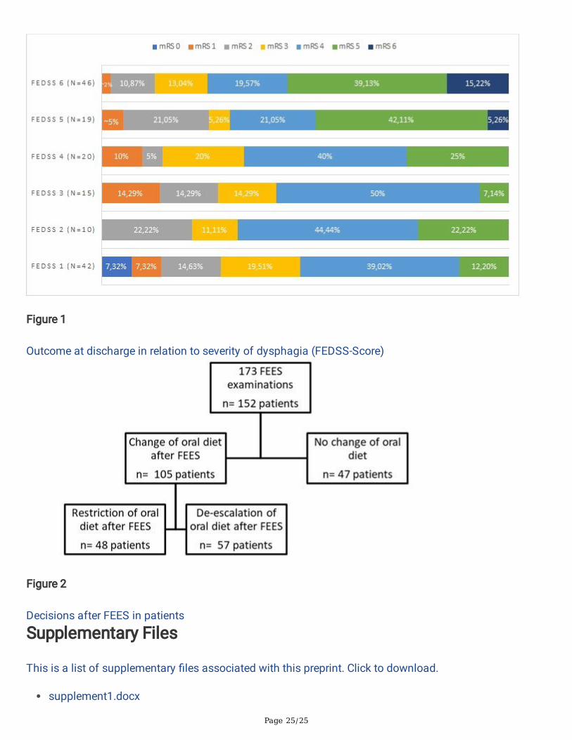
The median FEDSS in the entire study population was 4 (IQR 1-6) and the median time from admission to�rst FEES was 6 days (IQR 3-11days). FEES identi�ed 110 (72.4%) patients with dysphagia (FEDSS 2-6).A diet modi�cation was indicated in 105 patients (69.1%) with restriction of oral diet in 48 patients(31.6%) and lowering of restrictions in 57 (37.5%). NPO was indicated for the majority of patients (76.6%in this subgroup) without change in oral diet. 8 patients (5.3%) died during hospitalisation; all of themsuffered from dysphagia. NIHSS and mRS on admission and at discharge were higher in dysphagicpatients than non-dysphagic patients (admission: median NIHSS 11 [IQR 6-17] vs 7 [4-12], p=.013; medianmRS 5 [IQR 4-5] vs. 4 [IQR 3-5], p=.012; discharge: median NIHSS 7 [IQR 4-12] vs 6 [3-11], p=.05; medianmRS 4 [IQR 3-5] vs. 4 [IQR 2-4], p=.002). The outcome at discharge (mRS) in relation to the FEDSS issummarized in Figure 1. The same held true for the subgroup of ischemic stroke patients (admission:median NIHSS 11 [IQR 6-17] vs 6.5 [4-12], p=.017; median mRS 4 [IQR 4-5] vs. 4 [IQR 3-5], p=.017;discharge: median NIHSS 7 [IQR 4-13] vs 3 [1-7.5], p<.001; median mRS 4 [IQR 3-5] vs. 3 [IQR 2-4], p=.001).In ischemic stroke, dysphagia was more often diagnosed in right-hemispheric lesions (45.1% vs. 26.5%,p=.066) and was less frequently diagnosed in diabetic patients (19.8% vs. 41.2%, p=.021). Results aresummarized in Table 1 (all stroke patients) Table 2 (ischemic stroke patients only).
Differences in patients with and without change in oral diet
For patients that needed diet changes, the length of stay was shorter (median 16 days [IQR 11-25] vs. 22days [IQR 13-30], p=.027), intubation and mechanical ventilation were less frequently indicated (15.2 vs.31.9%, p=.028) and pneumonia as well as mortality rates were lower (pneumonia; 33.3% vs. 57.4%,p=.007; mortality: 1.9% vs 12.8%, p=.011). At discharge, mRS was lower in patients with diet changes(median 4 [IQR 3-4] vs. 4.5 [3-5], p=.006). A comparison of the intraindividual difference of mRS onadmission and at discharge revealed a better functional outcome in patients with a change in oral diet(p= .001); we observed no better outcome in patients without a change in oral diet (p= .583). In thesubgroup of ischemic strokes, mRS on admission and at discharge was lower in patients with a changein oral diet (admission: median 4 [IQR 3-5] vs. 5 [4-5], p=.048 discharge: median 4 [IQR 2-4] vs. 4 [3-5],p=.034). When comparing the intraindividual difference of mRS on admission with mRS at discharge inischemic stroke patients, the functional outcome was better in patients with a change in oral diet (p=.001); this effect could not be observed in ischemic stroke patients without change in oral diet (p= .151).Results are summarised in Table 3 (all stroke patients) and Table 4 (ischemic stroke patients only).
Binominal logistic regression analysis was signi�cant for the model (chi-square = 16.89, p= .002), and forpneumonia (p=.011) and mRS at discharge (p=.044) in the whole cohort. In case of a change in the oraldiet, the odds-ratio for pneumonia was 0.38 and 0.735 for mRS. In the subgroup of ischemic strokepatients, the analysis was signi�cant for the model (chi-square = 10.534, p= .032) and for pneumonia(p=.025), and there was a trend for mRS at discharge (p=.074). Odds-ratio for pneumonia was 0.399when patients’ oral diet was changed. Results are summarised in Table 5 (all stroke patients) and Table 6(ischemic stroke patients only).
Discussion

Page 8/25
In 72% of our stroke patients, FEES unveiled a relevant dysphagia, leading to an adjustment of oral diet.In those patients, we observed a better functional outcome at discharge and fewer complications, such asthe need for mechanical ventilation, a lower mortality rate and a lower rate of pneumonia.The most alarming result is that only 30.9% of our patients had an adequate oral diet prior to FEES,meaning more than two thirds of our patients needed adjustment of their oral diet. This demonstrates lowawareness of dysphagia and emphasises the need for instrumental diagnostics with a low threshold,proving that extensive clinical expertise avoids signi�cant complications. Screening and CSE arenecessary, but unable to detect all kinds of relevant swallowing disturbances, especially silent aspiration.Our results underline the necessity of performing FEES at a low threshold in the majority of strokepatients, irrespective of clinical examination and screening tests. This would be in accordance withintentions to change national guidelines as suggested by Lindner-P�eghar and co-workers[20]. FEES is asafe, fast and reliable tool, as we observed no side effects in about 1,730 minutes of examination.
When adjusting oral diet based on our �ndings in FEES, we observed in our patients a better outcome, areduced need of intubation and mechanical ventilation, a lower pneumonia rate, lower mortality and ashorter period of hospitalisation. In a meta-analysis, Steele and co-workers reported a signi�cantreduction in penetration and aspiration when thickening �uids [21]; this might be one factor explainingour results. One other factor contributing to a better outcome might be the increased mobility of patientsafter removal of a nasogastric feeding tube or an intravenous canulae for parenteral feeding, heighteningthe effects of physiotherapy. In case of a change in oral diet, the risk of pneumonia was reduced and thepatients’ chance of a better outcome increased. Our �ndings underline the value of FEES in �nding theadequate oral diet for stroke patients.
Dysphagic patients had a higher NIHSS and mRS on admission and at discharge. This is in agreementwith results by Dziewas and co-workers, who showed that patients with a NIHSS >3 had signs ofpenetration and aspiration [19]. Warnecke and co-workers showed that the degree of dysphagia ispredictive of functional outcome three months after the initial stroke [9]. Hence, the functional de�citseems to be predictive of dysphagia and vice versa.
Dysphagia was more often diagnosed in right hemispheric ischemia. Teismann and co-workers couldvisualize a time-dependent cortical activation of the hemispheres during swallowing usingmagnetencephalography; during the oral phase of swallowing there was a predominantly left-hemispheric activation, whereas right-sided activation was apparent during the pharyngeal phase ofswallowing [22]. As we detected penetration and aspiration (occurring during the pharyngeal phase ofswallowing) more often in our patients in right-hemispheric ischemia, our data are in accordance withthese �ndings. Right-sided brain lesions are also associated with neglect and lack of awareness,disposing patients to aspiration [23, 24]. This might be an additional explanation for our �ndings.
As far as we know, only one study has been published on the effect of FEES about the functionaloutcome in stroke patients [25]. Bax and co-workers found a reduction in the pneumonia rate and a higherrate of a normal diet at discharge after FEES implementation than before the procedure. The length of

Page 9/25
stay in their study was longer than in ours, and there were no differences in mortality when FEES wasperformed routinely. However, this study had potential �aws: (i) the authors compared their patients witha historical control group; (ii) in both groups, the majority of patients were not examined via FEES; and(iii) in terms of functional outcome, the scores of the commonly used NIHSS and mRS at discharge werenot reported. Thus, there is no evidence on the impact of FEES on the neurological functional outcomebased on these study results.
Our study shows associations between adjusting the diet based on FEES �ndings and the functionalneurological outcome, necessity for intubation and the rate of pneumonia and mortality. Our study designdoes not allow us to differentiate whether our results regarding better outcomes and fewer complicationsare based on our intervention (adjustment of oral diet based on FEES-�ndings) or fewer de�cits of thepatients. In our opinion, this effect could only be demonstrated by a randomised-controlled trial withpatients receiving FEES or no FEES. As we have demonstrated in our cohort that more than two thirds ofpatients lacked an adequate oral diet, it seems questionable to design a trial that withholds a potentiallybene�cial diagnostic test to one half of the study population. Potential selection bias of a large numberof intensive care patients needs consideration when interpreting our results; as these patients are moreseverely affected by stroke, this explains the median mRS of 4. Our �ndings would therefore overestimatethe number of neurological patients affected by dysphagia in this context, which might explain the highfrequency of pneumonia compared to other studies [26]. Because of ethical reasons, we used no controlgroup (without FEES), as the risk of pneumonia and pneumonia-related death would have been too high.Another circumstance to bear in mind when interpreting our results are the effects of rehabilitatory andcompensatory strategies that were chosen based on FEES �ndings. Those strategies might alsocontribute to fewer complications and better patient outcomes, meaning the change in oral diet might notbe the single factor for our results. However, all patients were treated by the same SLTs and when thepatient was able to use those techniques, he or she was trained accordingly and instructed to use them.Therefore, the effects of rehabilitatory and compensatory techniques might impact our results. However,these effects should also be present in the group without change in oral diet, as they were also instructedto use these techniques. The long period of 6 days from admission to FEES can be mainly attributed tothe large number of intensive care patients, as those patients could only undergo FEES after end ofsedation, mechanical ventilation and extubation. These are the study’s main limitations; however, thestudy design represents the clinical routine with a pre-selection of patients by using a screening followedby an instrumented diagnostic.
ConclusionsFEES can better identify acute stroke patients at risk than screening for dysphagia or CSE due to itsability to detect silent aspiration. It is a safe and fast procedure that led to an adjustment of oral diet inroughly two out of three patients, with potential positive consequences for the overall clinical outcome byavoiding pneumonias or mechanical ventilation. Based on our data, and despite the need for large-scaledand randomised-controlled studies, we recommend the use of FEES in stroke patients at a low threshold.

Page 10/25
AbbreviationsBSE bedside screening examination
CSE comprehensive swallowing examination
CT computed tomography
FEDSS �ber endoscopic dysphagia severity scale
FEES �exible endoscopic evaluation of swallowing
FOIS functional oral intake scale
GUSS Gugging Swallowing Screen
ICU intensive care unit
IQR interquartile range
MRI magnetic resonance imaging
mRS modi�ed Rankin-scale
NIHSS National Institute of Health stroke scale
NPO nil per os (no oral intake)
PEG percutaneous endoscopic gastrostomy tube
SLT speech and language therapist
VFS video�uoroscopy of swallowing
DeclarationsEthics approval and consent to participate: For the data acquisition and the use of �ndings for scienti�canalyses, an ethical approval was obtained from the local ethical committee (Justus-Liebig University,protocol number 208/16). The ethical committee waived the need for the patients’ consent to participate.
Consent for publication: Not applicable
Availability of data and material: The authors declare that the data supporting the �ndings of this studyare available within the article. The data that support the �ndings of this study are not publicallyavailable due to local medical data protection policies.

Page 11/25
Competing interests: None declared.
Funding This research received no speci�c grant from any funding agency in the public, commercial ornot-for-pro�t sectors.
Authors’ contributions: TB, MJ, MK and CT: Conceptualisation. TB, MV, MM and IR: FEES examinations.TB, MJ and MP: Analysis of data and statistics. TB, MJ and CT: Preparation of original draft. All authors:Review and editing; ICMJE criteria for authorship read and agree with manuscript results andconclusions.
Acknowledgements: The authors thank Kerstin Ulmrich-Braun for proof-reading the manuscript.
AppendixAppendix 1 - Functional oral intake scale [18]
1 Nothing by mouth (NPO)
2 Tube dependent with minimal attempts of food or liquid
3 Tube dependent with consistent oral intake of food or liquid
4 Total oral diet of a single consistency
5 Total oral diet with multiple consistencies, but requiring special preparation or compensations
6 Total oral diet with multiple consistencies without special preparation, but with speci�c foodlimitations
7 Total oral diet with no restrictions
Appendix 2 - FEDSS-Score [19]
Score Main �ndings
6 Handling ofsecretions/Saliva
Penetration or Aspiration
5 Puree consistency Penetration/aspiration without or insu�cient protective re�ex
4 Penetration/aspiration with su�cient protective re�ex
4 Liquids Penetration/aspiration without or insu�cient protective re�ex
3 Penetration/aspiration with su�cient protective re�ex
2 Soft solid food Penetration/aspiration or massive residues in valleculae or piriforms
1 No penetration/aspiration and no more than mild to moderateresidues in valleculae or piriforms

Page 12/25
References1. Singh S, Hamdy S. Dysphagia in stroke patients. Postgrad Med J. 2006;82:383–91.doi:10.1136/pgmj.2005.043281.
2. Martino R, Foley N, Bhogal S, Diamant N, Speechley M, Teasell R. Dysphagia after stroke: incidence,diagnosis, and pulmonary complications. Stroke. 2005;36:2756–63.doi:10.1161/01.STR.0000190056.76543.eb.
3. Heuschmann PU, Kolominsky-Rabas PL, Misselwitz B, Hermanek P, Leffmann C, Janzen RWC, et al.Predictors of in-hospital mortality and attributable risks of death after ischemic stroke: the German StrokeRegisters Study Group. Arch Intern Med. 2004;164:1761–8. doi:10.1001/archinte.164.16.1761.
4. Hajat C, Hajat S, Sharma P. Effects of poststroke pyrexia on stroke outcome: A meta-analysis of studiesin patients. Stroke. 2000;31:410–4.
5. Yoo S-H, Kim JS, Kwon SU, Yun S-C, Koh J-Y, Kang D-W. Undernutrition as a predictor of poor clinicaloutcomes in acute ischemic stroke patients. Arch Neurol. 2008;65:39–43.doi:10.1001/archneurol.2007.12.
6. Finestone HM, Greene-Finestone LS, Wilson ES, Teasell RW. Malnutrition in stroke patients on therehabilitation service and at follow-up: prevalence and predictors. Arch Phys Med Rehabil. 1995;76:310–6.
7. Bonilha HS, Simpson AN, Ellis C, Mauldin P, Martin-Harris B, Simpson K. The one-year attributable costof post-stroke dysphagia. Dysphagia. 2014;29:545–52. doi:10.1007/s00455-014-9543-8.
8. Smithard DG, Smeeton NC, Wolfe CDA. Long-term outcome after stroke: Does dysphagia matter? AgeAgeing. 2007;36:90–4. doi:10.1093/ageing/a�149.
9. Warnecke T, Ritter MA, Kroger B, Oelenberg S, Teismann I, Heuschmann PU, et al. Fiberoptic endoscopicDysphagia severity scale predicts outcome after acute stroke. Cerebrovasc Dis. 2009;28:283–9.doi:10.1159/000228711.
10. Arnold M, Liesirova K, Broeg-Morvay A, Meisterernst J, Schlager M, Mono M-L, et al. Dysphagia inAcute Stroke: Incidence, Burden and Impact on Clinical Outcome. PLoS One. 2016;11:e0148424.doi:10.1371/journal.pone.0148424.
11. Gonzalez-Fernandez M, Brodsky MB, Palmer JB. Poststroke Communication Disorders andDysphagia. Phys Med Rehabil Clin N Am. 2015;26:657–70. doi:10.1016/j.pmr.2015.06.005.
12. Braun T, Juenemann M, Viard M, Meyer M, Fuest S, Reuter I, et al. What is the value of �bre-endoscopic evaluation of swallowing (FEES) in neurological patients? A cross-sectional hospital-basedregistry study. BMJ Open. 2018;8:e019016. doi:10.1136/bmjopen-2017-019016.

Page 13/25
13. Trapl M, Enderle P, Nowotny M, Teuschl Y, Matz K, Dachenhausen A, Brainin M. Dysphagia bedsidescreening for acute-stroke patients: The Gugging Swallowing Screen. Stroke. 2007;38:2948–52.doi:10.1161/STROKEAHA.107.483933.
14. Langmore SE. Endoscopic evaluation and treatment of swallowing disorders. New York, NY: Thieme;2001.
15. Langmore SE, Schatz K, Olsen N. Fiberoptic endoscopic examination of swallowing safety: a newprocedure. Dysphagia. 1988;2:216–9.
16. Rosenbek JC, Robbins JA, Roecker EB, Coyle JL, Wood JL. A penetration-aspiration scale. Dysphagia.1996;11:93–8.
17. Daniels SK, Huckabee ML. Dysphagia following stroke. San Diego, CA: Plural Publishing Inc; 2014.
18. Crary MA, Mann GDC, Groher ME. Initial psychometric assessment of a functional oral intake scale fordysphagia in stroke patients. Arch Phys Med Rehabil. 2005;86:1516–20.doi:10.1016/j.apmr.2004.11.049.
19. Dziewas R, Warnecke T, Olenberg S, Teismann I, Zimmermann J, Kramer C, et al. Towards a basicendoscopic assessment of swallowing in acute stroke - development and evaluation of a simpledysphagia score. Cerebrovasc Dis. 2008;26:41–7. doi:10.1159/000135652.
20. Lindner-P�eghar B, Neugebauer H, Stosser S, Kassubek J, Ludolph A, Dziewas R, et al.Dysphagiemanagement beim akuten Schlaganfall: Eine prospektive Studie zur Uberprufung dergeltenden Empfehlungen. Nervenarzt. 2017;88:173–9. doi:10.1007/s00115-016-0271-1.
21. Steele CM, Alsanei WA, Ayanikalath S, Barbon CEA, Chen J, Cichero JAY, et al. The in�uence of foodtexture and liquid consistency modi�cation on swallowing physiology and function: A systematic review.Dysphagia. 2015;30:2–26. doi:10.1007/s00455-014-9578-x.
22. Teismann IK, Dziewas R, Steinstraeter O, Pantev C. Time-dependent hemispheric shift of the corticalcontrol of volitional swallowing. Hum Brain Mapp. 2009;30:92–100. doi:10.1002/hbm.20488.
23. Andre JM, Beis JM, Morin N, Paysant J. Buccal hemineglect. Arch Neurol. 2000;57:1734–41.
24. Parker C, Power M, Hamdy S, Bowen A, Tyrrell P, Thompson DG. Awareness of dysphagia by patientsfollowing stroke predicts swallowing performance. Dysphagia. 2004;19:28–35. doi:10.1007/s00455-003-0032-8.
25. Bax L, McFarlane M, Green E, Miles A. Speech-language pathologist-led �beroptic endoscopicevaluation of swallowing: functional outcomes for patients after stroke. J Stroke Cerebrovasc Dis.2014;23:e195-200. doi:10.1016/j.jstrokecerebrovasdis.2013.09.031.

Page 14/25
26. Hannawi Y, Hannawi B, Rao CPV, Suarez JI, Bershad EM. Stroke-associated pneumonia: Majoradvances and obstacles. Cerebrovasc Dis. 2013;35:430–43. doi:10.1159/000350199.
TablesTable 1. Baseline characteristics in stroke patients with normal swallowing function vs. patients withrelevant dysphagia.

Page 15/25
Totalcohort
(n=152)
Normalswallowingfunction
(n=42)
Relevantdysphagia
(n=110)
P
Sex
Male 94(61.8%)
25 (59.5%) 69 (62.7%) 0.427
Age median (IQR) 73(61.25-81)
71 (58.5-80) 74 (63-81) 0.198
Stroke entity
ischemic stroke 125(82.2%)
34 (81%) 91 (82.7%)
primary haemorrhage 27(17.8%)
8 (19%) 19 (17.3%)
Stroke severity on admission
NIHSS on admission; median (IQR) 10 (5-15.5)
7 (4-12) 11 (6-17) 0.013
mRS on admission; median (IQR) 4 (3-5) 4 (3-5) 5 (4-5) 0.012
Stroke severity at discharge
NIHSS at discharge; median (IQR) 6 (3-11) 4 (1-9.5) 7 (4-12) 0.05
mRS at discharge; median (IQR) 4 (3-5) 4 (2-4) 4 (3-5) 0.002
Time from admission to �rst FEES in days(median, IQR)
6 (3-11) 6 (2-10.25) 6 (3-11) 0.497
Length of stay in hospital in days (median,IQR)
17 (12-27.75)
15.5 (11.75-25.25)
18 (12-29) 0.225
Intensive care unit 61(48.8%)
14 (33.3%) 47 (42.7%) 0.378
Necessity for intubation & mechanicalventilation lasting longer than 24h
29(19.1%)
4 (9.5%) 25 (22.7%) 0.023
Pneumonia 62(40.8%)
16 (38.1%) 46 (41.8%) 0.715
Death 8(5.3%)
0 8 (7.2%) 0.107
PEG procedure 34(22.4%)
8 (19%) 26 (23.6%) 0.665
Diet after FEES

Page 16/25
No change in oral diet 47(30.9%)
7 (16.7%) 40 (36.4%) 0.019
Change in oral diet 105(69.1%)
35 (83.3%) 70 (63.6%) 0.019
Restriction 48(31,6%)
1 (2.4%) 47 (42.7%) <0.001
Lowering of restrictions 57(37,5%)
34 (81%) 23 (20.9%) <0.001
IQR: interquartile range
NIHSS: National institute of Health Stroke Scale
mRS: Modi�ed Rankin-Scale
PEG: percutaneous endoscopic gastrotomy tube
Table 2. Differences in baseline characteristics between patients with normal swallowing function versusthose with clinically relevant dysphagia in the subgroup of patients with ischemic stroke.

Page 17/25
Ischemicstrokepatients
(n=125)
Normalswallowingfunction
(n=34)
RelevantDysphagia
(n=91)
P
Sex
Male 76 (60.8%) 21 (61.8%) 55 (60.4%) >0.999
Age (median, IQR) 75 (62.5-81.5)
71 (58.5-80) 75 (65-82) 0.135
Stroke aetiology
Large artery atherosclerosis 32 (25.6%) 8 (23.5%) 24 (26.4%) 0.821
Cardioembolism 48 (38.4%) 15 (67.6%) 33 (36.3%) 0.536
Small vessel disease 14 (11.2%) 1 (2.9%) 13 (14.3%) 0.110
Other determined aetiology 6 (4.8%) 2 (5.9%) 4 (4.4%) 0.663
Undetermined aetiology 25 (20%) 8 (23.5%) 17 (18.7%) 0.617
Localisation of ischemic lesion
Left hemispheric (includes bilaterallesions)
52 (41.6%) 16 (47.1%) 36 (39.6%) 0.543
Right hemispheric (includes bilaterallesions)
50 (40%) 9 (26.5%) 41 (45.1%) 0.066
Bilateral 10 (8%) 3 (8.8%) 7 (7.7%) 0.705
Brain Stem 13 (10.4%) 4 (11.8%) 9 (9.9%) 0.184
Vascular territory
Arteria cerebri anterior 1 (0.8%) 0 1 (1.1%) >0.999
Arteria cerebri media 87 (71.3%) 20 (58.8%) 67 (73.6%) 0.121
Arteria cerebri posterior 1 (0.8%) 1 (2.9%) 0 0.27
Arteria cerebri media + anterior 3 (2.5%) 1 (2.9%) 2 (2.2%) >0.999
Vertebrobasilar 24 (19.7%) 9 (26.5%) 15 (16.5%) 0.209
Multiple vascular territories 6 (4.9%) 2 (5.9%) 4 (4.4%) 0.661
Risk factors
Hypertension 109 (87.2%) 27 (79.4%) 82 (90.1%) 0.135
Artrial �brillation 41 (32.8%) 10 (29.4%) 31 (34.1%) 0.674
Diabetes mellitus 32 (25.6%) 14 (41.2%) 18 (19.8%) 0.021

Page 18/25
Hyperlipidaemia 49 (39.2%) 11 (32.4%) 38 (41.8%) 0.412
Tobacco smoking* 21 (16.8%) 6 (17.6%) 15 (16.5%) >0.999
Cardiovascular disease† 30 (24%) 6 (17.6%) 24 (26.4%) 0.356
Previous stroke 26 (20.8%) 7 (20.6%) 19 (20.9%) >0.999
Stroke severity on admission
NIHSS on admission; median (IQR) 10 (5-16) 6.5 (4-12) 11 (6-17) 0.017
mRS on admission; median (IQR) 4 (3-5) 4 (3-5) 4 (4-5) 0.017
Stroke severity at discharge
NIHSS at discharge; median (IQR) 6 (3-21) 3 (1-7.5) 7 (4-13) <0.001
mRS at discharge; median (IQR) 4 (3-5) 3 (2-4) 4 (3-5) 0.001
Time from admission to �rst FEES 6 (2-9.5) 5.5 (2-9) 6 (3-10) 0.201
Length of stay in hospital in days(median, IQR)
16 (11.5-26) 14 (10.75-18.75)
18 (12-29) 0.062
Intensive care unit 34 (27.2%) 8 (23.5%) 26 (28.6%) 0.656
Necessity for intubation & mechanicalventilation lasting longer than 24h
21 (16.8%) 3 (8.8%) 18 (19.8%) 0.184
Pneumonia 49 (39.2%) 12 (35.3%) 37 (40.7%) 0.682
Death 4 (3.2%) 0 4 (4.4%) 0.574
PEG procedure 26 (20.8%) 5 (14.7%) 21 (23.1%) 0.458
Diet after FEES
No change in oral diet 38 (30.4%) 7 (20.6%) 31 (34.1%) 0.191
Change in oral diet 87 (69.6%) 27 (79.4%) 60 (65.9%) 0.191
Restriction 44 (35.2%) 1 (2.9%) 43 (47.3%) <0.001
Lowering of restrictions 43 (34.4%) 26 (76.5%) 17 (18.7%) <0.001
FEDSS: Fiberendoscopic Dysphagia Severity Scale
IQR: Interquartile range
NIHSS: National Institute of Health Stroke Scale
mRS: Modi�ed Rankin-Scale
PEG: Percutaneous endoscopic gastrotomy tube

Page 19/25
* Current smoker, or quit within last 5 years
† Cardiovascular disease includes one of the following conditions: ischemic coronary artery disease,myocardial infarctions, peripheral artery occlusive disease, congestive heart failure and valvular disease.
Table 3. Differences in baseline characteristics between stroke patients with and without change in theoral diet.

Page 20/25
Totalcohort
(n=152)
No change inoral diet
(n=47)
Change inoral diet
(n=105)
P
Sex
Male 94(61.8%)
30 (63.8%) 64 (61%) 0.857
Age median (IQR) 73(61.25-81)
75 (65-79) 72 (61-81.5)
0.657
Stroke entity
ischemic stroke 125(82.2%)
38 (80.9%) 87 (82.9%)
primary haemorrhage 27(17.8%)
9 (19.1%) 18 (17.1%)
Stroke severity on admission
NIHSS on admission; median (IQR) 10 (5-15.5)
11 (5.5-17.5) 9 (5-14) 0.237
mRS on admission; median (IQR) 4 (3-5) 5 (4-5) 4 (3-5) 0.087
Stroke severity at discharge
NIHSS at discharge; median (IQR) 6 (3-11) 8 (3-13.5) 6 (3-10) 0.172
mRS at discharge; median (IQR) 4 (3-5) 4.5 (3-5) 4 (3-4) 0.006
Time from admission to �rst FEES 6 (3-11) 6 (3-13) 6 (2-10) 0.297
Length of stay in hospital in days (median, IQR) 17 (12-27.75)
22 (13-30) 16 (11-25) 0.027
Intensive care unit 61 22 (46.8%) 31 (29.5%) 0.044
Necessity for intubation & mechanicalventilation lasting longer than 24h
31(20.4%)
15 (31.9%) 16 (15.2%) 0.028
Pneumonia 62(40.8%)
27 (57.4%) 35 (33.3%) 0.007
Death 8 (5.3%) 6 (12.8%) 2 (1.9%) 0.011
PEG procedure 34(22.4%)
13 (27.7%) 21 (20%) 0.3
IQR: Interquartile range
NIHSS: National Institute of Health Stroke Scale

Page 21/25
mRS: Modi�ed Rankin-Scale
PEG: Percutaneous endoscopic gastrotomy tube
Table 4. Differences in baseline characteristics between stroke patients with and without change in oraldiet in the subgroup of patients with ischemic stroke.

Page 22/25
Ischemic strokepatients (n=125)
No changein oral diet
(n=38)
Change inoral diet
(n=87)
p
Sex
Male 76 (60.8%) 22 (57.9%) 54(62.1%)
0.694
Age (median, IQR) 75 (62.5-81.5) 75 (65.75-79.5)
74 (62-82)
0.776
Stroke aetiology
Large artery atherosclerosis 32 (25.6%) 10 (26.3%) 22(25.3%)
>0.999
Cardioembolism 48 (38.4%) 18 (47.4%) 30(34.5%)
0.23
Small vessel disease 14 (11.2%) 1 (2.6%) 13(14.9%)
0.062
Other determined aetiology 6 (4.8%) 2 (5.3%) 4 (4.6%) >0.999
Undetermined aetiology 25 (20%) 7 (18.4%) 18(20.7%)
>0.999
Localisation of ischemic lesion
Left hemispheric (includes bilaterallesions)
52 (41.6%) 18 (47.4%) 34 (39.1) 0.436
Right hemispheric (includes bilaterallesions)
50 (40%) 15 (39.5%) 35(40.2%)
>0.999
Bilateral 10 (8%) 2 (5.3%) 7 (8%) 0.72
brain stem 13 (10.4%) 3 (7.9%) 10(11.5%)
0.753
Vascular territory
Arteria cerebri anterior 1 (0.8%) 0 1 (1.1%) >0.999
Arteria cerebri media 87 (71.3%) 27 (71.1%) 60 (69%) >0.999
Arteria cerebri posterior 1 (0.8%) 0 1 (1.1%) >0.999
Arteria cerebri media + anterior 3 (2.5%) 0 3 (3.4%) 0.551
Vertebrobasilar 24 (19.7%) 8 (21.1%) 16(18.4%)
0.809
Multiple vascular territories 6 (4.9%) 3 (7.9%) 3 (3.4%) 0.374
Risk factors

Page 23/25
Hypertension 109 (87.2%) 35 (92.1%) 74(85.1%)
0.387
Artrial �brillation 41 (32.8%) 16 (42.1%) 25(28.7%)
0.153
Diabetes mellitus 32 (25.6%) 10 (26.3%) 22(25.3%)
>0.999
Hyperlipidaemia 49 (39.2%) 17 (44.7%) 32(36.8%)
0.431
Tobacco smoking* 21 (16.8%) 5 (13.2%) 16(18.4%)
0.606
Cardiovascular disease† 30 (24%) 14 (36.8%) 16(18.4%)
0.039
Previous stroke 26 (20.8%) 7 (18.4%) 19(21.8%)
0.812
Stroke severity on admission
NIHSS on admission; median (IQR) 10 (5-16) 11 (6-17.75)
9 (4-14.25)
0.124
mRS on admission; median (IQR) 4 (3-5) 5 (4-5) 4 (3-5) 0.048
Stroke severity at discharge
NIHSS at discharge; median (IQR) 6 (3-21) 9 (3-14) 5 (2-10) 0.78
mRS at discharge; median (IQR) 4 (3-5) 4 (3-5) 4 (2-4) 0.034
Time from admission to �rst FEES 6 (2-9.5) 6 (2-9) 6 (2-9) 0.267
Length of stay in hospital in days(median, IQR)
16 (11.5-26) 20 (12-30) 15 (11-22)
0.013
Intensive care unit 34 (27.2%) 14 (36.8%) 20 (23%) 0.129
Necessity for intubation & mechanicalventilation lasting longer than 24h
21 (16.8%) 9 (23.7%) 12(13.8%)
0.198
Pneumonia 49 (39.2%) 21 (55.3%) 28(32.2%)
0.018
Death 4 (3.2%) 2 (5.3%) 2 (2.3%) 0.584
PEG procedure 26 (20.8%) 9 (23.7%) 17(19.5%)
0.636
IQR: Interquartile range
NIHSS: National Institute of Health Stroke Scale
mRS: Modi�ed Rankin-Scale

Page 24/25
PEG: Percutaneous endoscopic gastrotomy tube
* Current smoker, or quit within last 5 years
†Cardiovascular disease includes one of the following conditions: ischemic coronary artery disease,myocardial infarctions, peripheral artery occlusive disease, congestive heart failure and valvular disease.
Table 5 – Binominal logistic regression analysis in stroke patients for change in oral diet
P Odds-Ratio 95%- Con�dence interval
Age above 60 0.625 0.793 0.313 – 2.007
mRS at discharge 0.044 0.735 0.544 - 0.992
Intubation 0.135 0.505 0.206 – 1.237
Pneumonia 0.011 0.380 0.181 - 0.797
Constant <0.001 15.882
Table 6 – Binominal logistic regression analysis in ischemic stroke patients for change in oral diet
p Odds-Ratio 95%- Con�dence interval
Age above 60 0.735 0.839 0.302 - 2.325
mRS at discharge 0.074 0.752 0.550 – 1.028
Intubation 0.462 0.681 0.244 – 1.897
Pneumonia 0.025 0.399 0.179 - 0.890
Constant 0.001 11.902
Figures
Page 25/25
Figure 1
Outcome at discharge in relation to severity of dysphagia (FEDSS-Score)
Figure 2
Decisions after FEES in patients
Supplementary Files
This is a list of supplementary �les associated with this preprint. Click to download.
supplement1.docx