basic blood tests and information. composition of blood the avg. person circulates approx. 5l of...
TRANSCRIPT
Basic Blood Tests and Information

Composition of Blood
• The avg. person circulates approx. 5L of blood (1/13 of body wt.) of which 3L is plasma and 2L is cells
• Plasma fluid is derived from the intestines and lymphatic system and is the liquid portion of blood
• The cells are produced primarily by the bone marrow
• Cells include RBCs, WBCs, and platelets

Hematology Blood Tests
• Used to determine and address disorders of hemoglobin (Hb) and cell production (hematopoiesis), synthesis, and function
• Blood and bone marrow exams constitute the major means of determining certain blood disorders (anemia, leukemia, abnormal bleeding and clotting), inflammation, infection and inherited disorders of RBC, WBC, and platelets

Specimens
• Obtained through capillary skin puncture (finger, toe, heel), dried blood samples, arterial or venous sampling or bone marrow aspiration
• Proper collection techniques MUST be used including collection and transport/timing

Specimen Collection Tubes
• Most hematology tests use EDTA (ethylenediaminetetraacetic acid) as an anticoagulant. These tubes should be inverted 7 to 10 times after collection to ensure proper mixing of anticoagulant with blood to prevent clot formation.
• For coagulation studies, the tube must be filled to capacity to ensure proper blood-to-anticoagulant ratio.

Venipuncture• Most commonly, the antecubital
veins are used because of ease of access
• The area must be cleaned prior to blood collection
• A tourniquet or blood pressure cuff (inflated to a point between systolic and diastolic pressure values) can be used
• Needle is inserted bevel side up

Venipuncture
• Remove the tourniquet before removing the needle to prevent bruising
• Prolonged tourniquet application causes stasis and hemoconcentration and will alter test results

Alerts
• Never draw blood from the same extremity being used for IV medications, fluids, or tranfusions.
• IF no other site is available, make sure the venipuncture site is below the site.
• Avoid areas that are edematous, paralyzed, on the same side as a mastectomy, or have infections or skin conditions present
Bone Marrow Aspiration
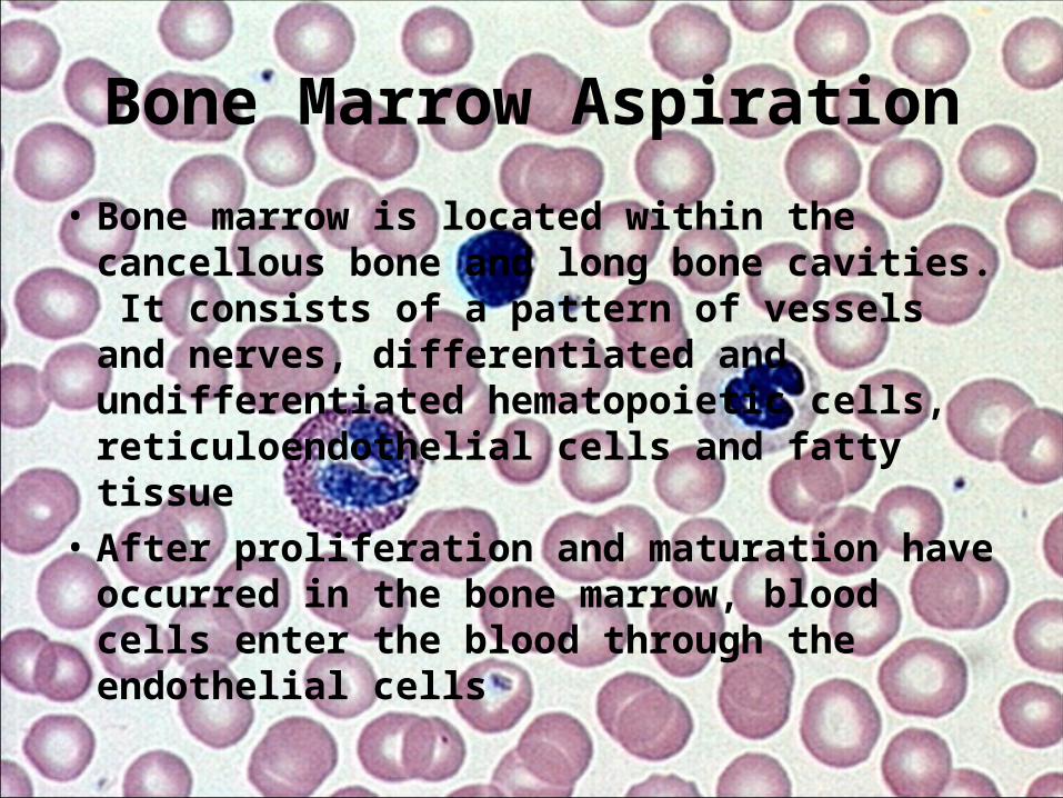
• Bone marrow is located within the cancellous bone and long bone cavities. It consists of a pattern of vessels and nerves, differentiated and undifferentiated hematopoietic cells, reticuloendothelial cells and fatty tissue
• After proliferation and maturation have occurred in the bone marrow, blood cells enter the blood through the endothelial cells
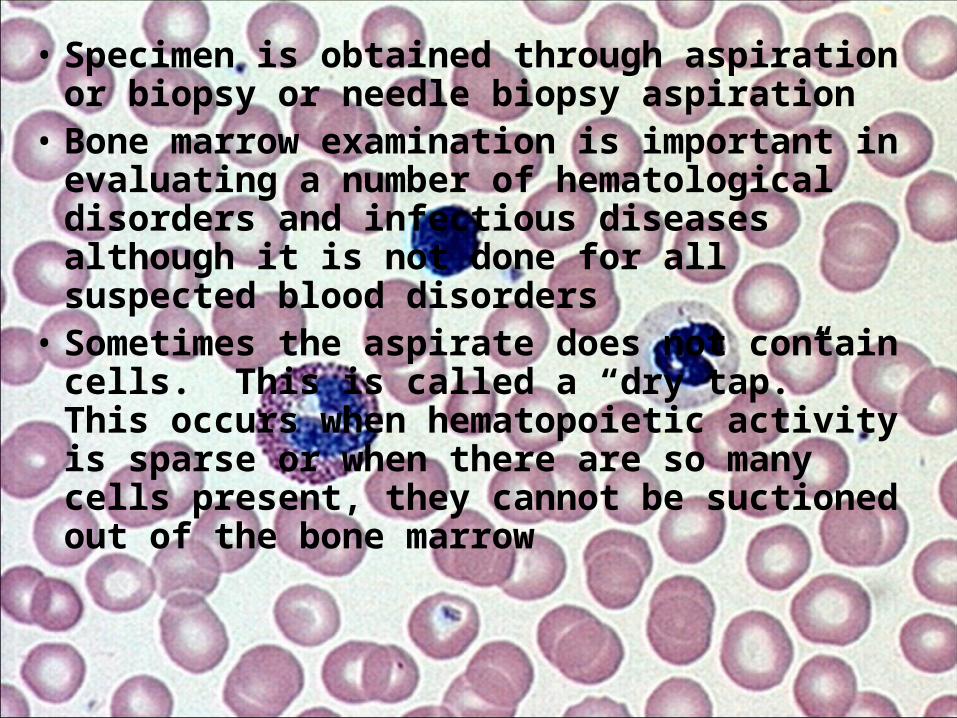
• Specimen is obtained through aspiration or biopsy or needle biopsy aspiration
• Bone marrow examination is important in evaluating a number of hematological disorders and infectious diseases although it is not done for all suspected blood disorders
• Sometimes the aspirate does not contain cells. This is called a “dry tap.” This occurs when hematopoietic activity is sparse or when there are so many cells present, they cannot be suctioned out of the bone marrow

• Patient must lie on back or side. – Posterior iliac crest is preferred for patients older
than 12 to 18 months.– Alternative sites include the anterior iliac crest,
sternum, spinous vertebral processes T10 through L4, the ribs, and the tibia in children. The sternum is not usually used in children because of the shallow bone cavity

Clinical Implications
• Bone marrow provides clues to many diseases through the presence, absence, and ratio of cells– Examples include:
• Multiple myeloma• Leukemia• Anemia• Neoplastic diseases• Agranulocytosis (decrease in wbc)• Platelet dysfunction• Infectious diseases – histoplasmosis and tuberculosis
Basic Blood Tests
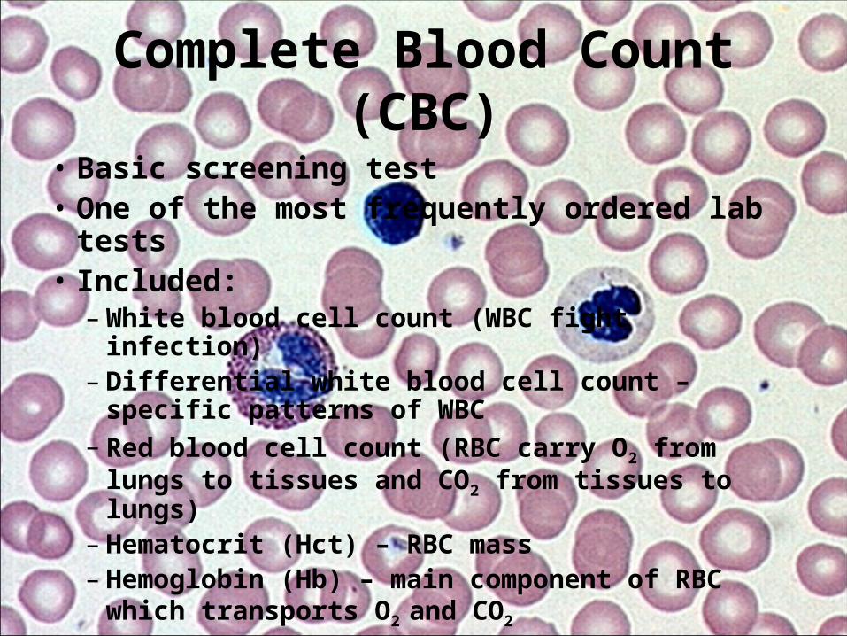
Complete Blood Count (CBC)
• Basic screening test• One of the most frequently ordered lab tests• Included:– White blood cell count (WBC fight infection)– Differential white blood cell count – specific
patterns of WBC– Red blood cell count (RBC carry O2 from lungs to
tissues and CO2 from tissues to lungs)– Hematocrit (Hct) – RBC mass– Hemoglobin (Hb) – main component of RBC
which transports O2 and CO2

• Platelet count – necessary for clotting and to control bleeding
• Dehydration and overhydration (from IV fluids) can give erroneous results
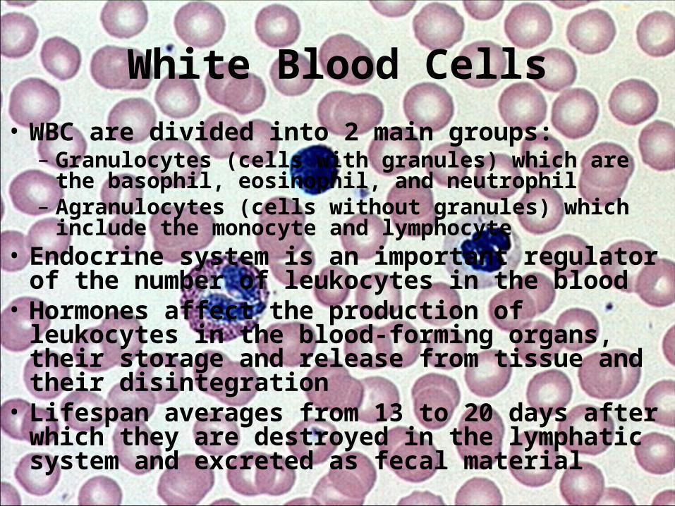
White Blood Cells• WBC are divided into 2 main groups:– Granulocytes (cells with granules) which are the basophil,
eosinophil, and neutrophil– Agranulocytes (cells without granules) which include the
monocyte and lymphocyte• Endocrine system is an important regulator of the
number of leukocytes in the blood• Hormones affect the production of leukocytes in the
blood-forming organs, their storage and release from tissue and their disintegration
• Lifespan averages from 13 to 20 days after which they are destroyed in the lymphatic system and excreted as fecal material
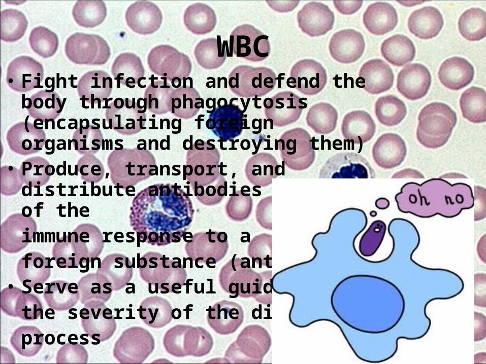
WBC• Fight infection and defend the body
through phagocytosis (encapsulating foreign organisms and destroying them)
• Produce, transport, and distribute antibodies as part of theimmune response to a foreign substance (antigen)
• Serve as a useful guide to the severity of the disease process
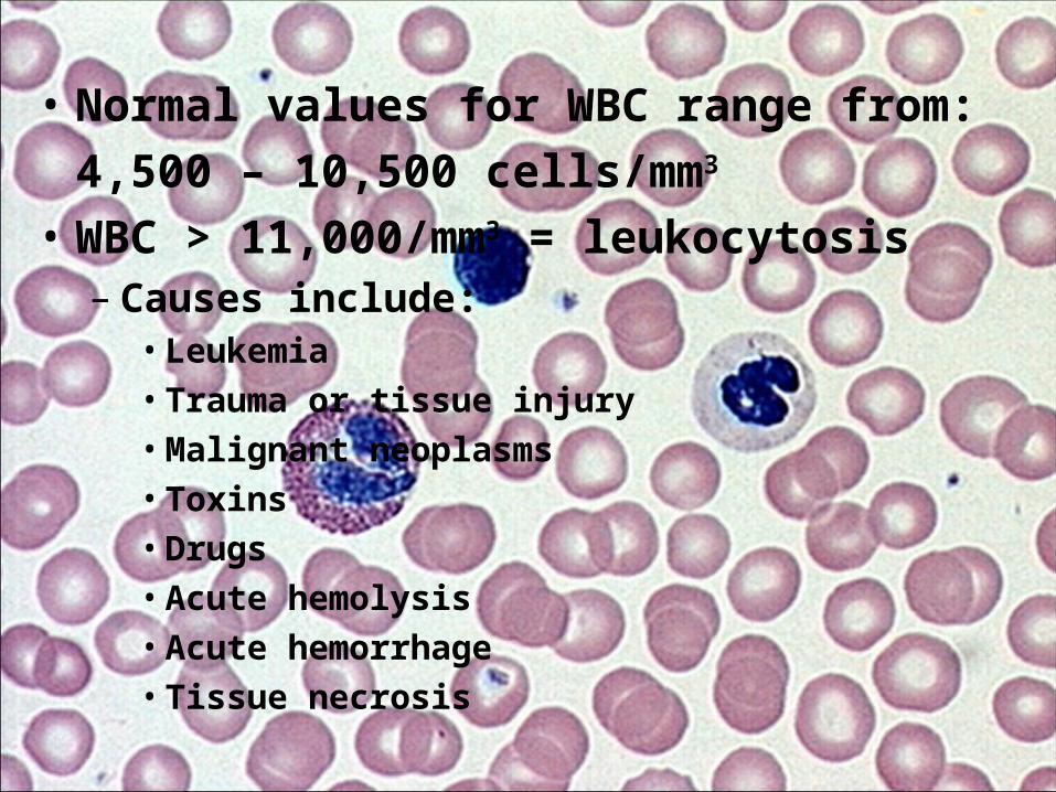
• Normal values for WBC range from:4,500 – 10,500 cells/mm3
• WBC > 11,000/mm3 = leukocytosis– Causes include:• Leukemia• Trauma or tissue injury• Malignant neoplasms• Toxins• Drugs• Acute hemolysis• Acute hemorrhage• Tissue necrosis
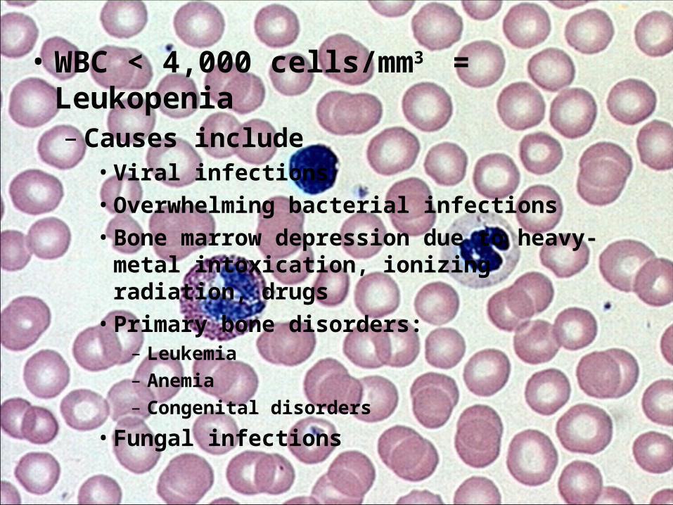
• WBC < 4,000 cells/mm3 = Leukopenia– Causes include• Viral infections• Overwhelming bacterial infections• Bone marrow depression due to heavy-metal
intoxication, ionizing radiation, drugs• Primary bone disorders:
– Leukemia– Anemia– Congenital disorders
• Fungal infections
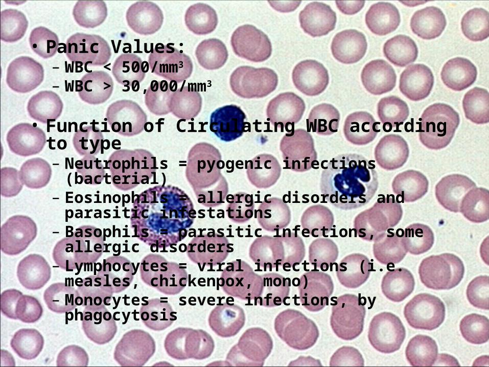
• Panic Values:– WBC < 500/mm3
– WBC > 30,000/mm3
• Function of Circulating WBC according to type– Neutrophils = pyogenic infections (bacterial)– Eosinophils = allergic disorders and parasitic
infestations– Basophils = parasitic infections, some allergic
disorders– Lymphocytes = viral infections (i.e. measles,
chickenpox, mono)– Monocytes = severe infections, by phagocytosis
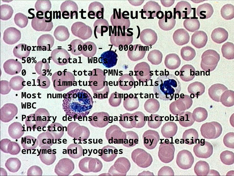
Segmented Neutrophils (PMNs)
• Normal = 3,000 – 7,000/mm3
• 50% of total WBCs• 0 – 3% of total PMNs are stab or band cells
(immature neutrophils)• Most numerous and important type of WBC• Primary defense against microbial infection• May cause tissue damage by releasing
enzymes and pyogenes

Eosinophils
• Normal = 0 – 0.7 x 10⁹/L• 0 – 3% of total WBCs• Capable of phagocytosis, ingest antigen-
antibody complexes and become active in later stages of inflammation
• Respond to allergic and parasitic diseases• Granules contain histamine (1/3 of all the
histamine in the body)

Basophils
• Normal = 15 – 50 / mm3
• 0 – 1% of total WBCs• Phagocytic• Granules contain heparin, histamine and
serotonin• Tissue basophils = mast cells – not seen in
peripheral blood; rarely seen in healthy bone marrow
• Used to study chronic inflammation

Monocytes• Normal = 100 – 500/mm3
• 3 – 7% of total WBCs• Largest cells of normal blood• 2nd line of defense against infection• Become large macrophagic phagocytes called
histiocytes; reversible transformation• Remove injured and dead cells, microorganisms,
and insoluble particles from circulating blood• Clear debris from the respiratory tract,
gastrointestinal tract and genitourinary organs• Produce an antiviral agent called interferon
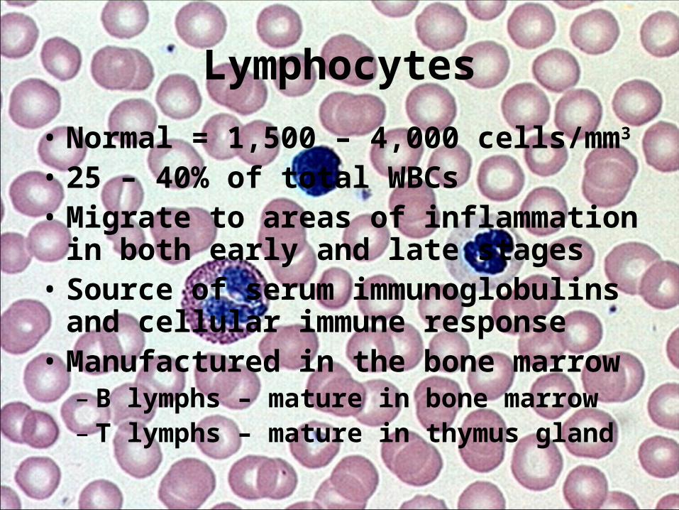
Lymphocytes
• Normal = 1,500 – 4,000 cells/mm3
• 25 – 40% of total WBCs• Migrate to areas of inflammation in both
early and late stages• Source of serum immunoglobulins and
cellular immune response• Manufactured in the bone marrow – B lymphs – mature in bone marrow– T lymphs – mature in thymus gland

T cells and B cells
• T-cells – the master immune cells, include CD4+ helper T cells, killer cells, cytotoxic cells, and CD8+ suppressor T cells
• B-cells – control antigen-antibody response that is specific to the offending antigen and is said to have “memory”
Red Blood Cell (RBC) Count
• Normal = – Men 4.2 – 5.4 x 10⁶/mm3
– Women 3.6 – 5.0 x 10⁶/mm3
– 16 – 18y/o 4.2 – 5.4 x 10⁶/mm3

Factors Affecting RBC values
• Posture – healthy recumbent person will have a 5% lower RBC value
• Dehydration – falsely increased value• Age – newborns have normally higher RBC
values than adults• Prolonged venous stasis can falsely increase
results• Stress can cause higher RBC values

• Altitude – higher altitude = greater increase in RBC
• Pregnancy – decreased value due to increase in body fluids
• Drugs can increase/decrease RBC values

Hematocrit (Hct)
• Normal =– Men 42% - 52%– Women 36% - 48%– 16 – 18 y/o 34% - 44%
• Indirect measure of RBC mass• Expressed in % by volume of packed RBCs in
whole blood

• ↓Hct – indicates anemia• Also may indicate:– Leukemias, lymphomas, Hodgkin’s disease– Adrenal insufficiency– Chronic disease– Acute and chronic blood loss– Hemolytic rxn
• ↑Hct may indicate:– Erythrocytosis– shock

• People who live at high altitudes have high Hct as well as ↑ Hb and RBC
• Normal values for Hct vary with age and gender; babies have higher normal values due to larger sized RBCs; female normal values are usually slightly lower than male
• Severe dehydration can cause a falsely elevated Hct

• A Hct <20% can lead to cardiac failure and death
• A Hct >60% is associated with spontaneous clotting of blood
Hemoglobin (Hb or Hgb)
• Normal = – Men 14.0 – 17.4 g/dL– Women 12.0 – 16.0 g/dL– 16-18 y/o 11.1 – 15.7g/dL

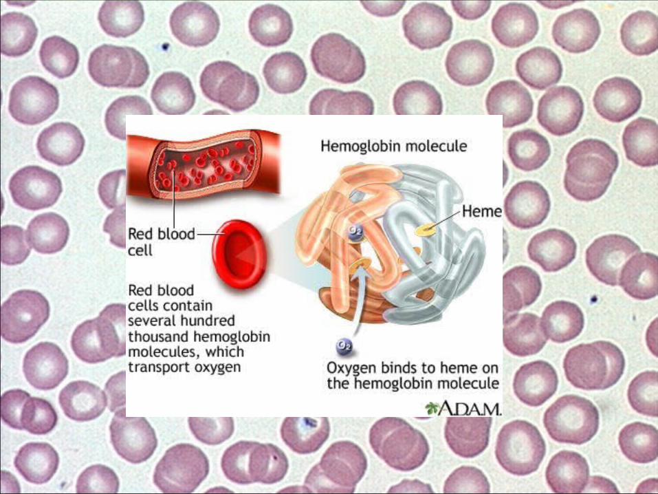
• Main component of RBCs• Composed of amino acids that form a single
protein called “globin” and a compound called “heme,” which contains iron atoms and a red pigment called porphyrin.
• The iron pigment combines readily with oxygen and gives blood its red color
• The oxygen-combining capacity is directly proportional to the Hb concentration within the RBCs

• Hb also serves as an important buffer in the extracellular fluid.
• Decreased levels – – Anemia– Hemorrhage– Liver disease
• Increased levels – – Congestive heart failure– Chronic obstructive pulmonary disease (COPD)
• <5.0 g/dL leads to heart failure and death• >20 g/dL leads to clogging of the capillaries

Red Blood Cell Indices
• Define the size and Hb content of RBC• Include:– Mean corpuscular volume (MCV)– Mean corpuscular hemoglobin concentration
(MCHC)– Mean corpuscular hemoglobin (MCH)
• Used to differentiate anemias• Help to characterize if RBCs are normal or
abnormal in volume and Hb content
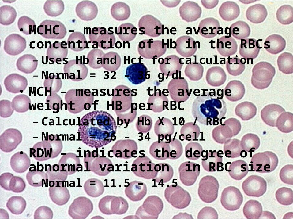
• MCHC – measures the average concentration of Hb in the RBCs– Uses Hb and Hct for calculation– Normal = 32 – 36 g/dL
• MCH – measures the average weight of HB per RBC– Calculated by Hb X 10 / RBC– Normal = 26 – 34 pg/cell
• RDW – indicates the degree of abnormal variation in RBC size– Normal = 11.5 – 14.5