basic lab procedures
TRANSCRIPT

Basic Laboratory Techniques
in Wildlife Rehabilitation
Melanie Gordon, Christina* Carrières,
BSc, RAHT AHT
Wildlife Rehabilitator Senior Wildlife Rehabilitator
BC SPCA Wild ARC BC SPCA Wild ARC
IWRC Symposium 2013

Overview Why and when you would perform lab
techniques
Equipment
Parasites and wildlife
Parasite families
Microscope and how to read a slide
Fecal procedures
Ectoparasite methods
Common parasites of avian and mammals
Microbiology
Mycology
Cytology
Hematology- WBCs, RBCs, platelets
Sending samples away
Urinalysis
Necropsy
What you can do in your rehab facility lab
Conclusion
What a birds feather looks like
under the microscope
www.flickriver.com

Why perform laboratory procedures?
To help detect any
underlying disease
processes- wildlife hide
signs
Provides information to
assess current condition
Provides information to
design a treatment plan
Helps evaluate prognosis
Tool to monitor progress
Bald Eagle with Lead Poisoning
http://www.zutrition.com/avian-toxicities/

When would you perform laboratory
procedures?
Upon intake if animal is stable
enough to handle it
Oiled animal
Seabird
Thin body condition
Crop not emptying
Eating but not gaining weight
Suspect poisoning, toxicity
Abnormal color, consistency, or
blood in feces
Anorexia
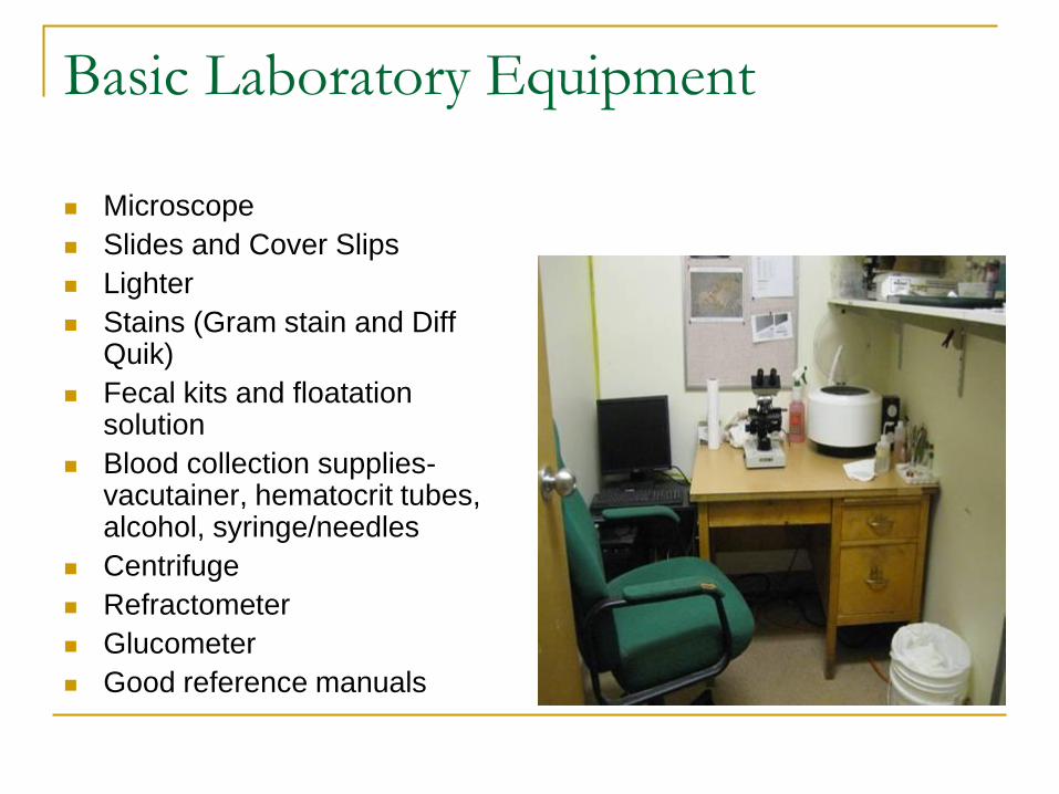
Basic Laboratory Equipment
Microscope
Slides and Cover Slips
Lighter
Stains (Gram stain and Diff Quik)
Fecal kits and floatation solution
Blood collection supplies- vacutainer, hematocrit tubes, alcohol, syringe/needles
Centrifuge
Refractometer
Glucometer
Good reference manuals

Parasites and Wildlife Parasites in low numbers are
normal in wildlife
Higher numbers may indicate a disease process or animal is immunocompromised
Some parasites are host-specific
Many of the parasites encountered are zoonotic so wear PPE
Know how to diagnose, treat, and prevent re-infection
Decrease stress, provide supportive care
Isolate if necessary
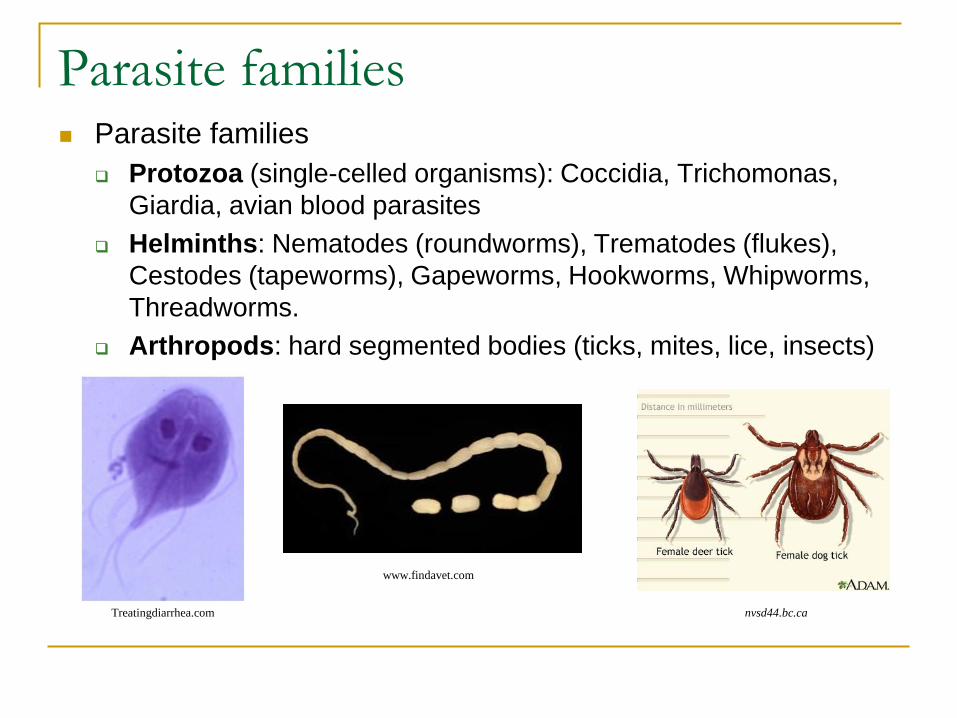
Parasite families Parasite families
Protozoa (single-celled organisms): Coccidia, Trichomonas,
Giardia, avian blood parasites
Helminths: Nematodes (roundworms), Trematodes (flukes),
Cestodes (tapeworms), Gapeworms, Hookworms, Whipworms,
Threadworms.
Arthropods: hard segmented bodies (ticks, mites, lice, insects)
www.findavet.com
Treatingdiarrhea.com nvsd44.bc.ca

Microscope and Reading a Slide
Low power objective (10x) first faster to read, less detail and
light needed
scan for platelet clumps, staining, large abnormal cells
High dry objective (40x) to examine object more closely
field of view is decreased, need more light
Oil immersion (100x) need lots of light,
small field of view
to see bacteria, yeasts, spores, and cell details
cal.vet.upenn.edu
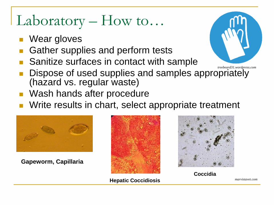
Laboratory – How to… Wear gloves
Gather supplies and perform tests
Sanitize surfaces in contact with sample
Dispose of used supplies and samples appropriately (hazard vs. regular waste)
Wash hands after procedure
Write results in chart, select appropriate treatment
treebeard31.wordpress.com
marvistavet.com
Gapeworm, Capillaria
Hepatic Coccidiosis
Coccidia

Fecal Analysis - Procedures
1. Gross examination of feces
2. Fecal floatation, centrifugation, sedimentation
3. Direct smear
4. Gram stain
startortoises.net

Fecal Analysis - Collection
Need a fresh sample- rapid development and changes can occur
Label with species, case number, date
Note feces color, consistency, and presence of (gross) parasites and/or blood
Avian- collect fecal part (dark colour) only, not the urates or urine (white or light green)
Can keep in the fridge up to 4 days
www.pet-informed-veterinary-advice-online.com...Remove frame
GWGU dropping

1. Gross examination of feces Consistency: liquid, soft, hard,
granular, gelatinous
Odor: normal vs abnormal for the species
Color: green, dark brown, black, red, etc.
Blood: dark black/brown and tarry stools (bleeding from upper GI)
red or maroon-coloured (bleeding from lower GI)
Mucus: intestinal inflammation, parasitism, or infection
Gross parasites: larvae or portions of parasites are sometimes visible to the naked eye (e.g. Tapeworm)
wideningcircle.blogspot.com/2011/01/poop-char...Remove frame

2. Fecal float Detects ova/oocytes from internal
parasites
If Negative: Perform fecal analysis for 3 consecutive days
If Positive: Perform fecal analysis 2-3 days after the last treatment
Uses a solution with a higher specific gravity than that of the eggs
Many floatation solutions: zinc sulfate, sodium nitrate, sugar, etc.
Allow 10-15 mins. for eggs to float
Read slide ideally within 30 mins.
Floatation not used for Trematodes (flukes) as too heavy for solution or Giardia as solution will lyse organism
atozvetsupply.com
Wildlife under the microscope
Ovatector, Fecalyzer and
Ovassay kits
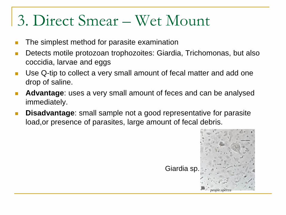
3. Direct Smear – Wet Mount The simplest method for parasite examination
Detects motile protozoan trophozoites: Giardia, Trichomonas, but also
coccidia, larvae and eggs
Use Q-tip to collect a very small amount of fecal matter and add one
drop of saline.
Advantage: uses a very small amount of feces and can be analysed
immediately.
Disadvantage: small sample not a good representative for parasite
load,or presence of parasites, large amount of fecal debris.
people.upei.ca
Giardia sp.

3. Direct Smear - Gram Stain Can detect bacteria, yeast
(purple colored), spores, Clostridium sp., Campylobacter sp. etc.
Differentiate between two types of bacteria: gram positive and gram negative
Evaluate the balance of bacterial flora (cocci vs. bacilli)
masaav.org

Fecal Analysis – Recording Results
Results either No Ova Observed (N.O.O) or Ova Observed (OVA).
Standardize techniques on how to record: 1+ = 1-2 ova per low power field (LPF) (light load)
2+ = 3-5 (moderate)
3+ = 6-8 (heavy)
4+ = > 9 (very heavy)
TNTC
Wet Mount interpretation
Gram stain interpretation

Ectoparasites Analysis - Procedures
Three methods used:
1. Squash Smear: to see tapeworm segments and ectoparasites (saline + cover slip)
2. Scotch tape method: to visualize lice, mites, and others
3. Skin scrape: to detect mites such as Mange (Sarcoptes, Demodex), Scaley leg mite
Preserve them in 70% ethanol or formalin
Find through physical examination except for mites (will see lesions)
Cheyletiella under the microscope
Scotch tape method for Cheyletiella
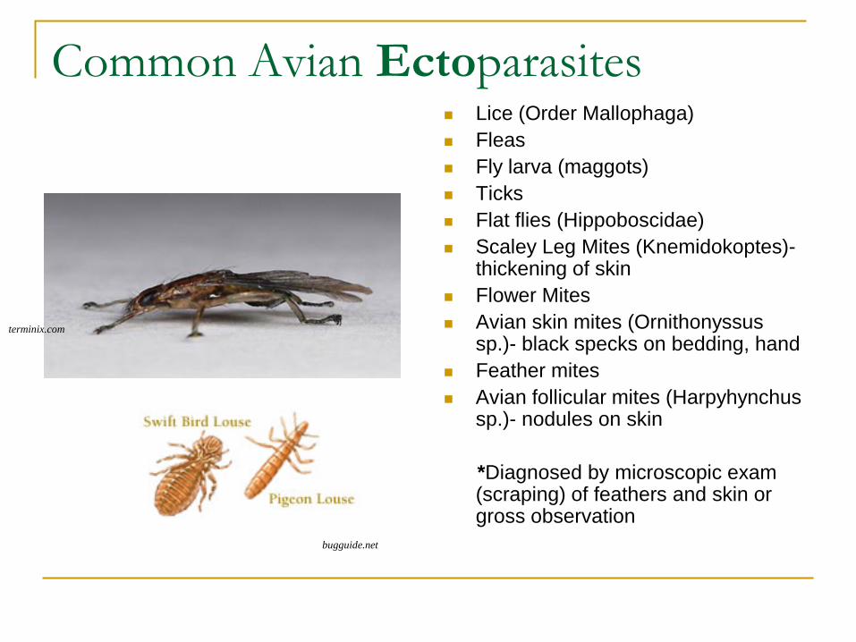
Common Avian Ectoparasites Lice (Order Mallophaga)
Fleas
Fly larva (maggots)
Ticks
Flat flies (Hippoboscidae)
Scaley Leg Mites (Knemidokoptes)- thickening of skin
Flower Mites
Avian skin mites (Ornithonyssus sp.)- black specks on bedding, hand
Feather mites
Avian follicular mites (Harpyhynchus sp.)- nodules on skin
*Diagnosed by microscopic exam (scraping) of feathers and skin or gross observation
terminix.com
bugguide.net

Common Avian Endoparasites
Fecal Analysis:
Capillaria sp. (Threadworm)
Syngamus sp. (Gapeworm)
Coccidia (Protozoa of genera Isospora or Eimeria)
Taenia sp. (Tapeworm)
Ascaridia (Roundworm)
Giardia (Protozoa) (direct smear)
Crop Swab:
Trichomonas sp. (Protozoa)
Yeasts (fungal infection)
Trichomonas sp. (Diff Quik)
Syngamus sp. (Direct smear)

Avian Blood Parasites
All protozoans
Plasmodium (Malaria)
Leukocytozoon
Hemoproteus: considered non-pathogenic in most avian species.
Transmission by biting arthropods
Anyone’s experience?
*Diagnosed by blood smear stained with Diff-Quik (not smaller than 25G needle for blood collection)
Plasmodium sp.
Hemoproteus sp. vet.uga.edu
Leukocytozoon sp.
and Hemoproteus sp.

Mammal Endoparasites
Taenia sp., Dipilydium sp.
(Tapeworm)
Toxocara sp., Toxascaris
sp., Baylisascaris sp.
(Roundworm)
Ancylostoma (Hookworm)
Trichuris (whipworm)
Coccidia
Giardia
sccvote.org
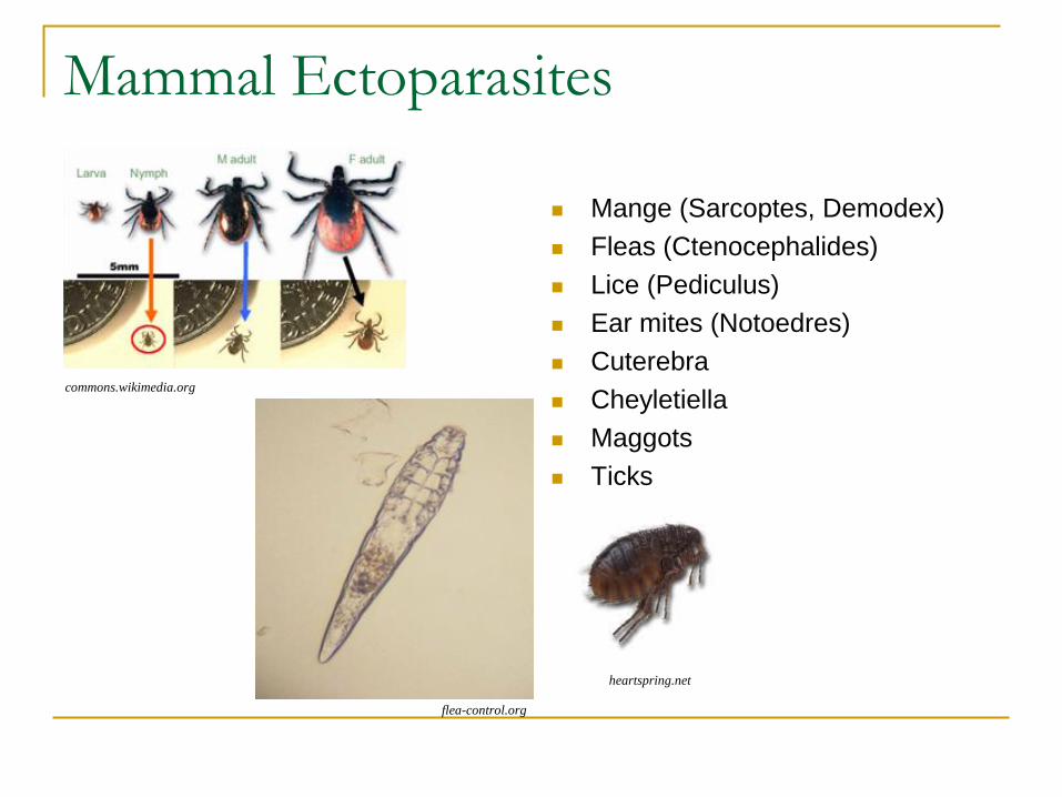
Mammal Ectoparasites
Mange (Sarcoptes, Demodex)
Fleas (Ctenocephalides)
Lice (Pediculus)
Ear mites (Notoedres)
Cuterebra
Cheyletiella
Maggots
Ticks
commons.wikimedia.org
flea-control.org heartspring.net

Microbiology
Gram stain
Remember normal flora: areas normal to have bacteria (digestive tract, mouth) vs. sterile areas
Sterile swab used to culture bacteria (ID and antibiotic sensitivity)
Fungassay (fungal culture)
http://loudoun.nvcc.edu/vetonline/vet132/micro/unit2/swabsmear.jpg

Cytology Cytology: Removing cells from tissue and examining them under the microscope
Evaluate cells in body fluids, purulent discharge, skin masses, and internal organs
Look for abnormal cells (neoplastic) or presence of inflammatory cells (WBC)
Can be tricky to identify cells- send to pathologist or consult with veterinarian
Techniques: fine needle aspirate of a lump, impression smear, swab, scraping
Stain with Diff Quik
Skin scrape
Impression smear
http://loudoun.nvcc.edu/vetonline/vet132/micro/unit2/
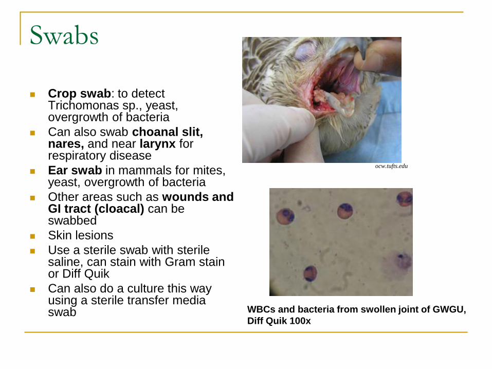
Swabs
Crop swab: to detect Trichomonas sp., yeast, overgrowth of bacteria
Can also swab choanal slit, nares, and near larynx for respiratory disease
Ear swab in mammals for mites, yeast, overgrowth of bacteria
Other areas such as wounds and GI tract (cloacal) can be swabbed
Skin lesions
Use a sterile swab with sterile saline, can stain with Gram stain or Diff Quik
Can also do a culture this way using a sterile transfer media swab
ocw.tufts.edu
WBCs and bacteria from swollen joint of GWGU,
Diff Quik 100x

Mycology When to consider investigating: visual observation of skin lesion,
foul smell, aural or oral accumulation of abnormal material
Yeast (Candida)
Ringworm (Dermatophyte): culture with fungassay, 50% of cases light up with a woods lamp if the fungus is Microsporum canis species
Sometimes fungi can be seen by stained direct smear or KOH smear (hair and skin scraping)
http://loudoun.nvcc.edu/vetonline/vet132/micro/unit2/microbiology_unit2_lesson3.htm
Positive flourecence
Positive fungassay
KOH direct smear
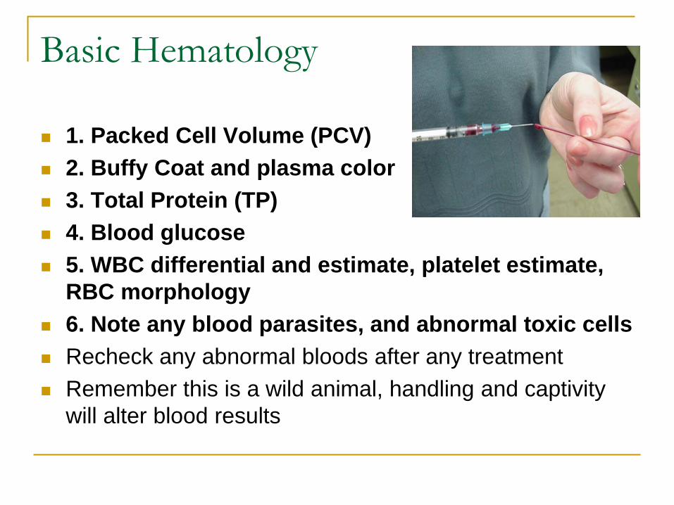
Basic Hematology
1. Packed Cell Volume (PCV)
2. Buffy Coat and plasma color
3. Total Protein (TP)
4. Blood glucose
5. WBC differential and estimate, platelet estimate,
RBC morphology
6. Note any blood parasites, and abnormal toxic cells
Recheck any abnormal bloods after any treatment
Remember this is a wild animal, handling and captivity
will alter blood results
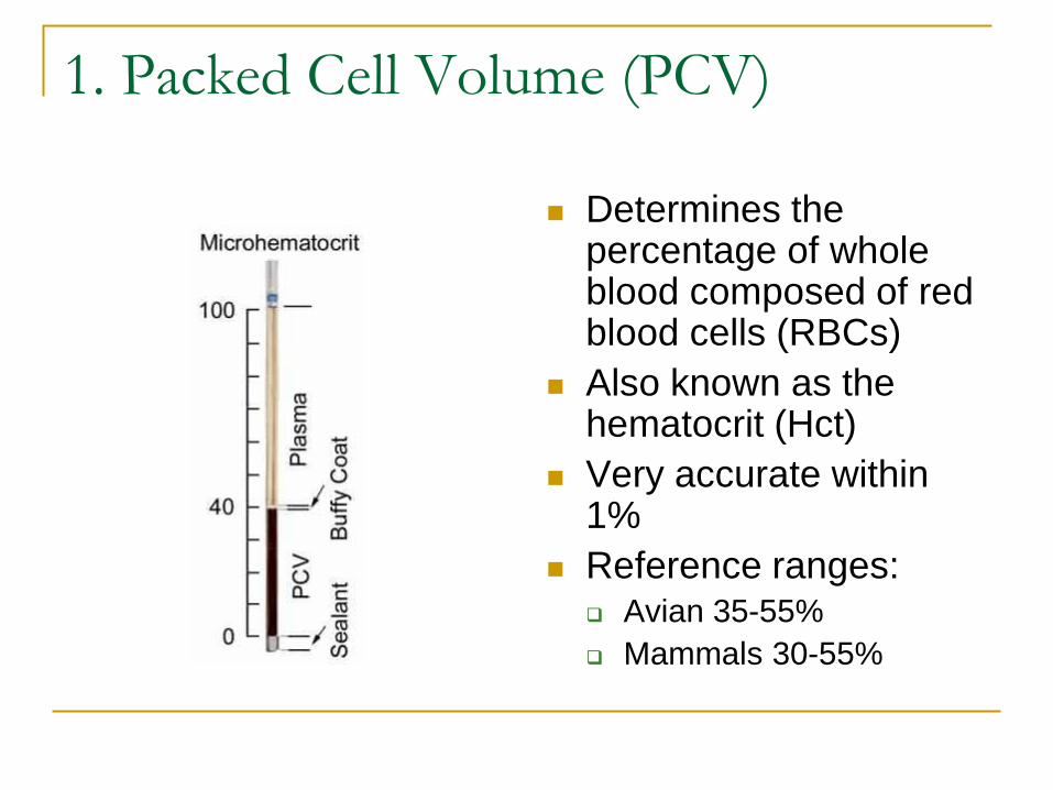
1. Packed Cell Volume (PCV)
Determines the percentage of whole blood composed of red blood cells (RBCs)
Also known as the hematocrit (Hct)
Very accurate within 1%
Reference ranges: Avian 35-55%
Mammals 30-55%

PCV Values
Low PCV < 30% indicates anemia caused by:
Hemorrhage
Parasites
Destruction of RBCs
Decreased production of RBCs
Treatment:
Iron dextran injection
Pentaspan or Hetastarch
Consider transfusion or euthanasia at < 15%
High PCV > 55 % can indicate:
Dehydration
Treatment: fluid therapy
Vetguru.com

2. Buffy Coat
Estimates the WBC and platelet level in mammals
Thicker buffy coat = increased number of WBCs
Whitish-gray layer just above the RBC layer
Healthy patient: <1%
Over 2% consider antibiotics, NO Iron administration
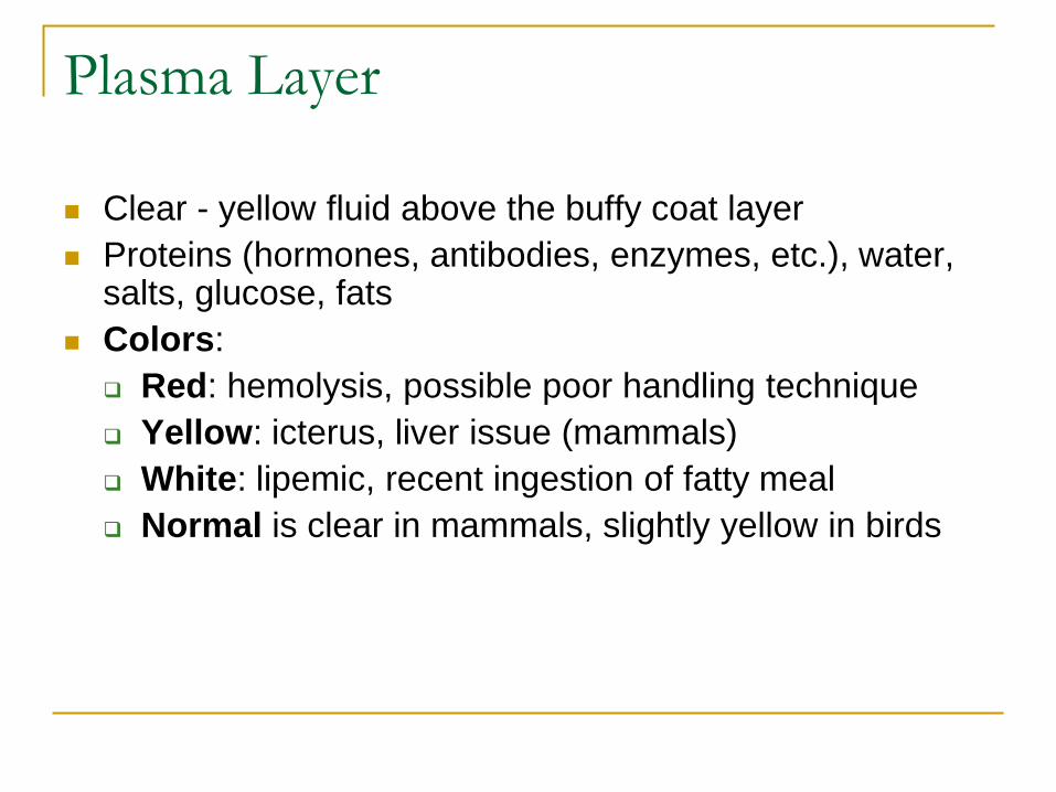
Plasma Layer
Clear - yellow fluid above the buffy coat layer
Proteins (hormones, antibodies, enzymes, etc.), water, salts, glucose, fats
Colors:
Red: hemolysis, possible poor handling technique
Yellow: icterus, liver issue (mammals)
White: lipemic, recent ingestion of fatty meal
Normal is clear in mammals, slightly yellow in birds

3. Total Protein (TP)
The total protein measures the total amount of protein in the liquid portion of the blood
Indicates general health status of the patient
Healthy avian patient should be between 3.0-6.0 g/dL
TP > 6 g/dL: indicates dehydration, chronic disease, infection
TP < 6 g/dL: hypoalbuminemia due to malnutrition, malabsorption, chronic liver disease, starvation
TP < 2.0 g/dL: poor prognosis, consider euthanasia
vetlab.com

Blood Glucose
Using a glucometer and a drop of blood
Most useful in mammals
Normal values: species specific
Hypoglycemia: Below normal range
Starvation, malnutrition
Treatment: dextrose
Hyperglycemia: Above normal range
Diabetes
Pancreatitis
Stress can elevate blood glucose levels
articlesbase.com
ikigai-de-crabahuteuse.over-blog.com

Blood Collection Sites
Mammals:
Jugular vein
Cephalic vein (forearm)
Saphenous (lateral side of back leg)
Femoral (medial side of back leg)
Marginal ear vein (rabbits, felines)
Avian:
Ulnar vein (ventral aspect of elbow)
Medial metatarsal vein (along the metatarsus)
Jugular vein (R one is two-thirds larger than the L)
Superficial digital veins through webbing (large species)
No toe nail clips
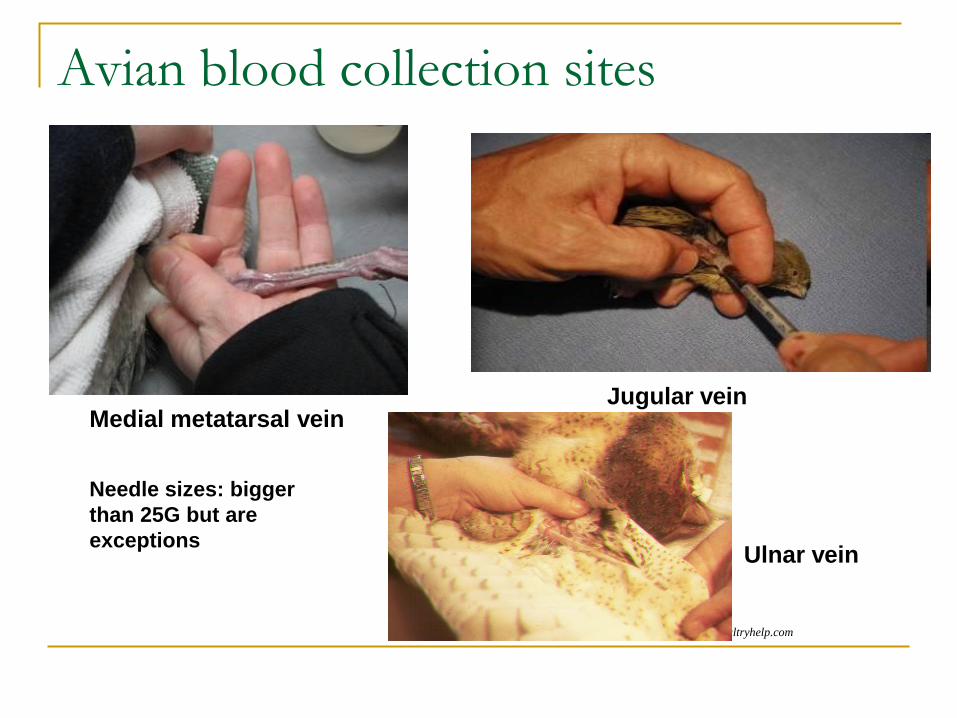
Avian blood collection sites
poultryhelp.com
Medial metatarsal vein Jugular vein
Ulnar vein
Needle sizes: bigger
than 25G but are
exceptions
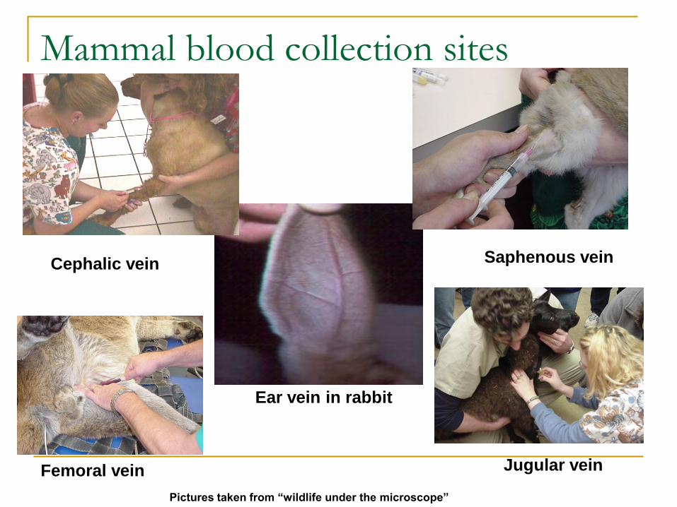
Mammal blood collection sites
Femoral vein
Cephalic vein
Ear vein in rabbit
Saphenous vein
Jugular vein
Pictures taken from “wildlife under the microscope”

Blood Volume
Maximum safe blood volume for birds and mammals: 1% of patients body weight
Equivalent to 10% of blood volume
Only applies to healthy animals
General rule of thumb: Birds: 1.0 ml/100 g
Example: 1000g bird has a total blood volume of 100 ml, safe maximum amount of blood taken is 10 ml
Effects of blood loss (hypovolemic shock)
host.web-print-design.com
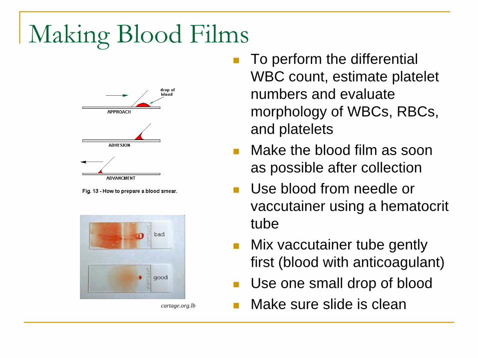
Making Blood Films To perform the differential
WBC count, estimate platelet
numbers and evaluate
morphology of WBCs, RBCs,
and platelets
Make the blood film as soon
as possible after collection
Use blood from needle or
vaccutainer using a hematocrit
tube
Mix vaccutainer tube gently
first (blood with anticoagulant)
Use one small drop of blood
Make sure slide is clean cartage.org.lb
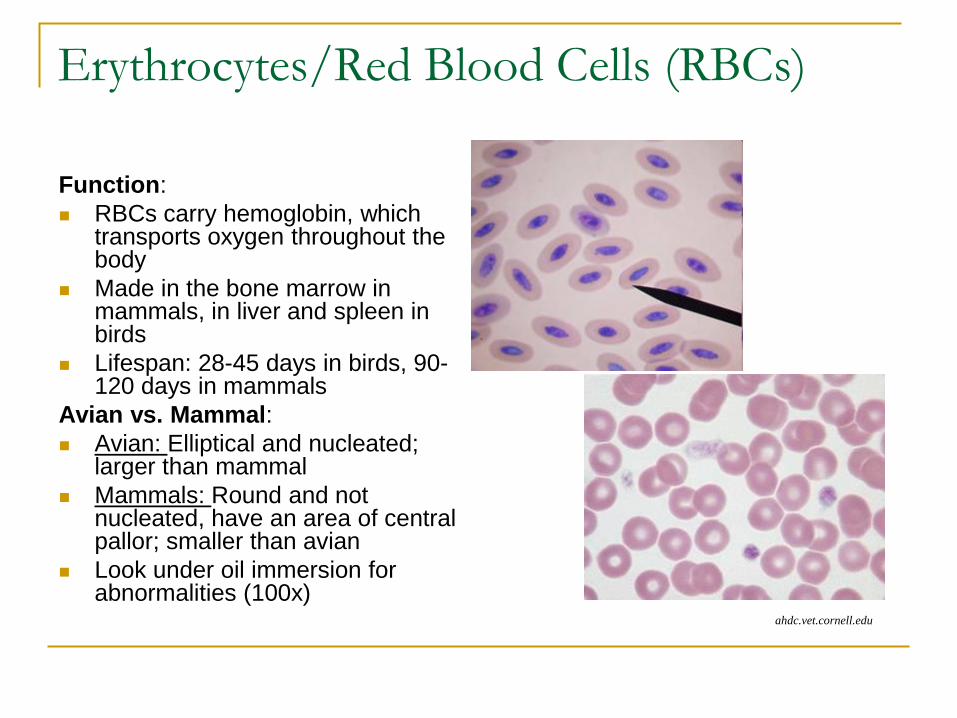
Erythrocytes/Red Blood Cells (RBCs)
Function:
RBCs carry hemoglobin, which transports oxygen throughout the body
Made in the bone marrow in mammals, in liver and spleen in birds
Lifespan: 28-45 days in birds, 90-120 days in mammals
Avian vs. Mammal:
Avian: Elliptical and nucleated; larger than mammal
Mammals: Round and not nucleated, have an area of central pallor; smaller than avian
Look under oil immersion for abnormalities (100x)
ahdc.vet.cornell.edu

Thrombocytes (Platelets)
Function:
Hemostasis, wound healing, phagocytosis, clotting
Can clump on a blood film
Avian vs. Mammal:
Avian: nucleated, small rim of gray cytoplasm
Mammals: not nucleated, much smaller than avian
Determine if they are normal, increased or decreased:
Normal: 1-5 platelets/ oil immersion field (100x)
Consider amount of clumping
http://www.marvistavet.com
http://www.spcollege.edu/hec/vt/vtde/avianhemo/avian1/21.jpg

White Blood Cells (WBCs)
Avian and mammals function similar: to defend the body against foreign invaders (immune system)
Neutrophil (mammals), Heterophil (avian, rabbits, some rodents): phagocytosis
Eosinophil: allergic reactions, parasites, phagocytosis
Basophil: initiation of immune and allergic reactions
Monocyte: phagocytosis and antigenic processing
Lymphocyte: antibody production and immunity
Leukocytosis: increase, due to inflammation, infection, tissue damage
Leukopenia: decrease due to virus, septicemia

Avian WBCs
http://www.exoticpetvet.com
Heterophil and smudge cells
Eosinophil
Lymphocyte Monocyte
Basophil
www.vet.uga.edu
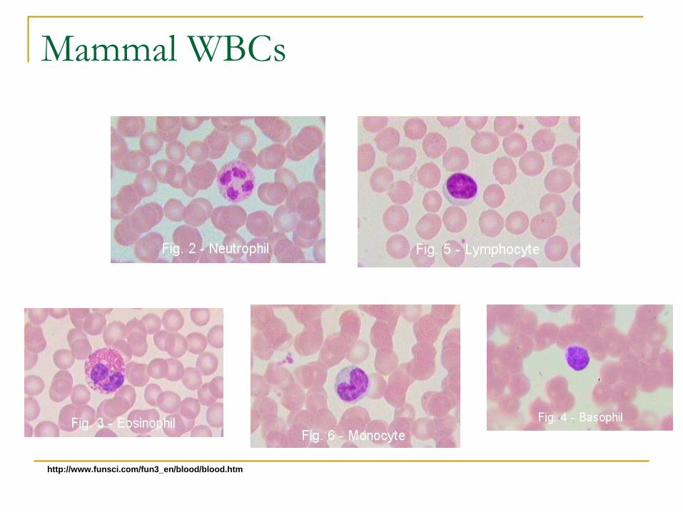
Mammal WBCs
http://www.funsci.com/fun3_en/blood/blood.htm
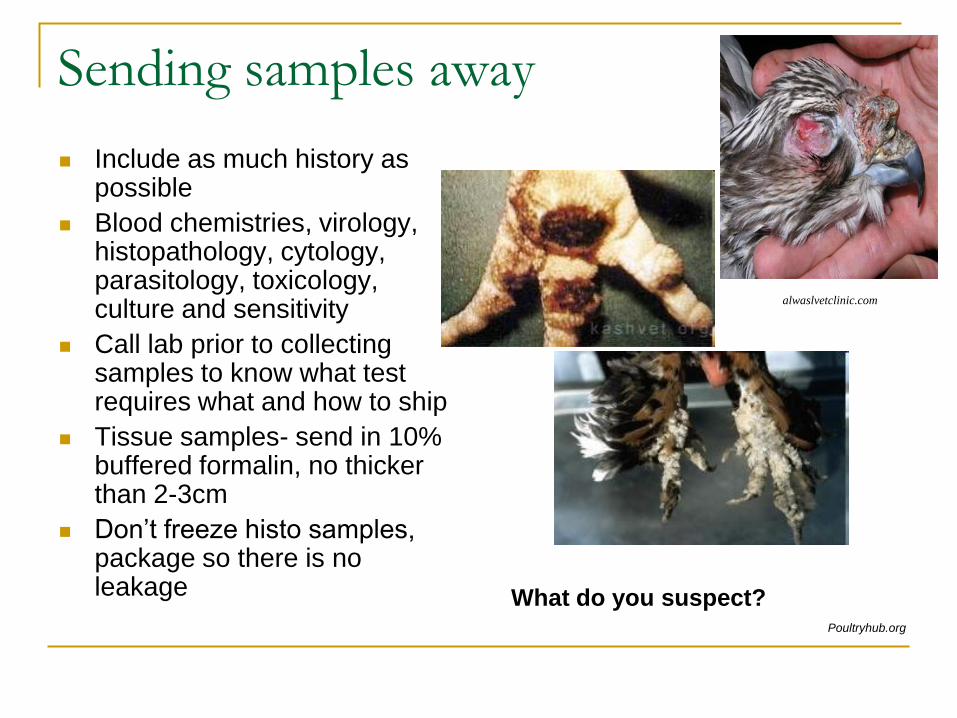
Sending samples away
Include as much history as possible
Blood chemistries, virology, histopathology, cytology, parasitology, toxicology, culture and sensitivity
Call lab prior to collecting samples to know what test requires what and how to ship
Tissue samples- send in 10% buffered formalin, no thicker than 2-3cm
Don’t freeze histo samples, package so there is no leakage
alwaslvetclinic.com
What do you suspect? Poultryhub.org

Necropsy samples
Important to send body,
tissue, or fluid samples
to lab after unexplained
death
Can give you information
on cause of death
Can be linked to
symptoms
Helps you learn from the
case and change
protocols for future cases
Population health
monitoring
http://www.unbc.ca/nlui/wildlife_diseases_bc/parvovirus.htm
Inflamed bowels indicative of
which mammal viral disease?
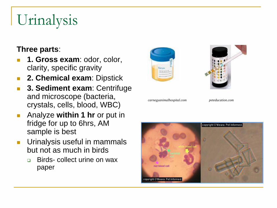
Urinalysis
Three parts:
1. Gross exam: odor, color, clarity, specific gravity
2. Chemical exam: Dipstick
3. Sediment exam: Centrifuge and microscope (bacteria, crystals, cells, blood, WBC)
Analyze within 1 hr or put in fridge for up to 6hrs, AM sample is best
Urinalysis useful in mammals but not as much in birds
Birds- collect urine on wax paper
peteducation.com carnegyanimalhospital.com

Things you can do in your center Laboratory Standard
Operating Procedures
(standardize lab work)
Create a chart with blood
values for each species to
learn species normals
(intake vs. pre-release)
Start a ‘library’ of interesting
slides/photos to learn from,
and use as a reference
Quality control: for
refractometer, glucometer,
any lab machines
Teach others!
Conclusion
OBSERVATION: notice abnormalities to know when to
test
Consult with your veterinarian, send lab work to the lab
for further answers
Most useful basic procedures: Fecal analysis, PCV, TP,
crop swabs, blood glucose in young mammals
Can use a few simple lab tests to help in patient
diagnosis, treatment, and prognosis
Standardize techniques for consistent values

References
Wynne, J. A review of avian hematozoan parasites. School of Veterinary Medicine, University of California Davis, Wildlife Journal, Vol 11 no 4.
Porter, S. Wildlife under the microscope. Blue Ridge community college, Weyers Cave, Virginia, Vol 20, no 1, spring/summer 2002.
Lancaster, E. Internal parasites of birds. Bayfield CO. Wildlife rehabilitation.
Campbell, Terry, Dein, F. Avian Hematology: The basics. Veterinary Clinics of North America: Small animal practice. Vol 14, No 2. 1984.
Colville, T and Bassert, J. Clinical Anatomy and Physiology for Veterinary Technicians. 2002.
Websites:
http://www.avianmedicine.net/ampa/36.pdf
http://www.ctdslab.co.uk/cytology.html
http://people.eku.edu/ritchisong/birdcirculatory.html
Plankton sample under the microscope
www.nationalgeographic.com
Acknowledgments
Lindsaye Akhurst
Kimberly Reid
Wild ARC Staff
University of Victoria for the
use of microscopes
IWRC