basic principles and applications of electrophoresis stephen k.w. tsui department of biochemistry
TRANSCRIPT
Basic Principles and Applications of Electrophoresis
Stephen K.W. Tsui
Department of Biochemistry
Order Form for Electrophoresis Kits and Reagents
Please click this sentence to download the form
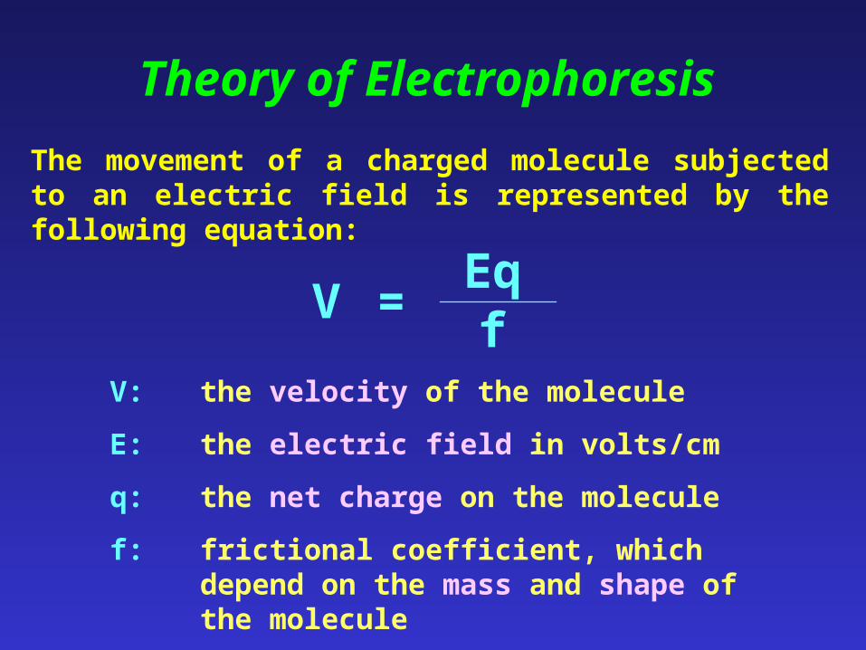
Theory of Electrophoresis
The movement of a charged molecule subjected to an electric field is represented by the following equation:
V =Eqf
V: the velocity of the molecule
E: the electric field in volts/cm
q: the net charge on the molecule
f: frictional coefficient, which depend on the mass and shape of the molecule
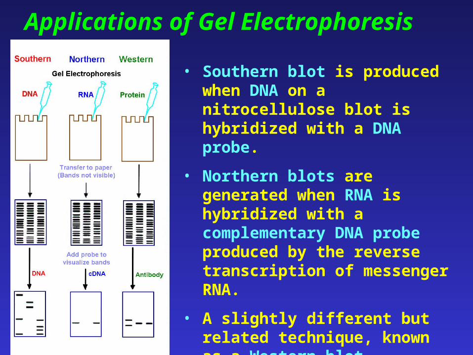
Applications of Gel Electrophoresis
• Southern blot is produced when DNA on a nitrocellulose blot is hybridized with a DNA probe.
• Northern blots are generated when RNA is hybridized with a complementary DNA probe produced by the reverse transcription of messenger RNA.
• A slightly different but related technique, known as a Western blot, involves separating proteins by gel electrophoresis and probing with labeled antibodies for specific proteins.
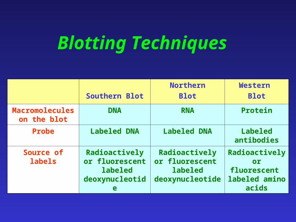
Southern Blot
Northern
Blot
Western
Blot
Macromolecules on the blot
DNA RNA Protein
Probe Labeled DNA Labeled DNA Labeled antibodies
Source of labels Radioactively or fluorescent
labeled deoxynucleotide
Radioactively or fluorescent
labeled deoxynucleotide
Radioactively or fluorescent
labeled amino acids
Blotting Techniques
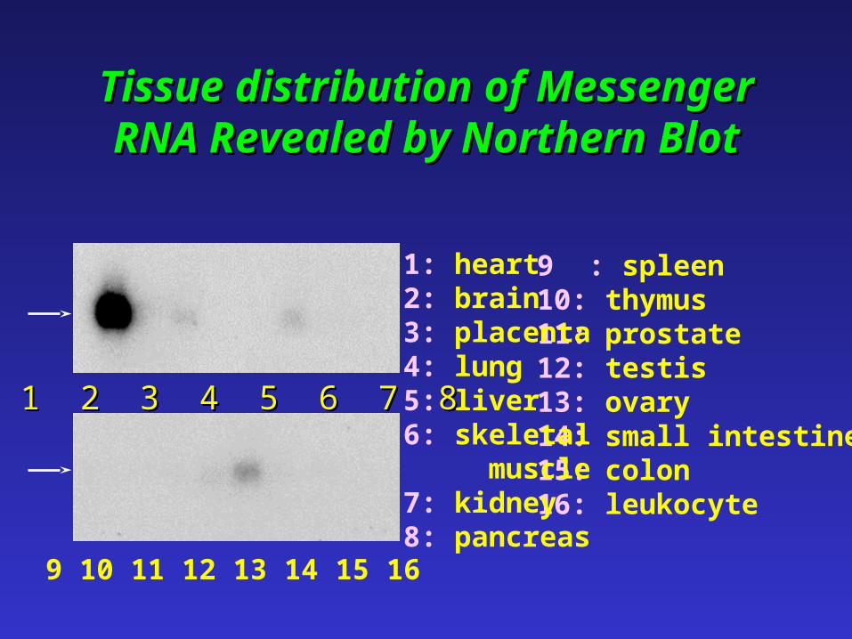
Tissue distribution Tissue distribution of Messenger of Messenger RNA RevealedRNA Revealed by Northern by Northern BlotBlot
1 2 3 4 5 6 7 81 2 3 4 5 6 7 8
9 : spleen10: thymus11: prostate12: testis13: ovary14: small intestine15: colon16: leukocyte
1: heart2: brain3: placenta4: lung5: liver6: skeletal muscle7: kidney8: pancreas
9 10 11 12 13 14 15 16
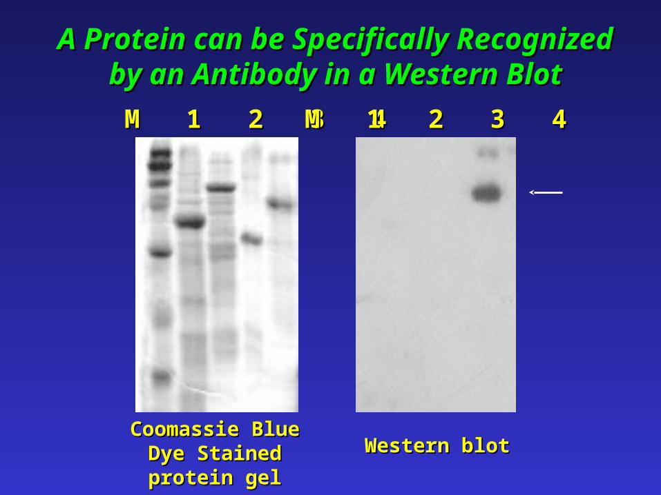
A Protein can be Specifically Recognized A Protein can be Specifically Recognized by an Antibody in a Western Blotby an Antibody in a Western Blot
M 1 2 3 4M 1 2 3 4 M 1 2 3 4M 1 2 3 4
Coomassie BlueCoomassie Blue Dye Dye Stained Stained protein gelprotein gel Western blotWestern blot
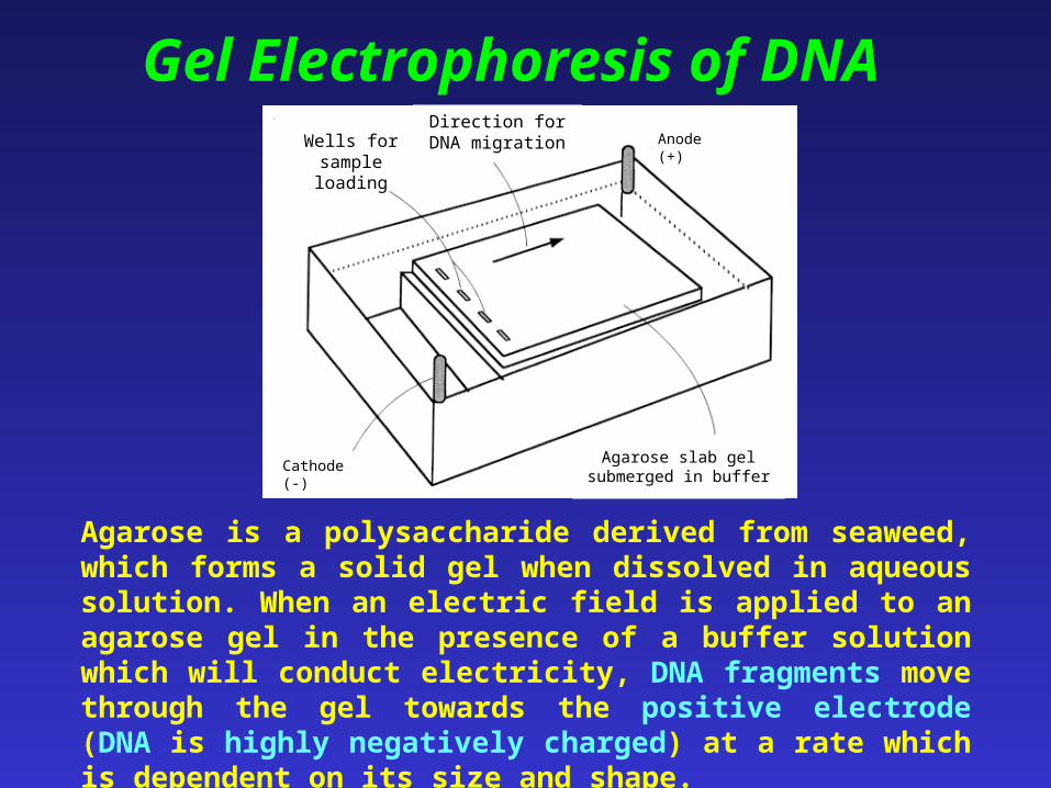
Gel Electrophoresis of DNA
Agarose slab gel submerged in buffer
Wells for sample loading
Cathode (-)
Direction for DNA migration Anode (+)
Agarose is a polysaccharide derived from seaweed, which forms a solid gel when dissolved in aqueous solution. When an electric field is applied to an agarose gel in the presence of a buffer solution which will conduct electricity, DNA fragments move through the gel towards the positive electrode (DNA is highly negatively charged) at a rate which is dependent on its size and shape.
Gel Electrophoresis of DNA
For linear DNA molecules, they have uniform shape and charge to mass ratio. The electrophoretic mobility of the DNA molecule is influenced primarily by the molecular size: The larger molecules are retarded by the molecular sieving effect of the gel, and the small molecules have greater mobility.
Gel Electrophoresis of DNA
• The DNA can be stained by the inclusion of ethidium bromide in the gel, or by soaking the gel in a solution of ethidium bromide after electrophoresis. The DNA shows up as an orange band on illumination by UV light. Alternatively, methylene blue can be used to stain DNA.
• Gels composed of polyacrylamide can separate DNA molecules that differ in length by only one nucleotide and are used to determine the base sequence of DNA. Agarose gels are used to separate DNA fragments that have larger size differences.
Procedures of DNA Fingerprinting
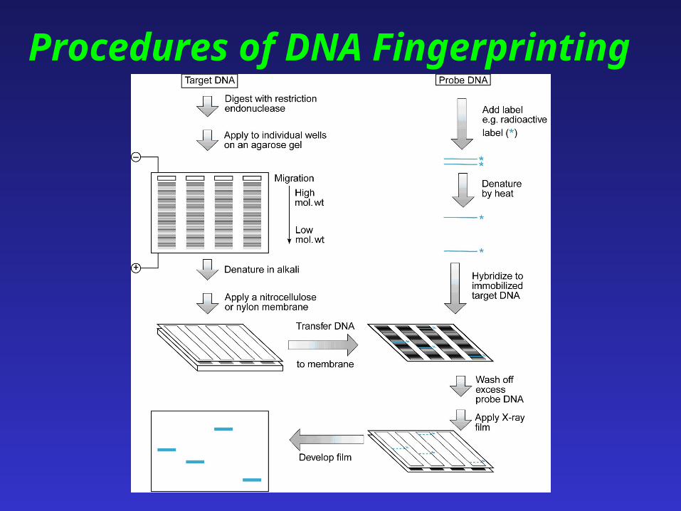
• In order to detect specific sequences, DNA is usually transferred to a solid support, such as a sheet of nitrocellulose or nylon paper.
• The paper is treated with an alkaline solution to denature DNA, that is, separate the two strands of each double helix.
• The single-stranded DNA can be hybridized with a probe, and the regions on the nitrocellulose blot containing DNA that base-pairs with the probe can be identified.
Procedures of DNA Fingerprinting
DNA Polymorphisms
• Polymorphisms are variations in DNA sequences. There may be millions of different polymorphisms in the human DNA.
• Polymorphisms in the human DNA serve as the basis for the diagnosis of diseases and the identity of individuals.
Detection of Polymorphism Restriction Fragment Length Polymorphisms
• Occasionally, a point mutation occurs in a recognition site for a restriction enzyme. The enzyme, therefore, can cut at other recognition sites but not at the site of the mutation. Consequently, the restriction fragment produced by the enzyme is larger for a person with the mutation than for a normal person.
• Mutations can also create restriction sites that are not present in the normal gene. In this case, restriction fragments will be smaller for the person with the mutation than for the normal individual. These variations in the length of restriction fragments are known as restriction fragment length polymorphisms (RFLPs).
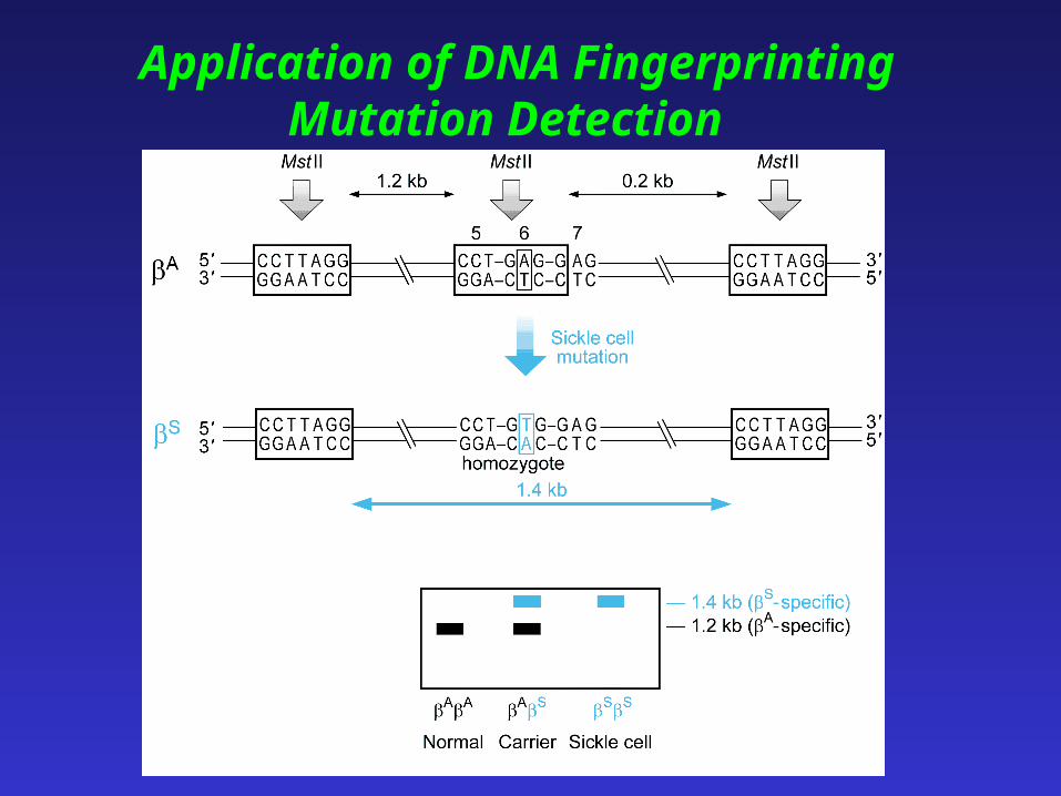
Application of DNA Fingerprinting Mutation Detection
Highly Variable Regions
• Human DNA contains many sequences that are repeated in tandem a variable number of times at certain loci in the genome. These regions are called hypervariable regions because they contain a variable number of tandem repeats (VNTR).
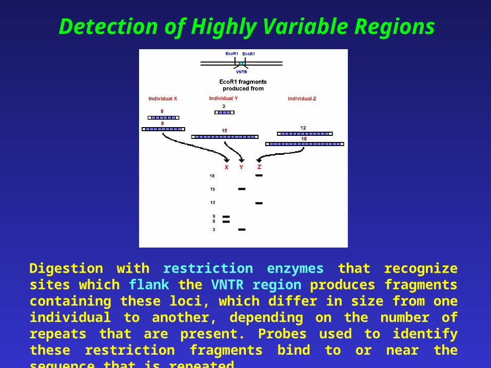
Digestion with restriction enzymes that recognize sites which flank the VNTR region produces fragments containing these loci, which differ in size from one individual to another, depending on the number of repeats that are present. Probes used to identify these restriction fragments bind to or near the sequence that is repeated.
Detection of Highly Variable Regions
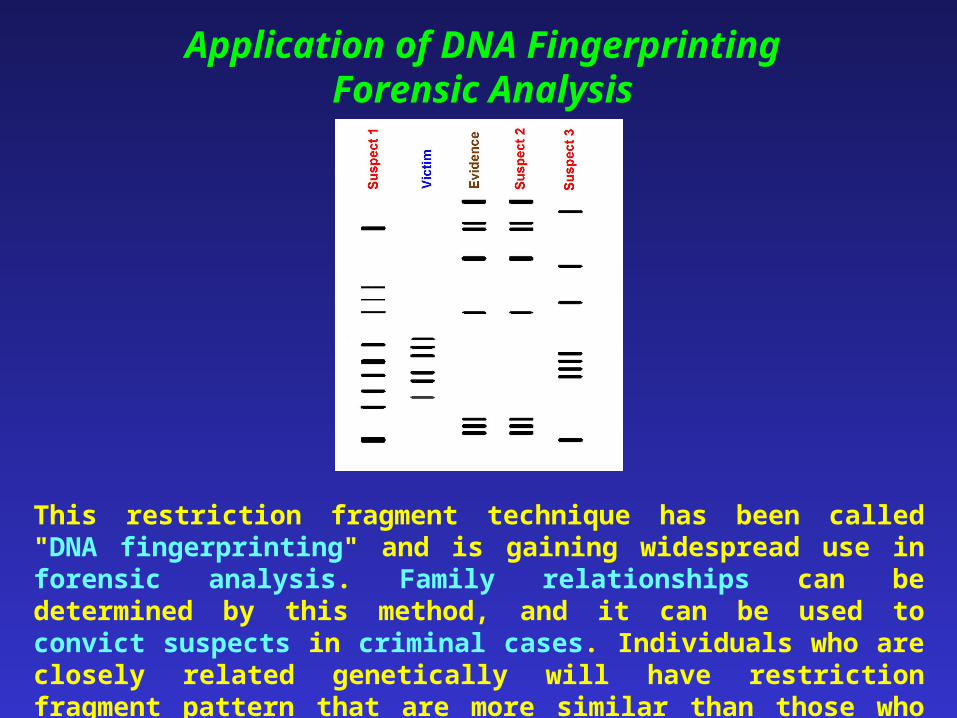
Application of DNA Fingerprinting Forensic Analysis
This restriction fragment technique has been called "DNA fingerprinting" and is gaining widespread use in forensic analysis. Family relationships can be determined by this method, and it can be used to convict suspects in criminal cases. Individuals who are closely related genetically will have restriction fragment pattern that are more similar than those who are more distantly related.
Other Applications of DNA Fingerprinting
• Parentage test
• Endangered species or Chinese herbs identification

Animation 1: Southern Blotting
http://www.dnalc.org/resources/BiologyAnimationLibrary.htm
Animation 2: DNA Detective
http://www.dnalc.org/resources/BiologyAnimationLibrary.htm

Online Courses: DNA from the Beginning
http://www.dnaftb.org/dnaftb/

Download Illustrations: Human Molecular Genetics
http://www.bios.co.uk/illustrations.asp

http://dlab.reed.edu/projects/vgm/vgm/VGMProjectFolder/VGM/RED/RED.ISG/
gel.html
Good Website: Gel Electrophoresis

Workshop
Agarose Gel Electrophoresis
Department of Biochemistry
(2001-2002)
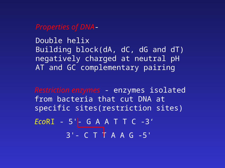
Restriction enzymes - enzymes isolated from bacteria that cut DNA at specific sites(restriction sites)
EcoRI - 5'- G A A T T C -3‘
3'- C T T A A G -5'
Properties of DNA-
Double helixBuilding block(dA, dC, dG and dT)negatively charged at neutral pHAT and GC complementary pairing
Baterial plasmid DNA
Plasmids are molecules of DNA that are found in bacteria separate from the bacterial chromosome.
They:
are small (a few thousand base pairs)
usually carry only one or a few genes
are circular
have a single origin of replication
+
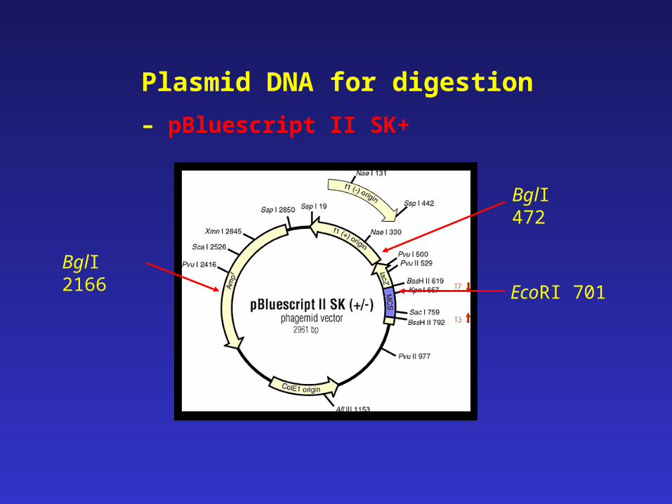
BglI 2166
BglI 472
EcoRI 701
Plasmid DNA for digestion
– pBluescript II SK+
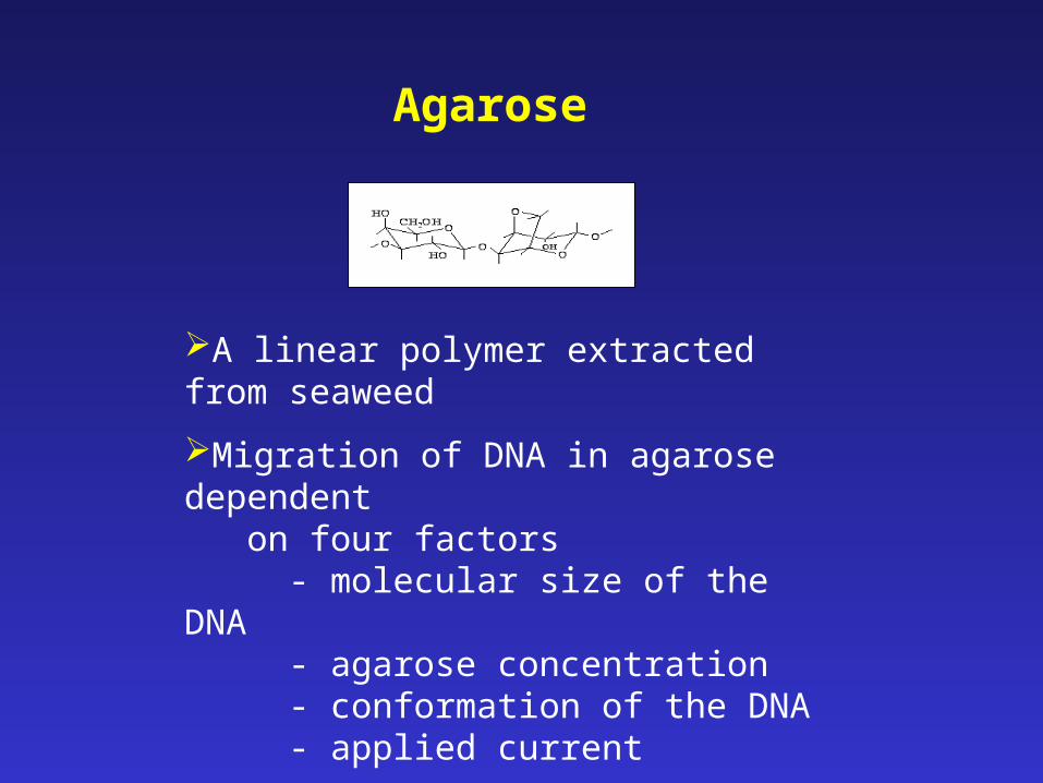
Agarose
A linear polymer extracted from seaweed
Migration of DNA in agarose dependent on four factors
- molecular size of the DNA- agarose concentration- conformation of the DNA- applied current
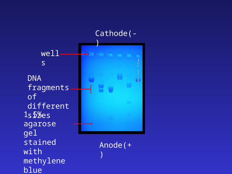
wells
Anode(+)
Cathode(-)
1.5% agarosegel stained with methylene blue
DNA fragments of different sizes
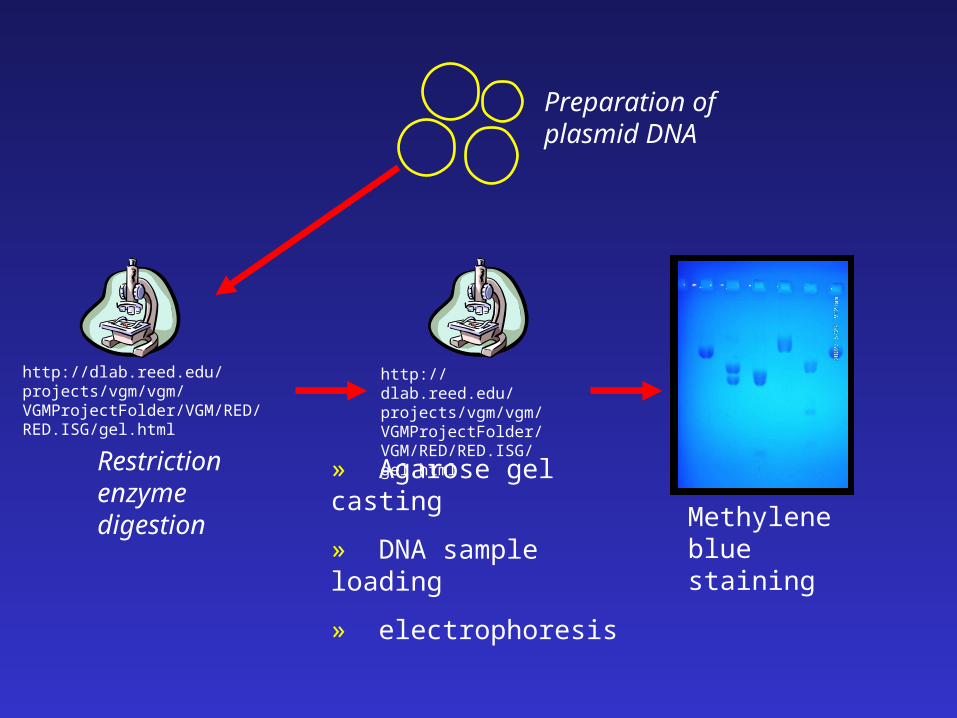
Preparation of plasmid DNA
Restriction enzyme digestion
» Agarose gel casting
» DNA sample loading
» electrophoresis
Methylene blue staining
http://dlab.reed.edu/projects/vgm/vgm/VGMProjectFolder/VGM/RED/RED.ISG/gel.html
http://dlab.reed.edu/projects/vgm/vgm/VGMProjectFolder/VGM/RED/RED.ISG/gel.html
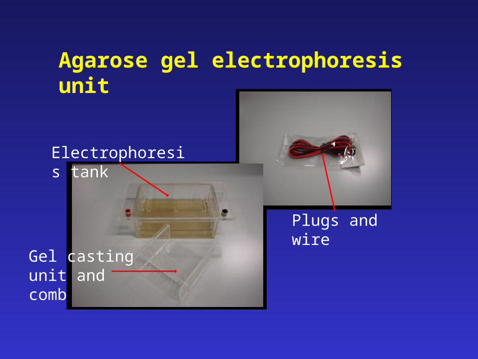
Agarose gel electrophoresis unit
Gel casting unit and comb
Electrophoresis tank
Plugs and wire
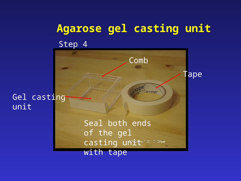
Seal both ends of the gel casting unit with tape
Agarose gel casting unitStep 4
Gel casting unit
Comb
Tape

Preparation of 1.5% agarose gelStep 5
http://dlab.reed.edu/projects/vgm/vgm/VGMProjectFolder/VGM/RED/RED.ISG/gel.html
Electrophoresis(5V/cm)
Sample loading, wash syringe with 1X TBE buffer between successive loading
Step 10
http://dlab.reed.edu/projects/vgm/vgm/VGMProjectFolder/VGM/RED/RED.ISG/gel.html
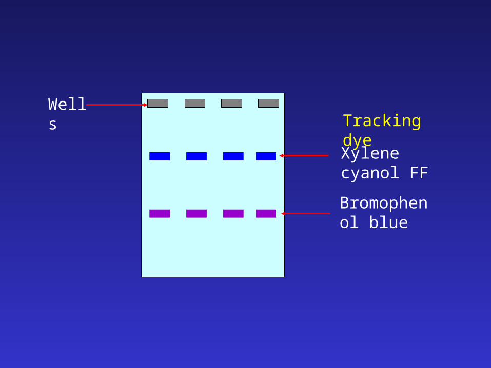
Wells
Bromophenol blue
Xylene cyanol FF
Tracking dye
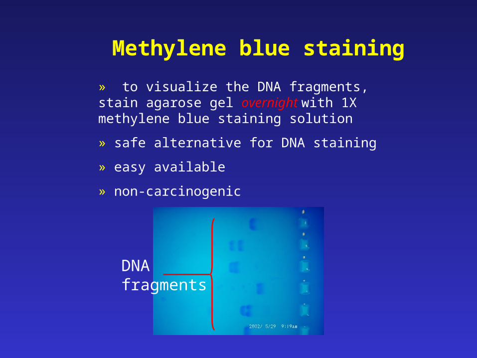
Methylene blue staining
» to visualize the DNA fragments, stain agarose gel overnight with 1X methylene blue staining solution
» safe alternative for DNA staining
» easy available
» non-carcinogenic
DNA fragments
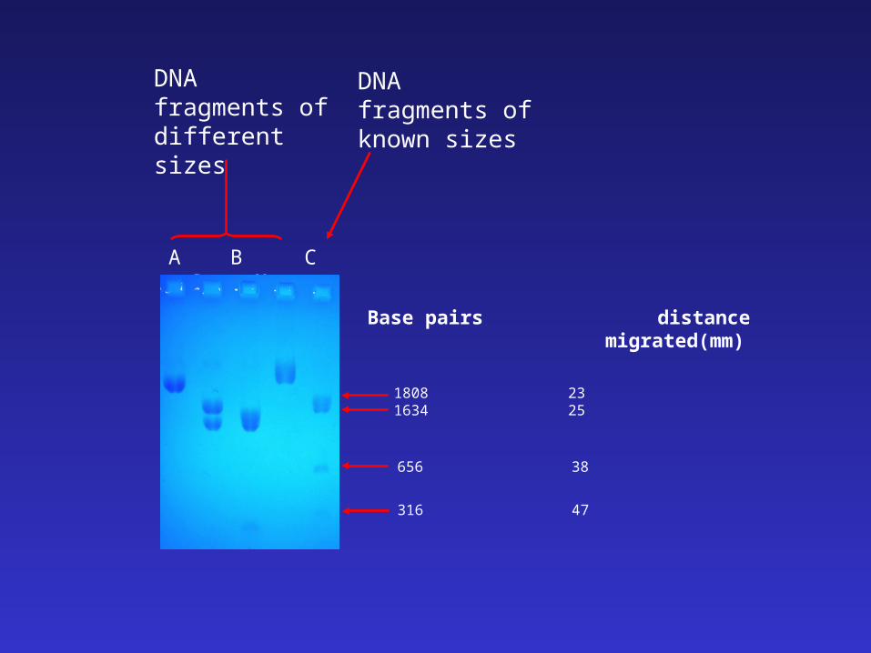
Base pairs distance migrated(mm)
A B C D M
316 47
656 38
1808 231634 25
DNA fragments of different sizes
DNA fragments of known sizes

Calibration curve for DNA size determination
http://www.pangloss.com/seidel/Protocols/webmap.html
Size determination of the candidate DNA fragments
http://www.pangloss.com/seidel/Protocols/webmap.html
Workshop
DNA Fingerprinting
&
Agarose Gel Electrophoresis
Department of BiochemistryCUHK
(TDC2003)
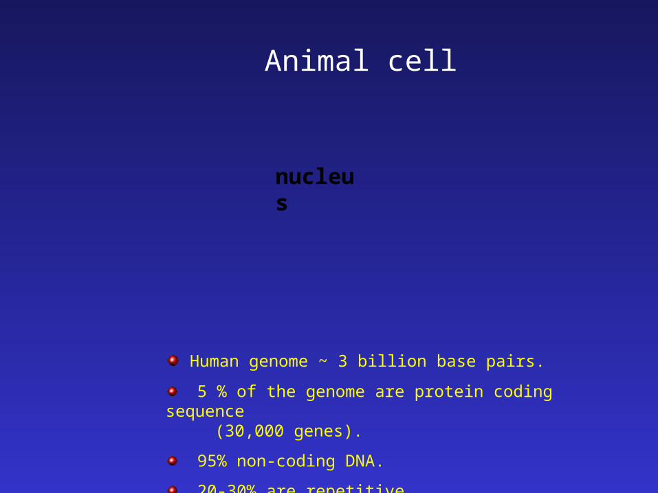
nucleus
Human genome ~ 3 billion base pairs.
5 % of the genome are protein coding sequence (30,000 genes).
95% non-coding DNA.
20-30% are repetitive.
Animal cell
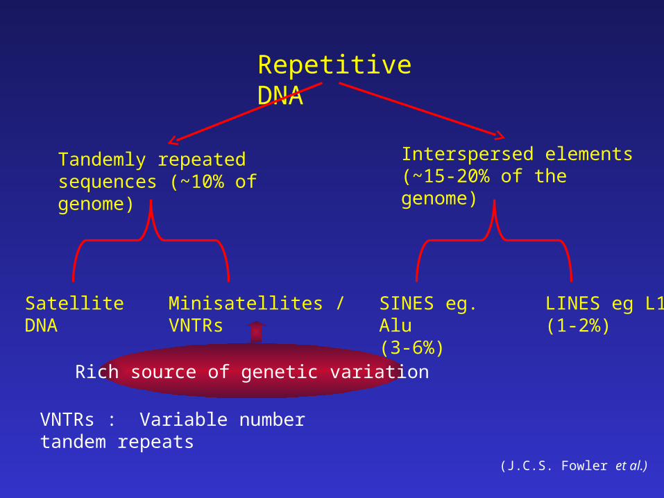
Repetitive DNA
Tandemly repeated sequences (~10% of genome)
Interspersed elements(~15-20% of the genome)
Satellite DNA Minisatellites / VNTRs SINES eg. Alu (3-6%)
LINES eg L1(1-2%)
(J.C.S. Fowler et al.)
VNTRs : Variable number tandem repeats
Rich source of genetic variation
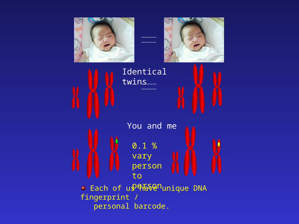
Identical twins
You and me
0.1 % vary person to person
Each of us have unique DNA fingerprint / personal barcode.
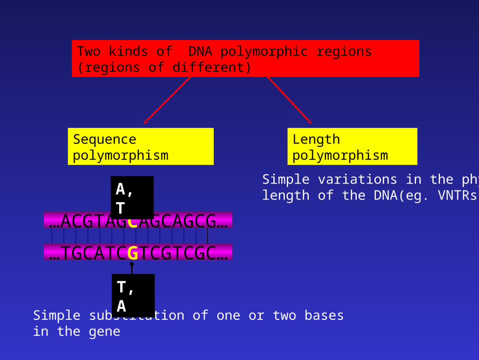
Two kinds of DNA polymorphic regions (regions of different)
Sequence polymorphism Length polymorphism
…ACGTAGCAGCAGCG…
…TGCATCGTCGTCGC…
A, T
Simple substitution of one or two bases in the gene
T, A
Simple variations in the physical length of the DNA(eg. VNTRs)
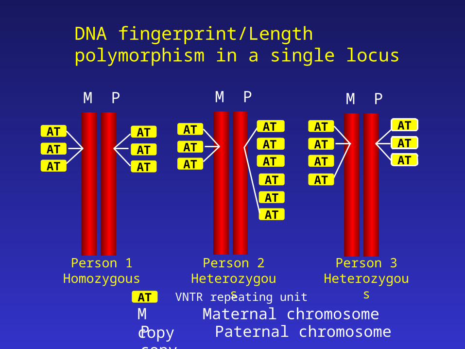
DNA fingerprint/Length polymorphism in a single locus
M P
AT
AT
AT
AT
AT
AT
M P
AT
AT
AT
AT
AT
AT
AT
AT
AT
AT
AT
AT
M P
AT
AT
AT
AT
AT
AT
AT
Person 1Homozygous
Person 2Heterozygous
Person 3Heterozygous
AT VNTR repeating unit
M Maternal chromosome copyP Paternal chromosome copy
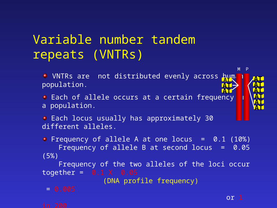
Variable number tandem repeats (VNTRs)
VNTRs are not distributed evenly across human population.
Each of allele occurs at a certain frequency in a population.
Each locus usually has approximately 30 different alleles.
Frequency of allele A at one locus = 0.1 (10%) Frequency of allele B at second locus = 0.05 (5%) Frequency of the two alleles of the loci occur together = 0.1 X 0.05 (DNA profile frequency) = 0.005
or 1 in 200
M P
ATATAT
ATATATATATAT
ATATAT
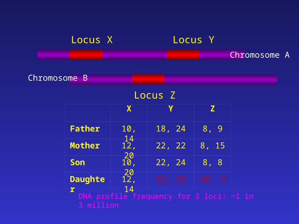
Locus X Locus Y
Locus Z X Y Z
Father 10, 14 18, 24 8, 9
Mother 12, 20 22, 22 8, 15
Son 10, 20 22, 24 8, 8
Daughter 12, 14 22, 22 26, 9
Chromosome A
Chromosome B
DNA profile frequency for 3 loci: ~1 in 3 million
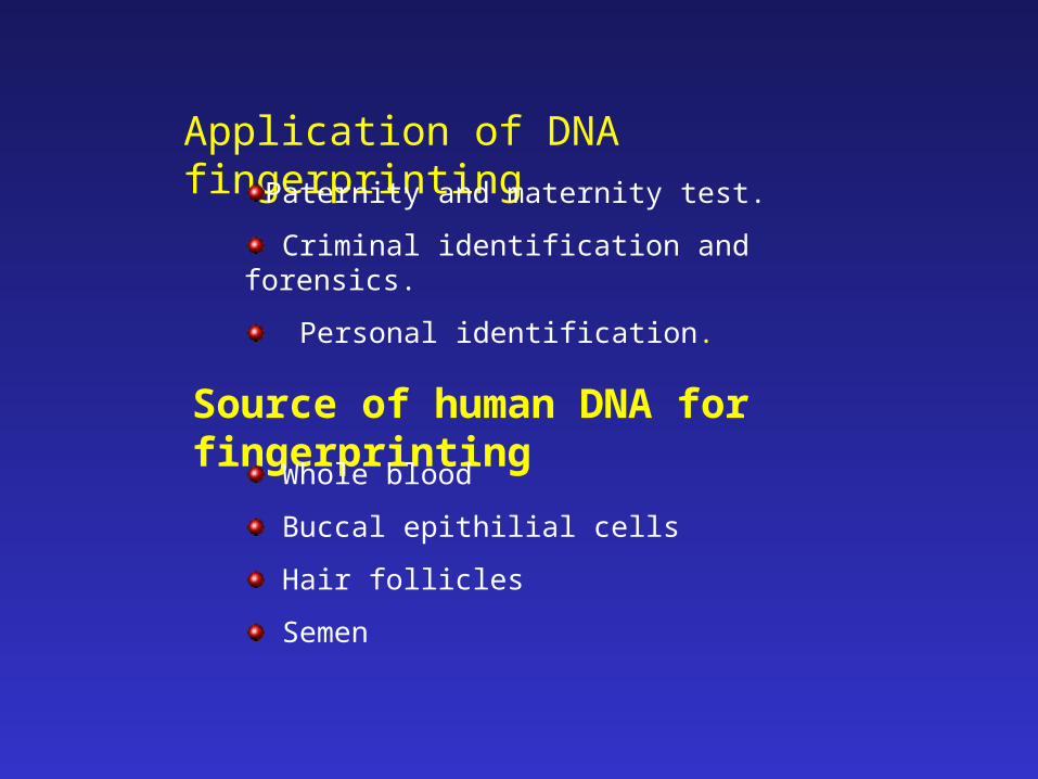
Application of DNA fingerprinting
Paternity and maternity test.
Criminal identification and forensics.
Personal identification.
Source of human DNA for fingerprinting
Whole blood
Buccal epithilial cells
Hair follicles
Semen

Double stranded target DNA
Two DNA targets available for PCR
PCR amplification
Cycle(35 – 40 cycles)
DNA denatured95oC
Step 1 : Denaturation
Primers bind to target DNA~55oC
Step 2 : Primer Annealing
Double stranded DNA duplicated72oC
Step 3 : DNA Extension
Amount of amplified DNA = 2n x Cwhere n = number of PCR cycles; and C = the initial number of copies of DNA template present in the tube.
So, you will get 1,048,576 copies of DNA after 20 cycles of PCR reaction even you start with only one copy of DNA template initially
Polymerase chain reaction
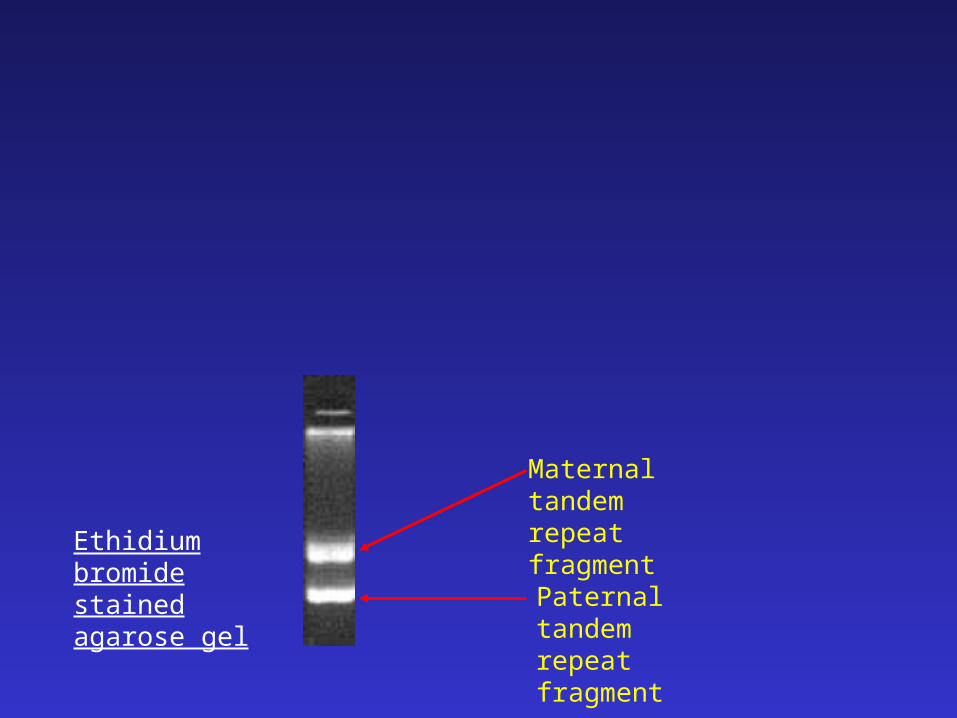
Maternal tandem repeat fragment
Paternal tandem repeat fragment
Ethidium bromide stained agarose gel
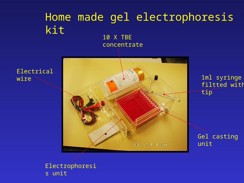
Electrical wire1ml syringe filtted with tip
10 X TBE concentrate
Electrophoresis unit
Gel casting unit
Home made gel electrophoresis kit
Agarose
A linear polymer extracted from seaweed
Migration of DNA in agarose dependent on four factors
- molecular size of the DNA- agarose concentration- conformation of the DNA- applied current
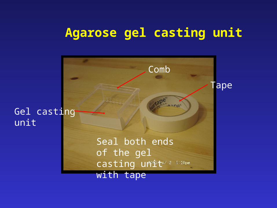
Seal both ends of the gel casting unit with tape
Agarose gel casting unit
Gel casting unit
Comb
Tape
Preparation of 1.5% agarose gel
Wells
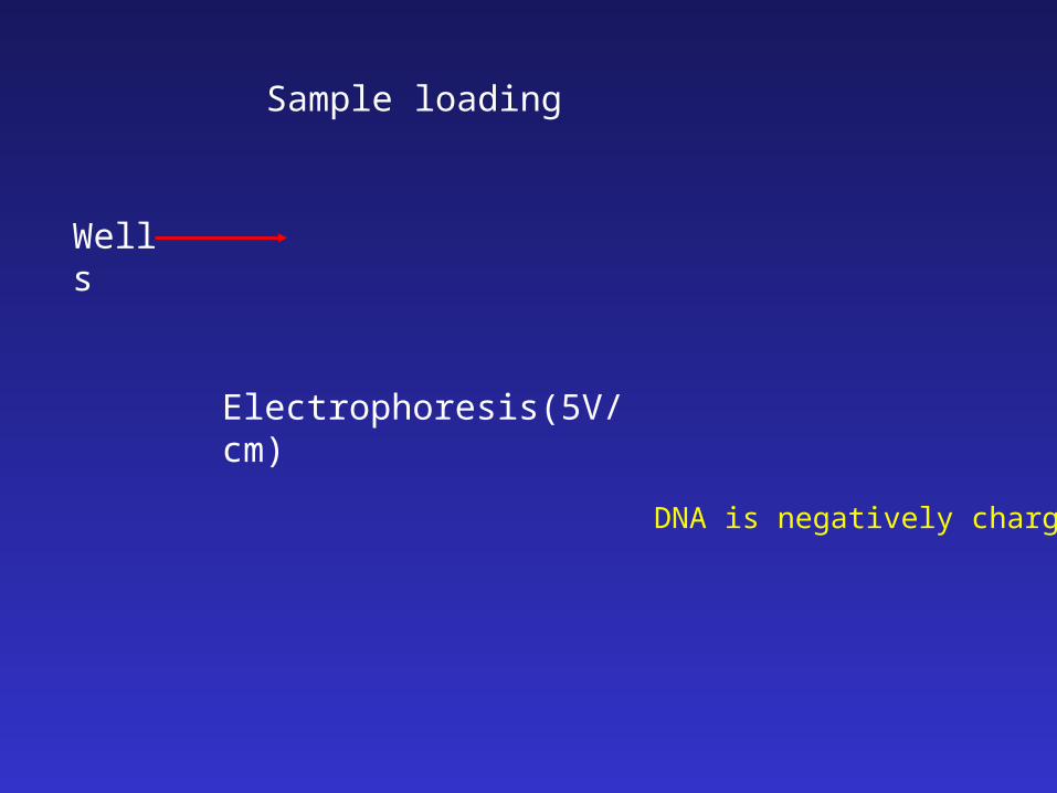
Electrophoresis(5V/cm)
Sample loading
DNA is negatively charged
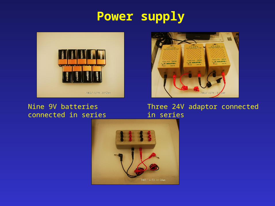
Power supply
Nine 9V batteries connected in series
Three 24V adaptor connected in series
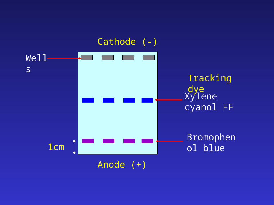
Wells
Bromophenol blue
Xylene cyanol FF
Tracking dye
1cm
Cathode (-)
Anode (+)
Methylene blue staining
» to visualize the DNA fragments, stain agarose gel overnight with 1X methylene blue staining solution and destain in distilled water for 3 – 4 hours
» non-toxic
» easy available
» non-carcinogenic
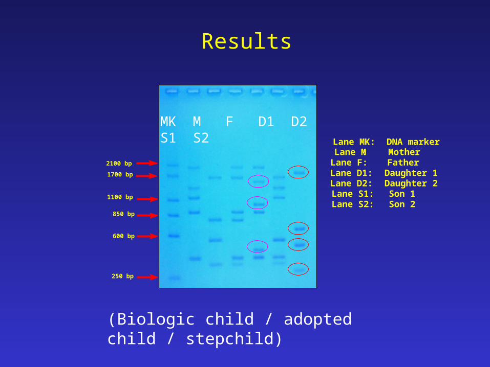
Lane MK: DNA marker Lane M: Mother Lane F: Father Lane D1: Daughter 1 Lane D2: Daughter 2 Lane S1: Son 1 Lane S2: Son 2
2100 bp
1700 bp
1100 bp
850 bp
600 bp
250 bp
MK M F D1 D2 S1 S2
Results
(Biologic child / adopted child / stepchild)
Calibration curve for DNA size determination
Size determination of the candidate DNA fragments
online size determination

THE END