basic transplant immunology - louisville
TRANSCRIPT

Basic Transplant Immunology & Immunosuppressive Drugs
Luis S. Marsano, MD Professor of Medicine
Division of Gastroenterology, Hepatology & Nutrition University of Louisville &
Louisville VAMC
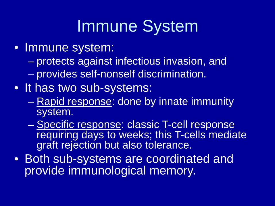
Immune System • Immune system:
– protects against infectious invasion, and – provides self-nonself discrimination.
• It has two sub-systems: – Rapid response: done by innate immunity
system. – Specific response: classic T-cell response
requiring days to weeks; this T-cells mediate graft rejection but also tolerance.
• Both sub-systems are coordinated and provide immunological memory.

Hyperacute Rejection
• Extremely rare. • Occurs hours to days after transplantation, • Target is vascular endothelium. • Antibody-mediated, & complement
dependent graft destruction by coagulative necrosis.
• Preformed antibodies specific to MHC. • Lack of lymphocytic infiltration.

Acute Rejection • Occurs in 45-70% of patients. • Days to months after transplant (usually initial 3 months). • Classical, cell-mediated rejection:
– Predominantly CD4 & CD8 T-cells. – Directed against donor MHC antigens (cholangiocytes &
vascular endothelium) • Target of current immunosuppression. • Diagnostic Triad:
– Portal inflammation – Bile-duct damage – Venular endothelium inflammation.

Acute Rejection
• Generally 5-30 days; 10-20% of patients. • Higher in: Autoimmune dz, females, young age,
DR mismatch, retransplant. • Usually asymptomatic unless late diagnosis.
– Fever, Abdominal pain, Ascites, Leukocytosis, Eosinophilia
• Biochemical abnormalities not specific (high GGT)

Management of Acute Rejection • Moderate to Severe: Corticosteroids. • Refractory:
– ATG; – Consider Antibody Mediated Rejection (C4D staining + Donor
Specific AB testing).
• Investigate the reason of rejection. • Optimize immunosuppression. • If already optimized, add MMF, AZA or SRL. • Consider long term prednisone in:
– autoimmune liver dz, or – after 2 episodes of severe AR.

Variants of Classic AR • Plasma cell hepatitis:
– Early, often with (-) auto-Abs (> 60%) – Centrilobular necrosis – ? Antibody mediated Rejection
• Idiopathic post-transplantation hepatitis: – 5-15% with fibrosis progressing to cirrhosis over 10 y. – Auto-Abs and plasma cell are associated with progression.
• Late Acute Rejection (> 6 months after LT): – May be histologically different. – Lobular activity, interface hepatitis, central perivenulitis (without
endothelitis); can mimic plasma cell hepatitis. – Possible evolution to chronic rejection with perivenular
hepatocyte drop out and loss of inflammation.

Antibody Mediated Rejection When to Consider
• RISK FACTORS: – Refractory Rejection (steroid resistant). – Re-transplant (sensitized). – HLA mismatch, positive x-match. – Necrosis or vascular injury.
• TREATMENT: In evolution; plasmapheresis + immunosuppression changes.
• PROPHYLAXIS: – blood transfusion minimization in cirrhosis. – Adherence to immunosuppression regimen.

Risk of Rejection in OLTx with Different Regimens
Regimen Acute (ACR)
Chronic (Ductopenic)
Pred + Aza 85 % 25 %
Pred + CyA 70 % 15 %
Pred + Tacr 55 % 6 %
Pred + CNI + MMF 45 % 1 %
Tacr + Rapa 18 % 1 %

Chronic Rejection • Occurs to 2-5% of patients. • Slow, indolent process months to years after transplantation. • Rise of cholestatic enzymes. • Has immune & non-immune components; poorly defined. • Causes ischemic injury and paucity of bile ducts. • Characterized by arteriole thickening & interstitial fibrosis. • Loss of small bile ducts +/- neo-intimal proliferation with
obliterative vasculopathy (foam cell obliterative arteriopathy). – Bile duct loss > 50%, or – bile duct atrophy/pyknosis in the majority of bile ducts, or – foam cell obliterative arteriopathy.

Risk Factors for CR
• Multiple AR episodes. • Severe AR with Centrilobular Necrosis. • Non-compliance. • Under immunosuppression.

CR Prognostic Factors
• Bile duct loss > 50% • Severe, bridging, perivenular fibrosis. • Foam cell clusters within the sinusoids. • Severe hyperbilirubinemia (TB >/= 25)
– Bili of </= 4.6 has higher resolution.

Management of CR
• Switch CyA to TAC while TB < 10 mg/dL – 50% success – Ductular reaction is a positive feature.
• Higher TAC levels • Add mTOR-I or MMF
– Consider infection prophylaxis • Avoid over-immunosuppression with late
cases of liver synthetic dysfunction.

GVHD post Liver Transplant • Less than 1%, but > 80% mortality. • Usually 2-8 weeks post-LT • TRIAD: Skin Rash + Cytopenia + Diarrhea, with normal
Liver Enzymes and allograft function. • Diagnosis: FISH X-Y chimerism, PBMC donor-recipient
chimerism, skin Bx, or rarely intestinal Bx. • Treatment:
– High dose steroids – ? Lymphodepletion – ? Stop immunosuppression – Stem Cell transplant.

T-cell Recognition of Alloantigen & T-cell Activation: Rejection
• Recipient T-lymphocytes recognize a donor alloantigen by: – a) Direct Path : native donor MHC molecule
expressed in donor APCs , – b) Indirect Path : donor alloantigen
peptides (from damaged cells or soluble MHC class I) presented by recipient APCs.
• “Direct path” dominates in “acute” rejection, and • “Indirect path” in chronic rejection and tolerance.
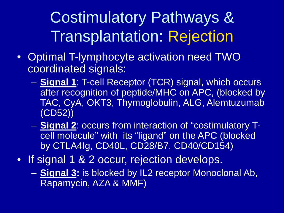
Costimulatory Pathways & Transplantation: Rejection
• Optimal T-lymphocyte activation need TWO coordinated signals: – Signal 1: T-cell Receptor (TCR) signal, which occurs
after recognition of peptide/MHC on APC, (blocked by TAC, CyA, OKT3, Thymoglobulin, ALG, Alemtuzumab (CD52))
– Signal 2: occurs from interaction of “costimulatory T-cell molecule” with its “ligand” on the APC (blocked by CTLA4Ig, CD40L, CD28/B7, CD40/CD154)
• If signal 1 & 2 occur, rejection develops. – Signal 3: is blocked by IL2 receptor Monoclonal Ab,
Rapamycin, AZA & MMF)

Effector Pathways of Graft Injury Rejection
• There is not a single mediator or cell type that is absolutely required for allograft rejection; there are several redundant and compensatory mechanisms contributing to rejection.
• After [T-cell Receptor signal + costimulatory signal, + cytokines], there is proliferation and maturation of CD4+ or CD8+ T-cells capable of graft injury; this will lead to: – T-cell mediated cytotoxicity – Delayed hypersensitivity – Antibody-mediated damage

Effector Pathways of Graft Injury Rejection
• T-cell mediated cytotoxicity: – A) CD8+ cytotoxic T-lymphocytes (CTLs)
specific for donor class I, cause apoptosis through biochemical mechanisms (perforin/granzyme B in a Ca++ dependent process, and Fas/FasL through caspase 8);
– B) NK cells, without need for activation or sensitization, which can cause apoptosis through FasL & granzyme B.

Effector Pathways of Graft Injury Rejection
• Delayed hypersensitivity: – CD4+ T-lymphocytes specific for donor class
II, release IFN gamma activating macrophages and cellular mediators.
• Antibody-mediated damage: – Antibodies against liver sinusoidal endothelial
cells (LSECs) indirectly promote acute rejection.

Immune System & Tolerance • Tolerance: Absence of destructive response to
an allograft in immunocompetent host. • Tolerance is accomplished by T-cell suppression
mediated by : – a) cell-contact dependent mechanism:
CD4+CD25+ cells, – b) cytokine mediated T-cell mechanism:
T regulatory-1 & T helper-3 (Th3), – c) antigen presentation dependent mechanism: by
liver-derived Dendritic Cells (DCs) and by Liver Sinusoidal Endothelial Cells (LSECs) which behave as immature DCs causing incomplete activation, inhibiting T-cell response.
– d) NK cells which give a “death signal” to recipient derived T-cell passing through the graft.

Costimulatory Pathways & Transplantation: Tolerance
• Optimal T-lymphocyte activation needs coordinated “signal-1” and “signal-2” stimuli.
• If only “signal 1” occurs, tolerance develops; • To prevent rejection and induce tolerance, you can
disrupt “signal 2”: – a) CTLA4 (cytotoxic T lymphocyte antigen 4) can compete with
CD28 for B7, and gives “negative costimulation”; CTLA4-Ig fusion protein has been used for this goal,
– b) anti-CD154 disrupts CD40/CD154 pathway. • Programmed death-1 (PD-1) is a molecule induced
upon T-cell activation and causes a “negative signal” similar to CTLA4, causing spontaneous tolerance. PD-1 binds to ligands PDL-1 & PDL-2.

Possible Mechanisms for Liver Tolerance
• 1) The liver produces large amounts of soluble MHC class I antigen, causing: – a) Passive blockade of alloantibodies & donor-specific
effectors, or – b) Activation-induced apoptosis of allospecific CTLs.
• 2) Liver suppressor factor-1: is produced by spontaneously tolerant recipients and prolongs rat cardiac allograft survival.
• 3) Liver produces a soluble Fas “incomplete variant”, which inhibits anti-Fas induced apoptosis and inhibits CTL function in vitro.

Possible Mechanisms for Liver Tolerance
• 4) Graft-derived Stem-cells migrate out of the liver and establish “microchimerism” with clonal exhaustion/deletion of host alloreactive T-cells.
• 5) Inmature “Dendritic Cells” (DCs) and “Liver Sinusoidal Endothelial Cells” (LSECs) do not express enough costimulatory molecules, hence facilitate tolerance.
• 6) Kupffer cells (APCs) express FasL which can induce apoptosis of host T-cells.

Costimulatory Pathways & Transplantation: Autoimmunity
• Deficiency in “Programmed death-1” (PD-1) molecule and/or PDL-1 causes autoimmune disorders and autoimmune hepatitis with large amounts of CD8 T-cells in the liver.

Immunosuppression in Liver Transplantation

Survival vs Rejection in OLTx
90 88 86 82 8277
8275
0102030405060708090
100
1 y 2 y 3 y 4 y
RejectionNo-Rejection
Rejection/No-Rejection RR = 0.7

Causes of Late Liver-Graft-Loss
Rejection < 5 %
De-novo Malignancy 15 %
Infections 16 %
Cardiovascular Disease 20 %
Recurrent Disease 35 %
Excessive Immunosuppression causes more problems than rejection

Impact of ACR Therapy on Survival
Patient Therapy RR Mortality
Non-HCV Steroids 0.5
HCV Steroids 2.9
HCV OKT3 5.4
DO NOT TREAT MILD REJECTION IN HCV

Long-Term Complications of Immunosuppression
Renal Dysfunction 80 %
Hypertension* 70 %
Hyperlipidemia* 50 %
Diabetes Mellitus* 20 %
Bone Disease* 20 %
Skin Cancer 40 %
Lymphoma 4 %
* Less if Steroids are withdrawn shortly after 3 months

Cyclosporin A • From Tolypocladium inflatum; approved in 1983. Is
calcineurin inhibitor. • Selective immunosuppression by inhibition of T-cell
activation. • CyA forms complex with cytoplasmic receptor
“cyclophilin” and inhibits calcium- & calmodulin-dependent phosphatase calcineurin.
• Inhibition of Ca+ dephosphorilation of NFAT (Nuclear Factor of activated Tcells) – Calcineurin is vital for the transcriptional process by which IL-
2 and other cytokines are activated, which is needed for T-helper cell mediated graft rejection.

Cyclosporin A
• Currently CyA comes as a microemulsion
in lipophilic solvent which is less dependent in bile flow (Neoral, Gengraf).
• CyA is metabolized in the liver by P450-3A pathway.

Cyclosporin A Toxicity
• Nephrotoxicity: can be acute or long term; renal failure in up to 20%; can cause hyperkalemia and hypomagnesemia.
• Hyperlipidemia, hyperglycemia, hypertension, gingival hyperplasia, hirsutism.
• 10-28% may have tremor, peripheral neuropathy, psychoses, hallucinations, motor weakness, or seizures.
• May cause Hemolytic Uremic Syndrome.

Cyclosporin A Dose & Target Levels
• Initial 10-15 mg/kg/d divided q 12h; check trough level after 24 h.
• New data indicates that level 2-h post dose represents better “total exposure”.
• Week 0-2: trough 250-350 ng/mL • Week 3-4: 200-300 • Week 5-24: 150-250
(850-1400 2h post) • Week 25-52: 100-200

Drugs that Increase Cyclosporin & Tacrolimus Levels
Calcium Channel Blockers
Antifungals Macrolide antibiotics
Pro-kinetics
Miscellaneous
Diltiazem Nicardipine Nifedipine Verapamil
Fluconazole Itraconazole Ketoconazole Voriconazole Clotrimazole
Clarithromycin Erythromycin Troleandomycin Azithromycin Telithromycin
Cisapride Metoclo_pramide
Amiodarone Cimetidine Methyl-prednisolone Omeprazole Protease inhibitors Nefazodone Ethinyl estradiol Grapefruit juice

Drugs that Decrease Cyclosporin & Tacrolimus Levels
Anticonvulsants Antibiotics Herbal Preparations
Miscellaneous
Carbamazepine Phenobarbital Phenytoin Fosphenytoin
Rifabutin Rifampin Rifapentin
St. John’s Wort Probucol Terbinafine

Tacrolimus
• From Streptomyces tsukubaensis. • It is 100-times stronger than CyA. • Binds to FKBP12 and the complex inhibits
calcineurin; this prevents transcription of IL-2, IL-3, IL-4, IL-8, and various chemotactic factors.
• It is absorbed in duodenum & jejunum without need for bile.
• Food decrease bioavailability. • Metabolized via P450-3A pathway.

Tacrolimus Toxicity & Dose
• More DM than CyA. More HUS than CyA. • Less HTN, dyslipidemia, hirsutism (TAC causes hair
loss), gum hyperplasia than CyA. • Similar hyperkalemia, tremor, hypomagnesemia,
infection, malignancies, & renal dysfunction than CyA. • Nausea, vomiting, diarrhea, headache. • Less rejection in 1st year in all, less steroid-resistant
rejection, and longer graft survival in Hepatitis C than CyA.
• Dose: 0.1-0.15 mg/kg/d divided q 12h p.o.; trough levels 10-15 ng/mL early; 8-10 later.

Calcineurin Inhibitors in OLTx Risk of Chronic Renal Failure
2.5 3.55
10
21
0
5
10
15
20
25
1 year 3 years 5 years 10 years 15 years
CRF

Risk Factors for CRF in Non-Renal Tx
Relative Risk
Post-Op ARF 2.13
Diabetes Mellitus 1.42
Age (per each 10 years) 1.36
Hypertension 1.18
Hepatitis C 1.15

Corticosteroids • Block T-cell-derived and antigen-presenting cell-derived
cytokine expression, decreasing IL-1, IL-2, IL-3, and IL-6 • Are used in reversing acute rejection and in
maintenance. • Side effects: hypertension, mental status changes,
dyslipidemia, poor wound healing, hyperglycemia, gastric ulcers, myopathy, osteoporosis, Cushing S., fungal/bacterial infections, pituitary axis suppression, fluid retention, cataracts.
• Dose: 500-1000 mg pre-op; then taper from 50 to minimal dose over a few months.

Beneficial Effect of Steroid-Withdrawal after 3 months post OLTx
Steroids No-Steroids P-value
Hypertension 58 % 15 % 0.0002
Diabetes 25 % 6 % 0.007
Infection 17 % 2 % 0.05
Bone Disease 9 % 0 % 0.05
Mean Cholesterol 253 mg/dL 183 mg/dL 0.001

Adverse Effects of Steroid-Withdrawal
• Recurrent AIH & PBC • Worsens HCV if done before 3rd month. • Flare up of Ulcerative Colitis • Arthralgias • Depression

Azathioprine (AZA)
• Antimetabolite; antagonises purine metabolism. Inhibits synthesis of DNA, RNA, and proteins.
• Used in < 5% US transplant centers. • Can cause myelosuppression and
hepatotoxicity. • Side effects: nausea, vomiting, diarrhea,
pancreatitis, anemia, leukopenia, thrombocytopenia, and weight loss.
• Usual dose: 1-2 mg/kg/d
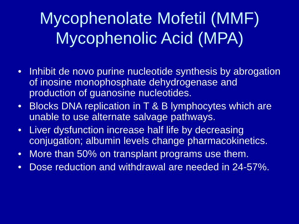
Mycophenolate Mofetil (MMF) Mycophenolic Acid (MPA)
• Inhibit de novo purine nucleotide synthesis by abrogation of inosine monophosphate dehydrogenase and production of guanosine nucleotides.
• Blocks DNA replication in T & B lymphocytes which are unable to use alternate salvage pathways.
• Liver dysfunction increase half life by decreasing conjugation; albumin levels change pharmacokinetics.
• More than 50% on transplant programs use them. • Dose reduction and withdrawal are needed in 24-57%.

MMF & MPA Toxicity & Dose
• Nausea, vomiting, abdominal pain, diarrhea, anemia, leukopenia, thrombocytopenia, hypercholesterolemia, hypokalemia, tremor, hypertension, edema.
• MMF: 2-3 g/day, divide q 12h • MPA: 720-1440 mg/d divided q 12h.

Drug-Drug Interaction Azathioprine & Mycophenolate
Increases AZA
Increases MMF
Decreases AZA & MMF
Allopurinol Methotrexate ACE inhibitors
Probenecid Tacrolimus
Cholestiramine Antacids Iron preparations

Triple Therapy Prednisone + CNI + MMF
• Improves patient & graft survival in HCV & Non-HCV.
• Lower ACR rate in HCV & Non-HCV • Less renal toxicity with lower level of CNI. • Does not increase risk of infection nor
malignancy.

Sirolimus/Everolimus • Macrocyclic triene antibiotic with immunosuppressive,
antitumor & antifungal properties • Binds to immunophilin FKBP12 but has different action
than TAC: blocks cell-cycle progression at the “G1 – S phase” junction; mTOR.
• No calcineurin inhibition, hence no increase in endothelin nor TGF beta that cause vasoconstriction and renal injury.
• Suppresses cytokine driven T Cell proliferation. • Increase risk of Hepatic Artery Thrombosis: “The safety
and efficacy of Sirolimus…has not been established in liver transplant patients, and therefore such use is not recommended”.
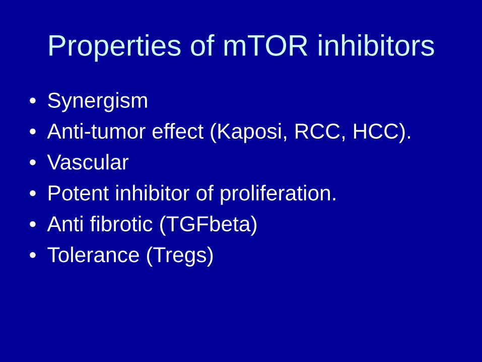
Properties of mTOR inhibitors
• Synergism • Anti-tumor effect (Kaposi, RCC, HCC). • Vascular • Potent inhibitor of proliferation. • Anti fibrotic (TGFbeta) • Tolerance (Tregs)

Sirolimus/Everolimus Toxicity & Dose
• Anemia, hypercholesterolemia, hypertrigliceridemia, hyperlipidemia, leukopenia, thrombocytopenia, interstitial lung disease, peripheral edema, wound dehiscence, lymphocele, oral ulcers.
• Dose Sirolimus: 2 mg/d, adjusted to maintain trough level of 4-10 ng/mL.
• Everolimus given BID due to short half-life.

Drugs that Increase Rapamycin Concentration
Calcium Channel Blockers
Antifungals Macrolide antibiotics
Pro-kinetics
Miscellaneous
Diltiazem Nicardipine Nifedipine Verapamil
Fluconazole Itraconazole Ketoconazole Voriconazole Clotrimazole
Clarithromycin Erythromycin Troleandomycin Azithromycin Telithromycin
Cisapride Metoclo_pramide
Amiodarone Cimetidine Omeprazole Methyl-prednisolone Protease inhibitors CyA Graprfruit juice

Drugs that Decrease Sirolimus Concentration
Anticonvulsants Antibiotics Herbal Preparations
Carbamazepine Phenobarbital Phenytoin Fosphenytoin
Rifabutin Rifampin Rifapentin
St. John’s Wort

Special Conditions to use Rapamycin
HCC Anti-tumor Effect
HCV & PSC Anti-fibrotic Effect
Renal Insufficiency
Spares CNI

Antithymocyte Globulin (ATG) • ATGAM (equine) and Thymoglobulin (rabbit) • Polyclonal Ab against T-cells epitopes (CD2, CD3, CD4,
CD8, CD28, & T-cell receptor), NK cells epitopes (CD16), and macrophages.
• Cause T-cell depletion by: apoptosis, antibody mediated cytolysis, and internalization of cell surface receptors.
• First dose can cause “cytokine release S”: fever, chills, tachycardia, chest pain, bronchospasm, GI disturbances, blood pressure changes. Steroids + Benadryl + acetaminophen helps.
• Used in 6% of US transplant programs. • Dose: 1.5-5 mg/kg/d over 4-6 h infusion, for 3-5 days.

Muramonab-CD3 (OKT3)
• Murine Ab against T-cell CD3 antigen; inactivates T-cell receptor.
• Cytokine release syndrome is very common 1-3 h after first dose. Sometimes life-threatening with pulmonary edema and shock.
• Re-exposure to OKT3 may decrease efficacy. • Dose: 5 mg IV q day x 10-14 days for steroid
resistant rejection.

IL-2 receptor antibodies Basiliximab & Daclizumab
• Basiliximab (Simulect) is chimeric, Daclizumab (Zenapax) is humanized;
• Bind to IL-2R alpha-chain present in activated T-lymphocytes. Causes competitive antagonism of IL-2 induced T-cell proliferation.
• Effect up to 3 weeks with Basiliximab, and 10 weeks with Daclizumab.
• Side effects are mild. • Dose:
– a) Basiliximab: 20 mg IV pre-op + 20 mg 4 d later. – b) Daclizumab: 1 mg/kg every 14 days x 5 doses.
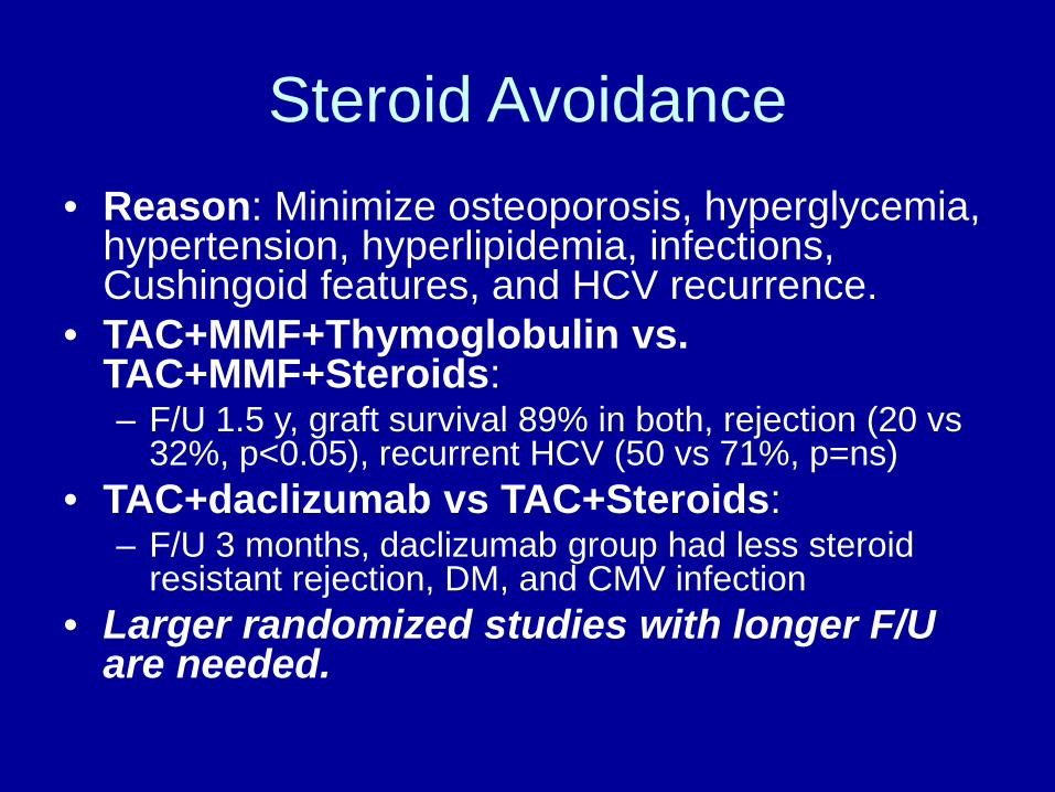
Steroid Avoidance • Reason: Minimize osteoporosis, hyperglycemia,
hypertension, hyperlipidemia, infections, Cushingoid features, and HCV recurrence.
• TAC+MMF+Thymoglobulin vs. TAC+MMF+Steroids: – F/U 1.5 y, graft survival 89% in both, rejection (20 vs
32%, p<0.05), recurrent HCV (50 vs 71%, p=ns) • TAC+daclizumab vs TAC+Steroids:
– F/U 3 months, daclizumab group had less steroid resistant rejection, DM, and CMV infection
• Larger randomized studies with longer F/U are needed.

Renal Sparing Protocols
• Up to 21% of LTx patients develop CRF within 5 years.
• 18% of patients have severe renal dysfunction after 13 years.
• Adding MMF and reducing dose of calcineurin inhibitor (CNI) can improve GFR by 15% in 50% of patients even if done > 1 y post-OLTx; if CNI is D/C, rejection risk is increased.

Conversion from CNI to Sirolimus
• 28 patients with creatinine > 1.8 mg/dL were converted; mean time= 2y post-LTx.
• Dose: 2 mg/d, titrated to 4-10 ng/mL. • 14 (50%) had improvement in GFR;
7 progressed to ESRD, and 6 did not tolerate the change.
• Large randomized trials are ongoing to evaluate proper time to change.


Effect of Steroid-Withdrawal after 3 months post OLTx
Steroids No-Steroids P-value Survival 82 % 83 % NS Hypertension 58 % 15 % 0.0002 Diabetes 25 % 6 % 0.007 Infections 17 % 2 % 0.05 Recurrent HCV 17 % 21 % NS Bone Disease 9 % 0 % 0.05 Acute Rejection 8 % 4 % Chronic Rejection 1 % 2 % Mean Cholesterol 253 mg/dL 183 mg/dL 0.001