bau1ch05pg125 166 b - pearson schweiz ag · pdf filetransfer of electrons between molecules....
TRANSCRIPT
125
MicrobialMetabolism
C H A P T E R 5
A worker prepares cacao seeds for fermentation.
The next time you bite into adelicious piece of chocolate,consider this: Microbial metabo-lism played a key role in howthat chocolate came to taste sogood.
Chocolate comes from cacaoseeds, found inside the pods ofTheobroma cacao trees. After thepods are split open, the seedsand the surrounding pulp arescooped out and placed in heapson top of plantain or bananaleaves. The heaps are then cov-ered and left to ferment for 2–7days. Fermentation occurs asmicroorganisms—includingyeast and several kinds ofbacteria—grow on the fleshy,sugary pulp. Bathed in ferment-ing pulp, the cacao seeds (whichstart off tasting bitter) begin todevelop the flavors and colorsthat we associate with choco-late. After fermentation, theseeds are dried, roasted, andthen processed further by choco-late manufacturers before be-coming one of our favoritedesserts.
In this chapter we will learnabout many metabolic processesof microorganisms, including fer-mentation.
C H A P T E R O U T L I N E
Basic Chemical ReactionsUnderlying Metabolism
Catabolism and AnabolismOxidation and Reduction ReactionsATP Production and Energy StorageThe Roles of Enzymes in Metabolism
Carbohydrate CatabolismGlycolysisAlternatives to GlycolysisCellular RespirationFermentation
Other Catabolic PathwaysLipid CatabolismProtein Catabolism
PhotosynthesisChemicals and StructuresLight-Dependent ReactionsLight-Independent Reactions
Other Anabolic Pathways Carbohydrate BiosynthesisLipid BiosynthesisAmino Acid BiosynthesisNucleotide Biosynthesis
Integration and Regulationof Metabolic Functions
MicroPrep Pre-Test: Take the pre-test for this chapter on the web. Visit: www.microbiologyplace.com.

126 CHAPTER 5 Microbial Metabolism
How do pathogens acquire energy and nutrientsat the expense of a patient s health? How does grape juiceturn into wine, and how does yeast cause bread to rise? Howdo disinfectants, antiseptics, and antimicrobial drugs work?When laboratory personnel perform biochemical tests toidentify unknown microorganisms and help diagnose dis-ease, what exactly are they doing?
The answers to all of these questions require anunderstanding of microbial metabolism,1 the collection ofcontrolled biochemical reactions that takes place within thecells of an organism. While it is true that metabolism in itsentirety is complex, consisting of thousands of chemical re-actions and control mechanisms, the reactions are neverthe-less elegantly logical and can be understood in a simpli edform. In this chapter we will concern ourselves only with thecentral metabolic pathways and energy metabolism.
Your study of metabolism will be manageable if youkeep in mind that the ultimate function of metabolism is toreproduce the organism, and that metabolic processes areguided by the following eight elementary statements:
1. Every cell acquires nutrients, which are the chemicalsnecessary for metabolism.
2. Metabolism requires energy from light or from thecatabolism (kaø-tab ło-lizm), or breakdown, of acquirednutrients.
3. Energy is stored in the chemical bonds of adenosinetriphosphate (ATP).
4. Using enzymes, cells catabolize nutrient molecules toform elementary building blocks called precursormetabolites.
5. Using these precursor metabolites, energy from ATP,and other enzymes, cells construct larger building blocksin anabolic (an-aø-bol ik), or biosynthetic, reactions.
6. Cells use enzymes and additional energy from ATP tolink building blocks together to form macromoleculesin polymerization reactions.
7. Cells grow by assembling macromolecules into cellularstructures such as ribosomes, membranes, and cellwalls.
8. Cells typically reproduce once they have doubled insize.We will discuss each aspect of metabolism in the chap-
ters that most directly apply. For instance, we discussed therst step of metabolism—the active and passive transport of
nutrients into cells—in Chapter 3. In this chapter we will ex-amine the importance of enzymes in catabolic and anabolicreactions, study the three ways that ATP molecules are syn-thesized, and show that catabolic and anabolic reactions arelinked. We will also examine the catabolism of nutrient mol-ecules; the anabolic reactions involved in the synthesis ofcarbohydrates, lipids, amino acids, and nucleotides; and a
few ways that cells control their metabolic activities. Geneticcontrol of metabolism and the polymerization of DNA,RNA, and proteins are discussed in Chapter 7, and thespeci cs of cell division are covered in Chapters 11 and 12.
Basic Chemical ReactionsUnderlying Metabolism
In the following sections we will examine the basic conceptsof catabolism, anabolism, and a special class of reactionscalled reduction and oxidation reactions. The latter involve thetransfer of electrons between molecules. Then we will turnour attention brie y to the synthesis of ATP and energy stor-age before we discuss the organic catalysts called enzymes,which make metabolism possible.
Catabolism and Anabolism
Learning Objective
� Distinguish among metabolism, anabolism, and catabolism.
Metabolism, which is all of the chemical reactions in anorganism, can be divided into two major classes of reactions:catabolism and anabolism (Figure 5.1). A series of reactionsis called a pathway. Cells have catabolic pathways, whichbreak larger molecules into smaller products; and anabolicpathways, which synthesize large molecules from the smaller
Figure 5.1 Metabolism is composed of catabolic and anabolicreactions. Some energy released in catabolism is storedin ATP molecules, but most is lost as heat. Anabolicreactions require energy, typically provided by ATP.There is some heat loss in anabolism as well. Theproducts of catabolism provide many of the buildingblocks for anabolic reactions; other anabolic buildingblocks must be acquired directly as nutrients.
➤
1From Greek metabole, meaning change.
CHAPTER 5 Microbial Metabolism 127
proton and one electron. (New Frontiers 5.1 describes aninteresting example of how some prokaryotes are able toreduce gold dissolved in solution.) In contrast, a moleculemay be oxidized in one of three ways: by losing a simpleelectron, by losing a hydrogen atom, or by gaining anoxygen atom. Because biological oxidations often involvethe loss of hydrogen atoms, such reactions are also calleddehydrogenation (deł -hıł droł -jen-ał shuøn) reactions.
Electrons rarely exist freely in cytoplasm; instead, theyorbit atomic nuclei. Therefore, cells use electron carrier mol-ecules to carry electrons (often in hydrogen atoms) from onelocation in a cell to another. Three important electron carriermolecules, which are derived from vitamins, are nicotin-
amide adenine dinucleotide (NAD�), nicotinamide ade-
nine dinucleotide phosphate (NADP�), and flavine
adenine dinucleotide (FAD). Cells use each of these mole-cules in speci c metabolic pathways to carry pairs of elec-trons. One of the electrons carried by either NAD� orNADP� is part of a hydrogen atom, forming NADH andNADPH. FAD carries two electrons as hydrogen atoms(FADH2). Many metabolic pathways, including those thatsynthesize ATP, require such electron carrier molecules.
ATP Production and Energy Storage
Learning Objective
� Compare and contrast the three types of ATP phosphorylation.
Nutrients contain energy, but that energy is spread through-out their chemical bonds and generally is not concentratedenough for use in anabolic reactions. During catabolism or-ganisms release energy from nutrients that can then be con-centrated and stored in high-energy phosphate bonds ofmolecules such as ATP. This happens by a general processcalled phosphorylation (fos foł r-i-lał shuøn), in which inorganicphosphate (PO4
2�) is added to a substrate. For example,
products of catabolism. Even though catabolic and anabolicpathways are intimately linked in cells, it is often useful tostudy the two types of pathways as if they were separate.
When catabolic pathways break down large molecules,they release energy; that is, catabolic pathways are exergonic(ek-ser-gon ik). Cells store some of this released energy inthe bonds of ATP, though much of it is lost as heat. Anotherresult of the breakdown of large molecules by catabolicpathways is the production of numerous smaller molecules,some of which are precursor metabolites of anabolism.Some organisms, such as Escherichia coli (esh-øe-rik�łe-øa kło�lłı),can synthesize everything in their cells from these precursormetabolites; other organisms must acquire some anabolicbuilding blocks as nutrients. Note that catabolic pathways,but not necessarily individual catabolic reactions, produce ATPand metabolites; a given catabolic pathway may produceATP, or metabolites, or both. An example of a catabolic path-way is the breakdown of lipids into glycerol and fatty acids.
Anabolic pathways are functionally the opposite of cata-bolic pathways in that they synthesize macromolecules andcellular structures. Because building anything requires en-ergy, anabolic pathways are endergonic (en-der-gon ik); thatis, they require more energy than they release. The energyrequired for anabolic pathways usually comes from ATPmolecules produced during catabolism. An example of ananabolic pathway is the synthesis of lipids for cell mem-branes from glycerol and fatty acids.
To summarize, then, a cell s metabolism involves bothcatabolic pathways that break down macromolecules tosupply molecular building blocks and energy in the form ofATP, and anabolic pathways that use the building blocksand ATP to synthesize macromolecules needed for growthand reproduction.
Oxidation and Reduction Reactions
Learning Objective
� Contrast reduction and oxidation reactions.
Many metabolic reactions involve the transfer of electronsfrom a molecule that donates an electron (called an electrondonor) to a molecule that accepts an electron (called an elec-tron acceptor). Such electron transfers are called oxidation-
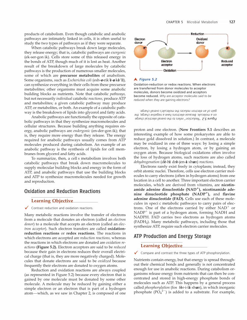
reduction reactions or redox reactions. The reactions inwhich electrons are accepted are reduction reactions, whereasthe reactions in which electrons are donated are oxidation re-actions (Figure 5.2). Electron acceptors are said to be reducedbecause their gain in electrons reduces their overall electri-cal charge (that is, they are more negatively charged). Mole-cules that donate electrons are said to be oxidized becausefrequently their electrons are donated to oxygen atoms.
Reduction and oxidation reactions are always coupled(as represented in Figure 5.2) because every electron that isgained by one molecule must be donated by some othermolecule. A molecule may be reduced by gaining either asimple electron or an electron that is part of a hydrogenatom—which, as we saw in Chapter 2, is composed of one
Figure 5.2 Oxidation-reduction or redox reactions. When electronsare transferred from donor molecules to acceptormolecules, donors become oxidized and acceptorsbecome reduced. Why are acceptor molecules said to bereduced when they are gaining electrons?
➤
Figure 5.2“Reduction” refers to the overall electrical chargeon a molecule. Because electrons have a negative charge, thegain of an electron reduces the molecule’s overall charge.
128 CHAPTER 5 Microbial Metabolism
cells phosphorylate adenosine diphosphate (ADP), whichhas two phosphate groups, to form adenosine triphosphateATP, which has three phosphate groups (Figure 5.3).
As we will examine in the following sections, cells phos-phorylate ADP to form ATP in three speci c ways:
¥ Substrate-level phosphorylation (see page 139), whichinvolves the transfer of phosphate to ADP fromanother phosphorylated organic compound
¥ Oxidative phosphorylation (see page 147), in whichenergy from redox reactions of respiration (describedshortly) is used to attach inorganic phosphate to ADP
¥ Photophosphorylation (see page 154), in which lightenergy is used to phosphorylate ADP with inorganicphosphate
We will investigate each of these in more detail as we pro-ceed through the chapter.
In summary, after ADP is phosphorylated to produceATP, anabolic pathways use some energy of ATP by break-ing a phosphate bond (which re-forms ADP). Thus the cycli-cal interconversion of ADP and ATP functions somewhatlike rechargeable batteries: ATP molecules store energy fromlight (in photosynthetic organisms) and from catabolic reac-tions and then release stored energy to drive cellularprocesses (including anabolic reactions, active transport,and movement). Reformed ADP molecules can berecharged to ATP again and again (Figure 5.3).
The Roles of Enzymes in Metabolism
Learning Objectives
� Draw a table listing the six basic types of enzymes, their activities,and an example of each.
� Describe the components of a holoenzyme, and contrast proteinand RNA enzymes.
� Define activation energy, enzyme, apoenzyme, cofactor, coen-zyme, active site, and substrate, and describe their roles inenzyme activity.
� Describe how temperature, pH, substrate concentration, andcompetitive and noncompetitive inhibition affect enzyme activity.
As we saw in Chapter 2, reactions occur when chemicalbonds are broken or formed between atoms. In catabolic re-actions, a bond must be destabilized before it will break,whereas in anabolic reactions reactants must collide withsuf cient energy before bonds will form between them. Inanabolism, increasing either the concentrations of reactantsor ambient temperatures will increase the number of colli-sions and produce more chemical reactions; however, in liv-ing organisms, neither reactant concentration nortemperature is usually high enough to ensure that bondswill form. Therefore, the chemical reactions of life dependupon catalysts, which are chemicals that increase thelikelihood of a reaction but are not permanently changed inthe process. Organic catalysts are known as enzymes.
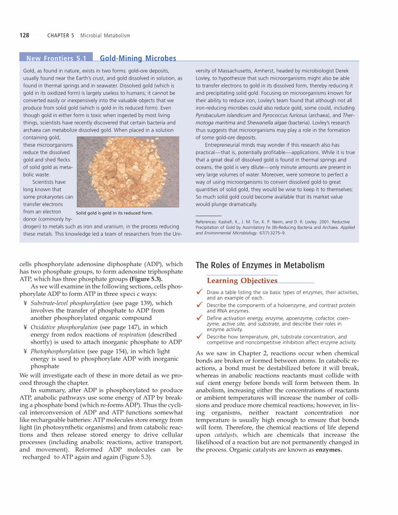
Gold, as found in nature, exists in two forms: gold-ore deposits,usually found near the Earth’s crust, and gold dissolved in solution, asfound in thermal springs and in seawater. Dissolved gold (which isgold in its oxidized form) is largely useless to humans; it cannot beconverted easily or inexpensively into the valuable objects that weproduce from solid gold (which is gold in its reduced form). Eventhough gold in either form is toxic when ingested by most livingthings, scientists have recently discovered that certain bacteria andarchaea can metabolize dissolved gold. When placed in a solutioncontaining gold,these microorganismsreduce the dissolvedgold and shed flecksof solid gold as meta-bolic waste.
Scientists havelong known thatsome prokaryotes cantransfer electronsfrom an electrondonor (commonly hy-drogen) to metals such as iron and uranium, in the process reducingthese metals. This knowledge led a team of researchers from the Uni-
versity of Massachusetts, Amherst, headed by microbiologist DerekLovley, to hypothesize that such microorganisms might also be ableto transfer electrons to gold in its dissolved form, thereby reducing itand precipitating solid gold. Focusing on microorganisms known fortheir ability to reduce iron, Lovley’s team found that although not alliron-reducing microbes could also reduce gold, some could, includingPyrobaculum islandicum and Pyrococcus furiosus (archaea), and Ther-motoga maritima and Shewanella algae (bacteria). Lovley’s researchthus suggests that microorganisms may play a role in the formationof some gold-ore deposits.
Entrepreneurial minds may wonder if this research also haspractical—that is, potentially profitable—applications. While it is truethat a great deal of dissolved gold is found in thermal springs andoceans, the gold is very dilute—only minute amounts are present invery large volumes of water. Moreover, were someone to perfect away of using microorganisms to convert dissolved gold to greatquantities of solid gold, they would be wise to keep it to themselves:So much solid gold could become available that its market valuewould plunge dramatically.
References: Kashefi, K., J. M. Tor, K. P. Nevin, and D. R. Lovley. 2001. ReductivePrecipitation of Gold by Assimilatory Fe (III)-Reducing Bacteria and Archaea. Appliedand Environmental Microbiology. 67(7):3275–9.
New Frontiers 5.1 Gold-Mining Microbes
Solid gold is gold in its reduced form.

CHAPTER 5 Microbial Metabolism 129
do not add or remove anything (so they are neithercatabolic nor anabolic).
3. Ligases or polymerases join two molecules together (andare thus anabolic). They often use energy supplied byATP.
4. Lyases split large molecules (and are thus catabolic)without using water in the process.
5. Oxidoreductases remove electrons from (oxidize) or addelectrons to (reduce) various substrates. They are usedin both catabolic and anabolic pathways.
6. Transferases transfer functional groups, such as an aminogroup (NH2), a phosphate group, or a two-carbon(acetyl) group, between molecules. Transferases can beanabolic.
Naming and Classifying Enzymes
The names of enzymes usually end with the suf x -ase,and the name of each enzyme often incorporates the name ofthat enzyme s substrate, which is the molecule the enzymeacts upon. Based on their mode of action, enzymes can begrouped into six basic categories:
1. Hydrolases catabolize molecules by adding water in adecomposition process known as hydrolysis. Hydrolasesare used primarily in the depolymerization of macro-molecules.
2. Isomerases2 rearrange the atoms within a molecule but
Figure 5.3 Phosphorylation of ADP to form ATP. Cells add phosphate via a high energy bond toADP, making ATP. During anabolism, ATP gives up its energy and phosphate to becomeADP, which is then recycled to ATP.
➤
2An isomer is a compound with the same molecular formula as another molecule, butwith a different arrangement of atoms.
130 CHAPTER 5 Microbial Metabolism
Table 5.1 summarizes these types of enzymes and gives ex-amples of each.
The Makeup of Enzymes
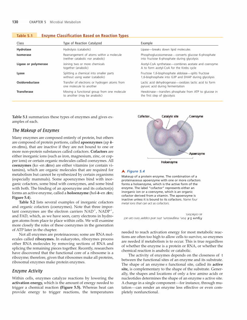
Many enzymes are composed entirely of protein, but othersare composed of protein portions, called apoenzymes (ap oł -en-zıłms), that are inactive if they are not bound to one ormore non-protein substances called cofactors. Cofactors areeither inorganic ions (such as iron, magnesium, zinc, or cop-per ions) or certain organic molecules called coenzymes. Allcoenzymes (ko -en zıłms) are either vitamins (or contain vi-tamins), which are organic molecules that are required formetabolism but cannot be synthesized by certain organisms(especially mammals). Some apoenzymes bind with inor-ganic cofactors, some bind with coenzymes, and some bindwith both. The binding of an apoenzyme and its cofactor(s)forms an active enzyme, called a holoenzyme (hol-oł -en zıłm;Figure 5.4).
Table 5.2 lists several examples of inorganic cofactorsand organic cofactors (coenzymes). Note that three impor-tant coenzymes are the electron carriers NAD�, NADP�,and FAD, which, as we have seen, carry electrons in hydro-gen atoms from place to place within cells. We will examinemore closely the roles of these coenzymes in the generationof ATP later in the chapter.
Not all enzymes are proteinaceous; some are RNA mol-ecules called ribozymes. In eukaryotes, ribozymes processother RNA molecules by removing sections of RNA andsplicing the remaining pieces together. Recently, researchershave discovered that the functional core of a ribosome is aribozyme; therefore, given that ribosomes make all proteins,ribosomal enzymes make protein enzymes.
Enzyme Activity
Within cells, enzymes catalyze reactions by lowering theactivation energy, which is the amount of energy needed totrigger a chemical reaction (Figure 5.5). Whereas heat canprovide energy to trigger reactions, the temperatures
needed to reach activation energy for most metabolic reac-tions are often too high to allow cells to survive, so enzymesare needed if metabolism is to occur. This is true regardlessof whether the enzyme is a protein or RNA, or whether thechemical reaction is anabolic or catabolic.
The activity of enzymes depends on the closeness of tbetween the functional sites of an enzyme and its substrate.The shape of an enzyme s functional site, called its active
site, is complementary to the shape of the substrate. Gener-ally, the shapes and locations of only a few amino acids ornucleotides determines the shape of an enzyme s active site.A change in a single component—for instance, through mu-tation—can render an enzyme less effective or even com-pletely nonfunctional.
Table 5.1 Enzyme Classification Based on Reaction Types
Class Type of Reaction Catalyzed Example
Hydrolase Hydrolysis (catabolic) Lipase—breaks down lipid molecules
Isomerase Rearrangement of atoms within a molecule Phosphoglucoisomerase—converts glucose 6-phosphate(neither catabolic nor anabolic) into fructose 6-phosphate during glycolysis
Ligase or polymerase Joining two or more chemicals Acetyl-CoA synthetase—combines acetate and coenzymetogether (anabolic) A to form acetyl-CoA for the Krebs cycle
Lyase Splitting a chemical into smaller parts Fructose 1,6-bisphosphate aldolase—splits fructosewithout using water (catabolic) 1,6-bisphosphate into G3P and DHAP during glycolysis
Oxidoreductase Transfer of electrons or hydrogen atoms from Lactic acid dehydrogenase—oxidizes lactic acid to formone molecule to another pyruvic acid during fermentation
Transferase Moving a functional group from one molecule Hexokinase—transfers phosphate from ATP to glucose into another (may be anabolic) the first step of glycolysis
Figure 5.4 Makeup of a protein enzyme. The combination of aproteinaceous apoenzyme with one or more cofactorsforms a holoenzyme, which is the active form of theenzyme. The label “cofactor” represents either aninorganic ion or a coenzyme, which is an organiccofactor derived from a vitamin. The apoenzyme isinactive unless it is bound to its cofactors. Name fourmetal ions that can act as cofactors.
➤
Figure 5.4Iron, magnesium, zinc and copper ions can actas cofactors.

CHAPTER 5 Microbial Metabolism 131
In some cases, several different enzymes possess activesites that are complementary to various portions of a singlesubstrate molecule. For example, an important precursormetabolite called phosphoenolpyruvic acid (PEP) is the sub-strate for at least ve enzymes; depending on the enzyme in-volved, various products are produced from PEP. Forinstance, in a catabolic pathway PEP is converted to pyruvicacid, whereas in an anabolic pathway PEP is converted tothe amino acid phenylalanine.
Although the exact ways that enzymes lower activationenergy are not known, it appears that several mechanismsare involved. Some enzymes appear to bring reactants intosuf ciently close proximity to enable a bond to form,whereas other enzymes change the shape of a reactant, in-ducing a bond to be broken. In any case, enzymes increasethe likelihood that bonds will form or break.
The activity of enzymes is believed to follow the processillustrated in Figure 5.7, which depicts the catabolic lysis ofa molecule called fructose 1,6-bisphosphate:
An enzyme associates with a speci c substrate mole-cule having a shape that is complementary to thatenzyme s active site.The enzyme and its substrate bind to form a temporaryintermediate compound called an enzyme-substratecomplex. The binding of the substrate induces theenzyme to t the shape of the substrate even moreclosely.
This enzyme-substrate specificity, which is critical to en-zyme activity, has been likened to the t between a lock andkey. This analogy is not completely apt because enzymeschange shape slightly when they bind to their substrate, al-most as if a lock could grasp its key once it has been inserted.This latter description of enzyme-substrate speci city iscalled the induced fit model (Figure 5.6).
Table 5.2 Representative Cofactors of Enzymes
SubstanceExample of Use in Transferred in Vitamin Source
Cofactors Enzymatic Activity Enzymatic Activity (of Coenzyme)
Inorganic (metal ion)
Magnesium (Mg2�) Forms bond with ADP Phosphate Noneduring phosphorylation
Organic (coenzymes)
Nicotinamide adenine Carrier of reducing Two electrons Niacindinucleotide (NAD�) power and a hydrogen ion
Nicotinamide adenine Carrier of reducing Two electrons Niacindinucleotide phosphate power and a hydrogen ion(NADP�)
Flavine adenine dinucleotide Carrier of reducing Two hydrogen Riboflavin(FAD) power atoms
Tetrahydrofolate Used in synthesis of One-carbon Folic acidnucleotides and some moleculeamino acids
Coenzyme A Formation of acetyl-CoA Two-carbon Pantothenic acidin Krebs cycle and moleculebeta-oxidation
Pyridoxal phosphate Transaminations in the Amine group Pyridoxinesynthesis of amino acids
Thiamine pyrophosphate Decarboxylation of Aldehyde group Thiaminepyruvic acid (CHO)
Figure 5.5The effect of enzymes on chemical reactions. Enzymescatalyze reactions by lowering the activation energy;that is, the energy needed to trigger the reaction.
➤

132 CHAPTER 5 Microbial Metabolism
Bonds within the substrate are broken, forming two(and in some other reactions, more than two) products.(Note that in an anabolic reaction, instead of the break-age of a bond, two reactants are linked together to forma single product.)The enzyme disassociates from the newly formed mol-ecules, which diffuse away from the site of the reaction,and the enzyme resumes its original con guration andis ready to associate with another substrate molecule.Many factors in uence the rate of enzymatic reactions,
including temperature, pH, enzyme and substrate concen-trations, and the presence of inhibitors.
Temperature As mentioned, higher temperatures tend toincrease the rate of most chemical reactions because mole-cules are moving faster and collide more frequently, whichencourages bonds to form or break. However, this is not en-tirely true of enzymatic reactions, because the active sites ofenzymes change shape as temperature changes. If the tem-perature rises too high or falls too low, an enzyme is often nolonger able to achieve a t with its substrate.
Each enzyme has an optimal temperature for its activity(Figure 5.8a). The optimum temperature for the enzymes inthe human body is about 37¡C, which is normal body tem-perature. Part of the reason certain pathogens can cause dis-ease in humans is that the optimal temperature for theenzymes in those microorganisms is also 37¡C. The enzymesof some other microorganisms, however, function best atmuch higher temperatures; this is the case for hyperther-mophiles, organisms that grow best at temperatures above80¡C.
If temperature rises beyond a certain critical point, thenoncovalent bonds within an enzyme (such as the hydrogenbonds between amino acids) will break, and the enzyme willdenature (Figure 5.9). Denatured enzymes lose their speci cthree-dimensional structure, so they are no longer func-tional. Denaturation is said to be permanent when an enzymecannot regain its original three-dimensional structure onceconditions return to normal, much like the irreversible so-l idi cation of the protein albumin when egg whites arecooked and then cooled. In other cases denaturation isreversible—the denatured enzyme s noncovalent bonds re-form upon the return of normal conditions.
CRITICAL THINKING
Explain why thermophiles do not cause disease in humans.
pH Extremes of pH also denature enzymes when ions re-leased from acids and bases interfere with hydrogen bond-ing and distort and disrupt an enzyme s secondary and ter-tiary structures. Therefore, each enzyme has an optimal pH(see Figure 5.8b).
Changing the pH provides a way to control the growthof unwanted microorganisms by denaturing their proteins.
Figure 5.6(a) The induced-fit model of enzyme-substrateinteraction. The enzyme’s active site is generallycomplementary to the shape of its substrate, but aperfect fit between them does not occur until thesubstrate and enzyme bind to form an enzyme-substrate complex. (b) Space-filling models of anenzyme binding to a substrate.
➤

CHAPTER 5 Microbial Metabolism 133
Enzyme and Substrate Concentration Another factorthat determines the rate of enzymatic activity within cells isthe concentration of substrate present (see Figure 5.8c). Assubstrate concentration increases, enzymatic activity in-creases as more and more enzyme active sites bind more andmore substrate molecules. Eventually, when all enzyme ac-tive sites have bound substrate, the enzymes have reachedtheir saturation point, and the addition of more substrate willnot increase the rate of enzymatic activity.
Obviously, the rate of enzymatic activity is also affectedby the concentration of enzyme within cells. In fact, one waythat organisms regulate their metabolism is by controllingthe quantity and timing of enzyme synthesis. In otherwords, many enzymes are produced in the amounts and atthe times they are needed to maintain metabolic activity.Chapter 7 discusses the role of genetic mechanisms in theregulation of enzyme synthesis. Additionally, eukaryotic
For example, vinegar (acetic acid, pH 3.0) acts as a preserva-tive in dill pickles, and ammonia (pH 11.5) can be used as adisinfectant.
CRITICAL THINKING
In addition to extremes in temperature and pH, other chemical andphysical agents, including ionizing radiation, alcohol, enzymes, andheavy-metal ions denature proteins. For example, the firstantimicrobial drug, salvarsan, contained the heavy metal arsenic andwas used to inhibit the enzymes of the bacterium Treponemapallidum, the causative agent of syphilis.
Given that both human and bacterial enzymes are denatured byheavy metals, how was salvarsan used to treat syphilis withoutpoisoning the patient? Why is syphilis no longer treated with arsenic-containing compounds?
Figure 5.7The process of enzymatic activity. Shown here is the lysisof fructose 1,6-bisphosphate by the enzyme fructose 1,6-bisphosphate aldolase (a catabolic reaction). After theenzyme associates with the substrate ➀ , the twomolecules bind to form an enzyme-substrate complex➁ . As a result of binding, the enzyme’s active site isinduced to fit the substrate even more closely. Next,bonds within the substrate are broken ➂ , after whichthe enzyme dissociates from the two new products ➃ .The enzyme resumes its initial configuration and is thenready to associate with another substrate molecule.
➤

134 CHAPTER 5 Microbial Metabolism
cells control some enzymatic activities by compartmentaliz-ing enzymes inside membranes so that certain metabolicreactions proceed physically separated from the rest of thecell. For example, white blood cells catabolize phagocytizedpathogens using enzymes packaged within lysosomes.
Inhibitors Enzymatic activity can be in uenced by avariety of inhibitory substances that block an enzyme s activesite. Enzymatic inhibitors, which may be either competitiveor noncompetitive, do not denature enzymes.
Competitive inhibitors are shaped such that they tinto an enzyme s active site and thus prevent the normal
substrate from binding (Figure 5.10a). However, such in-hibitors do not undergo a chemical reaction to form prod-ucts. Competitive inhibitors can bind permanently orreversibly to an active site. Permanent binding results inpermanent loss of enzymatic activity; reversible competitioncan be overcome by an increase in the concentration of sub-strate molecules, which increases the likelihood that activesites will be lled with substrate instead of inhibitor (Figure
5.10b).
A good example of competitiveinhibition is the action of sulfanil-amide (found in sulfa drugs), whichhas a shape similar to that of para-aminobenzoic acid (PABA).
Sulfanilamide has great af nityfor the active site of an enzyme re-quired in the conversion of PABA
Figure 5.9 Denaturation of protein enzymes. Breakage ofnoncovalent bonds (such as hydrogen bonds) causes theprotein to lose its secondary and tertiary structure andbecome denatured; as a result, the enzyme is no longerfunctional.
➤
Figure 5.8 The effects of temperature, pH, and substrate concentration on enzyme activity.(a) Rising temperature enhances enzymatic activity to a point, but above some optimaltemperature the enzyme denatures and loses function. (b) Enzymes typically have someoptimal pH, at which point enzymatic activity reaches a maximum. (c) At lowersubstrate concentrations, enzyme activity increases as the substrate concentrationincreases and as more and more active sites are utilized. At the substrate concentrationat which all active sites are utilized, termed the saturation point, enzymatic activityreaches a maximum, and any additional increase in substrate concentration has noeffect on enzyme activity. What is the optimal pH of the enzyme shown in part (b)?
➤
Figure 5.8The enzyme’s optimal pH is approximately 7.

CHAPTER 5 Microbial Metabolism 135
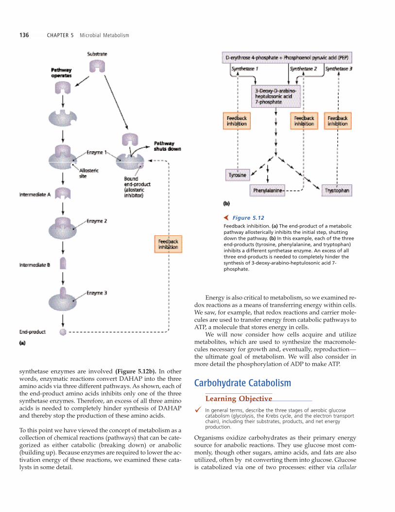
much the way a thermostat controls a heater. As the roomgets warmer, a sensor inside the thermostat changes shapeand sends an electrical signal that turns off the ame or elec-trical coil in the heater. Similarly, in metabolic feedback inhi-bition, the end-product of a series of reactions is an allostericinhibitor of an enzyme in an earlier part of the pathway (Fig-
ure 5.12a). Because the product of each reaction in the path-way is the substrate for the next reaction, inhibition of the
rst enzyme in the series inhibits the entire pathway, therebysaving the cell energy. For example, in Escherichia coli, thepresence of the amino acid isoleucine allosterically inhibitsthe rst enzyme in the metabolic pathway that producesisoleucine. In this manner, the bacterium prevents the accu-mulation of isoleucine (and intermediate products) whenthe amino acid is available from the environment. When en-vironmental isoleucine is depleted, the rst metabolic en-zyme is no longer inhibited, and isoleucine productionresumes.
Feedback inhibition can occur in even more complexways. For instance, even though the rst step in the synthe-sis of the amino acids tyrosine, phenylalanine, and trypto-phan is the same—the linkage of phosphoenol pyruvic acid(PEP) and erythrose 4-phosphate to form 3-deoxy-arabino-heptulosonic acid 7-phosphate (DAHAP)—three different
into the B vitamin folic acid, which is essential for DNA syn-thesis. Once sulfanilamide is bound to the enzyme, it staysbound. As a result, it prevents synthesis of folic acid. Sul-fanilamide effectively inhibits bacteria that make folic acidfrom PABA without harming people because humans lackthe necessary enzymes; they must acquire folic acid in theirdiets.
Noncompetitive inhibitors do not bind to the active sitebut instead prevent enzymatic activity by binding to anallosteric (al-oł -stał r�ik) site located elsewhere on the enzyme.Binding at an allosteric site alters the shape of the active siteso that substrate cannot be bound. Allosteric control of en-zyme activity can take two forms: inhibitory and excitatory.Allosteric (noncompetitive) inhibition halts enzymatic activityin the manner just described (Figure 5.11a). In excitatory al-losteric control, the binding of certain activator molecules(such as a heavy-metal ion cofactor) to an allosteric sitecauses a change in shape of the active site, which activatesan otherwise inactive enzyme (Figure 5.11b). Some enzymeshave several allosteric sites, both inhibitory and excitatory,which allows their function to be closely regulated.
Cells often control the action of enzymes through feed-
back inhibition (also called negative feedback or end-prod-uct inhibition). Allosteric feedback inhibition functions in
Figure 5.10 Competitive inhibition of enzyme activity. (a) Inhibitorymolecules, which are similar in shape to substratemolecules, compete for and block active sites. (b) Reversible inhibition can be overcome by an increasein substrate concentration.
➤
Figure 5.11 Allosteric control of enzyme activity. (a) Allosteric(noncompetitive) inhibition results from a change in theshape of the active site when an inhibitor binds to anallosteric site. (b) Allosteric activation results when thebinding of an activator molecule to an allosteric sitecauses a change in the active site that makes it capableof binding substrate.
➤
136 CHAPTER 5 Microbial Metabolism
synthetase enzymes are involved (Figure 5.12b). In otherwords, enzymatic reactions convert DAHAP into the threeamino acids via three different pathways. As shown, each ofthe end-product amino acids inhibits only one of the threesynthetase enzymes. Therefore, an excess of all three aminoacids is needed to completely hinder synthesis of DAHAPand thereby stop the production of these amino acids.
To this point we have viewed the concept of metabolism as acollection of chemical reactions (pathways) that can be cate-gorized as either catabolic (breaking down) or anabolic(building up). Because enzymes are required to lower the ac-tivation energy of these reactions, we examined these cata-lysts in some detail.
Energy is also critical to metabolism, so we examined re-dox reactions as a means of transferring energy within cells.We saw, for example, that redox reactions and carrier mole-cules are used to transfer energy from catabolic pathways toATP, a molecule that stores energy in cells.
We will now consider how cells acquire and utilizemetabolites, which are used to synthesize the macromole-cules necessary for growth and, eventually, reproduction—
the ultimate goal of metabolism. We will also consider inmore detail the phosphorylation of ADP to make ATP.
Carbohydrate Catabolism
Learning Objective
� In general terms, describe the three stages of aerobic glucosecatabolism (glycolysis, the Krebs cycle, and the electron transportchain), including their substrates, products, and net energyproduction.
Organisms oxidize carbohydrates as their primary energysource for anabolic reactions. They use glucose most com-monly, though other sugars, amino acids, and fats are alsoutilized, often by rst converting them into glucose. Glucoseis catabolized via one of two processes: either via cellular
Figure 5.12Feedback inhibition. (a) The end-product of a metabolicpathway allosterically inhibits the initial step, shuttingdown the pathway. (b) In this example, each of the threeend-products (tyrosine, phenylalanine, and tryptophan)inhibits a different synthetase enzyme. An excess of allthree end-products is needed to completely hinder thesynthesis of 3-deoxy-arabino-heptulosonic acid 7-phosphate.
➤
CHAPTER 5 Microbial Metabolism 137
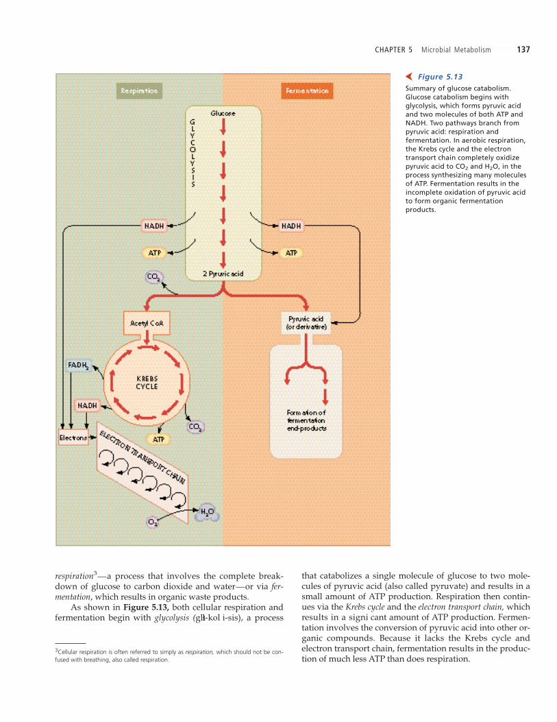
that catabolizes a single molecule of glucose to two mole-cules of pyruvic acid (also called pyruvate) and results in asmall amount of ATP production. Respiration then contin-ues via the Krebs cycle and the electron transport chain, whichresults in a signi cant amount of ATP production. Fermen-tation involves the conversion of pyruvic acid into other or-ganic compounds. Because it lacks the Krebs cycle andelectron transport chain, fermentation results in the produc-tion of much less ATP than does respiration.
respiration3—a process that involves the complete break-
down of glucose to carbon dioxide and water—or via fer-mentation, which results in organic waste products.
As shown in Figure 5.13, both cellular respiration andfermentation begin with glycolysis (glıł-kol i-sis), a process
Figure 5.13 Summary of glucose catabolism.Glucose catabolism begins withglycolysis, which forms pyruvic acidand two molecules of both ATP andNADH. Two pathways branch frompyruvic acid: respiration andfermentation. In aerobic respiration,the Krebs cycle and the electrontransport chain completely oxidizepyruvic acid to CO2 and H2O, in theprocess synthesizing many moleculesof ATP. Fermentation results in theincomplete oxidation of pyruvic acidto form organic fermentationproducts.
➤3Cellular respiration is often referred to simply as respiration, which should not be con-fused with breathing, also called respiration.
138 CHAPTER 5 Microbial Metabolism
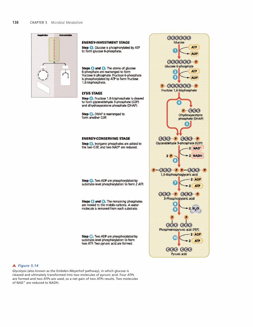
Figure 5.14 Glycolysis (also known as the Embden-Meyerhof pathway), in which glucose iscleaved and ultimately transformed into two molecules of pyruvic acid. Four ATPsare formed and two ATPs are used, so a net gain of two ATPs results. Two moleculesof NAD� are reduced to NADH.
➤

CHAPTER 5 Microbial Metabolism 139
level phosphorylations occur in metabolism. As you mightexpect, each type has its own enzyme that recognizes bothits substrate molecule and ADP.
In glycolysis, two ATP molecules are invested bysubstrate-level phosphorylation to prime glucose for lysis,and four molecules of ATP are produced, also by substrate-level phosphorylation. Therefore, a net gain of two ATP mol-ecules occurs for each molecule of glucose that is oxidized topyruvic acid. Glycolysis also yields two molecules ofNADH.
Alternatives to Glycolysis
Learning Objective
� Compare the pentose phosphate pathway and the Entner-Doudoroff pathway with glycolysis in terms of energy productionand products.
The initial part of the catabolism of glucose can also proceedvia two alternate pathways: the pentose phosphate pathwayand the Entner-Doudoroff pathway. Though they yieldfewer molecules of ATP than glycolysis, these alternatepathways reduce coenzymes and yield different substratemetabolites that are needed in anabolic pathways. Next webrie y examine each of these alternate pathways.
Pentose Phosphate Pathway
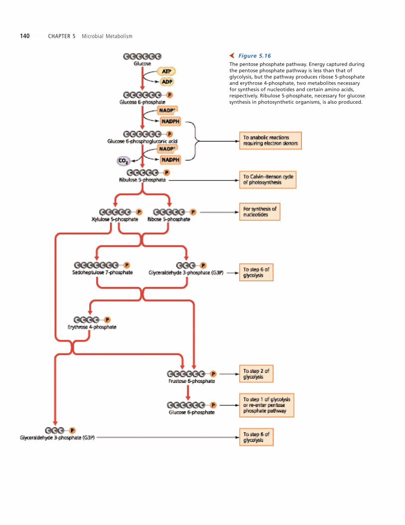
The pentose phosphate pathway is named for the phos-phorylated pentose ( ve-carbon) sugars—ribulose, xylu-lose, and ribose—that are formed from glucose 6-phosphateby enzymes in the pathway (Figure 5.16). The pentose phos-phate pathway is primarily used for the production of pre-cursor metabolites used in anabolic reactions, including thesynthesis of nucleotides for nucleic acids, of certain aminoacids, and of glucose by photosynthesis (described in a later
The following is a simpli ed discussion of glucosecatabolism. Details of the substrates and enzymes involvedare provided in the Appendix on page A-1. To help under-stand the basic reactions in each of the pathways of glucosecatabolism, pay special attention to three things: the numberof carbon atoms in each of the intermediate products, the rel-ative numbers of ATP molecules produced in each pathway,and the changes in the coenzymes NAD� and FAD as theyare reduced and then oxidized back to their original forms.
GlycolysisGlycolysis4, also called the Embden-Meyerhof pathway afterthe scientists who discovered it, is the rst step in the catab-olism of glucose via both respiration and fermentation.Glycolysis occurs in most cells. In general, as its name im-plies, glycolysis involves the splitting of a six-carbon glu-cose molecule into two three-carbon sugar molecules. Whenthese three-carbon molecules are oxidized to pyruvic acid,some of the energy released is stored in molecules of ATP.
Glycolysis, which occurs in the cytoplasm, can be di-vided into three stages involving a total of 10 steps (Figure
5.14), each of which is catalyzed by its own enzyme. Thethree stages of glycolysis are:
1. Energy-investment stage (steps – ). As with money,one must invest before a pro t can be made. In thiscase, the energy in two molecules of ATP is invested tophosphorylate a six-carbon glucose molecule andrearrange its atoms to form fructose 1,6-bisphosphate.
2. Lysis stage (steps and ). Fructose 1,6-bisphos-phate is cleaved into glyceraldehyde 3-phosphate(G3P)5 and dihydroxyacetone phosphate (DHAP). Eachof these compounds contains three carbon atoms andis freely convertible into the other.
3. Energy-conserving stage (steps – ). G3P is oxi-dized to pyruvic acid, yielding two ATP molecules.DHAP is converted to G3P and also oxidized to pyru-vic acid, yielding another two ATP molecules, for atotal of four ATP molecules.Our study of glycolysis provides our rst opportunity to
study substrate-level phosphorylation (see steps , , and in Figure 5.14). Let s examine this important process
more closely by considering the 10th and nal step of gly-colysis.
Each of the two phosphoenolpyruvic acid (PEP) mole-cules produced in step of glycolysis is a three-carboncompound containing a high-energy phosphate bond. In thepresence of a speci c holoenzyme (which requires a Mg2�
cofactor), the high-energy phosphate in PEP (one substrate)is transferred to an ADP molecule (a second substrate) toform ATP (Figure 5.15); the direct transfer of the phosphatebetween the two substrates is the reason the process is calledsubstrate-level phosphorylation. A variety of substrate-
Figure 5.15 Substrate-level phosphorylation, in which high-energyphosphate bonds are transferred from one substrate toanother. What role does Mg2� play in this reaction?
➤
4From Greek glykys, meaning sweet, and lysein, meaning to loosen.5G3P is also known as phosphoglyceraldehyde or PGAL.
Figure 5.15Mg2�
is a cofactor of the enzyme.
140 CHAPTER 5 Microbial Metabolism
Figure 5.16 The pentose phosphate pathway. Energy captured duringthe pentose phosphate pathway is less than that ofglycolysis, but the pathway produces ribose 5-phosphateand erythrose 4-phosphate, two metabolites necessaryfor synthesis of nucleotides and certain amino acids,respectively. Ribulose 5-phosphate, necessary for glucosesynthesis in photosynthetic organisms, is also produced.
➤

CHAPTER 5 Microbial Metabolism 141
section). The pathway also reduces two molecules ofNADP� to NADPH and nets a single molecule of ATP fromeach molecule of glucose. NADPH is a necessary coenzymefor anabolic enzymes that synthesize DNA nucleotides,steroids, and fatty acids.
CRITICAL THINKING
Examine the biosynthetic pathway for the production of the aminoacids tryptophan, tyrosine, and phenylalanine in Figure 5.12b. Wheredo the initial reactants (PEP and erythrose 4-phosphate) originate?
Entner-Doudoroff Pathway
Most bacteria use glycolysis and the pentose phosphatepathway, but a few substitute the Entner-Doudoroff
pathway (Figure 5.17) for glycolysis. This pathway, namedfor its discoverers, is a series of reactions that catabolize glu-cose to pyruvic acid using different enzymes from thoseused in either glycolysis or the pentose phosphate pathway.
Among organisms, only a very few bacteria use theEntner-Doudoroff pathway. These include the Gram-negative Pseudomonas aeruginosa (soo-doł -moł nas ła-røu-ji-nło-søa), and the Gram-positive bacterium Enterococcus faecalis(en-te-roł -kok kus feł -kał lis). Like the pentose phosphatepathway, the Entner-Doudoroff pathway nets only a singlemolecule of ATP for each molecule of glucose, but it doesyield precursor metabolites and NADPH. The latter is un-available from glycolysis.
CRITICAL THINKING
Even though Pseudomonas aeruginosa and Enterococcus faecalis usu-ally grow harmlessly in the body, they can cause disease. Because thesebacteria use the Entner-Doudoroff pathway instead of glycolysis to ca-tabolize glucose, investigators sometimes use clinical tests that provideevidence of the Entner-Doudoroff pathway to identify the presence ofthese potential pathogens.
Suppose you were able to identify the presence of any specificorganic compound. Name a substrate molecule you would find inPseudomonas and Enterococcus cells, but not in human cells.
Cellular Respiration
Learning Objectives
� Discuss the roles of acetyl-CoA, the Krebs cycle, and electrontransport in carbohydrate catabolism.
� Contrast electron transport in aerobic and anaerobic respiration.
� Identify four classes of carriers in electron transport chains.
� Describe the role of chemiosmosis in oxidative phosphorylation ofATP.
After glucose has been oxidized via glycolysis or one of thealternate pathways, a cell uses the resultant pyruvic acidmolecules to complete either cellular respiration or fermen-tation (which we will discuss in a later section). Our topichere—cellular respiration—is a metabolic process that in-volves the complete oxidation of substrate molecules and
Figure 5.17Pseudomonas aeruginosaand Enterococcus faecalisuse theEntner-Doudoroff pathway.
Figure 5.17 Entner-Doudoroff pathway, analternate pathway for the oxidationof glucose to pyruvic acid. Whatpotential pathogens use this pathway?
➤

142 CHAPTER 5 Microbial Metabolism
then production of ATP by a series of redox reactions. Thethree stages of cellular respiration are: (1) synthesis ofacetyl-CoA, (2) the Krebs cycle, and (3) a nal series of redoxreactions, which collectively constitute an electron transportchain.
Synthesis of Acetyl-CoA
Before pyruvic acid (generated by glycolysis, the pentosephosphate pathway, and the Entner-Doudoroff pathway)can enter the Krebs cycle for respiration, it must rst be con-verted to acetyl-coenzyme A or acetyl-CoA (as e-til koł -ał ; seeFigure 5.13). Enzymes remove one carbon from pyruvic acidas CO2 and join the remaining two-carbon acetate tocoenzyme-A with a high-energy bond (Figure 5.18). The re-moval of CO2, called decarboxylation, requires a coenzymederived from the vitamin thiamine. One molecule of NADHis also produced during this reaction.
Recall that two molecules of pyruvic acid were derivedfrom each molecule of glucose. Therefore, at this stage, twomolecules of acetyl-CoA, two molecules of CO2, and twomolecules of NADH are produced.
The Krebs Cycle
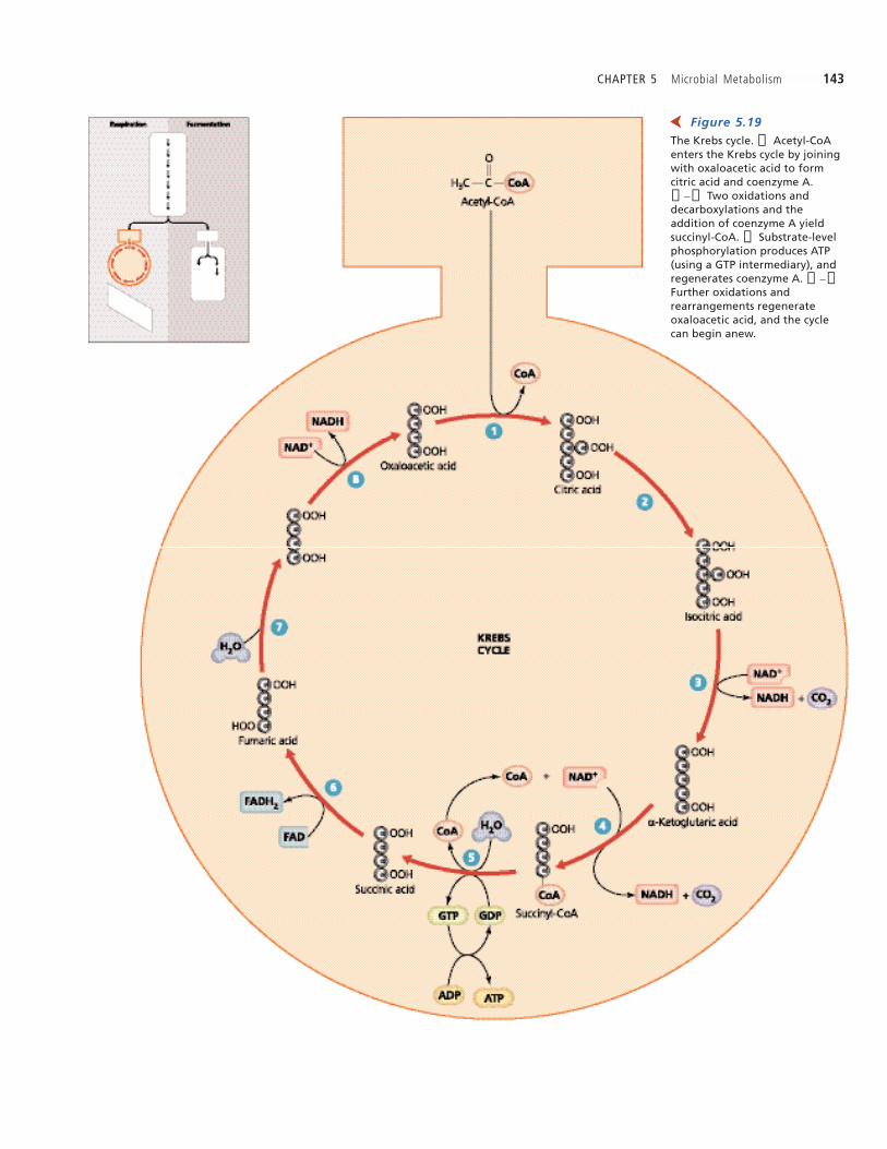
At this point in the catabolism of a molecule of glucose, agreat amount of energy remains in the bonds of acetyl-CoA.The Krebs cycle6 is a series of eight enzymatically catalyzedreactions that transfer much of this stored energy to thecoenzymes NAD� and FAD. The two carbons in acetate areoxidized, and the coenzymes are reduced. The Krebs cycle,which occurs in the cytoplasm in prokaryotes and in thematrix of mitochondria in eukaryotes, is diagrammed inFigure 5.19 and presented in more detail in the Appendix onpage B-1. It is also known as the tricarboxylic acid (TCA)cycles, because many of its compounds have three carboxyl(—COOH) groups, and as the citric acid cycle, for the rstcompound formed in the cycle.
There are ve types of reactions in the Krebs cycle:1. Anabolism of citric acid (step )2. Isomerization reactions (steps , , and )3. Redox reactions (steps , , , and )4. Decarboxylations (steps and )5. Substrate-level phosphorylation (step )
In the rst step of the Krebs cycle, the splitting of thehigh-energy bond between acetate and coenzyme A releasesenough energy to enable the binding of the freed two-carbonacetate to a four-carbon compound called oxaloacetic acid,forming the six-carbon compound citric acid.
As you study Figure 5.19, notice that after isomerization(step ), the decarboxylations of the Krebs cycle releasetwo molecules of CO2 for each acetyl-CoA that enters (steps
and ). Thus, for every two carbon atoms that enter thecycle, two are lost to the environment. At this juncture in therespiration of a molecule of glucose, six carbon atoms havebeen lost to the environment: two as CO2 molecules pro-duced in decarboxylation of two molecules of pyruvic acidto form two acetyl-CoA molecules, and four in CO2 mole-cules produced in decarboxylations in the two turns throughthe Krebs cycle. (One molecule of acetyl-CoA enters the cy-cle at a time).
A small amount of ATP is also produced in the Krebscycle. For every two molecules of acetyl-CoA that passthrough the Krebs cycle, two molecules of ATP are gener-ated by substrate-level phosphorylation (step ). A mole-cule of guanosine triphosphate (GTP), which is similar toATP, serves as an intermediary in this process.
Redox reactions reduce FAD to FADH2 (step ) andNAD� to NADH (steps , , and ), so that forevery two molecules of acetyl-CoA that move through thecycle, six molecules of NADH and two of FADH2 are formed.In the Krebs cycle, little energy is captured directly in high-energy phosphate bonds, but much energy is transferred viaelectrons to NADH and FADH2. These coenzymes are themost important molecules of respiration because they carry alarge amount energy that is subsequently used to phospho-rylate ADP to ATP.
CRITICAL THINKING
We have examined the total ATP, NADH, and FADH2 production in theKrebs cycle for each molecule of glucose coming through Embden-Meyerhof glycolysis. How many of each of these molecules would beproduced if the Entner-Doudoroff pathway were used instead ofglycolysis?
Electron Transport
Some scientists estimate that each day an average humansynthesizes his or her own weight in ATP molecules anduses them for metabolism, responsiveness, growth, and cellreproduction. ATP turnover in prokaryotes is relatively as
Figure 5.18Formation of acetyl-CoA. Theresponsible enzyme acts in a stepwisemanner to ➀ remove CO2 from pyruvicacid, ➁ attach the remaining two-carbon acetate to coenzyme A, and ➂reduce a molecule of NAD� to NADH.
➤
6Named for biochemist Sir Hans Krebs (1900–1981), who elucidated its reactions in the1940s.
CHAPTER 5 Microbial Metabolism 143
Figure 5.19 The Krebs cycle. ➀ Acetyl-CoAenters the Krebs cycle by joiningwith oxaloacetic acid to formcitric acid and coenzyme A.➁ –➃ Two oxidations anddecarboxylations and theaddition of coenzyme A yieldsuccinyl-CoA. ➄ Substrate-levelphosphorylation produces ATP(using a GTP intermediary), andregenerates coenzyme A. ➅ –➇Further oxidations andrearrangements regenerateoxaloacetic acid, and the cyclecan begin anew.
➤

144 CHAPTER 5 Microbial Metabolism
copious. The most signi cant production of ATP does notoccur through glycolysis or the Krebs cycle, but ratherthrough the stepwise release of energy from a series of redoxreactions between molecules known as an electron trans-
port chain (Figure 5.20).
An electron transport chain consists of a series ofmembrane-bound carrier molecules that pass electrons fromone to another and ultimately to a final electron acceptor. Typ-ically, as we have seen, electrons come from the catabolismof an organic molecule such as glucose; however, microor-ganisms called chemolithotrophs (kem oł -lith -oł -troł fs) acquireelectrons from inorganic sources such as H2, NO2�, or Fe2�.(Chemolithotrophs are discussed further in Chapter 6.) Inany case, electrons are passed down the chain like buckets ina re brigade to the nal acceptor. As with a bucket brigade,the nal step of electron transport is irreversible. Energyfrom the electrons is used to actively transport (pump)
protons (H�) across the membrane, establishing a protongradient that generates ATP via a process called chemiosmosis,which we will discuss shortly.
To avoid getting lost in the details of electron transport,keep the following critical concepts in mind:
¥ Electrons pass sequentially from one membrane-bound carrier molecule to another, and eventually toa nal acceptor molecule.
¥ The electrons energy is used to pump protons acrossthe membrane.Electron transport chains are located in the inner mito-
chondrial membranes (cristae) of eukaryotes and in the cy-toplasmic membrane of prokaryotes (Figure 5.21). ThoughNADH and FADH2 donate electrons as hydrogen atoms(electrons and protons), many carrier molecules pass onlythe electrons down the chain. There are four categories ofcarrier molecules in electron transport chains.
Figure 5.20 An electron transport chain. ATP production is indicated at the approximate point inthe chain that energy is captured as electrons move down the chain, but themolecules of the chain do not actually synthesize ATP.
➤
CHAPTER 5 Microbial Metabolism 145
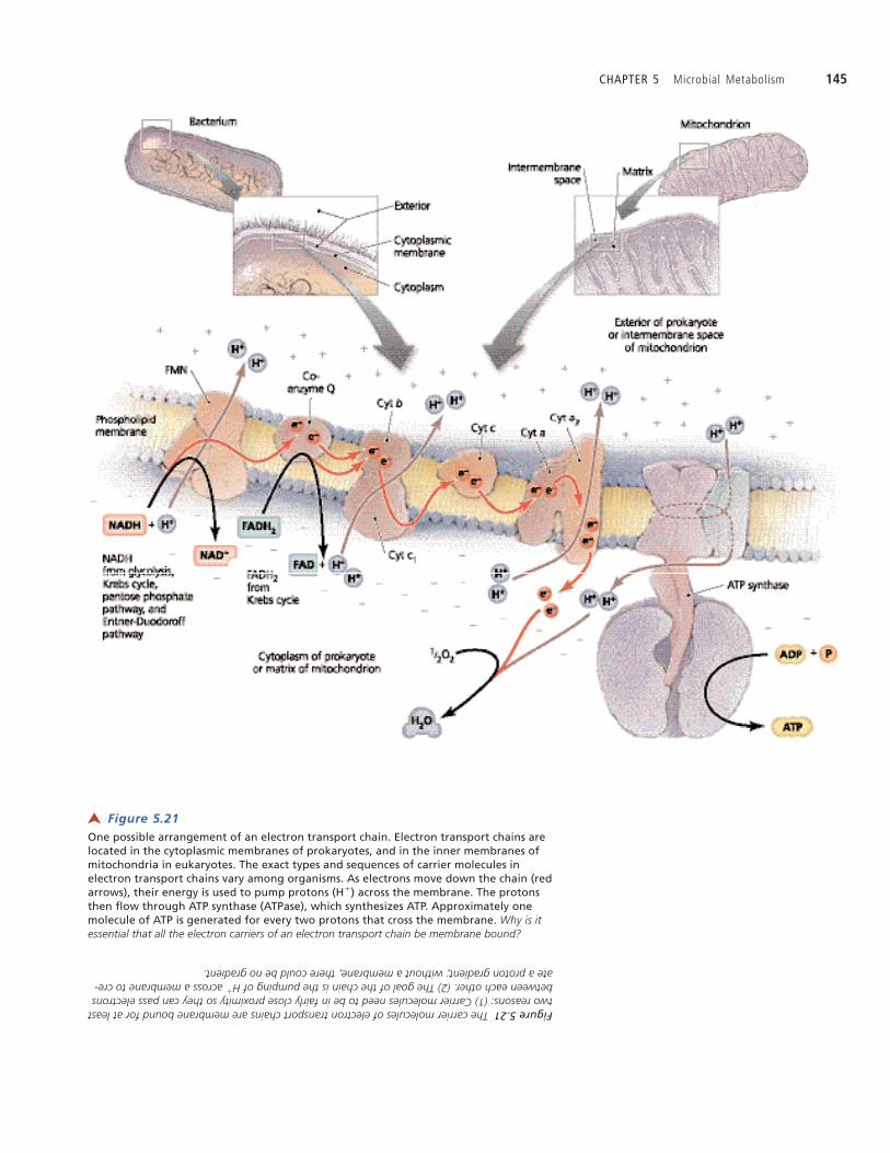
Figure 5.21One possible arrangement of an electron transport chain. Electron transport chains arelocated in the cytoplasmic membranes of prokaryotes, and in the inner membranes ofmitochondria in eukaryotes. The exact types and sequences of carrier molecules inelectron transport chains vary among organisms. As electrons move down the chain (redarrows), their energy is used to pump protons (H�) across the membrane. The protonsthen flow through ATP synthase (ATPase), which synthesizes ATP. Approximately onemolecule of ATP is generated for every two protons that cross the membrane. Why is itessential that all the electron carriers of an electron transport chain be membrane bound?
➤
Figure 5.21The carrier molecules of electron transport chains are membrane bound for at leasttwo reasons: (1) Carrier molecules need to be in fairly close proximity so they can pass electronsbetween each other. (2) The goal of the chain is the pumping of H
�across a membrane to cre-
ate a proton gradient; without a membrane, there could be no gradient.
146 CHAPTER 5 Microbial Metabolism
They are:1. Flavoproteins are integral membrane proteins, many of
which contain avin, a coenzyme derived fromribo avin (vitamin B2). One form of avin is avinmononucleotide (FMN), which is the initial carriermolecule of electron transport chains of mitochondria.The familiar FAD is a coenzyme for other avoproteins.Like all carrier molecules in the electron transportchain, avoproteins alternate between the reduced andoxidized states.
2. Ubiquinones (yuł -bik wi-noł ns) are lipid-soluble, nonpro-tein carriers that are so named because they are ubiq-uitous in all cells. Ubiquinones are derived fromvitamin K. In mitochondria, the ubiquinone is calledcoenzyme Q.
3. Metal-containing proteins are a mixed group of integralproteins with a wide-ranging number of iron, sulfur,and copper atoms that can alternate between thereduced and oxidized states. Iron-sulfur proteins occurin various places in electron transport chains of variousorganisms. Copper proteins are found only in electrontransport chains involved in photosynthesis (discussedshortly).
4. Cytochromes (sıł toł -kroł ms) are integral proteins associ-ated with heme, which is the same iron-containing, non-protein, pigmented molecule found in the hemoglobin
of blood. Iron can alternate between a reduced (Fe2�)state and an oxidized (Fe3�) state. Cytochromes areidenti ed by letters and numbers based on the order inwhich they were identi ed, so their sequence in elec-tron transport chains does not always seem logical.The carrier molecules in electron transport chains are
diverse—bacteria typically have different carrier moleculesarranged in different sequences than do archaea or the mito-chondria of eukaryotes. Some prokaryotes, including E. coli,can even vary their carrier molecules under different envi-ronmental conditions. Even among bacteria, the makeup ofcarrier molecules can be variable. For example, thepathogens Neisseria (nıł-se reł -aø) and Pseudomonas containtwo cytochromes, a and a3—together called cytochrome oxi-dase—that oxidize cytochrome c; such bacteria are said to beoxidase positive. In contrast, other bacterial pathogens, suchas Escherichia, Salmonella, and Proteus (proł teł -uøs), lack cy-tochrome oxidase and are thus considered to be oxidasenegative.
Electrons carried by NADH enter the transport chain ata avoprotein, and those carried by FADH2 are introducedvia a ubiquinone. This explains why more molecules of ATPare generated from NADH than from FADH2. Researchersdo not agree on which carrier molecules are the actual pro-ton pumps, nor on the number of protons that are pumped.Figure 5.21 shows one possibility.
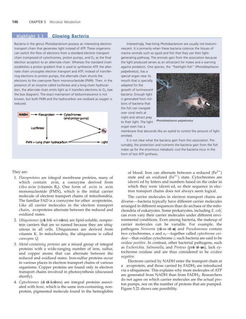
Bacteria in the genus Photobacterium possess an interesting electrontransport chain that generates light instead of ATP. These organismscan switch the flow of electrons from a standard electron transportchain (composed of cytochromes, proton pumps, and O2 as the finalelectron acceptor) to an alternate chain. Whereas the standard chainestablishes a proton gradient that is used to synthesize ATP, the alter-nate chain uncouples electron transport and ATP; instead of transfer-ring electrons to proton pumps, the alternate chain shunts theelectrons to the coenzyme flavin mononucleotide (FMN). Then, in thepresence of an enzyme called luciferase and a long-chain hydrocar-bon, the alternate chain emits light as it transfers electrons to O2 (seethe box diagram). The exact mechanism of bioluminescence is notknown, but both FMN and the hydrocarbon are oxidized as oxygen isreduced.
Interestingly, free-living Photobacterium are usually not biolumi-nescent. It is primarily when these bacteria colonize the tissues ofmarine animals such as squid and fish that they use their light-generating pathway. The animals gain from the association becausethe light produced serves as an attractant for mates and a warningagainst predators. One species, the “flashlight fish” (Photoblepharonpalpebratus), has aspecial organ near itsmouth that is speciallyadapted for thegrowth of luminescentbacteria. Enough lightis generated from mil-lions of bacteria thatthe fish can navigateover coral reefs atnight and attract preyto their light. The lightorgan even has amembrane that descends like an eyelid to control the amount of lightemitted.
It is not clear what the bacteria gain from this association. Pre-sumably, the protection and nutrients the bacteria gain from the fishmake up for the enormous metabolic cost the bacteria incur in theform of lost ATP synthesis.
Highlight 5.1 Glowing Bacteria
Photoblepharon palpebratus

CHAPTER 5 Microbial Metabolism 147
Recall that chemicals diffuse from areas of high concen-tration to areas of low concentration, and toward an electri-cal charge opposite their own. We call the composite ofdifferences in concentration and charge an electrochemicalgradient. Chemicals diffuse down their electrochemical gra-dients. Recall as well that membranes of cells and organellesare impermeable to most chemicals unless a speci c proteinchannel allows their passage across the membrane. A mem-brane maintains an electrochemical gradient by keeping oneor more chemicals in a higher concentration on one side. Theblockage of diffusion creates potential energy, like waterbehind a dam.
Chemiosmosis uses the potential energy of an electro-chemical gradient to phosphorylate ADP into ATP. Eventhough chemiosmosis is a general principle with relevance toboth oxidative phosphorylation and photophosphorylation, herewe consider it as it relates to oxidative phosphorylation.
As we have seen, cells use the energy released in the re-dox reactions of electron transport chains to actively trans-port protons (H�) across a membrane. Theoretically, anelectron transport chain pumps three pairs of protons foreach pair of electrons contributed by NADH, and pumpstwo pairs of protons for each electron pair delivered byFADH2. This difference results from the fact that FADH2
delivers electrons farther down the chain than does NADH;therefore, energy carried by FADH2 is used to transport one-third fewer protons (see Figure 5.20). Because lipid bilayersare impermeable to protons, the transport of protons to oneside of the membrane creates an electrochemical gradientknown as a proton gradient, which has potential energyknown as a proton motive force.
Hydrogen ions, propelled by the proton motive force,ow down their electrochemical gradient through protein
channels, called ATP synthases (ATPases), that phosphory-late molecules of ADP to ATP (see Figure 5.21). Such phos-phorylation is called oxidative phosphorylation becausethe proton gradient is created by the oxidation of compo-nents of an electron transport chain.
In the past, scientists attempted to calculate the exactnumber of ATP molecules synthesized per pair of electronsthat travel down an electron transport chain. However, it isnow apparent that phosphorylation and oxidation are notdirectly coupled. In other words, chemiosmosis does notrequire exact constant relationships among the number ofmolecules of NADH and FADH2 reduced, the number ofelectrons that move down an electron transport chain, andthe number of molecules of ATP that are synthesized. Addi-tionally, cells use proton gradients for other cellularprocesses, including active transport and bacterial agellarmotion, so not every transported electron results in ATPproduction.
Nevertheless, about 34 molecules of ADP per moleculeof glucose are oxidatively phosphorylated to ATP viachemiosmosis: three from each of the 10 molecules ofNADH generated from glycolysis, the synthesis of acetyl-CoA, and the Krebs cycle, and two from each of the two
In some organisms, the nal electron acceptors areoxygen atoms, which, with the addition of hydrogen ions,generate H2O; these organisms conduct aerobic7 respira-
tion and are called aerobes. Other organisms, called anaer-obes,8 use other inorganic molecules (or rarely an organicmolecule) instead of oxygen as the nal electron acceptorand perform anaerobic respiration. The anaerobic bac-terium Desulfovibrio (deł sul-foł -vib-reł -oł ), for example, re-duces sulfate (SO4
2�) to hydrogen sul de gas (H2S),whereas other anaerobes in the genera Bacillus (ba-sil uøs)and Pseudomonas utilize nitrate (NO3
�) to produce nitriteions (NO2
�), nitrous oxide (N2O), or nitrogen gas (N2). Someprokaryotes—particularly archaea called methanogens—
reduce carbonate (CO32�) to methane gas (CH4). Highlight
5.1 describes an unusual bacterial electron transport systemthat produces light instead of ATP.
Laboratory technologists test for products of anaerobicrespiration, such as nitrite, to aid in identi cation of somespecies of bacteria. As discussed more fully in Chapter 26,anaerobic respiration is also critical for the recycling of ni-trogen and sulfur in nature.
CRITICAL THINKING
Cyanide is a potent poison because it irreversibly blocks cytochromea3. What effect would its action have on the rest of the electrontransport chain? What would be the redox state (reduced or oxidized)of coenzyme Q in the presence of cyanide?
In summary, glycolysis, the pentose phosphate pathway, theEntner-Doudoroff pathway, and the Krebs cycle strip elec-trons, which carry energy, from glucose molecules andtransfer them to molecules of NADH and FADH2. In turn,NADH and FADH2 pass the electrons to an electron trans-port chain. As the electrons move down the electron trans-port chain, proton pumps use the electrons energy toactively transport protons (hydrogen ions) across the mem-brane.
Recall, however, that the signi cance of electron trans-port is not merely that it pumps protons across a membrane,but that it ultimately results in the synthesis of ATP. We turnnow to the process by which cells synthesize ATP using theconcentration gradient produced by the large concentrationof protons on one side of a membrane.
Chemiosmosis
Chemiosmosis is a general term for the use of ion gradientsto generate ATP; that is, ATP is synthesized utilizing energyreleased by the ow of ions down their electrochemical gra-dient across a membrane. The term should not be confusedwith osmosis of water. To understand chemiosmosis, weneed to review several concepts from Chapter 3 concerningdiffusion and phospholipid membranes.
7From Greek aer, meaning air (that is, oxygen), and bios, meaning life.8The Greek prefix an means not.
148 CHAPTER 5 Microbial Metabolism
molecules of FADH2 generated in the Krebs cycle. Giventhat glycolysis produces a net two molecules of ATP bysubstrate-level phosphorylation, and that the Krebs cycleproduces two more, the complete aerobic oxidation of onemolecule of glucose by a prokaryote can theoretically yield anet total of 38 molecules of ATP (Table 5.3). The theoreticalnet maximum for eukaryotic cells is generally given as 36molecules of ATP because the energy from two ATP mole-cules is required to transport NADH generated in glycolysisinto the mitochondria.
CRITICAL THINKING
Suppose you could insert a tiny pH probe into the space betweenmitochondrial membranes. Would the pH be above or below 7.0?Why?
Fermentation
Learning Objectives
� Describe fermentation and contrast it with respiration.
� Identify three useful end-products of fermentation, and explainhow fermentation reactions are used in the identification ofbacteria.
� Discuss the use of biochemical tests for metabolic enzymes andproducts in the identification of bacteria.
Sometimes cells cannot completely oxidize glucose by cellu-lar respiration. For instance, they may lack suf cient nalelectron acceptors, as is the case, for example, for an aerobicbacterium in the anaerobic environment of the colon. Elec-trons cannot ow down an electron transport chain unlessoxidized carrier molecules are available to receive them.Our bucket brigade analogy can help clarify this point.
Suppose the last person in the brigade did not throw thewater, but instead held onto two full buckets. What wouldhappen? The entire brigade would soon consist of re ght-ers holding full buckets of water. The analogous situationoccurs in an electron transport chain: All the carrier mole-cules are forced to remain in their reduced states when thereis not a nal electron acceptor. Without the movement ofelectrons down the chain, protons cannot be transported, theproton motive force is lost, and oxidative phosphorylationof ADP to ATP ceases. Without suf cient ATP, a cell is unableto anabolize, grow, or divide.
ATP could be synthesized in glycolysis and the Krebscycle by substrate-level phosphorylation. After all, togetherthese pathways produce four molecules of ATP per mole-cule of glucose. However, careful consideration reveals thatglycolysis and the Krebs cycle require a continual supply ofoxidized NAD� molecules (see Figures 5.14 and 5.19). Inrespiration, electron transport produces the required NAD�,but without a nal electron acceptor, this source of NAD�
ceases to be available. A cell in such a predicament must usean alternate source of NAD� provided by alternative meta-bolic pathways, called fermentation pathways.
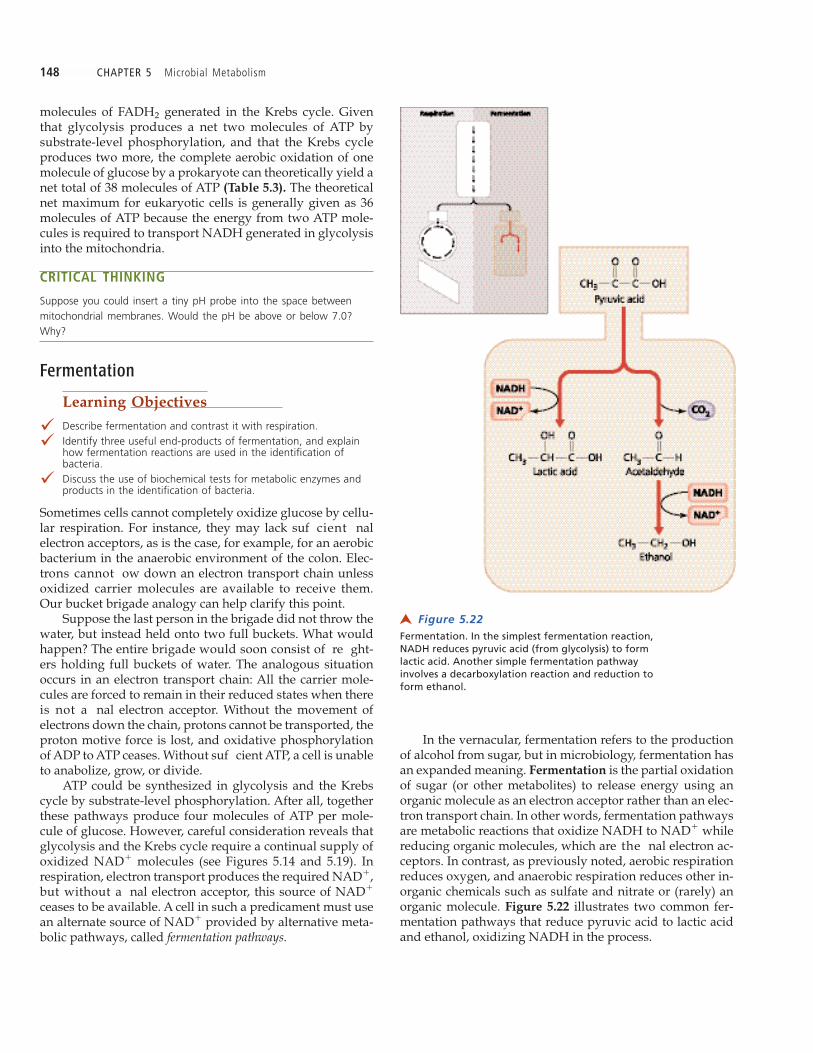
In the vernacular, fermentation refers to the productionof alcohol from sugar, but in microbiology, fermentation hasan expanded meaning. Fermentation is the partial oxidationof sugar (or other metabolites) to release energy using anorganic molecule as an electron acceptor rather than an elec-tron transport chain. In other words, fermentation pathwaysare metabolic reactions that oxidize NADH to NAD� whilereducing organic molecules, which are the nal electron ac-ceptors. In contrast, as previously noted, aerobic respirationreduces oxygen, and anaerobic respiration reduces other in-organic chemicals such as sulfate and nitrate or (rarely) anorganic molecule. Figure 5.22 illustrates two common fer-mentation pathways that reduce pyruvic acid to lactic acidand ethanol, oxidizing NADH in the process.
Figure 5.22 Fermentation. In the simplest fermentation reaction,NADH reduces pyruvic acid (from glycolysis) to formlactic acid. Another simple fermentation pathwayinvolves a decarboxylation reaction and reduction toform ethanol.
➤

CHAPTER 5 Microbial Metabolism 149
In this section on carbohydrate catabolism, we havespent some time examining glycolysis, alternatives to gly-colysis, the Krebs cycle, and electron transport because thesepathways are central to metabolism. They generate all of theprecursor metabolites and most of the ATP needed for an-abolism. We have seen that some ATP is generated in respi-ration by substrate-level phosphorylation (in both glycolysisand the Krebs cycle), and that most ATP is generated by ox-idative phosphorylation via chemiosmosis utilizing the re-ducing power of NADH and FADH2. We also saw that somemicroorganisms use fermentation to provide an alternatesource of NAD� when conditions are not suitable for respi-ration.
Thus far we have concentrated on the catabolism of glu-cose as a representative carbohydrate, but microorganismscan also use other molecules as energy sources. In the nextsection we will examine catabolic pathways that utilizelipids and proteins.
Other Catabolic Pathways
Lipid and protein molecules contain abundant energy intheir chemical bonds and can also be converted into precur-sor metabolites. These molecules are rst catabolized to pro-duce their constituent monomers, which serve as substratesin glycolysis and the Krebs cycle.
Lipid Catabolism
Learning Objective
� Explain how lipids are catabolized for energy and metaboliteproduction.
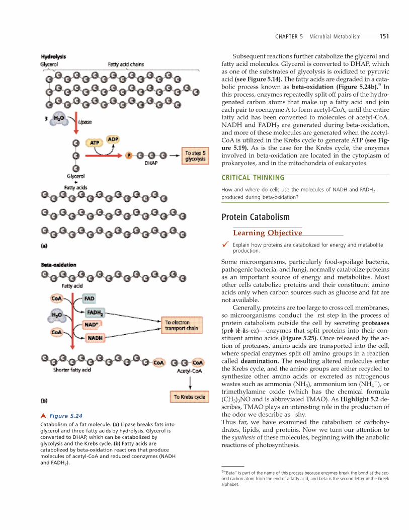
The most common lipids involved in ATP and metaboliteproduction are fats, which, as we saw in Chapter 2, consistof glycerol and fatty acids. In the rst step of fat catabolism,enzymes called lipases hydrolyze the bonds attaching theglycerol to the fatty acid chains (Figure 5.24a on page 151).
The essential function of fermentation is the regenera-tion of NAD� for glycolysis, so that ADP molecules can bephosphorylated to ATP. Even though fermentation path-ways are not as energetically ef cient as respiration becausemuch of the potential energy stored in glucose remains inthe bonds of fermentation products, the major bene t offermentation is that it allows ATP production to continue inthe absence of cellular respiration. Microorganisms withfermentation pathways can colonize anaerobic environ-ments, which are unavailable to strictly aerobic organisms.Table 5.4 on page 150 compares fermentation to aerobic andanaerobic respiration with respect to four crucial aspects ofthese processes.
Microorganisms produce a variety of fermentationproducts depending on the enzymes and substrates avail-able to each. Though fermentation products are wastes tothe cells that make them, many are useful to humans, in-cluding ethanol (drinking alcohol), acetic acid (vinegar), andlactic acid (used in the production of cheese, sauerkraut, andpickles) (Figure 5.23).
Other fermentation products are harmful to humanhealth and industry. For example, fermentation products ofthe bacterium Clostridium perfringens (klos-tri deł -um per-frin jens) are involved in the necrosis (death) of muscle tis-sue associated with gangrene. Also, as you may recall fromChapter 1, Pasteur discovered that bacterial contaminants ingrape juice fermented the sugar into unwanted productssuch as acetic acid and lactic acid, which spoiled the wine.
Laboratory personnel routinely use the detection of fer-mentation products in the identi cation of microbes. For ex-ample, Proteus ferments glucose but not lactose, whereasEscherichia and Enterobacter ferment both. Further, glucosefermentation by Escherichia produces mixed acids (acetic,lactic, succinic, and formic), while Enterobacter produces 2,3-butanediol. Common fermentation tests contain a carbohy-drate and a pH indicator, which is a molecule that changescolor as the pH changes. An organism that utilizes the car-bohydrate causes a change in pH, causing the pH indicatorto change color.
Table 5.3 Summary of Prokaryotic Aerobic Respiration of One Molecule of Glucose
ATP NADH FADH2Pathway Produced ATP Used Produced Produced Comments
Glycolysis 4 2 2 0 Two molecules of water produced
Synthesis of 2 0 8 2 Two molecules of water used, six acetyl-CoA molecules of CO2 producedand Krebscycle
Electron 34 0 0 0 Ten molecules of NADH, twotransport molecules of FADH2, and sixchain molecules of O2 used, six
molecules of water produced
Total 40 2
Net total 38
150 CHAPTER 5 Microbial Metabolism
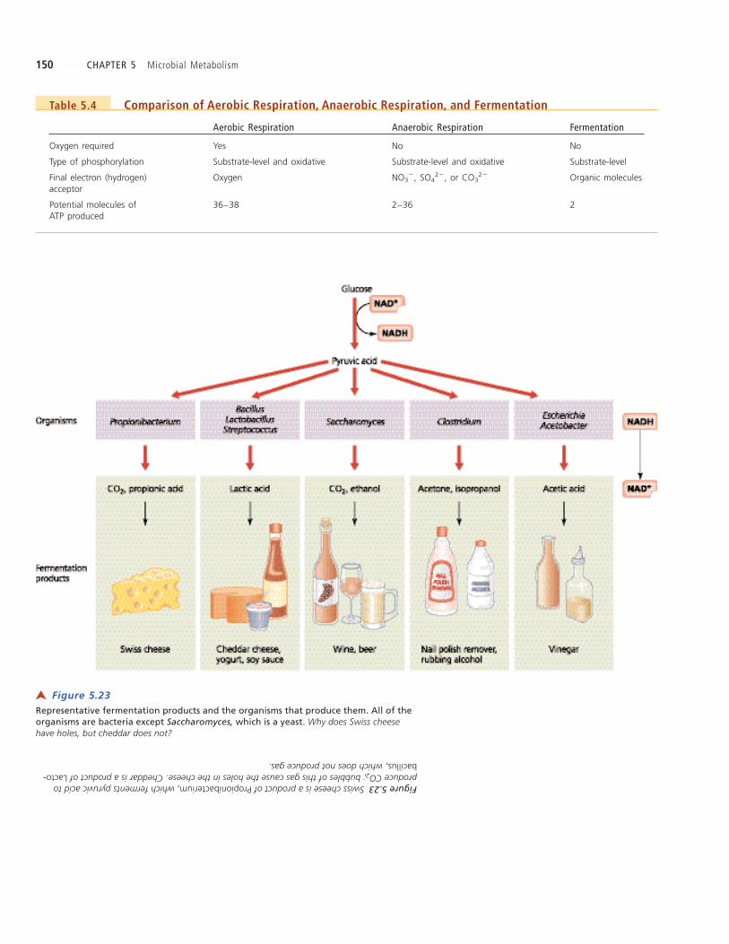
Table 5.4 Comparison of Aerobic Respiration, Anaerobic Respiration, and Fermentation
Aerobic Respiration Anaerobic Respiration Fermentation
Oxygen required Yes No No
Type of phosphorylation Substrate-level and oxidative Substrate-level and oxidative Substrate-level
Final electron (hydrogen) Oxygen NO3�, SO4
2�, or CO32� Organic molecules
acceptor
Potential molecules of 36–38 2–36 2ATP produced
Figure 5.23 Representative fermentation products and the organisms that produce them. All of theorganisms are bacteria except Saccharomyces, which is a yeast. Why does Swiss cheesehave holes, but cheddar does not?
➤
Figure 5.23Swiss cheese is a product of Propionibacterium,which ferments pyruvic acid toproduce CO2; bubbles of this gas cause the holes in the cheese. Cheddar is a product of Lacto-bacillus,which does not produce gas.
CHAPTER 5 Microbial Metabolism 151
Subsequent reactions further catabolize the glycerol andfatty acid molecules. Glycerol is converted to DHAP, whichas one of the substrates of glycolysis is oxidized to pyruvicacid (see Figure 5.14). The fatty acids are degraded in a cata-bolic process known as beta-oxidation (Figure 5.24b).9 Inthis process, enzymes repeatedly split off pairs of the hydro-genated carbon atoms that make up a fatty acid and joineach pair to coenzyme A to form acetyl-CoA, until the entirefatty acid has been converted to molecules of acetyl-CoA.NADH and FADH2 are generated during beta-oxidation,and more of these molecules are generated when the acetyl-CoA is utilized in the Krebs cycle to generate ATP (see Fig-
ure 5.19). As is the case for the Krebs cycle, the enzymesinvolved in beta-oxidation are located in the cytoplasm ofprokaryotes, and in the mitochondria of eukaryotes.
CRITICAL THINKING
How and where do cells use the molecules of NADH and FADH2
produced during beta-oxidation?
Protein Catabolism
Learning Objective
� Explain how proteins are catabolized for energy and metaboliteproduction.
Some microorganisms, particularly food-spoilage bacteria,pathogenic bacteria, and fungi, normally catabolize proteinsas an important source of energy and metabolites. Mostother cells catabolize proteins and their constituent aminoacids only when carbon sources such as glucose and fat arenot available.
Generally, proteins are too large to cross cell membranes,so microorganisms conduct the rst step in the process ofprotein catabolism outside the cell by secreting proteases
(proł teł -ał s-ez)—enzymes that split proteins into their con-stituent amino acids (Figure 5.25). Once released by the ac-tion of proteases, amino acids are transported into the cell,where special enzymes split off amino groups in a reactioncalled deamination. The resulting altered molecules enterthe Krebs cycle, and the amino groups are either recycled tosynthesize other amino acids or excreted as nitrogenouswastes such as ammonia (NH3), ammonium ion (NH4
�), ortrimethylamine oxide (which has the chemical formula(CH3)3NO and is abbreviated TMAO). As Highlight 5.2 de-scribes, TMAO plays an interesting role in the production ofthe odor we describe as shy.Thus far, we have examined the catabolism of carbohy-drates, lipids, and proteins. Now we turn our attention tothe synthesis of these molecules, beginning with the anabolicreactions of photosynthesis.
Figure 5.24Catabolism of a fat molecule. (a) Lipase breaks fats intoglycerol and three fatty acids by hydrolysis. Glycerol isconverted to DHAP, which can be catabolized byglycolysis and the Krebs cycle. (b) Fatty acids arecatabolized by beta-oxidation reactions that producemolecules of acetyl-CoA and reduced coenzymes (NADHand FADH2).
➤
9“Beta” is part of the name of this process because enzymes break the bond at the sec-ond carbon atom from the end of a fatty acid, and beta is the second letter in the Greekalphabet.
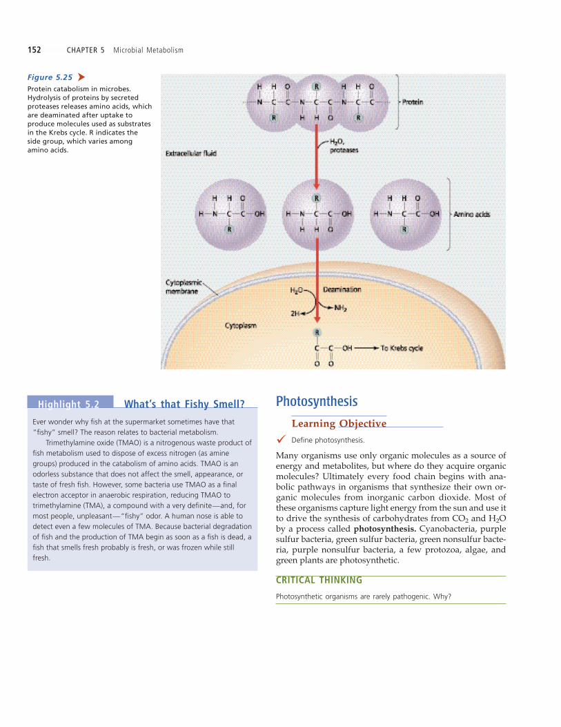
152 CHAPTER 5 Microbial Metabolism
Photosynthesis
Learning Objective
� Define photosynthesis.
Many organisms use only organic molecules as a source ofenergy and metabolites, but where do they acquire organicmolecules? Ultimately every food chain begins with ana-bolic pathways in organisms that synthesize their own or-ganic molecules from inorganic carbon dioxide. Most ofthese organisms capture light energy from the sun and use itto drive the synthesis of carbohydrates from CO2 and H2Oby a process called photosynthesis. Cyanobacteria, purplesulfur bacteria, green sulfur bacteria, green nonsulfur bacte-ria, purple nonsulfur bacteria, a few protozoa, algae, andgreen plants are photosynthetic.
CRITICAL THINKING
Photosynthetic organisms are rarely pathogenic. Why?
Ever wonder why fish at the supermarket sometimes have that“fishy” smell? The reason relates to bacterial metabolism.
Trimethylamine oxide (TMAO) is a nitrogenous waste product offish metabolism used to dispose of excess nitrogen (as aminegroups) produced in the catabolism of amino acids. TMAO is anodorless substance that does not affect the smell, appearance, ortaste of fresh fish. However, some bacteria use TMAO as a finalelectron acceptor in anaerobic respiration, reducing TMAO totrimethylamine (TMA), a compound with a very definite—and, formost people, unpleasant—“fishy” odor. A human nose is able todetect even a few molecules of TMA. Because bacterial degradationof fish and the production of TMA begin as soon as a fish is dead, afish that smells fresh probably is fresh, or was frozen while stillfresh.
Highlight 5.2 What’s that Fishy Smell?
Figure 5.25Protein catabolism in microbes.Hydrolysis of proteins by secretedproteases releases amino acids, whichare deaminated after uptake toproduce molecules used as substratesin the Krebs cycle. R indicates theside group, which varies amongamino acids.
➤

CHAPTER 5 Microbial Metabolism 153
Chemicals and Structures
Learning Objective
� Compare and contrast the basic chemicals and structures involvedin photosynthesis in prokaryotes and eukaryotes.
Photosynthetic organisms capture light energy with pig-ment molecules, the most important of which are chloro-
phylls. Chlorophyll molecules are composed of ahydrocarbon tail attached to a light-absorbing active site cen-tered around a magnesium ion (Mg2�) (Figure 5.26a). Activesites are structurally similar to the cytochrome moleculesfound in electron transport chains, except chlorophylls useMg2� rather then Fe2�. Chlorophylls, typically designatedwith letters—for example, chlorophyll a, chlorophyll b, andbacteriochlorophyll a—vary slightly in the lengths andstructures of their hydrocarbon tails and in the atoms thatextend from their active sites. Green plants, algae, photo-synthetic protozoa, and cyanobacteria principally usechlorophyll a, whereas green and purple bacteria use bac-teriochlorophylls.
The slight structural differences among chlorophyllscause them to absorb light of different wavelengths. For ex-ample, chlorophyll a from algae best absorbs light withwavelengths of about 425 nm and 660 nm (violet and red),whereas bacteriochlorophyll a from purple bacteria best ab-sorbs light with wavelengths of about 350 nm and 880 nm(ultraviolet and infrared). Because they best use light withdiffering wavelengths, algae and purple bacteria success-fully occupy different ecological niches.
Cells arrange numerous molecules of chlorophyll andother pigments within a protein matrix to form light-harvesting matrices called photosystems that are embeddedin cellular membranes called thylakoids. Thylakoids ofphotosynthetic prokaryotes are invaginations of the cyto-plasmic membranes (Figure 5.26b). The thylakoids of eu-karyotes appear to be formed from infoldings of the innermembranes of chloroplasts, though thylakoid membranesand the inner membranes are not connected in maturechloroplasts (see Figure 3.37). Thylakoids of chloroplasts arearranged in stacks called grana. An outer chloroplast mem-brane surrounds the grana, and the space between the outermembrane and the thylakoid membrane is known as thestroma.
There are two types of photosystems, named photosys-tem I (PS I) and photosystem II (PS II), in the order of theirdiscovery. Photosystems absorb light energy and use redoxreactions to store the energy in molecules of ATP andNADPH. Because they depend upon light energy, these re-actions of photosynthesis are classi ed as light-dependent
reactions. Photosynthesis also involves light-independent
reactions that actually synthesize glucose from carbon diox-ide and water. Historically, these reactions were called lightand dark reactions, but this older terminology implies thatlight-independent reactions occur only in the dark, which isnot the case. We rst consider the light-dependent reactionsof the two photosystems.
Figure 5.26 Photosynthetic structures in aprokaryote. (a) Chlorophyll moleculesare grouped together in thylakoidmembranes to form photosystems.Chlorophyll contains a light-absorbingactive site that is connected to a longhydrocarbon tail. The chlorophyllshown here is bacteriochlorophyll a;other chlorophyll types differ in theside chains protruding from the centralring. (b) Prokaryotic thylakoids areinfoldings of the cytoplasmicmembrane.
➤

154 CHAPTER 5 Microbial Metabolism
Light-Dependent Reactions
Learning Objectives
� Describe the components and function of the two photosystems,PS I and PS II.
� Contrast cyclic and noncyclic photophosphorylation.
The pigments of photosystem I absorb light energy andtransfer it to a neighboring molecule within the photosys-tem until the energy eventually arrives at a special chloro-phyll molecule called the reaction center chlorophyll
(Figure 5.27). Light energy from hundreds of such transfersexcites electrons in the reaction center chlorophyll, whichpasses its excited electrons to the reaction center s electronacceptor, which is the initial carrier of an electron transportchain. As electrons move down the chain, their energy isused to pump protons across the membrane, creating a pro-ton motive force. In prokaryotes, protons are pumped out ofthe cell; in eukaryotes they are pumped from the stroma intothe interior of the thylakoids.
The proton motive force is used in chemiosmosis, inwhich, you should recall, protons ow down theirelectrochemical gradient through ATPases, which generateATP. In photosynthesis, this process is called photophosphory-lation, which can be either cyclic or noncyclic.
Cyclic Photophosphorylation
Electrons moving from one carrier molecule to another in athylakoid must eventually pass to a nal electron acceptor.In cyclic photophosphorylation, which occurs in all photo-synthetic organisms, the nal electron acceptor is the origi-nal reaction center chlorophyll that donated the electrons(Figure 5.28a). In other words, when light energy exciteselectrons in PS I, they pass down an electron transport chainand return to PS I. The energy from the electrons is used toestablish a proton gradient that drives the phosphorylationof ADP to ATP by chemiosmosis.
Noncyclic Photophosphorylation
Some photosynthetic bacteria and all plants and algae alsoutilize noncyclic photophosphorylation. (Exceptions aregreen and purple sulfur bacteria.) Noncyclic photophos-phorylation, which requires both PS I and PS II, not onlygenerates molecules of ATP, but also reduces molecules ofcoenzyme NADP� to NADPH (Figure 5.28b).
When light energy excites electrons of PS II, they arepassed to PS I through an electron transport chain. (Notethat photosystem II occurs rst in the pathway, and photo-system I second, a result of the fact that the photosystemswere named for the order in which they were discovered,not the order in which they operate.) PS I further energizesthe electrons with additional light energy and transfersthem through an electron transport chain to NADP�, whichis thereby reduced to NADPH. Hydrogen ions in NADPHcome from the stroma or cytosol. NADPH subsequently par-ticipates in the synthesis of glucose in the light-independentreactions, which we will examine in the next section.
In noncyclic photophosphorylation, a cell must con-stantly replenish electrons to the reaction center of photo-system II. Oxygenic (oxygen-producing) organisms, such asalgae, green plants, and cyanobacteria, derive electrons fromthe dissociation of H2O (see Figure 5.28b). In these organ-isms, two molecules of water give up their electrons, pro-ducing molecular oxygen (O2) as a waste product during
Figure 5.28The light-dependent reactions of photosynthesis: cyclicand noncyclic photophosphorylation. The red arrowsindicate the flow of electrons excited by light energy.(a) In cyclic photophosphorylation, electrons excited bylight striking photosystem I travel from its reactioncenter, down an electron transport chain, and thenreturn to the reaction center. The proton gradientproduced drives the phosphorylation of ADP by ATPsynthase (chemiosmosis). (b) In noncyclicphotophosphorylation, light striking photosystem IIexcites electrons that are passed down an electrontransport chain to photosystem I, simultaneouslyestablishing a proton gradient that is used tophosphorylate ADP to ATP. Light energy collected byphotosystem I further excites the electrons, which areused to reduce NADP� to NADPH via an electrontransport chain. In oxygenic organisms, new electronsare provided by the lysis of H2O (bottom left).
➤
Figure 5.27 Reaction center of a photosystem.Light energy absorbed by pigmentsanywhere in the photosystem istransferred to the reaction centerchloroplast, where it is used to exciteelectrons for delivery to the reactioncenter’s electron acceptor.
➤

CHAPTER 5 Microbial Metabolism 155
CRITICAL THINKING
We have seen that of the two ways that ATP is generated viachemiosmosis—photophosphorylation and oxidative phosphorylation—
the former can be cyclical, but the latter is never cyclical. Why can’toxidative phosphorylation be cyclical; that is, why aren’t electronspassed back to the molecules that donated them?
photosynthesis. Anoxygenic photosynthetic bacteria get elec-trons from inorganic compounds such as H2S, which resultsin a nonoxygen waste such as sulfur.
Table 5.5 on page 156 compares photophosphorylationto substrate-level and oxidative phosphorylation.

156 CHAPTER 5 Microbial Metabolism
To this point, we have examined the use of photosyntheticpigments and thylakoid structure to harvest light energy toproduce both ATP and reducing power in the form ofNADPH. Next we examine the light-independent reactionsof photosynthesis.
Light-Independent Reactions
Learning Objectives
� Contrast the light-dependent and light-independent reactions ofphotosynthesis.
� Describe the reactants and products of the Calvin-Benson cycle.
Light-independent reactions of photosynthesis do notrequire light directly; instead, they use ATP and NADPHgenerated by the light-dependent reactions. The key reactionof the light-independent pathway of photosynthesis iscarbon fixation by the Calvin-Benson cycle,10 which involvesthe attachment of molecules of CO2 to molecules of a ve-carbon organic compound called ribulose 1,5-bisphosphate(RuBP). RuBP is derived from phosphorylation of a precur-sor metabolite produced by the pentose phosphate pathway(see Figure 5.16).
The Calvin-Benson cycle is reminiscent of the Krebs cy-cle in that the substrates of the cycle are regenerated. It ishelpful to notice the number of carbon atoms during eachpart of the three steps of the Calvin-Benson cycle (Figure
5.29):
Fixation of CO2. Enzymes attach three molecules ofcarbon dioxide (3 carbon atoms) to three molecules ofRuBP (15 carbon atoms), which are split to form sixmolecules of 3-phosphoglyceric acid (18 carbon atoms).Reduction. NADPH reduces the six molecules of3-phosphoglyceric acid to form six molecules of glyc-eraldehyde 3-phosphate (G3P) (18 C). These reactionsrequire six molecules each of ATP and NADPH gener-ated by the light-dependent reactions.Regeneration of RuBP. The cell regenerates three mol-ecules of RuBP (15 C) fro m ve molecules of G3P(15 C); it also uses the remaining molecule of glycer-
aldehyde 3-phosphate to synthesize glucose by revers-ing the reactions of glycolysis.
In summary, ATP and NADPH from the light-dependentreactions drive the synthesis of glucose from CO2 in thelight-independent reactions of the Calvin-Benson cycle. Forevery three molecules of CO2 that enter the Calvin-Bensoncycle, a molecule of glyceraldehyde 3-phosphate (G3P)leaves. Glycolysis is subsequently reversed to anabolicallysynthesize glucose 6-phosphate from two molecules of G3P.A more detailed account of the Calvin-Benson cycle is givenin the Appendix on page C-1.
The processes of oxygenic photosynthesis and aerobicrespiration complement one another to complete both a car-bon cycle and an oxygen cycle. During the synthesis of glu-cose in oxygenic photosynthesis, water and carbon dioxideare used, and oxygen is released as a waste product; in aero-bic respiration, oxygen serves as the nal electron acceptorin the oxidation of glucose to carbon dioxide and water.
In this section we examined an essential anabolic path-way for life on Earth—photosynthesis, which produces glu-cose. Next we will consider anabolic pathways involved inthe synthesis of other organic molecules.
Other Anabolic Pathways
Learning Objective
� Define amphibolic reaction.
Anabolic reactions are synthesis reactions. As such, theyrequire energy and a source of metabolites. Energy for anab-olism is provided by ATP generated in the catabolic reac-tions of aerobic respiration, anaerobic respiration, andfermentation; and by the initial redox reactions of photosyn-thesis. Glycolysis, the Krebs cycle, and the pentose phos-phate pathway provide twelve basic precursor metabolitesfrom which all macromolecules and cellular structures canbe made. Some microorganisms, such as E. coli, can synthe-size all twelve precursors (Table 5.6 on page 160); others,such as humans, must acquire some chemicals in their diets.
Many anabolic pathways are the reversal of the cata-bolic pathways we have discussed; therefore, much of thematerial that follows has in a sense been discussed in the sec-
Table 5.5 A Comparison of the Three Types of Phosphorylation
Source of Location in Location inPhosphate Source of Energy Eukaryotic Cell Prokaryotic Cell
Substrate-level Organic molecule High-energy bond Cytosol and mitochondrial Cytosolphosphorylation between donor matrixand phosphate
Oxidative phosphorylation Inorganic phosphate Proton motive force Inner membrane of Cell membrane(PO4
2�) mitochondrion
Photophosphorylation Inorganic phosphate Proton motive force Thylakoid of chloroplast Thylakoid of cell(PO4
2�) membrane
10Named for the men who elucidated its pathways.

CHAPTER 5 Microbial Metabolism 157
Carbohydrate Biosynthesis
Learning Objective
� Describe the biosynthesis of carbohydrates.
As we have seen, anabolism begins in photosyntheticorganisms with carbon xation by the enzymes of theCalvin-Benson cycle to form molecules of G3P. Enzymes useG3P as the starting point for synthesizing sugars, complex
tions on catabolism. Reactions that can proceed in either di-rection—toward catabolism or toward anabolism—are saidto be amphibolic. The following sections discuss the synthe-sis of carbohydrates, lipids, amino acids, and nucleotides.The polymerizations of amino acids into proteins, and of nu-cleotides into RNA and DNA, are closely linked to genetics,so they are covered in Chapter 7.
Figure 5.29 Simplified diagram of the Calvin-Benson cycle ➀ . Three molecules of RuBP combinewith three molecules of CO2 ➁ . The resulting molecules are reduced to form sixmolecules of G3P ➂ . Five molecules of G3P are converted to three molecules of RuBP,which completes the cycle. Two turns of the cycle also yield two molecules of G3P, whichare polymerized to synthesize glucose 6-phosphate.
➤

158 CHAPTER 5 Microbial Metabolism
polysaccharides such as starch, cellulose for cell walls in al-gae, and peptidoglycan for cell walls of bacteria. Animalsand protozoa synthesize the storage molecule glycogen.
Some cells are able to synthesize sugars from noncarbo-hydrate precursors such as amino acids, glycerol, and fattyacids by pathways collectively called gluconeogenesis11
(gluł koł -neł -oł -jen eø-sis; Figure 5.30). Most of the reactions ofgluconeogenesis are amphibolic, using enzymes of glycoly-sis in reverse, but four of the reactions require unique en-zymes. Gluconeogenesis is highly endergonic and canproceed only if there is an adequate supply of energy.
Lipid Biosynthesis
Learning Objective
� Describe the biosynthesis of lipids.
As we saw in Chapter 2, lipids are a diverse group of organicmolecules that function as energy-storage compounds andas components of membranes. Carotenoids, which are red-
dish pigments found in many bacterial and plant photosys-tems, are also lipids.
Because of their variety, it is not surprising that lipidsare synthesized by a variety of routes. For example, fats aresynthesized in anabolic reactions that are the reverse of theircatabolism—they polymerize glycerol and three fatty acids(Figure 5.31). Glycerol is derived from G3P generated by theCalvin-Benson cycle and glycolysis; the fatty acids are pro-duced by the linkage of two-carbon acetyl-CoA molecules toone another by a sequence of endergonic reactions that ef-fectively reverse the catabolic reactions of beta-oxidation.Other lipids, such as steroids, are synthesized in complexpathways involving polymerizations and isomerizations ofsugar and amino acid metabolites.
Mycobacterium tuberculosis (mıł koł -bak-teł r eł -uøm tuł -ber-kyuł -loł -sis), the pathogen that causes tuberculosis, makes co-pious amounts of a waxy lipid called mycolic acid, which isincorporated into its cell wall. The arduous, energy-intensive process of making long lipid chains explains whythis organism grows slowly, and why tuberculosis requires along course of treatment with antimicrobial drugs.
11From Greek glykys, meaning sweet, neo, meaning new, and genesis, meaninggenerate.
Figure 5.30The role of gluconeogenesis in the biosynthesis ofcomplex carbohydrates. Complex carbohydrates aresynthesized from simple sugar molecules such asglucose, glucose-6 phosphate, and fructose-6 phosphate.Starch and cellulose are found in algae; glycogen isfound in animals and protozoa; and peptidoglycan isfound in bacteria.
➤

CHAPTER 5 Microbial Metabolism 159
baø-sil uøs), a bacterium that ferments milk and producessome cheeses. This microorganism cannot synthesize any ofits amino acids; it acquires all of them by catabolizing pro-teins in its environment.
Precursor metabolites are converted to amino acids bythe addition of an amine group. This process is called ami-
nation when the amine group comes from ammonia (NH3);an example is the formation of aspartic acid from NH3 andthe Krebs cycle intermediate oxaloacetic acid (Figure 5.32a).
Amination reactions are the reverse of the catabolic deami-nation reactions we discussed previously. More commonly,however, a cell moves the amine group from one amino acidand adds it to a metabolite, producing a different amino
Amino Acid Biosynthesis
Learning Objective
� Describe the biosynthesis of amino acids.
Amino acids are synthesized from precursor metabolites de-rived from glycolysis, the Krebs cycle, and the pentose phos-phate pathway, and from other amino acids. Someorganisms (such as E. coli, and most plants and algae) syn-thesize all their amino acids from precursor metabolites.Other organisms, including humans, must acquire so-calledessential amino acids in their diets because they cannot syn-thesize them. One extreme example is Lactobacillus (lak-toł -
Figure 5.31Biosynthesis of fat, a lipid. A fat molecule is synthesizedfrom glycerol and three molecules of fatty acid, theprecursors of which are produced in glycolysis.
➤
Figure 5.32 The synthesis of amino acids via amination andtransamination. (a) In amination, an amine group fromammonia is added to a precursor metabolite. In thisexample, oxaloacetic acid is converted into the aminoacid aspartic acid. (b) In transamination, the aminegroup is derived from an existing amino acid. In theexample shown here, the amino acid glutamic aciddonates its amine group to oxaloacetic acid, which isconverted into aspartic acid. Note that transamination isa reversible reaction.
➤

160 CHAPTER 5 Microbial Metabolism
acid. This process is called transamination because theamine group is transferred from one amino acid to another(Figure 5.32b). All transamination enzymes use a coenzyme,pyridoxal phosphate, which is derived from vitamin B6.
Ribozymes of ribosomes polymerize amino acids intoproteins. This energy-demanding process is examined inChapter 7 because it is intimately linked with genetics.
CRITICAL THINKING
What class of enzyme is involved in amination reactions? What classof enzyme catalyzes transaminations?
Nucleotide Biosynthesis
Learning Objective
� Describe the biosynthesis of nucleotides.
Recall from Chapter 2 that the building blocks of nucleicacids are nucleotides, each of which consists of a ve-car-bon sugar, a phosphate group, and a purine or pyrimidinebase (see Figure 2.24a). Nucleotides are produced from pre-cursor metabolites of glycolysis and the Krebs cycle (Figure
5.33):
¥ The ve-carbon sugars—ribose in RNA and deoxyri-bose in DNA—are derived from ribose-5 phosphatefrom the pentose phosphate pathway.
¥ The phosphate group is derived ultimately from ATP.¥ Purines and pyrimidines are synthesized in a series
of ATP-requiring reactions from the amino acids glut-amine and aspartic acid derived from Krebs cycleintermediates, ribose-5 phosphate, and folic acid.(which is a vitamin for humans but can be synthe-sized by many bacteria and protozoa).
The anabolic reactions by which polymerases polymerize nu-cleotides to form DNA and RNA are discussed in Chapter 7.
Integration and Regulationof Metabolic Functions
Learning Objectives
� Describe interrelationships between catabolism and anabolism interms of ATP and substrates.
� Discuss regulation of metabolic activity.
As we have seen, catabolic and anabolic reactions interactwith one another in several ways. First, ATP molecules
Table 5.6 The Twelve Precursor Metabolites
Pathway that Generates Examples of Macromoleculethe Metabolite Synthesized from Metabolitea Examples of Functional Use
Glucose 6-phosphate Glycolysis Lipopolysaccharide Outer membrane of cell wall
Fructose 6-phosphate Glycolysis Peptidoglycan Cell wall
Glyceraldehyde-3-phosphate (G3P) Glycolysis Glycerol portion of lipids Fats–energy storage
Phosphoglyceric acid Glycolysis Amino acids: cysteine, Enzymesglycine, and serine
Phosphoenolpyruvic acid (PEP) Glycolysis Amino acids: phenylalanine, Enzymestryptophan, and tyrosine
Pyruvic acid Glycolysis Amino acids: alanine, Enzymesleucine, and valine
Ribose 5-phosphate Pentose phosphate DNA, RNA, amino acid and Genome, enzymespathway histidine
Erythrose 4-phosphate Pentose phosphate Amino acids: phenylalanine, Enzymespathway tryptophan, and tyrosine
Acetyl-CoA Krebs cycle Fatty acid portion of lipids Cell membrane
�-Ketogluteric acid Krebs cycle Amino acids: arginine, Enzymesglutamic acid, glutamine, and proline
Succinyl-CoA Krebs cycle Heme Cytochrome electron carrier
Oxaloacetate Krebs cycle Amino acids: aspartic acid, Enzymesasparagine, isoleucine, lysine, methionine, and threonine
aExamples given apply to the bacterium E. coli.

CHAPTER 5 Microbial Metabolism 161
excess of aspartic acid will cease the amination ofoxaloacetic acid (see Figure 5.32).
¥ Eukaryotic cells keep metabolic processes from inter-ferring with each other by isolating particularenzymes within membrane-bounded organelles. Forexample, proteases sequestered within lysosomesdigest phagocytized proteins without destroying vitalproteins in the cytosol.
¥ Cells use inhibitory and excitatory allosteric sites onenzymes to control the activity of enzymes (seeFigure 5.11).
¥ Feedback inhibition slows or stops anabolic pathwayswhen the product is in abundance (see Figure 5.12).
¥ Cells regulate catabolic and anabolic pathways thatuse the same substrate molecules by requiring differ-ent coenzymes for each. For instance, NADH is usedalmost exculsively with catabolic enzymes, whereasNADPH is typically used for anabolism.
Note that these regulatory mechanisms are generally of twotypes: control of gene expression, in which cells control theamount and timing of protein (enzyme) production, andcontrol of metabolic expression, in which cells control the activ-ity of proteins (enzymes) once they have been produced.
Numerous interrelationships among the metabolicpathways discussed in this chapter are diagrammedschematically in Figure 5.34.
produced by catabolism are used to drive anabolic reactions.Second, precursor metabolites produced by catabolicpathways are used as substrates for anabolic reactions.Additionally, most metabolic pathways are amphibolic; theyfunction as part of either catabolism or anabolism as needed.
Cells regulate metabolism in a variety of ways to maxi-mize ef ciency in growth and reproductive rate. Among themechanisms involved are the following:
¥ Cells synthesize or degrade channel and transportproteins to increase or decrease the concentration ofchemicals in the cytosol and organelles.
¥ Cells often synthesize the enzymes needed to catabo-lize a particular substrate only when that substrate isavailable. For instance, the enzymes of beta-oxidationare not produced when there are no fatty acids tocatabolize.
¥ If two energy sources are available, cells catabolizethe more energy ef cient of the two. For example, abacterium growing in the presence of both glucoseand lactose will produce enzymes only for the trans-port and catabolism of glucose. Once the supply ofglucose is depleted, lactose-utilizing proteins are pro-duced.
¥ Cells synthesize the metabolites they need, but theytypically cease synthesis if a metabolite is available asa nutrient. For instance, bacteria grown with an
Figure 5.33The biosynthesis of nucleotides.
➤
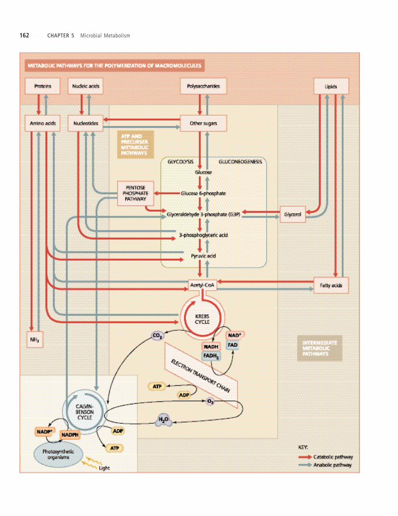
162 CHAPTER 5 Microbial Metabolism

CHAPTER 5 Microbial Metabolism 163
CHAPTER SUMMARY
Basic Chemical Reactions Underlying Metabolism(pp. 126-136)
1. Metabolism is the sum of complex biochemical reactionswithin the cells of an organism, including catabolism, whichbreaks down molecules and releases energy, and anabolism,
which synthesizes molecules and uses energy.
2. Precursor metabolites, often produced in catabolic reactions,are used to synthesize all other organic compounds.
3. Reduction reactions are those in which the electrical chargeon a chemical is made more negative by the addition of elec-trons. The molecule that donates an electron is oxidized. Ifthe electron is part of a hydrogen atom, an oxidation reactionis also called dehydrogenation. Oxidation and reduction reac-tions always occur in pairs called oxidation-reduction
(redox) reactions.
4. Three important electron carrier molecules are nicotinamide
adenine dinucleotide (NAD�), nicotinamide adenine dinu-
cleotide phosphate (NADP�), and flavine adenine dinu-
cleotide (FAD).
5. Phosphorylation is the addition of phosphate to a molecule.Three types of phosphorylation form ATP: Substrate-level
phosphorylation involves the transfer of phosphate to ADPfrom another phosphorylated organic compound. In oxida-
tive phosphorylation, energy from redox reactions of respi-ration is used to attach inorganic phosphate (PO4
2�) to ADP.Photophosphorylation is the phosphorylation of ADP withinorganic phosphate using energy from light.
6. Catalysts increase the rates of chemical reactions and are notpermanently changed in the process. Enzymes, which areorganic catalysts, are often named for their substrates, themolecules upon which they act. Enzymes can be classi edaccording to their mode of action into hydrolases, isomerases,ligases (polymerases), lyases, oxidoreductases, or trans-ferases.
7. Apoenzymes are the protein portions of protein enzymes thatmay require one or more cofactors such as inorganic ions ororganic cofactors are called coenzymes. The combination ofboth apoenzyme and its cofactors is a holoenzyme. RNAmolecules functioning as enzymes are called ribozymes.
8. Activation energy is the amount of energy required to initi-ate a chemical reaction.
9. Substrates t into the speci cally shaped active sites of theenzymes that catalyze their reactions.
10. Enzymes may be denatured by physical and chemical factorssuch as heat and pH. Denaturation may be reversible or per-manent.
11. Enzyme activity proceeds at a rate proportional to the con-centration of substrate molecules until all the active sites are
lled to saturation.
12. Competitive inhibitors block active sites and thereby blockenzyme activity. In allosteric inhibition, noncompetitiveinhibitors attach to an allosteric site on an enzyme, alteringthe active site so that it is no longer functional.
13. Feedback inhibition (negative feedback) occurs when thenal product of a series of reactions is an allosteric inhibitor
of some previous step in the series. Thus, accumulation of theend-product feeds back into the series a signal that stopsthe process.
Carbohydrate Catabolism (pp. 136-149)
1. Glycolysis involves the splitting of a glucose molecule in athree-stage, 10-step process that ultimately results in twomolecules of pyruvic acid and a net gain of two ATP and twoNADH molecules.
2. The pentose phosphate and Entner-Doudoroff pathways arealternative means for the catabolism of glucose that yieldfewer ATP molecules than does glycolysis. However, theyproduce precursor metabolites not produced in glycolysis.
3. Cellular respiration is a metabolic process that involves thecomplete oxidation of substrate molecules and the productionof ATP following a series of redox reactions.
4. Two carbons from pyruvic acid join coenzyme A to formacetyl-coenzyme A (acetyl-CoA), which then enters the Krebs
cycle, a series of eight enzymatic steps that transfer energyfrom acetyl-CoA to coenzymes NAD� and FAD.
5. An electron transport chain is a series of redox reactions thatpass electrons from one membrane-bound carrier to another,and then to a nal electron acceptor. The energy from theseelectrons is used to pump protons across the membrane.
6. The four classes of carrier molecules in electron transport sys-tems are avoproteins, ubiquinones, metal-containing pro-teins, and cytochromes.
7. Aerobes use oxygen atoms as nal electron acceptors of theirelectron transport chains in a process known as aerobic res-
piration, whereas anaerobes use other inorganic molecules(such as NO3
�, SO42�, and CO3
2�, or rarely an organic mol-ecule) as the nal electron acceptor in anaerobic respiration.
Figure 5.34 Integration of cellular metabolism. Cells possess three major categories of metabolicpathways: pathways for the polymerization of macromolecules (proteins, nucleic acids,polysaccharides, and lipids), intermediate pathways, and ATP and precursor pathways(glycolysis, Krebs cycle, the pentose phosphate pathway, and the Entner-Doudoroffpathway [not shown]). Cells of photosynthetic organisms also have the Calvin-Bensoncycle
➤

164 CHAPTER 5 Microbial Metabolism
8. In chemiosmosis, ions ow down their electrochemical gra-dient across a membrane through ATP synthase (ATPase) tosynthesize ATP.
9. A proton gradient is an electrochemical gradient of hydrogenions across a membrane. It has potential energy known as aproton motive force.
10. Oxidative phosphorylation and photophosphorylation usechemiosmosis.
11. Fermentation is the partial oxidation of sugar to releaseenergy using an organic molecule rather than an electrontransport chain as the nal electron acceptor. End-products offermentation, which are often useful to humans and aid inidenti cation of microbes in the laboratory, include acids,alcohols, and gases.
Other Catabolic Pathways (pp. 149-152)
1. Lipids and proteins can be catabolized into smaller mole-cules, which can be used as substrates for glycolysis and theKrebs cycle.
2. Beta-oxidation is a catabolic process in which enzymes splitpairs of hydrogenated carbon atoms from a fatty acid and jointhem to coenzyme A to form acetyl-CoA.
3. Proteases secreted by microorganisms digest proteins outsidethe microbe s cell wall. The resulting amino acids are movedinto the cell and used in anabolism, or deaminated and catab-olized for energy.
Photosynthesis (pp. 152-156)
1. Amphibolic reactions are metabolic reactions that arereversible.
2. Photosynthesis is a process in which light energy is capturedby pigment molecules called chlorophylls (bacteriochloro-phylls in some bacteria) and transferred to ATP and metabo-lites. Photosystems are networks of light-absorbing chloro-phyll molecules and other pigments held within a proteinmatrix on membranes called thylakoids.
3. The redox reactions of photosynthesis are classi ed as light-
dependent reactions and light-independent reactions.
4. A reaction center chlorophyll is a special chlorophyll mole-cule in a photosystem in which electrons are excited by lightenergy and passed to an acceptor molecule of an electrontransport chain.
5. In cyclic photophosphorylation, the electrons return to theoriginal reaction center after passing down the electron trans-port chain.
6. In noncyclic photophosphorylation, photosystem II workswith photosystem I, and the electrons are used to reduceNADP� to NADPH. In oxygenic photosynthesis, cyanobac-teria, algae, and green plants replenish electrons to the reac-tion center by dissociation of H2O molecules, resulting in therelease of O2 molecules. Anoxygenic bacteria derive electronsfrom inorganic compounds such as H2S, producing wastesuch as sulfur.
7. In the light-independent pathway of photosynthesis, carbon
fixation occurs in the Calvin-Benson cycle, in which CO2 isreduced to produce glucose.
Other Anabolic Pathways (pp. 156-160)
1. Some cells are able to synthesize glucose from amino acids,glycerol, and fatty acids via a process called gluconeogenesis.
2. Amination reactions involve adding an amine group fromammonia to a metabolite to make an amino acid. Transami-
nation occurs when an amine group is transferred from oneamino acid to another.
3. Nucleotides are synthesized from precursor metabolites pro-duced by glycolysis, the Krebs cycle, and the pentose phos-phate pathway.
Integration and Regulation of Metabolic Functions(pp. 160-163)
1. Cells regulate metabolism by control of gene expression ormetabolic expression. They control the latter in a variety ofways, including synthesizing or degrading channel proteinsand enzymes, sequestering reactions in membrane-boundedorganelles (seen only in eukaryotes), and feedback inhibition.
QUESTIONS FOR REVIEW
Multiple Choice
For each of the phrases in Questions 1–7, indicate the type of me-tabolism referred to, using the following choices:
a. anabolism onlyb. both anabolism and catabolism (amphibolic)c. catabolism only
1. Breaks a large molecule into smaller ones
2. Includes dehydration synthesis reactions
3. Is exergonic
4. Is endergonic
5. Involves the production of cell membrane constituents
6. Includes hydrolytic reactions
7. Includes metabolism
8. Redox reactionsa. transfer energy.b. transfer electrons.c. involve oxidation and reduction.d. are involved in all of the above.
9. A reduced molecule a. has gained electrons.b. has become more positive in charge.

CHAPTER 5 Microbial Metabolism 165
c. has lost electrons.d. is an electron donor.
10. Activation energya. is the amount of energy required during an activity such
as agellar motion.b. requires the addition of nutrients in the presence of water.c. is usually lowered by the action of organic catalysts.d. results from the movement of molecules.
11. Coenzymesa. are types of apoenzymes.b. are proteins.c. are inorganic cofactors.d. are organic cofactors.
12. Which of the following statements best describes ribozymes?a. Ribozymes are proteins that aid in the production of ribo-
somes.b. Ribozymes are nucleic acids that produce ribose sugars.c. Ribozymes store enzymes in ribosomes.d. Ribozymes process RNA molecules in eukaryotes.
13. Which of the following does not affect the function ofenzymes?a. ubiquinoneb. substrate concentration c. temperatured. competitive inhibitors
14. Most oxidation reactions in bacteria involve thea. removal of hydrogen ions and electrons.b. removal of oxygen.c. addition of hydrogen ions and electrons.d. addition of hydrogen ions.
15. Under ideal conditions, the fermentation of one glucose mol-ecule by a bacterium allows a net gain of how many ATPmolecules?a. 2b. 4c. 38d. 0
16. Under ideal conditions, the complete aerobic oxidation of onemolecule of glucose by a bacterium allows a net gain of howmany ATP molecules?a. 2b. 4c. 38d. 0
17. Which of the following statements is not true of the Entner-Doudoroff pathway?a. It is a series of reactions that synthesizes glucose.b. Its products are sometimes used to determine the presence
of Pseudomonas.c. It is a pathway of chemical reactions that catabolizes
glucose.d. It is an alternative pathway to glycolysis.
18. Reactions involved in the light-independent reactions of pho-tosynthesis constitute the a. Krebs cycle.b. Entner-Doudoroff pathway.c. Calvin-Benson cycle.d. pentose phosphate pathway.
Matching
19. Occurs when energy froma compound containingphosphate reacts with ADP to form ATP
20. Involves formation of ATPvia reduction of coenzymesin the electron transportchain
21. Begins with glycolysis
22. Occurs when all activesites on substrate moleculesare lled
Fill in the Blanks
23. The nal electron acceptor in cyclic photophosphorylationis .
24. Two ATP molecules are used to initiate glycolysis. Enzymesgenerate molecules of ATP for each molecule of glucose thatundergoes glycolysis. Thus a net gain of molecules of ATP is produced in glycolysis.
25. The initial catabolism of glucose occurs by glycolysisand/or the and pathways.
26. is a cyclic series of eight reactionsinvolved in the catabolism of acetyl-CoA that yields eightmolecules of NADH and two molecules of FADH2.
27. The final electron acceptor in aerobic respiration is.
28. Three common electron acceptors in anaerobic respirationare , , and
.
29. Chemolithotrophs acquire electrons from (organic/inor-ganic) compounds.
30. Complete the following chart:
Category of Enzymes Description
Catabolyzes substrate by adding water
Isomerase
Ligase/polymerase
Moves functional groups such as an acetyl group
Adds or removes electrons
Lyase
Short Answer
1. Why are enzymes necessary for anabolic reactions to occur inliving organisms?
2. How do organisms control the rate of metabolic activities intheir cells?
3. How does a noncompetitive inhibitor at an allosteric siteaffect a whole pathway of enzymatic reactions?
4. Explain the mechanism of negative feedback with respect toenzyme action.
a. Saturation
b. Oxidativephosphorylation
c. Substrate-levelphosphorylation
d. Photophosphorylation
e. Carbohydratecatabolism

166 CHAPTER 5 Microbial Metabolism
1. Why does an organism that uses glycolysis and the Krebs cy-cle also need the pentose phosphate pathway?
2. Describe how bacterial fermentation causes milk to sour.
3. Giardia lamblia and Entamoeba histolytica are protozoa that livein the colons of mammals and can cause life-threatening di-arrhea. Interestingly, these microbes lack mitochondria.What kind of pathway must they have for carbohydrate ca-tabolism?
4. Two cultures of a facultative anaerobe are grown in the sametype of medium, but one is exposed to air and the other ismaintained under anaerobic conditions. Which of the twocultures will contain more cells at the end of a week? Why?
5. What is the maximum number of molecules of ATP that canbe generated by a bacterium after the complete aerobic oxi-dation of a fat molecule containing three 12-carbon chains?(Assume that all the available energy released during catabo-lism goes to ATP production.)
6. In terms of its effects on metabolism, why is a fever over 40¡ Coften life threatening?
7. Using the categories de ned in the text on page 129, classifyeach of the enzymes of glycolysis (see page 138), the Krebscycle (see page 143), fat biosynthesis (see Figure 5.31 on page158), and transamination (see Figure 5.32 on page 159).
5. Facultative anaerobes can live under either aerobic or anaer-obic conditions. What metabolic pathways allow these organ-isms to continue to harvest energy from sugar molecules inthe absence of oxygen?
6. How does oxidation of a molecule occur without oxygen?
7. List at least four groups of microorganisms that are photo-synthetic.
8. Why do we breathe oxygen and give off carbon dioxide?
9. Why do cyanobacteria and algae take in carbon dioxide andgive off oxygen?
10. What happens to the carbon atoms in sugar catabolized byE. coli?
11. How do yeast cells make alcohol and cause bread to rise?
12. Where speci cally does the most signi cant production ofATP occur in prokaryotic and eukaryotic cells?
13. Why are vitamins essential metabolic factors for microbialmetabolism?
14. A laboratory scientist notices that a certain bacterium doesnot utilize lactose when glucose is available in its environ-ment. Describe a cellular regulatory mechanism that wouldexplain this observation.
8. How are photophosphorylation and oxidative phosphoryla-tion similar? How are they different?
9. Members of the pathogenic bacterial genus Haemophilus re-quire NAD� and heme from their environment. For whatpurpose does Haemophilus use these growth factors?
10. Compare and contrast aerobic respiration, anaerobic respira-tion, and fermentation.
11. Scientists estimate that up to one-third of Earth s biomass iscomposed of methanogenic prokaryotes in ocean sediments(Science 295:2067–2070). Describe the metabolism of these or-ganisms.
12. A young student was troubled by the idea that a bacterium isable to control its diverse and complex metabolic activities,even though it lacks a brain. How would you explain theirmetabolic control.
13. If a bacterium uses beta-oxidation to catabolize a molecule ofthe fatty acid arachidic acid, which contains 20 carbon atoms,how many acetyl-CoA molecules will be generated?
CRITICAL THINKING