bbaassiicc ccoonncceeppttss aanndd...
TRANSCRIPT

BBAASSIICC CCOONNCCEEPPTTSS AANNDD
IIMMPPOORRTTAANNCCEE OOFF VVAARRIIOOUUSS
PPHHAARRMMAACCOOKKIINNEETTIICC
PPAARRAAMMEETTEERRSS
INTRODUCTION:
Pharmacokinetics, sometimes abbreviated as PK, (from Ancient
Greek pharmakon "drug" and kinetikos "to do with motion") is dedicated to the
determination of the fate of substances administered externally to a living organism.
Pharmacokinetics is the science of the kinetics of drug absorption, distribution, and
elimination (ie, excretion and metabolism). The description of drug distribution and
elimination is often termed drug disposition.
Pharmacokinetics is often studied in conjunction with pharmacodynamics.
Pharmacodynamics explores what a drug does to the body, whereas
pharmacokinetics explores what the body does to the drug.
Pharmacokinetics includes the study of the mechanisms of absorption and
distribution of an administered drug, the rate at which a drug action begins and the
duration of the effect, the chemical changes of the substance in the body (e.g. by
enzymes) and the effects and routes of excretion of the metabolites of the drug.
Pharmacokinetics is divided into several areas which includes the extent and rate of
Absorption, Distribution, Metabolism and Excretion. This is commonly referred to as
the ADME scheme. However recent understanding about the drug-body interactions
brought about the inclusion of new term Liberation. Now Pharmacokinetics can be
better described as LADME.
Liberation is the process of release of drug from the formulation.
Absorption is the process of a substance entering the body.
Distribution is the dispersion or dissemination of substances throughout the
fluids and tissues of the body.
Metabolism is the irreversible transformation of parent compounds into
daughter metabolites.
Excretion is the elimination of the substances from the body. In rare cases,
some drugs irreversibly accumulate in a tissue in the body.
Pharmacokinetics describes how the body affects a specific drug after administration.
Pharmacokinetic properties of drugs may be affected by elements such as the site of
administration and the concentration in which the drug is administered. These may affect
the absorption rate.
To study these phases many pharmacokinetic parameters are required. Few of them are discussed here.
1. BIOLOGICAL HALF LIFE (t1/2) Synonym: Elimination half life
The biological half-life or elimination half life of a substance is the time it takes for a substance (drug, radioactive nuclide, or other) to lose half of its pharmacologic, physiologic, or radiologic activity.
In a medical context, half-life may also describe the time it takes for the blood plasma concentration of a substance to halve ("plasma half-life") of its steady-state.
The relationship between the biological and plasma half-lives of a substance can be complex, due to factors including accumulation in tissues, active metabolites, and receptor interactions.
EXAMPLES:
Substance Half-life
Amiodarone 25 days
Cisplatin 30 to 100 h
Chlorambucil 1.53 h
Digoxin 24 to 36 h
Fluoxetine
1 to 6 days The active metabolite of fluoxetine is lipophilic and
migrates slowly from the brain to the blood. The metabolite has a biological half-life of 4 to 16 days.
Methadone 15 to 60 h, in rare cases up to 190 h.
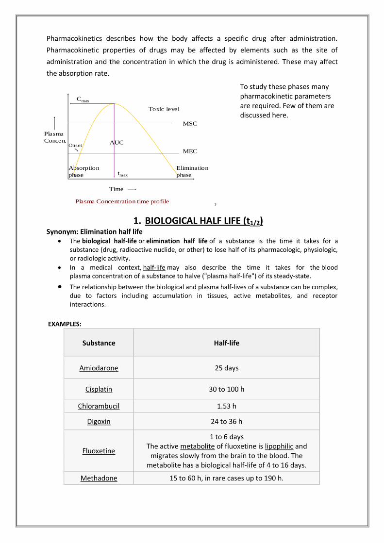
3
MEC
MSC
Time
Plasma
Concen.
tmax
Cmax
Toxic level
Onset
Elimination
phase
Absorption
phase
AUC
Plasma Concentration time profile
Oxaliplatin 14 min.
Salbutamol 7 h
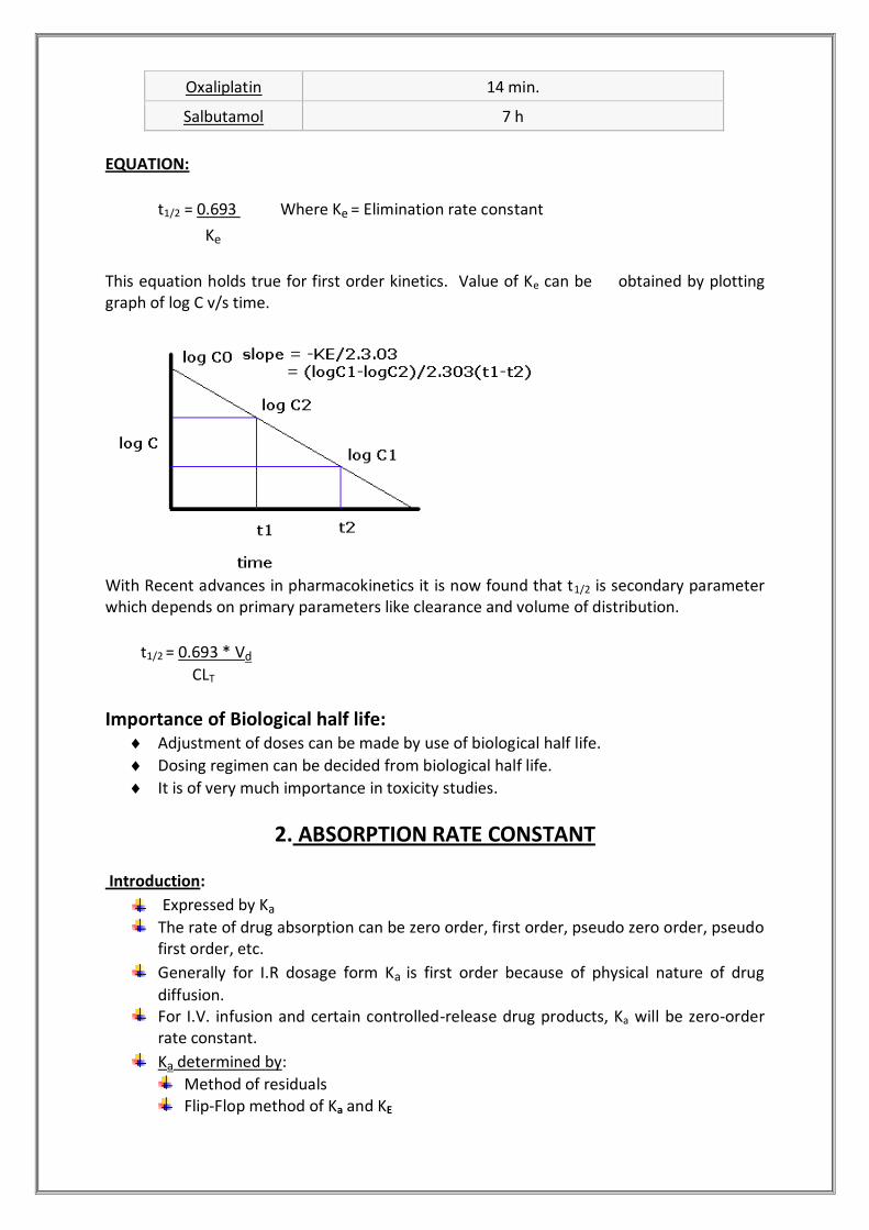
EQUATION:
t1/2 = 0.693 Where Ke = Elimination rate constant
Ke
This equation holds true for first order kinetics. Value of Ke can be obtained by plotting graph of log C v/s time.
With Recent advances in pharmacokinetics it is now found that t1/2 is secondary parameter which depends on primary parameters like clearance and volume of distribution.
t1/2 = 0.693 * Vd
CLT
Importance of Biological half life: Adjustment of doses can be made by use of biological half life.
Dosing regimen can be decided from biological half life.
It is of very much importance in toxicity studies.
2. ABSORPTION RATE CONSTANT
Introduction:
Expressed by Ka
The rate of drug absorption can be zero order, first order, pseudo zero order, pseudo first order, etc.
Generally for I.R dosage form Ka is first order because of physical nature of drug
diffusion. For I.V. infusion and certain controlled-release drug products, Ka will be zero-order
rate constant.
Ka determined by:
Method of residuals Flip-Flop method of Ka and KE

Wagner – Nelson Method Loo – Riegelman method
Importance:
Designing a multiple-dosage regimen Ka and Ke helps to predict peak and trough plasma drug concentrations following
multiple dosing. In bioequivalence studies, drug products are given in chemically equivalent (i.e.
pharmaceutical equivalent) doses. The respective rates of systemic absorption may not differ markedly. For these studies, tmax or time of peak drug concentration can be very useful in comparing the respective rates of absorption of a drug from chemically equivalent drug products.
3. ELIMINATION RATE CONSTANT (Ke)
KE is summation of rate constants for each process like urinary excretion, metabolism, biliary excretion, pulmonary excretion, etc.
KE = Km + Kb + Kl +..........
FOR, Zero order rate of elimination is constant irrespective of plasma concentration:
Er = KE.
FOR, First order: Rate of elimination proportional to plasma concentration. Constant
Fraction of drug eliminated per unit time.
Er = dC/dt = - KE C
IMPORTANCE OF ELIMINATION RATE CONSTANT: Determination of Ke is important for selection of dose regimen. Also in dose adjustment in renal impairment.
4. VOLUME OF DISTRIBUTION
The volume of distribution (VD) , also known as apparent volume of distribution, is
used to quantify the distribution of a medication between plasma and the rest of the
body after oral or parenteral dosing.
It is defined as the volume in which the amount of drug would need to be
uniformly distributed to produce the observed blood concentration.
Volume of distribution may be increased by renal failure (due to fluid retention)
and liver failure (due to altered body fluid and plasma protein binding). Conversely
it may be decreased in dehydration.

EQUATION:
The volume of distribution is given by the following equation:
Therefore the dose required to give a certain plasma concentration can be
determined if the VD for that drug is known. The VD is not a physiologic value; it is
more a reflection of how a drug will distribute throughout the body depending on
several physicochemical properties, e.g. solubility, charge, size, etc.
The units for Volume of Distribution are typically reported in (ml or liter)/kg body
weight.
The fact that VD is a ratio of a theoretical volume to a fixed unit of body weight
explains why the VD for children is typically higher than that for adults, even though
children are smaller and weigh less. As body composition changes with age,
VD decreases.
EXAMPLES:
Drug VD Comments
Warfarin 8L Reflects a high degree of plasma protein binding.
Theophylline, Ethanol 30L Represents distribution in total body water.
Chloroquine 15000L Shows highly lipophilic molecules which sequester into
total body fat.
NXY-059 8L Highly-charged hydrophilic molecule.
Vd rarely corresponds to a real volume & thus also called APPARENT volume of distribution…. Approx physiological volumes for a 70 kg man:
• Plasma~3 L • ECF ~ 16 L • TBW ~ 42 L
Vd can exceed physiological volumes as seen in table above.
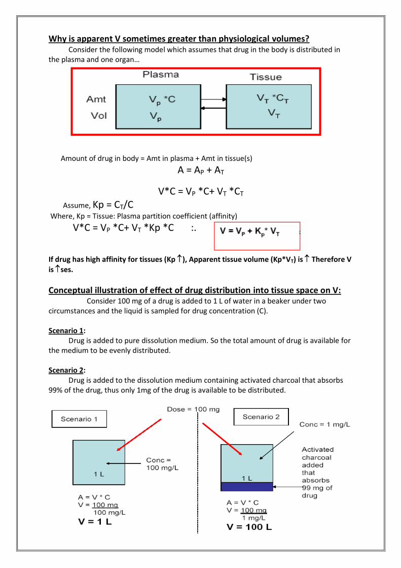
Why is apparent V sometimes greater than physiological volumes? Consider the following model which assumes that drug in the body is distributed in the plasma and one organ…
Amount of drug in body = Amt in plasma + Amt in tissue(s)
A = AP + AT
V*C = VP *C+ VT *CT
Assume, Kp = CT/C
Where, Kp = Tissue: Plasma partition coefficient (affinity)
V*C = VP *C+ VT *Kp *C :.
If drug has high affinity for tissues (Kp ), Apparent tissue volume (Kp*VT) is Therefore V
is ses.
Conceptual illustration of effect of drug distribution into tissue space on V: Consider 100 mg of a drug is added to 1 L of water in a beaker under two circumstances and the liquid is sampled for drug concentration (C). Scenario 1: Drug is added to pure dissolution medium. So the total amount of drug is available for the medium to be evenly distributed. Scenario 2: Drug is added to the dissolution medium containing activated charcoal that absorbs 99% of the drug, thus only 1mg of the drug is available to be distributed.
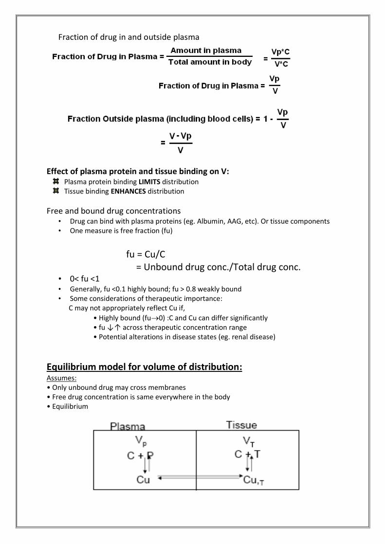
Fraction of drug in and outside plasma
Effect of plasma protein and tissue binding on V:
Plasma protein binding LIMITS distribution Tissue binding ENHANCES distribution
Free and bound drug concentrations • Drug can bind with plasma proteins (eg. Albumin, AAG, etc). Or tissue components • One measure is free fraction (fu)
fu = Cu/C = Unbound drug conc./Total drug conc.
• 0< fu <1 • Generally, fu <0.1 highly bound; fu > 0.8 weakly bound • Some considerations of therapeutic importance: C may not appropriately reflect Cu if,
• Highly bound (fu0) :C and Cu can differ significantly • fu ↓↑ across therapeutic concentration range • Potential alterations in disease states (eg. renal disease)
Equilibrium model for volume of distribution: Assumes: • Only unbound drug may cross membranes • Free drug concentration is same everywhere in the body • Equilibrium
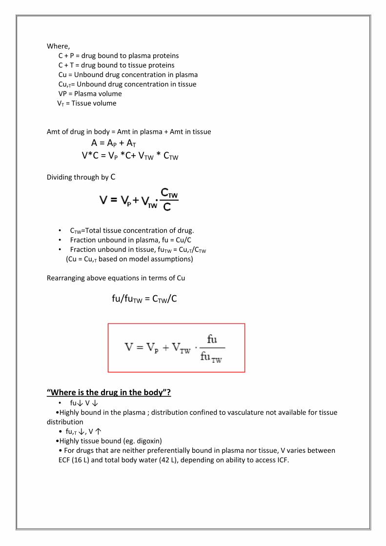
Where,
C + P = drug bound to plasma proteins C + T = drug bound to tissue proteins Cu = Unbound drug concentration in plasma Cu,T= Unbound drug concentration in tissue VP = Plasma volume
VT = Tissue volume Amt of drug in body = Amt in plasma + Amt in tissue
A = AP + AT
V*C = VP *C+ VTW * CTW
Dividing through by C
• CTW=Total tissue concentration of drug. • Fraction unbound in plasma, fu = Cu/C • Fraction unbound in tissue, fuTW = Cu,T/CTW
(Cu = Cu,T based on model assumptions) Rearranging above equations in terms of Cu
fu/fuTW = CTW/C
“Where is the drug in the body”? • fu↓ V ↓
•Highly bound in the plasma ; distribution confined to vasculature not available for tissue distribution
• fu,T ↓, V ↑ •Highly tissue bound (eg. digoxin)
• For drugs that are neither preferentially bound in plasma nor tissue, V varies between ECF (16 L) and total body water (42 L), depending on ability to access ICF.

5. RENAL CLEARANCE
Definition: It is the volume of blood from which the drug is totally removed in unit time through renal excretion.
Expressed as CLR
It has units : mL/min Major Organ for Excretion of Drugs is the Kidney. Functional Units are
– Nephron – Bowman’s Capsule – Proximal Tubule
– Loop of Henle – Distal Tubule – Collecting Duct
EQUATION: CLR = Rate of urinary excretion Plasma drug concentration Physiologically,
CLR = Rate of + Rate of - Rate of
Filtration Secretion Reabsorption Plasma drug concentration (C)
Renal function is determined by measuring GFR, Renal Blood Flow,& Urine flow. Normal values of:
Renal blood flow (RBF) = 1200 ml/min Glomerular filtration rate (GFR) = 125 ml/min Urine flow = 1.5 ml/min. GFR is measured by exogeneous/endogenous markers like Inulin/Creatine. Inulin
clearance is accurate measurement of GFR but tedious method while creatinine clearance widely used clinically for assessment of renal function.
Creatinine clearance:
CLcr = Rate of creatinine excretion in urine
Serum creatinine in mg%
In Males:
CLcr = (140 – Age)*W
72 * Scr
For Females:
CLcr = (140 – Age)*W = 0.9*Clcr of males
85 * Scr

Where, CLcr = creatinine clearance in ml/min Scr = serum creatinine in mg% W = weight in kgs. Age = in terms of years.
Normal creatinine clearance values: 120-130 ml/min Value of 20-50 ml/min : moderate renal failure Value of < 10 ml/min : severe renal impairment
Inulin Clearance: This is for inulin, and yields the glomerular filtration rate. • Value for normal males:
124.5 ± 9.7 ml/min • Value for normal females:
108.8 ± 13.5 ml/min
Factors affecting renal clearance:
1. Physiological properties of drug: I. Molecular size:
II: Pka:
Pka and ionized drug are poorly absorbed passively and excreted rapidly. III. Lipid Solubility:
Urinary excretion of unchanged drug is inversely proportional to the lipophilicity.
2. Distribution & Binding Characteristics of drug: Drug extensilvely bound to proteins have long half life because renal clearance is
small and urine flow rate is just 1-2ml/min.
Example: ClR of oxytetracycline (66% unbound) is 99ml/min.
ClR of doxycycline (7% unbound) is 16 ml/min.
3. Plasma Concentration of Drugs: Glomerular filtration and reabsorption are directly affected by plasma drug
concentration as both are passive processes. Drug not bound to plasma proteins and excreted by filtration only, shows a linear
relationship between rate of excretion and plasma drug concentration.
MOLECULAR SIZE EXCRETION BY KIDNEY
< 300 daltons, water soluble Readily excreted
300-500 daltons Excreted
> 500 daltons Lesser extent

4. Blood Flow to Kidneys: It is important for the drugs excreted by the glomerular filtration and those that are
actively secreted. For actively secreted drugs, increased perfusion increases the contact of drug with
secretory sites and enhances their elimination. Renal Clearance in such cases is called as perfusion rate limited.
5. Biological Factors: Renal clearance is approx. 10% lower in females than in males. In new borns renal function is 30-40% less than the adults and attains maturity
between 2.5 – 5 months age. In old age, GFR decreases and tubular function is altered thus prolongs the half life of
the administered drug.
6. Drug Interactions: Any drug interaction that results in alteration of binding characteristics, renal blood
flow, active secretion, urine pH and intrinsic clearance and forced diuresis would alter the renal clearance of drug.
Example: Gentamycin induced nephrotoxicity by Furosemide. Furosemide displaces Gentamycin from the binding sites. The free concentration of Gentamycin increases and accelerates its clearance.
7. Disease States: Renal Impairment Renal dysfunction & uremia impairs elimination of drugs that are primarily excreted by the kidneys & ultimately leads to increase in the half life of the drugs.
8. Dose Adjustment in Renal Failure: No need to alter dose if fraction of unchanged drug excreted (fu) ≤ 0.3 and renal function (R.F.) is ≥0.7 of the normal. But if not then dose required = normal dose * renal failure.

9. Effect of Exercise: Exhaustive exercise reduced RBF (Renal Blood Flow) by 53.4% compared to the pre-exercise values, and returned to 82.5% and 78.9% of the pre-exercise values at 30 and 60 min into the recovery period, respectively. As RBF decreases, CLR decreases.
Renal Extraction Ratio:
The fraction of a drug that is excreted when it passes through the kidneys is called the extraction ratio (ER) of the drug. By definition, a drug that is not excreted at all has an ER of 0 and a drug that is completely removed after a single passage has an ER of 1.
The table below shows the extraction ratios for a number of drugs that are excreted in the urine (> 30% by this route):
Extraction ratio:
Low (< 0.2) Intermediate High
Acetazolamide Procainamide Glucuronides
Chlorporpamide Quaternary ammonia compounds Penicillins
Diazoxide Sulfates
Digoxin Glycine conjugates
Furosemide
Gentamicin
Kanamycin
Phenobarbital
Sulfasoxazole
Tetracycline
6. TOTAL CLEARANCE
Synonym: Total systemic clearance
Definition: . It is the volume of plasma completely cleared of drug per unit time by all routes
and mechanisms. It is the sum of all the individual clearances by all the eliminating organs. i.e. It is the sum of all the individual clearances by all the eliminating organs…
CLT = CLR + CLH + CLP + …
Where, CLR = Renal clearance, CLH =Hepatic clearance, CLP =Pulmonary clearance etc.
Expressed in terms of ml/min.

Total Clearance can be expressed in following way:
1. From Rate and Concentration: where, dx/dt = Rate of elimination Cp = Concentration
2. From Dose and AUC:
3. From ke and V:
ROUTES OF CLEARANCE OTHER THAN RENAL
Biliary Clearance:
Quantitatively important excretory route for drugs and their metabolites which are actively transported by hepatocyte; once in small intestine, compounds with sufficient lipophilicity are reabsorbed and cleared again by liver (enterohepatic circulation), more polar substances may be biotransformed by bacteria (e.g. hydrolysis of drug conjugates) and products reabsorbed; unabsorbed drugs and metabolites are excreted in feces
Metabolic Clearance:
Removal process is metabolism. Fluid is usually blood (rarely plasma or serum), Denoted by CLm. When the metabolic organ is liver, it is known as Hepatic Clearance.
Effect of Exercise on Hepatic Clearance
Exercise increases cardiac output, but diverts blood flow away from the liver and could decrease the hepatic clearance of drugs. According to the degree of the hepatic extraction ratio, drugs may be classified as high, intermediate and low extracted drugs. In general, a highly-cleared drug is efficiently removed by the liver, and its elimination is blood flow dependent.
Minor routes • Sweat • Tears
• Reproductive fluids • Milk

• Generally pH-dependent passive diffusion of lipophilic drugs; • Can be of toxicologic significance e.g. exposure of infants to drugs in milk.
Importance: Clearance is an important pharmacokinetic parameter that describes how quickly
drugs are eliminated, metabolized or distributed throughout the body.
It can be viewed as the proportionality constant relating the rate of these processes and drug concentration.
The clearance of a drug can be used to understand the processes involved in drug elimination, distribution and metabolism.
Relating clearance to a patient renal or hepatic function can be used in the
determination of suitable drug dosage regimens. Dose adjustment based on total body clearance:
Cssavg = F (1/CLT) (Xo/T) If CLT’, Xo’ & T’ represents the value for renal failure patients then equation for dose
adjustment is given by: Cssavg = (Xo’/CLT’ T’) = (Xo/ CLT T)
Rearranging in terms of dose and dose interval to be adjusted the equation is : (Xo’/T’) = (CLT’ Xo)/(CLT T)
7. PLASMA PROTIEN BINDING Introduction:
It is binding of drug to plasma protein in blood component. Binding of drugs to plasma proteins is reversible. Various plasma protein to which drug binds includes Albumin, α1-Acid glycoprotein
(orosomucoid), lipoprotein, globulins, etc.
Order of binding of drugs to various plasma proteins:
Albumin > α1-Acid glycoprotein (orosomucoid) > Lipoproteins > Globulins
Such bound drug is both pharmacokinetically as well as pharmacodynamically inert i.e. a
protein bound drug is neither metabolized nor excreted nor it is pharmacologically active.
A bound drug is also restricted since it remains confined to a particular tissue for which it has greater affinity. Moreover, such bound drug because of its enormous size cannot undergo membrane transport & thus its half life is increased.
However this binding is rapidly reversible and non-specific – that is many drugs may bind to the same protein. Drug–plasma protein binding forms a "reservoir" of drug, but only the free (unbound) drug is available to the tissues to exert a therapeutic effect.
Binding of drug generally is reversible process (hydrogen bond , hydrophobic bond ,ionic bond ,van der waal’s forces ). Irreversible drug binding though rare (covalent binding) is often a reason for carcinogenicity or tissue toxicity of drug ;for example covalent binding of paracetamol metabolities to liver results in hepatotoxicity .
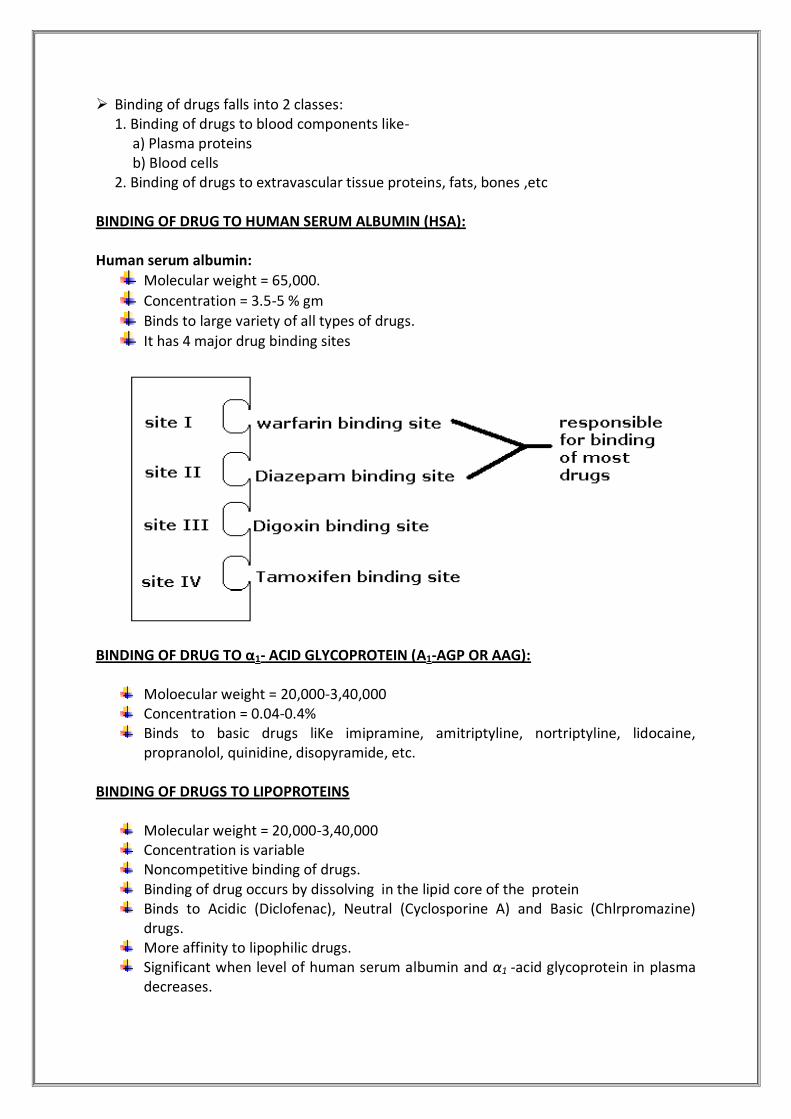
Binding of drugs falls into 2 classes: 1. Binding of drugs to blood components like- a) Plasma proteins b) Blood cells 2. Binding of drugs to extravascular tissue proteins, fats, bones ,etc
BINDING OF DRUG TO HUMAN SERUM ALBUMIN (HSA): Human serum albumin:
Molecular weight = 65,000. Concentration = 3.5-5 % gm Binds to large variety of all types of drugs. It has 4 major drug binding sites
BINDING OF DRUG TO α1- ACID GLYCOPROTEIN (Α1-AGP OR AAG):
Moloecular weight = 20,000-3,40,000 Concentration = 0.04-0.4% Binds to basic drugs liKe imipramine, amitriptyline, nortriptyline, lidocaine,
propranolol, quinidine, disopyramide, etc. BINDING OF DRUGS TO LIPOPROTEINS
Molecular weight = 20,000-3,40,000 Concentration is variable Noncompetitive binding of drugs. Binding of drug occurs by dissolving in the lipid core of the protein Binds to Acidic (Diclofenac), Neutral (Cyclosporine A) and Basic (Chlrpromazine)
drugs. More affinity to lipophilic drugs. Significant when level of human serum albumin and α1 -acid glycoprotein in plasma
decreases.
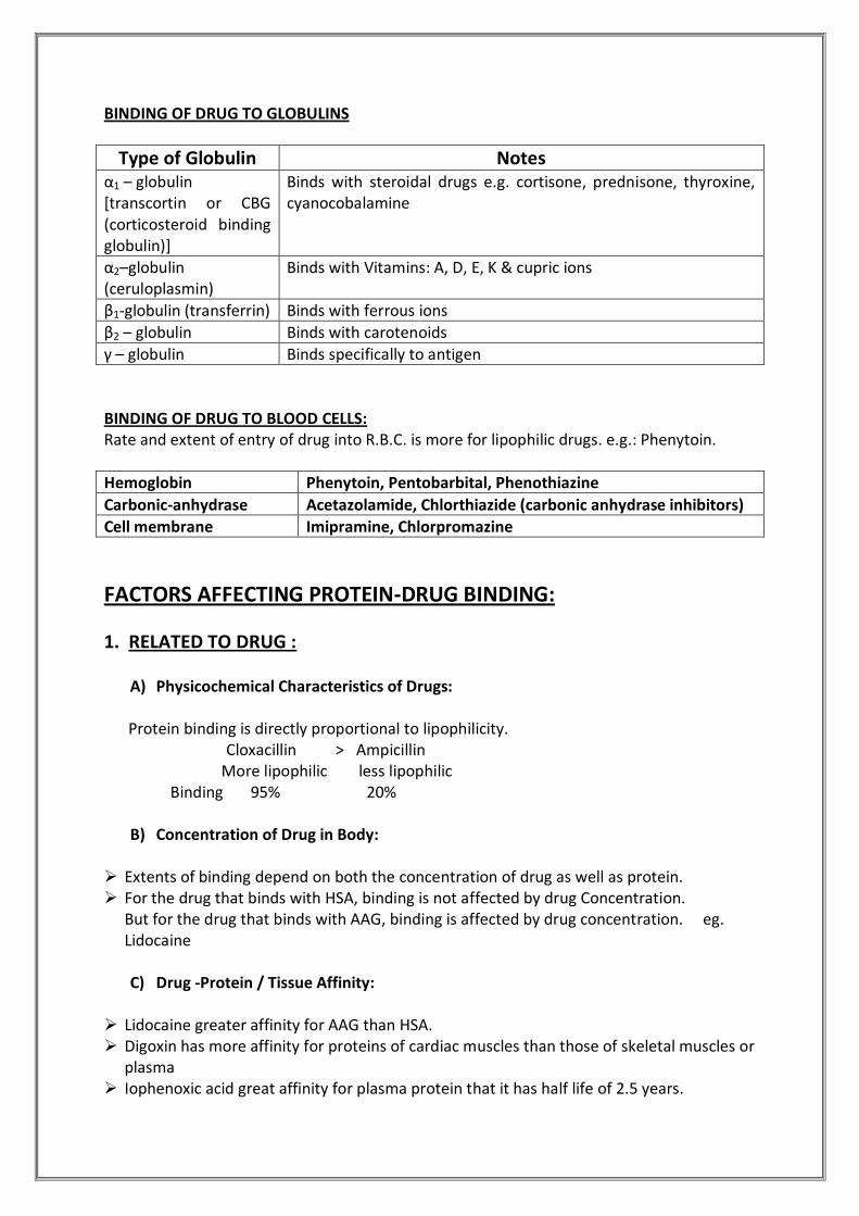
BINDING OF DRUG TO GLOBULINS
Type of Globulin Notes α1 – globulin [transcortin or CBG (corticosteroid binding globulin)]
Binds with steroidal drugs e.g. cortisone, prednisone, thyroxine, cyanocobalamine
α2–globulin (ceruloplasmin)
Binds with Vitamins: A, D, E, K & cupric ions
β1-globulin (transferrin) Binds with ferrous ions
β2 – globulin Binds with carotenoids
γ – globulin Binds specifically to antigen
BINDING OF DRUG TO BLOOD CELLS: Rate and extent of entry of drug into R.B.C. is more for lipophilic drugs. e.g.: Phenytoin.
Hemoglobin Phenytoin, Pentobarbital, Phenothiazine
Carbonic-anhydrase Acetazolamide, Chlorthiazide (carbonic anhydrase inhibitors)
Cell membrane Imipramine, Chlorpromazine
FACTORS AFFECTING PROTEIN-DRUG BINDING:
1. RELATED TO DRUG :
A) Physicochemical Characteristics of Drugs:
Protein binding is directly proportional to lipophilicity. Cloxacillin > Ampicillin More lipophilic less lipophilic Binding 95% 20%
B) Concentration of Drug in Body: Extents of binding depend on both the concentration of drug as well as protein. For the drug that binds with HSA, binding is not affected by drug Concentration.
But for the drug that binds with AAG, binding is affected by drug concentration. eg. Lidocaine C) Drug -Protein / Tissue Affinity:
Lidocaine greater affinity for AAG than HSA. Digoxin has more affinity for proteins of cardiac muscles than those of skeletal muscles or
plasma Iophenoxic acid great affinity for plasma protein that it has half life of 2.5 years.

2. RELATED TO PROTEINS:
A) Physicochemical Properties of Proteins: Lipoproteins and adipose tissue tends to bind with lipophilic drugs Physiologic pH determines presence of active anionic or cationic groups on the albumin
molecules to bind a variety of drug. B) Concentration of Plasma Proteins:
Binding predominantly occurs with albumin as it is present in a higher concentration in
comparision to other plasma proteins. Concentration of various proteins may change during diseased state that can alter extent
of binding.
C) No. Of Binding Sites on the Protein: HSA has more binding sites than AAG. Drug may bind with more than one site on protein.eg. Indomethacin is known to bind 3
different sites. AAG is a protein with limited binding capacity because of its low concentration & low
molecular size
3. DRUG INTERACTIONS When 2 or more drugs Can bind to same site , competition between them for interaction
with binding site results .Such a drug-drug interaction for common binding site is called as DISPLACEMENT INTERACTION.
Administration of phenylbutazone to a patient on a warfarin therapy results in displacement of latter from its binding site ,free warfarin cause the adverse hemorrahagic reactions which may be lethal
The concentration & affinity for binding to site will determine the extent to which displacement will occur
The drug with large Vd , redistributes into large volume of fluid & clinical effects may be insignificant. where as drug of small Vd ,remains confined to blood compartments shows serious toxic reactions.
EXAMPLE: Assume that Drug A and Drug B are both protein-bound drugs. If Drug A is given, it will bind to the plasma proteins in the blood. If Drug B is also given, it can displace Drug A from the protein, thereby increasing Drug A's fraction unbound. This may increase the effects of Drug A, since only the unbound fraction may exhibit activity.
Before Displacement After Displacement % increase in unbound fraction
Drug A
% bound 95 90
% unbound 5 10 +100

Drug B
% bound 50 45
% unbound 50 55 +10
For Drug A, the % increase in unbound fraction is 100%-- hence, Drug A's pharmacologic
effect has doubled. This change in pharmacologic effect could have adverse consequences.
This effect of protein binding is most significant with drugs that are highly protein-bound (>95%) and have a low therapeutic index, such as warfarin. A low therapeutic index indicates that there is a high risk of toxicity when using the drug. Since warfarin is an anticoagulant with a low therapeutic index, warfarin may cause bleeding if the correct degree of pharmacologic effect is not maintained. If a patient on warfarin takes another drug that displaces warfarin from plasma protein, it could result in an increased risk of bleeding.
4. Competition between Drugs and Normal Body Constituents
Bilirubin binding to HAS can be impaired by certain drugs & is of great concern in
neonates whose BBB & Bilirubin metabolizing capacity are not efficient. Acidic drugs such as Sodium salicylate, sodium benzoate & sulfonamides displace
bilirubin from its binding site. Free Bilirubin is no conjugated by the liver of neonates and thereby precipitates “ KERNICTERUS” (characterized by degeneration of brain & mental retardation )
5. Allosteric Changes in protein molecule It involves alteration of the protein structure by the drug thereby modifying its binding
capacity e.g aspirin acetylates the lysine fraction of albumin thereby modifying its capacity to bind NSAIDS like phenylbutazone (increased affinity ) & flufenamic acid (decreased affinity).
6. Patient related factors:
Physiological conditions: Species Age Pregnancy Ethnicity
Gender Smoking Obesity Nutritional status
7. Disease state:
DISEASE INFLUENCE ON PLASMA
PROTEINS INFLUENCE ON PROTEIN-DRUG
BINDING
Renal failure (uremia)
Decreased albumin content
Decreased binding of acidic drugs. Neutral & basic drugs are
unaffected.
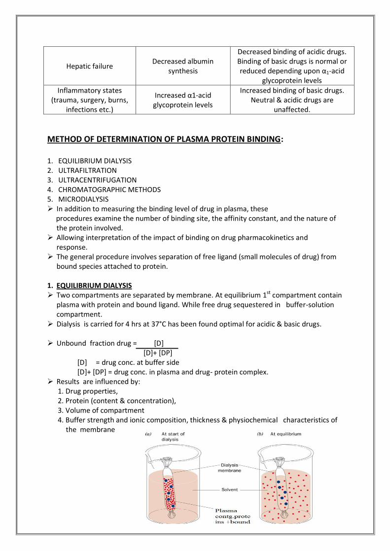
Hepatic failure Decreased albumin
synthesis
Decreased binding of acidic drugs. Binding of basic drugs is normal or reduced depending upon α1-acid
glycoprotein levels
Inflammatory states (trauma, surgery, burns,
infections etc.)
Increased α1-acid glycoprotein levels
Increased binding of basic drugs. Neutral & acidic drugs are
unaffected.
METHOD OF DETERMINATION OF PLASMA PROTEIN BINDING: 1. EQUILIBRIUM DIALYSIS 2. ULTRAFILTRATION 3. ULTRACENTRIFUGATION 4. CHROMATOGRAPHIC METHODS 5. MICRODIALYSIS In addition to measuring the binding level of drug in plasma, these procedures examine the number of binding site, the affinity constant, and the nature of
the protein involved. Allowing interpretation of the impact of binding on drug pharmacokinetics and
response. The general procedure involves separation of free ligand (small molecules of drug) from
bound species attached to protein. 1. EQUILIBRIUM DIALYSIS Two compartments are separated by membrane. At equilibrium 1st compartment contain
plasma with protein and bound ligand. While free drug sequestered in buffer-solution compartment.
Dialysis is carried for 4 hrs at 37°C has been found optimal for acidic & basic drugs.
Unbound fraction drug = [D] [D]+ [DP] [D] = drug conc. at buffer side [D]+ [DP] = drug conc. in plasma and drug- protein complex. Results are influenced by: 1. Drug properties, 2. Protein (content & concentration), 3. Volume of compartment 4. Buffer strength and ionic composition, thickness & physiochemical characteristics of
the membrane

Disadvantages: 1. Volume shift if the drug takes vary long time than dilution or volume shift can occur due
to concentration gradients. 2. Adsorption of drug on membrane less for acidic & lipophilic drugs & more for lipophilic
,basic drugs 3. Poorly water soluble drugs are difficult to study due to agglomeration and adherence to membrane. 2. ULTRAFILTRATION Separation of the protein and bound drug from free drug in solution occurs using a
suitable membrane which retains the proteins and is assisted by positive pressure or centrifugation.
Principal advantage : Speed (as little as 15 min) Disadvantages: Both equilibrium dialysis and Ultrafilration need radioisotopes for low Concentration or highly bound drug to provide sensitivity for quantization.
3. ULTRACENTRIFUGATION Based on differential sedimentation of solutes, based on
their molecular weights. High speed for long period (24h) Protein and bound drug are forced to the lower layer in the
tube and the free drug is bound in the “ middle cut.” & with any lipoprotein –bound drug near surface.
Advantages: 1. No membrane so no absorption of drug. Used for lipophilic drug. 2. Tubes are made up of non-absorptive materials like nitrocellulose and polyallomer no
adsorption & allowed the binding of water insoluble ,hydrophobic cyclosporine to be determined.
Disadvantages: 1. High cost 2. Long period 3. Cosedimentation of free and bound drug 4. Possibility of disturbing the equilibrium 4 . CHROMATOGRAPHIC METHODS 1. Size exclusion gel permeation chromatography (slow & detection difficulty) 2. HPLC (reduced time & extended scope of procedure)
Disadvantage: Plugging up of the column due to high concentration of proteins.

5. MICRODIALYSIS It is performed by perfusing of small diameter dialysis tubing with a carrier solution
.small molecules in the sample , such as free drugs , diffuse in the fiber & are transported to collection vials for analysis .large molecules such as protein & drug-protein bound drugs are excluded by dialysis membrane. The dialysate can be analyzed by standard technique
Microdialysis perfusion is rapid as ultrafiltration but the sample do not suffer from reequlibration during separation of free from bound drug
Relative recovery = Dialysate conc. Surrounding conc. Relative recovery is the mean value of determinations before and after each experiment
Unbound Fraction = (dialysate conc./ relative recovery)
Plasma conc.
It depends on 1. The perfusion flow rate 2. The diffusion characteristic of analyte 3. The nature of matrix 4. Properties and dimension of dialysis membrane.
KINETICS OF PROTEIN-DRUG BINDING: r = Ka * [D] (Applied only when 1 binding site on protein + Protein- Ka [D] +1 Drug complex is of 1:1 type) r = N*Ka*[D] (Applied only when > 1 binding site on protein + Ka*[D] + 1 Protein-Drug complex is other than 1:1 type) Where D = Free drug concentration Ka = Association rate constant r = number of moles of drug bound to total moles of proteins.

Using above equation values of Ka and N can be obtained by plotting above equations in three different ways:
1.Plot of r [D]
2. Scatchard Plot: linear form(r /[D] - r):
3. Double Reciprocal Plot (Line weaver-Burk Plot):
Importance of Protein or Tissue Binding of Drugs:
1. ABSORPTION
The absorption equilibrium is attained by transfer of free drug from site of administration
into systemic circulation & when conc. in these 2 compts.conc. become equal following eqlibrium, the process may stop.
However, binding of the absorbed drug to plasma protein decreases free drug concentration & disturbs such equilibrium. Thus, sink conditions & concentration gradient are re-established which act as a driving force for further absorption .
This is particularly useful in case of ionized drugs which are transported with difficulty.
2. SYSTEMIC SOLUBILITY OF DRUGS Water insoluble drugs, neutral endogenous macromolecules such as heparin & several steroids & oil soluble vitamins are circulated & distributed to tissues by binding especially to lipoproteins which act as a vehicle for such hydrophobic compounds.
3. DISTRIBUTION
Plasma protein binding restricts the entry of drugs that have specific affinity for certain tissues .
This prevents accumulation of a large fraction of drug in such tissues & thus, subsequent toxic reactions.
Plasma protein drug binding thus favors uniform distribution of drug throughout the body by its buffer function (maintains equilibrium between the free & the bound drug).
A protein bound drug in particular does not cross BBB, the placental barrier & glomerulus.
4. TISSUE BINDING, APPARENT VOLUME OF DISTRIBUTION & DRUG STORAGE A drug that s extensively bound to blood components remain confined to blood .such a
drug has a small volume of distribution & thus have small volume of distribution. A drug that shows extracellular tissue binding has a large volume of distribution .
A tissue or blood component that has great affinity for a particular drug act as depot or storage site for that drug .e.g. RBC is storage site for lipophilic compound tetrahydrocannabinol
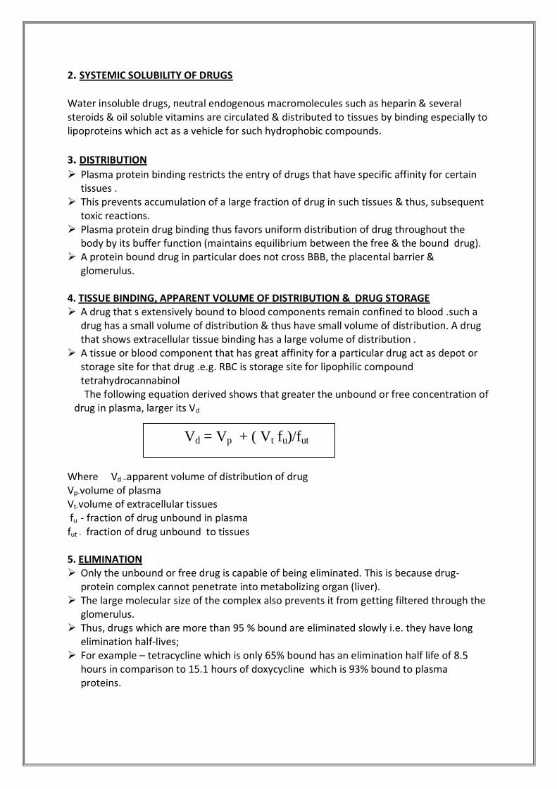
The following equation derived shows that greater the unbound or free concentration of drug in plasma, larger its Vd
Where Vd –apparent volume of distribution of drug Vp-volume of plasma Vt-volume of extracellular tissues fu - fraction of drug unbound in plasma fut - fraction of drug unbound to tissues 5. ELIMINATION Only the unbound or free drug is capable of being eliminated. This is because drug-
protein complex cannot penetrate into metabolizing organ (liver). The large molecular size of the complex also prevents it from getting filtered through the
glomerulus. Thus, drugs which are more than 95 % bound are eliminated slowly i.e. they have long
elimination half-lives; For example – tetracycline which is only 65% bound has an elimination half life of 8.5
hours in comparison to 15.1 hours of doxycycline which is 93% bound to plasma proteins.
Vd = Vp + ( Vt fu)/fut
However, penicillin have short elimination half-lives despite being extensively bound to plasma proteins .this is because rapid equilibrium occurs between free & bound drug & the free drug is equally rapidly excreted by active secretion in renal tubules
6. DISPLACEMENT INTERACTIONS & TOXICITY More significant in case of drugs which are more than 95% bound .a displacement of just
1% of a 99% bound drug results in doubling of the free drug conc. i.e. a 100% rise. For a drug i.e. bound to a lesser extent e.g. 90 % ,displacement of 1 % results in only a 10 % rise in free drug conc.which may be insignificant clinically.
Kernicterus in infants is an example of a disorder caused by displacement of bilirubin from albumin binding sites by the NSAIDS.
With a drug of large Vd such as DIGOXIN, even a substantial increase in the degree of displacement of drug in plasma may not effect a large increase in free drug concentration & dose adjustment may not be required.
Because :1) only a small fraction of such a drug is present in plasma whereas most of it is localized in extravascular tissues, 2) following displacement the free drug ,because of its large Vd redistributes in a large pool of extravascular tissues.
7. DIAGNOSIS The chlorine atom of chloroquine when replaced with radio labeled I-131 can be used to visualize melanomas of the eye since chloroquine has a tendency to interact with melanin of eyes. The thyroid gland has a great affinity for iodine containing compounds, hence any disorder by tagging such compound with radioisotope of iodine. 8. THERAPY AND DRUG TARGETING The binding of drugs to lipoproteins can be used for site specific delivery of hydrophilic
moieties. This is particularly useful in cancer therapies since certain tumor cells have greater
affinity for LDL than normal tissues. Thus binding of a suitable antineoplastic to it can be used as a therapeutic tool.
HDL is similarly transported more to adrenal and testes .an example of site specific drug delivery in cancer treatment is that of estramustine.
Estradiol binds selectively & strongly to prostrate & thus prostrate cancer can be treated by attaching nitrogen mustard to estradiol for targeting of prostrate gland. drug targeting prevents normal cells from getting destroyed.
9. DESIGN OF DOSAGE REGIMEN FROM PLASMA CONCENTRATION:
If the therapeutic range, apparent Vd and clearance or half life of a drug is known, then dosage regimen can be designed to maintain drug concentration within the specified therapeutic range.
Tmax = 3.32 t1/2 log (Cupper/C lower) Max maintanence dose = Vd (Cupper- C lower)
F After a convenient dosing interval t, smaller than tmax has been selected, the
maintanence dose is given as : Xo = (Xo max / tmax) t

Cssavg = (Cupper - C lower) 2.303 log (C upper /C lower)
Effect of protein binding on Drug Pharmacodynamic Effects:
For a drug showing little protein binding, the plasma acts simply as a watery solution in which the drug is dissolved. Where protein binding does occur, the behaviour of the drug may be influenced in several ways:
1. Extensive plasma protein binding will increase the amount of drug that has to be absorbed before effective therapeutic levels of unbound drug are reached. For example, acidic dugs (such as acetyl salicylic acid – aspirin) are often substantially bound to albumin.
2. Elimination of a highly bound drug may be delayed. Since the concentration of free drug is low, drug elimination by metabolism and excretion may be delayed. This effect is responsible for prolonging the effect of the drug digoxin
3. Changes in the concentration of plasma proteins will influence the effect of a highly bound drug. A low plasma protein level may occur in old age or malnutrition. It may also be caused by illness such as liver disease (remember that most plasma proteins are made in the liver), or chronic renal failure where there is excessive excretion of albumin. In each case the result is a smaller proportion of drug in bound form and more free drug in the plasma. The greater amount of free drug is able to produce a greater therapeutic effect and reduced drug dosages may be indicated in these cases
4. Drugs may compete for binding with plasma proteins leading to interactions. This is
significant for highly bound drugs such as the anticoagulant warfarin since even a small change in binding will greatly affect the amount of free drug. Such an effect is produced by the concurrent administration of aspirin, which displaces warfarin and increases the amount of free anticoagulant.
STEREOSELECTIVITY associated with protein binding : Stereoselectivity was demonstrated for both albumin-binding sites 1 & 2 . stereoselective binding was reported for Ibuprofen enantiomers ,unbound fraction of R(-
) Enantiomer (0.419 %)being significantly less than that S(+) enantiomer (0.643%). Stereoselective binding in humans was reported for acidic drugs such as
etodolac,warfarin,pentobarbital,& for basic drugs like chloroquine,propanolol,methadone,etc
Stereoselectivity in protein binding of enantiomer can also differ between species . for propanolol ,a basic drug bound to AAG ,R-enantiomer binds less than S-isomer in humans & dogs,the reverse is observed in rats
A difference in the binding of 2 isomers , Quinine & Quinidine , fu was 7.5 & 12.3 % respectively .
STUDY QUESTIONS:
State importance of absorption rate constant. Importance of elimination half life Suppose volume of distribution of a particular drug is more, then what does that it
mean? How can you design an effective dosage regimen based on plasma concentrations? What are the factors affecting renal clearance?
WWhhaatt iiss pprrootteeiinn bbiinnddiinngg ?? DDeessccrriibbee bbiinnddiinngg ooff ddrruugg wwiitthh vvaarriioouuss pprrootteeiinnss.. State consequences of high plasma protein binding
DDeessccrriibbee tthhee mmeetthhooddss ooff ddeetteerrmmiinnaattiioonn ooff pprrootteeiinn bbiinnddiinngg..
DDeessccrriibbee ffaaccttoorrss aaffffeeccttiinngg pprrootteeiinn bbiinnddiinngg
HHooww pprrootteeiinn bbiinnddiinngg aaffffeeccttss tthhee PPhhaarrmmaaccooddyynnaammiicc cchhaarraacctteerriissttiicc ooff ddrruugg..
EExxppllaaiinn tthhee ssiiggnniiffiiccaannccee ooff ddrruugg--pprrootteeiinn bbiinnddiinngg
REFERENCES:
“Applied Biopharmaceutics & Pharmacokinetics” fourth edition: Leon Shargel, Andrew B.C. YU. “Biopharmaceutics & Pharmacokinetics”: a treatise: D. M. Brahmankar, S. B.
Jaiswal Biopharmaceutics and Clinical Pharmaceutics by Robert Notari, p.g. 290-333,48-98 Textbook of Biopharmaceutics and Pharmacokinetics by Dr Shobha Rani, p.g 84—205 Gibaldi, M. 1984 "Biopharmaceutics and Clinical Pharmacokinetics", 3rd ed., Lea &
Febiger, Chapter 12, page 214. Rowland and Tozer, Ch 3, 10, 19 (From an Internet Article) IUPAC Compendium of Chemical Terminology 2nd Edition (1997) J Pharm Sci 88:292-302, 2000 Encyclopedia volume – 13 www.nottingham.ac.uk/nursing/sonet/rlos/bioproc/ www.ricerca.com/pages/virtua www.medscape.com www.wikipedia.com www.ncbi.nlm.nih.gov