be-flare: a fluorescent reporter of base editing activity
TRANSCRIPT
METHODOLOGY ARTICLE Open Access
BE-FLARE: a fluorescent reporter of baseediting activity reveals editingcharacteristics of APOBEC3A andAPOBEC3BMatthew A. Coelho1*, Songyuan Li2, Luna Simona Pane2, Mike Firth1, Giovanni Ciotta1, Jonathan D. Wrigley1,Maria Emanuela Cuomo1, Marcello Maresca2 and Benjamin J. M. Taylor1*
Abstract
Background: Base Editing is a precise genome editing method that uses a deaminase-Cas9 fusion protein to mutatecytidine to thymidine in target DNA in situ without the generation of a double-strand break. However, the efficientenrichment of genetically modified cells using this technique is limited by the ability to detect such events.
Results: We have developed a Base Editing FLuorescent Activity REporter (BE-FLARE), which allows for the enrichmentof cells that have undergone editing of target loci based on a fluorescence shift from BFP to GFP. We used BE-FLARE toevaluate the editing efficiency of APOBEC3A and APOBEC3B family members as alternatives deaminase domains to therat APOBEC1 domain used in base editor 3 (BE3). We identified human APOBEC3A and APOBEC3B as highly efficientcytidine deaminases for base editing applications with unique properties.
Conclusions: Using BE-FLARE to report on the efficiency and precision of editing events, we outline workflows for theaccelerated generation of genetically engineered cell models and the discovery of alternative base editors.
Keywords: Base editing, Fluorescent reporter, CRISPR/Cas9, APOBEC, Gene editing
BackgroundExperimental and therapeutic modification of genomicDNA has become a more rapid and efficient process dueto the development of CRISPR-Cas-based technologies.Base editing is a recently developed derivative ofCRISPR-Cas-mediated genome editing [1, 2]. The thirditeration of the Base Editor protein (BE3) is a fusion ofthree enzymes: rat APOBEC1 cytidine deaminase, Cas9D10A nickase, and uracil DNA glycosylase inhibitor(UGI) [1]. This multi-enzyme complex can introducehigh-frequency C to T mutations (or G to A on thecomplementary strand) through enzymatic deaminationof cytidine to uracil at the targeted locus. Replicationacross the uracil will lead to incorporation of a thymi-dine at this position due to the misrecognition of uracil
as thymidine by DNA polymerases. The base excisionrepair pathway enzyme, uracil DNA glycosylase, couldrecognise and remove the uracil; however, the UGI com-ponent in BE3 provides local inhibition of such repair.Cas9 nickase allows for guide RNA-mediated targeting,and through nicking of the non-edited strand, engendersrepair using the edited strand as a template [3].Introduction or correction of mutations using CRISPR-Cas9
generally depends on DNA double-strand breaks andhomology-directed repair (HDR) using an exogenous DNArepair template. This can be a very inefficient process,dependent upon the cell type and cell cycle phase [4–6]. Fur-thermore, DNA double-strand breaks generated by Cas9 areresolved in an unpredictable manner, often leading to undesir-able outcomes such as insertions and deletions (InDels) andtranslocations [7]. Base editing has unique advantages in thisrespect; independence from DNA double-strand break forma-tion and HDR leads to reduced rates of InDel formation and ahigh efficiency of editing in a broader range of cellular
* Correspondence: [email protected];[email protected] Sciences, IMED Biotech Unit, AstraZeneca, Cambridge, UKFull list of author information is available at the end of the article
© The Author(s). 2018 Open Access This article is distributed under the terms of the Creative Commons Attribution 4.0International License (http://creativecommons.org/licenses/by/4.0/), which permits unrestricted use, distribution, andreproduction in any medium, provided you give appropriate credit to the original author(s) and the source, provide a link tothe Creative Commons license, and indicate if changes were made. The Creative Commons Public Domain Dedication waiver(http://creativecommons.org/publicdomain/zero/1.0/) applies to the data made available in this article, unless otherwise stated.
Coelho et al. BMC Biology (2018) 16:150 https://doi.org/10.1186/s12915-018-0617-1
contexts [3]. However, producing genetically engineered cellmodels using base editing still depends on single-cell cloningand sequencing of genomic DNA to find successfully editedcells; this is often the rate-limiting step in the procedure, andgains in the efficiency of this process have the potential togreatly reduce timelines in cell model generation.Fluorescent reporters developed to discriminate between
CRISPR-Cas9-mediated HDR or NHEJ events have facili-tated the enrichment of cells with desired DNA repair out-comes and led to improvements in increasing HDR rates ingenome engineering [8, 9]. In addition, T2A self-cleavingpeptide fusions with fluorescent proteins are common forselecting enriched pools of transfected cells in gene editingexperiments. However, a system for reporting on base editorpoint mutation activity in mammalian cells, which allows foredited cell enrichment and refinement of base editor archi-tecture, has yet to be demonstrated. We used base editing tointroduce a well-documented single amino acid substitution
in enhanced Blue Fluorescent Protein (eBFP) that leads to aspectral shift associated with a transition to Green Fluores-cent Protein (GFP) [10]. By fluorescently marking BE-activecells, we quantitatively assessed efficiencies of different BEvariants incorporating alternative APOBEC enzymes anddemonstrate FACS-based enrichment of genetically modifiedcells including gene knock-outs and clinically relevant pointmutations. We predict that our reporter will expedite cellmodel generation with base editing.
ResultsValidation of a Base Editing FLuorescent Activity REporter(BE-FLARE)We generated a mammalian expression construct for aversion of eBFP that was modified to contain thenecessary NGG protospacer adjacent motif (PAM) forStreptococcus pyrogenes Cas9, downstream of the targetcodon histidine 66 (CAC) (Fig. 1a). We termed this
Fig. 1 A fluorescent reporter detects base editing activity. a Diagram of the BE-FLARE reporter comprised of a modified BFP (BFP) and gRNA sequenceused to transition BFP to GFP through base editing (BE). Codon 66 (CAC) encoding histidine is targeted and converted to tyrosine (codons TAT or TAC),resulting in GFP expression. Codon conversion to CAT is synonymous for His, thus the protein remains as BFP. b BFP to GFP conversion in HEK293 andPC9 cells. Cells were co-transfected with the BE-FLARE and a plasmid expressing BE3 and either a non-targeting guide (NT-BE) or a BFP targeting guide(BFP-BE). BFP and GFP positive cells were quantified by flow cytometry 72 h after transfection. Data are representative of three independent experiments. cPC9 cells from the experiment described in (b) were sorted based on BFP (unedited) or GFP fluorescence. Five days later, DNA was extracted for ampliconsequencing of the BFP locus. Data represent a gene browser view of aligned reads in IGV and are representative of two independent experiments. Rawdata can be found in Additional file 2
Coelho et al. BMC Biology (2018) 16:150 Page 2 of 11
reporter the Base Editing FLuorescent Activity REporter(BE-FLARE). We designed a guide RNA (gRNA) target-ing eBFP to mutate histidine 66 to tyrosine. Given thatBE3 can target neighbouring cytosines in the protospa-cer within a window of approximately five nucleotides[1], we considered outcomes of other likely editingevents. Out of the possible base edits at this codon, twoout of three mutations cause a histidine to tyrosine sub-stitution (CAC to TAC or TAT), and the other is syn-onymous with the wild-type histidine (CAT; Fig. 1a). Weintroduced a cassette for gRNA expression under thehuman U6 promoter into the BE3 expression vector togenerate a single delivery construct for targeted baseediting. Next, we tested BE-FLARE using transient trans-fections in vitro. We used HEK293 cells and theEGFR-mutant lung cancer cell line, PC9. In both celllines, GFP signal was detectable after 72 h by flow cy-tometry only in cells transfected with the construct en-coding BE3 together with the gRNA targeting BFP H66,but not a non-targeting control gRNA (non-targeting,NT; Fig. 1b). BE-FLARE was therefore able to reportspecifically on base editing activity and allows forBE-active cells to be tracked by flow cytometry or mi-croscopy (Additional file 1: Figure S1). To confirmwhich nucleotides are targeted by BE in the reporter, weperformed next-generation amplicon sequencing ofBE-FLARE from GFP-positive PC9 cells produced afterbase editing. As expected, we found that the predomin-ant result of editing codon H66 was CAC->TAT, sug-gesting that both cytosines within the optimal baseediting window are efficiently edited in cells (Fig. 1c).In addition to transient expression of BE-FLARE, we
could stably integrate BE-FLARE using ObLiGaRe-medi-ated integration into the AAVS1 safe-harbour locus [11],thus allowing for permanent fluorescent demarcation ofedited cells. A time-course of digital droplet PCR andmicroscope imaging of PC9-BE-FLARE cells after editingshowed DNA editing of BE-FLARE as early as 18 h andedited cells expressing GFP protein from 48 to 72 hpost-transfection (Additional file 1: Figure S2).
Enrichment of edited cells using BE-FLAREWe evaluated whether BE-FLARE would allow enrich-ment for simultaneous co-editing at a secondary locus. Asa proof of principle, we generated a cell model with a clin-ically relevant point mutation; in this instance, the T790M gate-keeper mutation in human Epidermal GrowthFactor Receptor (EGFR), which can be generated by C>Tsubstitution. This mutation confers resistance to theEGFR tyrosine kinase inhibitor gefitinib [12]. Parental PC9cells are dependent upon oncogenic EGFR signalling andthus are sensitive to gefitinib [13]. We co-transfectedPC9-BE-FLARE cells with a BE3 expression construct alsoencoding a gRNA targeting BFP H66, and a second
plasmid expressing a gRNA targeting EGFR T790. Strik-ingly, selection with gefitinib enriched for GFP-positivecells by ~ 3.5-fold (Fig. 2a and b). We confirmed the suc-cessful introduction of the T790M mutation in thedrug-resistant PC9 population by Sanger sequencing(Fig. 2c). In addition to the T790M base edit, we observeda 5′ bystander C->T mutation within the BE3 activity win-dow. In contrast, the 3′ proximal bystander cytosineremained unedited. Upon inspection of the coding se-quence, we noted that the 5′ bystander mutation is syn-onymous, whereas the 3′ bystander results in a prematurestop codon (TAG), which is much less likely to be toler-ated in this EGFR-dependent cancer cell line.In a reciprocal approach, we used flow cytometry to sort
for GFP-positive cells following simultaneous base editingof EGFR and BE-FLARE and then quantified gefitinib re-sistance in GFP-positive versus mock-sorted cells (total vi-able population; Fig. 2d). We observed a similarco-enrichment using this approach; GFP-positive cells ex-hibited enhanced levels of resistance to gefitinib relative tomock-sorted controls, with a ~ 4.5-fold increase in cellgrowth observed after 5 days of gefitinib treatment(Fig. 2d). There was no observable resistance conferredfrom a non-targeting gRNA in GFP-sorted cells. Thus, anintegrated BE-FLARE can be used to enrich for geneticco-editing events at secondary loci.We next determined whether we could use BE-FLARE
as a transient reporter to obviate the need for generatingstable cell lines and allow ‘scarless’ co-selection of genetic-ally engineered cells. We tested this hypothesis by generat-ing point mutations in two genes implicated inoncogenesis: EGFR and BRAF. Specifically, we generatedthe EGFR T790M mutation, or an observed clinical muta-tion in BRAF (T57I), or BRAF knock-out (KO), with sim-ultaneous editing of BE-FLARE. The BRAF mutationswere generated using a single gRNA designed to introducea premature stop codon at position Q58, or a subsitutionat T57. In all cases, a non-targeting gRNA served as anegative control. We FACS sorted GFP-positive cells andused Sanger sequencing to quantify editing efficiencies(Fig. 3a). For BRAF and EGFR, GFP-sorted cells had astriking increase in editing compared with transfectedcells that were mock-sorted. For BRAF, average editingwas increased from ~ 9 to 55% at T57 and from ~ 4 to 20% at Q58, and for EGFR T790M, from ~ 1 to 18% (Fig. 3b).The lower levels of base editing for EGFR may reflect theincreased EGFR allele copy number in PC9 cells [14].Taken together, these data demonstrate the use of transi-ent expression of BE-FLARE in the enrichment ofco-editing events in mammalian cells.Next, we sought to determine how BE-FLARE would
compare with marking BE-transfected cells with a fluores-cent reporter (TurboRFP) coded in the BE transcript via aself-cleaving T2A peptide (Fig. 3c). Interestingly, the
Coelho et al. BMC Biology (2018) 16:150 Page 3 of 11
majority of cells that had undergone base editing ofBE-FLARE (i.e. GFP-positive cells) remained RFP-negative(Fig. 3d). Thus, expression of BE3-T2A-RFP below thelevel detectable by flow cytometry is still functional incells, implying that using RFP expression for selectionwould significantly underestimate the number of editedcells. Moreover, the high levels of BE3 expression apparentin the RFP-positive population may not be desirable formany applications due to the possibility of increasingoff-target editing.
Activity measurement of APOBEC-Cas9 fusion variantsusing BE-FLARESeveral reports have tried using alternative cytidinedeaminase domains to drive base editing [2, 3, 15].We sought to use BE-FLARE to provide a sensitiveassessment of alternative domains by monitoring thefrequency of BFP to GFP transitions. Specifically, wereplaced the original rat APOBEC-1 (rA1) cytidinedeaminase domain of BE3 with human APOBEC3A(A3A), or human APOBEC3B (A3B) C-terminal
Fig. 2 BE-FLARE facilitates reciprocal enrichment of co-edited cells. a Enrichment for BFP editing by selection for EGFR T790 M co-editing. PC9cells stably expressing the BE-FLARE reporter were co-transfected with a construct expressing BE3 and gRNAs targeting EGFR T790 and BFP. Cellswere treated with 100 nM gefitinib 72 h after transfection to select for EGFR T790 M mutants. GFP-positive cells were quantified by flow cytometry4 days after addition of gefitinib. b Quantification of three independent experiments from (a), with each data point shown and mean representedas a bar. Unpaired Student’s t test; ***P < 0.001. c Sanger sequencing of the EGFR T790 locus from dual EGFR and BE-FLARE base edited cells aftergefitinib selection. Percentage editing was estimated from sequencing chromatograms using EditR [31]. d Enrichment for EGFR T790 M editing byselection for BFP co-editing. PC9 cells stably expressing BE-FLARE were co-transfected as above. Cells were mock-sorted (all viable cells) or sortedfor GFP expression by FACS 72 h after transfection, and 100 nM gefitinib was added 24 h later. Cell growth was quantified by Incucyte. Data arerepresentative of three independent experiments. Raw data can be found in Additional file 2
Coelho et al. BMC Biology (2018) 16:150 Page 4 of 11
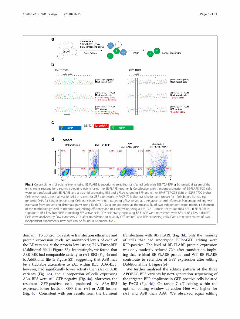
domain. To control for relative transfection efficiency andprotein expression levels, we monitored levels of each ofthe BE versions at the protein level using T2A-TurboRFP(Additional file 1: Figure S3). Interestingly, we found thatA3B-BE3 had comparable activity to rA1-BE3 (Fig. 4a andb, Additional file 1: Figure S3), suggesting that A3B maybe a tractable alternative to rA1 within BE3. A3A-BE3,however, had significantly lower activity than rA1 or A3Bvariants (Fig. 4b), and a proportion of cells expressingA3A-BE3 were still GFP-negative (Fig. 4a). Moreover, theresultant GFP-positive cells produced by A3A-BE3expressed lower levels of GFP than rA1 or A3B fusions(Fig. 4c). Consistent with our results from the transient
transfections with BE-FLARE (Fig. 3d), only the minorityof cells that had undergone BFP->GFP editing wereRFP-positive. The level of BE-FLARE protein expressionwas only modestly reduced 72 h after transfection, imply-ing that residual BE-FLARE protein and WT BE-FLAREcontribute to retention of BFP expression after editing(Additional file 1: Figure S4).We further analysed the editing pattern of the three
APOBEC-BE3 variants by next-generation sequencing ofthe targeted BFP amplicons in GFP-positive cells isolatedby FACS (Fig. 4d). On-target C->T editing within theoptimal editing window at codon H66 was higher forrA1 and A3B than A3A. We observed equal editing
Fig. 3 Co-enrichment of editing events using BE-FLARE is superior to selecting transfected cells with BE3-T2A-RFP. a Schematic diagram of theenrichment strategy for genomic co-editing events using the BE-FLARE reporter. b Co-selection with transient expression of BE-FLARE. PC9 cellswere co-transfected with BE-FLARE and a plasmid expressing BE3 and gRNAs targeting BFP and either BRAF T57/Q58 (left) or EGFR T790 (right).Cells were mock-sorted (all viable cells) or sorted for GFP expression by FACS 72 h after transfection and grown for 120 h before harvestinggenomic DNA for Sanger sequencing. Cells transfected with non-targeting gRNA served as a negative control reference. Percentage editing wasestimated from sequencing chromatograms using EditR [31]. Data are expressed as the mean ± SD of two independent experiments. c Schematicof the methodology used to monitor base editing efficiency and BE3 expression using a BE3-T2A-TurboRFP construct (BE3-RFP). d BE-FLARE issuperior to BE3-T2A-TurboRFP in marking BE3-active cells. PC9 cells stably expressing BE-FLARE were transfected with BE3 or BE3-T2A-turboRFP.Cells were analysed by flow cytometry 72 h after transfection to quantify GFP (edited) and RFP-expressing cells. Data are representative of twoindependent experiments. Raw data can be found in Additional file 2
Coelho et al. BMC Biology (2018) 16:150 Page 5 of 11

frequency of the CAC H66 codon for rA1 and A3A buthigher stringency observed for A3B, which seems morecapable of discriminating between these two proximalcytosine targets within the optimal editing window. Weobserved similarly low levels of non-C->T mutations, in-cluding C->G and C->A variants for all three APOBECvariants. Interestingly, A3A produced more bystanderC->T and G->A (on the complementary strand) muta-tions than either rA1 or A3B versions of BE3. Moreover,we observed low-frequency G->A mutations as far as −
86 (1.3 ± 0.04% allele frequency) and + 76 (1.3 ± 0.1% al-lele frequency) relative to the protospacer start position,exclusively in the A3A-edited samples (not shown). Not-ably, one of the bystander mutations produced a prema-ture stop codon (21.8 ± 2.2% allele frequency; Fig. 4d),which likely explains the reduction in GFP expressionobserved in A3A-BE3 edited cells.To better understand the potential mechanisms be-
hind the differing mutational characteristics of eachAPOBEC-BE3 variant, we analysed the protein
Fig. 4 Evaluation of base editor variants with BE-FLARE. a Comparison of APOBEC-BE3-T2A-TurboRFP variants with BE-FLARE. PC9 BE-FLARE stable cells weretransiently transfected with BE3-T2A-RFP versions where the cytosine deaminase domain of BE3 was rA1, A3A or A3B co-expressing non-targeting gRNA (NT)or BFP gRNA (BFP gRNA). The frequency of GFP-positive cells was quantified by flow cytometry after 72 h. Data are representative of three independentexperiments. b Quantification of the experiments described in (a). Mean± SD from three independent experiments. Unpaired Student’s t test; *P< 0.05. cRepresentative histograms from flow cytometry analysis of GFP fluorescent signal from the experiments described in (b). Data are representative of threeindependent experiments. d Base editing of BE-FLARE by rA1-BE3 (left), A3A-BE3 (middle) or A3B-BE3 (right) transfected cells assessed by amplicon sequencingof the BFP locus. GFP-positive cells were FACS sorted 72 h after transfection, and 5 days later, genomic DNA was taken for amplicon sequencing. BE with anon-targeting guide served as a negative control (see Additional file 1: Table S3). Boxed in red is a bystander mutation leading to the introduction of apremature stop codon. Data represent the mean± SD from two independent experiments and include SNPs found at ≥ 1% of total reads per sample. Readcounts can be found in Additional file 1: Table S3. Data from these experiments are also part of Fig. 1c. e Immunoblot analysis of PC9 BE-FLARE cells 24, 48 and72 h post-transfection with the indicated base editor variants (not T2A-RFP). Base editor expression over time was tracked by Cas9 immunodetection. Data arerepresentative of two independent experiments. Raw data can be found in Additional file 2
Coelho et al. BMC Biology (2018) 16:150 Page 6 of 11

expression of the base editors (Fig. 4e). A3A-BE3 hadthe highest expression of all at the time points tested,perhaps explaining the increased levels of undesired by-stander mutations. rA1-BE3 levels were comparable tothat of A3B-BE3 but decayed more rapidly over time. In-deed, at 72 h post-transfection, rA1-BE3 was undetect-able by Western blotting, whereas A3B-BE3 was still atlevels comparable to 24 h post-transfection. Taken to-gether, these results suggest that these APOBEC-Cas9fusions have drastically different protein expression and/or stability in mammalian cells, which may partially ex-plain base editing characteristics such as efficiency andprecision. Notably, we used a codon-optimised versionof rat APOBEC1 in BE3 throughout this report, as wefound that the codon-optimised version was expressedat much higher levels than the native sequence whenassessing RFP expression in a BE3-T2A-truboRFP sys-tem (Additional file 1: Figure S5), which is consistentwith recent reports [16, 17].To further analyse the editing profile of A3A and A3B
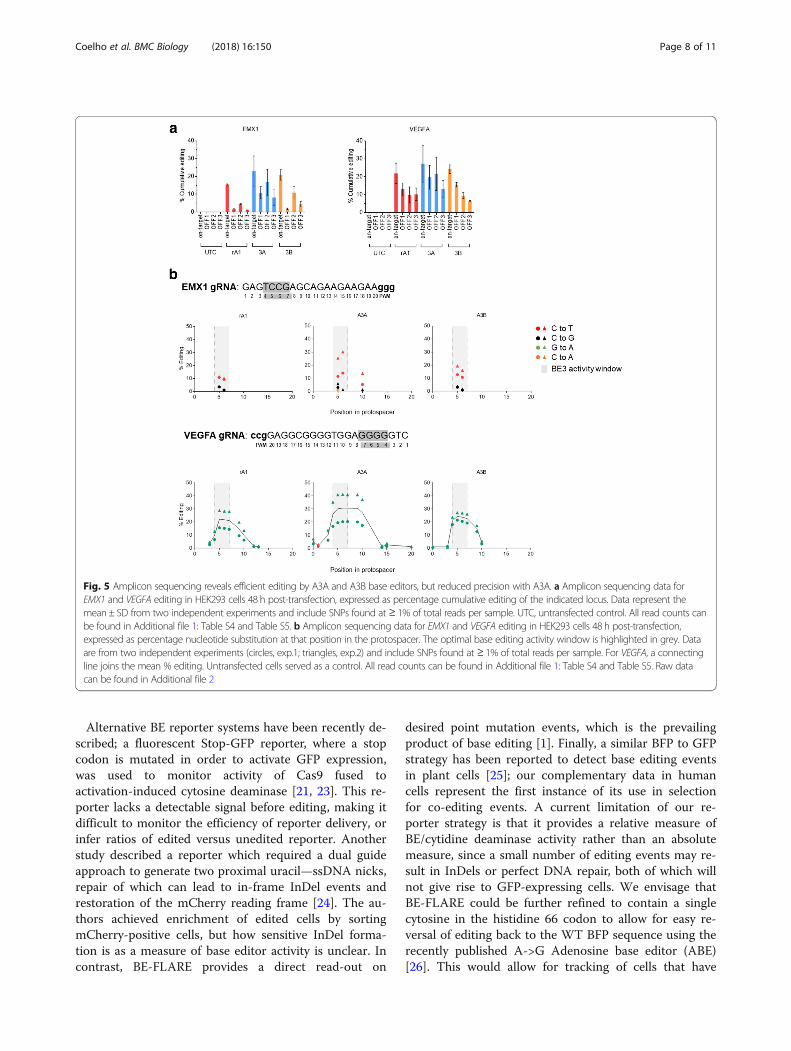
base editor variants, we set out to analyse on-target andoff-target editing by next-generation sequencing of PCRamplicons. Using HEK293 cells, we targeted EMX1 andVEGFA loci and measured on-target editing, three previ-ously reported off-target sites for each gRNA (Additionalfile 1: Table S4 and Table S5), and bystander mutationswithin the gRNA binding site. We found on-target edit-ing of EMX1 and VEGFA to be modestly higher for A3Awhen compared with rA1 and A3B base editor variants;however, A3A-BE3 generally produced higher frequen-cies of off-target events within 6/6 of the off-target locitested (Fig. 5a). Furthermore, when analysing the profileof on-target editing of EMX1 and VEGFA loci, we notedan increased frequency of bystander C->T and G->Aediting events in the A3A-BE3 samples, implying abroader editing window within the gRNA binding se-quence (Fig. 5b). For example, only A3A-BE3 producedbystander mutations at position + 10 in the EMX1 pro-tospacer and beyond position + 13 in the VEGFA proto-spacer. In contrast, rA1-BE3 and A3B-BE3 displayednarrow activity windows within the protospacer se-quence, with comparable on-target editing efficiencies.In conclusion, our deep sequencing analyses are broadly
consistent with results generated from editing BE-FLARE.We demonstrate that A3A-BE3 has a broader mutationalprofile leading to higher bystander mutation rates which isconsistent with loss of GFP fluorescence in the BE-FLAREsystem. Thus seen, BE-FLARE is a valuable tool for meas-uring the efficiency and precision of novel base editors.
DiscussionWe employed BE-FLARE to evaluate the efficiency andprecision of different base editor variants. Replacementof rat APOBEC1 in BE3 with human APOBEC3B
resulted in a similar level of activity and specificity asthe original, codon-optimised rat APOBEC1. Surpris-ingly, A3A-BE3 induced greater bystander mutations,which we further confirmed at multiple genomicon-target and off-target sites. Although A3A and A3BC-terminal domain share ~ 90% similarity in protein se-quence, the active site in A3A is open whereas in A3B itis partially occluded by the flexible loop 1 region (Add-itional file 1: Figure S6) [18–20]. Whilst these structuraldifferences could explain the increased bystander ratesof A3A-BE3, we also observed a significant increase inprotein expression over time, which suggests there couldbe a fine balance between protein abundance/stabilityand precision of base editors. This is supported by therecent finding that codon optimisation of BE3 constructssignificantly increase activity [16, 17], suggesting thatprotein expression from the original BE3 constructs is alimiting factor. Whilst undesirable for precision applica-tions, the increased bystander editing frequency ofA3A-BE3 may prove beneficial for targeted mutagenesisapproaches similar to the CRISPR-X system [21, 22]. Arecent study demonstrated that several point mutationsin A3A can reduce bystander mutation rates ofA3A-BE3 [15], showing that this highly active cytidinedeaminase can be rationally refined for gene editing.Our findings imply that A3B may also prove to be an ex-cellent starting point to develop more precise base edi-tors with increased editing efficiency, whilst thereduction of immunogenic peptides compared to rA1could be beneficial in therapeutic settings.We have developed a capability to enrich genetically
edited cells through selection based upon a fluorescentreporter of base editing activity. Transient expressionand selection of BE-FLARE-positive cells allowed sig-nificant enrichment of base editing at secondary sites.This methodology is broadly applicable and can helpwhen generating a modified cell line by reducing thenumber of clones screened to identify the desired geno-type, especially when no phenotypic selection is pos-sible or where the desired mutation has deleteriouseffects on cell fitness. The importance of this benefit ishighlighted by the low level of base editing of EGFR ob-served in mock-selected pools compared to the signifi-cant increase after BE-FLARE enrichment. This lowlevel of editing is likely a result of the high EGFR copynumber in PC9 cells, which are dependent upon mu-tant EGFR signalling for survival [13]. As gene copynumber alterations are common in cancer cell lines, en-richment before generation of single cell clones offersan invaluable tool to improve the success of cell modelgeneration. At certain sites, BE-FLARE enrichmentgenerated very high levels of editing in bulk pools,which in some cases may avoid the need to usesingle-cell clones entirely.
Coelho et al. BMC Biology (2018) 16:150 Page 7 of 11
Alternative BE reporter systems have been recently de-scribed; a fluorescent Stop-GFP reporter, where a stopcodon is mutated in order to activate GFP expression,was used to monitor activity of Cas9 fused toactivation-induced cytosine deaminase [21, 23]. This re-porter lacks a detectable signal before editing, making itdifficult to monitor the efficiency of reporter delivery, orinfer ratios of edited versus unedited reporter. Anotherstudy described a reporter which required a dual guideapproach to generate two proximal uracil—ssDNA nicks,repair of which can lead to in-frame InDel events andrestoration of the mCherry reading frame [24]. The au-thors achieved enrichment of edited cells by sortingmCherry-positive cells, but how sensitive InDel forma-tion is as a measure of base editor activity is unclear. Incontrast, BE-FLARE provides a direct read-out on
desired point mutation events, which is the prevailingproduct of base editing [1]. Finally, a similar BFP to GFPstrategy has been reported to detect base editing eventsin plant cells [25]; our complementary data in humancells represent the first instance of its use in selectionfor co-editing events. A current limitation of our re-porter strategy is that it provides a relative measure ofBE/cytidine deaminase activity rather than an absolutemeasure, since a small number of editing events may re-sult in InDels or perfect DNA repair, both of which willnot give rise to GFP-expressing cells. We envisage thatBE-FLARE could be further refined to contain a singlecytosine in the histidine 66 codon to allow for easy re-versal of editing back to the WT BFP sequence using therecently published A->G Adenosine base editor (ABE)[26]. This would allow for tracking of cells that have
Fig. 5 Amplicon sequencing reveals efficient editing by A3A and A3B base editors, but reduced precision with A3A. a Amplicon sequencing data forEMX1 and VEGFA editing in HEK293 cells 48 h post-transfection, expressed as percentage cumulative editing of the indicated locus. Data represent themean ± SD from two independent experiments and include SNPs found at ≥ 1% of total reads per sample. UTC, untransfected control. All read counts canbe found in Additional file 1: Table S4 and Table S5. b Amplicon sequencing data for EMX1 and VEGFA editing in HEK293 cells 48 h post-transfection,expressed as percentage nucleotide substitution at that position in the protospacer. The optimal base editing activity window is highlighted in grey. Dataare from two independent experiments (circles, exp.1; triangles, exp.2) and include SNPs found at ≥ 1% of total reads per sample. For VEGFA, a connectingline joins the mean % editing. Untransfected cells served as a control. All read counts can be found in Additional file 1: Table S4 and Table S5. Raw datacan be found in Additional file 2
Coelho et al. BMC Biology (2018) 16:150 Page 8 of 11
undergone a transition from BFP to GFP and then backto BFP, facilitating genetic rescue experiments on en-dogenous genes.Using an alternative approach, we marked transfected
cells with a co-expressed fluorescent protein. Whilst thissystem is suitable for transient transfection, the largesize of the final expression cassettes at nearly 8.5 kb pre-cludes such delivery by lentivirus, where cargo sizes arelimited [27]. Importantly, BE-FLARE directly reports onbase editor activity rather than simply the expression ofBE3, providing a more functional read-out. Separate de-livery of BE-FLARE allows for maximum flexibility andapplicability in multiple cell systems and with variousbase editor versions. Finally, cell lines stably expressingthe BE-FLARE allow for tracking of edited cells overtime to monitor phenotype, which is not possible withtransient expression of a fluorescent protein. Indeed, asGFP fluorescence is amenable to detection by fluores-cence microscopy, BE-FLARE can be applied to detectbase editing in high-throughput functional geneticscreens. This reporter may also be employed in thera-peutic genome editing, where it is important to selectfor rare editing events in primary cells without introdu-cing a permanent genetic marker.
ConclusionsIn conclusion, we present BE-FLARE as a rapidly imple-mentable system for tracking and selecting base editedcells and refining the next generation of base editors.
MethodsCell cultureHEK 293 and PC9 (both from ATCC) cells were main-tained at 5% CO2, 95% air in RPMI, 10% FCS, 1 X Gluta-MAX (ThermoFisher). Transfections were performedusing FuGENE HD (Promega) using a 3:1 ratio of trans-fection reagent to DNA according to the manufacturer’sinstructions. Cell lines were STR profiled and verified asmycoplasma-free.
Cloning and plasmidsThe BE3 expression cassette was synthesised (Thermo-Fisher) and cloned into pcDNA3.1(+). We introduced acassette into the Mlu site containing an AarI guide clon-ing region with ccdB for selection, and the human U6 pro-moter driving gRNA expression. gRNA sequences werecloned into the AarI site using complementary primerpairs, which were annealed, phosphorylated, and ligatedinto the linearised vector. Primers can be found in Add-itional file 1: Table S1. For the BFP reporter construct, agBlock encoding eBFP was synthesised (IDT) and intro-duced by Gibson assembly (NEB) into an expression vec-tor under the human EF-1 alpha promoter. The vectorcontains sequences to allow ObLiGaRe-mediated
integration into the human AAVS1 ‘safe harbour’ locus[11]. All sequences of the synthesised cassettes and guideRNAs are listed in Additional file 1: SupplementaryMethods. VEGFA, EMX1 and non-targeting guide RNAsare published [15, 28, 29].
Generation of stable BE-FLARE cell linesHEK293 and PC9 cells were transfected with BE-FLAREplasmid and a construct encoding zinc-fingers targetingthe AAVS1 safe-harbour locus, essentially as described[11], and subsequently selected for 3 days with puro-mycin (1 μg/ml).
Flow cytometryFACS was carried out on a FACSJazz (BD Biosciences),and flow cytometry analysis was carried out on a For-tessa (BD Biosciences). Briefly, cells were transfectedwith the indicated constructs and, 3 days later, harvestedby trypsinisation for flow cytometry analysis or FACS.
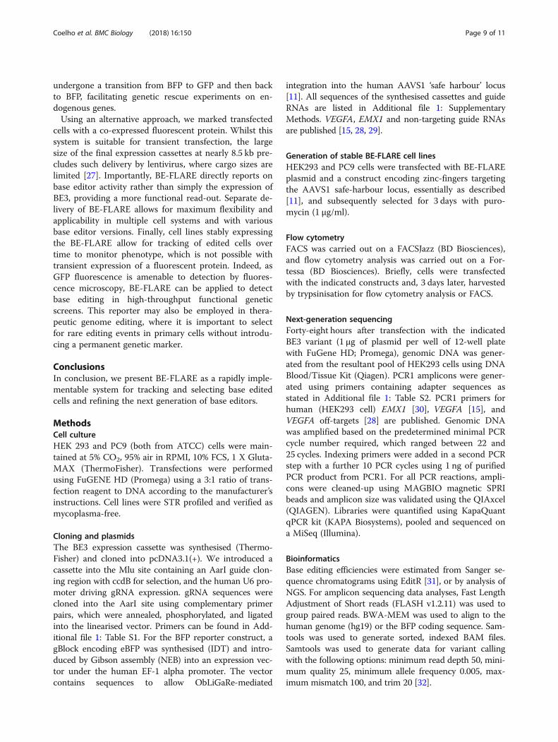
Next-generation sequencingForty-eight hours after transfection with the indicatedBE3 variant (1 μg of plasmid per well of 12-well platewith FuGene HD; Promega), genomic DNA was gener-ated from the resultant pool of HEK293 cells using DNABlood/Tissue Kit (Qiagen). PCR1 amplicons were gener-ated using primers containing adapter sequences asstated in Additional file 1: Table S2. PCR1 primers forhuman (HEK293 cell) EMX1 [30], VEGFA [15], andVEGFA off-targets [28] are published. Genomic DNAwas amplified based on the predetermined minimal PCRcycle number required, which ranged between 22 and25 cycles. Indexing primers were added in a second PCRstep with a further 10 PCR cycles using 1 ng of purifiedPCR product from PCR1. For all PCR reactions, ampli-cons were cleaned-up using MAGBIO magnetic SPRIbeads and amplicon size was validated using the QIAxcel(QIAGEN). Libraries were quantified using KapaQuantqPCR kit (KAPA Biosystems), pooled and sequenced ona MiSeq (Illumina).
BioinformaticsBase editing efficiencies were estimated from Sanger se-quence chromatograms using EditR [31], or by analysis ofNGS. For amplicon sequencing data analyses, Fast LengthAdjustment of Short reads (FLASH v1.2.11) was used togroup paired reads. BWA-MEM was used to align to thehuman genome (hg19) or the BFP coding sequence. Sam-tools was used to generate sorted, indexed BAM files.Samtools was used to generate data for variant callingwith the following options: minimum read depth 50, mini-mum quality 25, minimum allele frequency 0.005, max-imum mismatch 100, and trim 20 [32].
Coelho et al. BMC Biology (2018) 16:150 Page 9 of 11

Western blottingWhole cell lysates were generated using RIPA buffer(ThermoFisher Scientific), and Western blotting wasperformed using standard methods, with secondary anti-bodies conjugated to horseradish peroxidase (GEHealthcare). Cas9 (#14697; RRID: AB_2750916) andGAPDH (#2118; RRID: AB_561053) antibodies werefrom Cell Signaling Technology.
Digital droplet PCR (ddPCR)Base editing over time was estimated by extraction ofgenomic DNA with DNAeasy Blood & Tissue kit (Qia-gen) followed by ddPCR with ddPCR Supermix forprobes no dUTP (BioRad) according to the manufac-turer’s instructions. Probes were labelled with FAM andare listed in Additional file 1: Supplementary Methods.
Experimental design and statisticsThe exact value of sample size (n), statistical tests used,and the number of independent experiments performedare given in the figure legends. Unless otherwise stated,error bars represent standard deviation and an unpairedStudent’s t test was used to assess statistical significance(P < 0.05).
Additional files
Additional file 1: Figure S1–S6, Table S1–S5, Supplementarymethods. Figure S1. BE-FLARE facilitates visual tracking of base editedcells by microscopy. Figure S2. BE-FLARE base editing over time revealsdynamics of base editing and tracking of edited cells with GFP. FigureS3. Raw data relating to Fig. 4a b and c, and quantification of turbo-RFPpositive cells. Figure S4. BE-FLARE expression after editing. Figure S5. Ex-pression of native vs codon-optimised rat APOBEC-1 BE3 reveals superiorexpression after codon optimisation. Figure S6. Sequence alignment ofAPOBECs highlights divergent loop1 region. Supplementary methods.Table S1. Primers for guide RNA cloning. Table S2. Primers for ampliconsequencing. Table S3. Amplicon sequencing summary: BE-FLARE (BFP).Table S4. Amplicon sequencing summary: EMX1. Table S5. Amplicon se-quencing summary: VEGFA. Digital droplet PCR probes. Sequences of constructs.(DOCX 1133 kb)
Additional file 2: Raw data relating to Figs. 1, 2, 3, 4 and 5, Figure S2,S3, S4 and S5. (XLSX 34 kb)
AcknowledgementsThanks to the Discovery Biology IMED Biotech Unit, AstraZeneca, for thehelpful discussion. We thank Daniel O’Neill for providing valuable adviceregarding the editing of EGFR and the NGS team for their assistance withDNA sequencing. MC is a fellow of the AstraZeneca postdoc programme.
FundingThis work was funded by AstraZeneca plc.
Availability of data and materialsAll data generated or analysed during this study are included in thispublished article, its supplementary information files, and publicly availablerepositories. Sequencing data is available from the NCBI Sequence ReadArchive database, accession: SRP153020. Raw data relating to figures andsupplemental figures can be found in Additional file 2.
Authors’ contributionsMAC and BJMT designed the study. MAC carried out the experiments. MFprovided bioinformatics support for NGS data analysis. SL, LSP and GCprovided reagents. EC, JDW, MM and BJMT provided conceptual advice. MACand BJMT wrote the manuscript. All authors read and approved the finalmanuscript.
Ethics approval and consent to participateNot applicable.
Consent for publicationNot applicable.
Competing interestsThe authors declare no competing interests.
Publisher’s NoteSpringer Nature remains neutral with regard to jurisdictional claims inpublished maps and institutional affiliations.
Author details1Discovery Sciences, IMED Biotech Unit, AstraZeneca, Cambridge, UK.2Discovery Sciences, IMED Biotech Unit, AstraZeneca, Gothenburg, Sweden.
Received: 28 October 2018 Accepted: 3 December 2018
References1. Komor AC, Kim YB, Packer MS, Zuris JA, Liu DR. Programmable editing of a
target base in genomic DNA without double-stranded DNA cleavage.Nature. 2016;533:420–4. https://doi.org/10.1038/nature17946.
2. Nishida K, Arazoe T, Yachie N, Banno S, Kakimoto M, Tabata M, et al.Targeted nucleotide editing using hybrid prokaryotic and vertebrateadaptive immune systems. Science. 2016;353. https://doi.org/10.1126/science.aaf8729.
3. Komor AC, Badran AH, Liu DR, Guilinger JP, Bessen JL, Hu JH, et al. CRISPR-based technologies for the manipulation of eukaryotic genomes. Cell. 2017;168:20–36. https://doi.org/10.1016/j.cell.2016.10.044.
4. Cong L, Ran FA, Cox D, Lin S, Barretto R, Habib N, et al. Multiplex genomeengineering using CRISPR/Cas systems. Science. 2013;339:819–23. https://doi.org/10.1126/science.1231143.
5. Miyaoka Y, Berman JR, Cooper SB, Mayerl SJ, Chan AH, Zhang B, et al.Systematic quantification of HDR and NHEJ reveals effects of locus,nuclease, and cell type on genome-editing. Sci Rep. 2016;6:23549. https://doi.org/10.1038/srep23549.
6. Saleh-Gohari N, Helleday T. Conservative homologous recombinationpreferentially repairs DNA double-strand breaks in the S phase of the cellcycle in human cells. Nucleic Acids Res. 2004;32:3683–8. https://doi.org/10.1093/nar/gkh703.
7. Hsu PD, Lander ES, Zhang F. Development and applications of CRISPR-Cas9for genome engineering. Cell. 2014;157:1262–78.
8. Certo MT, Ryu BY, Annis JE, Garibov M, Jarjour J, Rawlings DJ, et al. Trackinggenome engineering outcome at individual DNA breakpoints. Nat Methods.2011;8:671–6.
9. Zhou Y, Liu Y, Hussmann D, Brøgger P, Al-Saaidi RA, Tan S, et al. Enhancedgenome editing in mammalian cells with a modified dual-fluorescentsurrogate system. Cell Mol Life Sci. 2016;73(13):2543–63.
10. Heim R, Prasher DC, Tsien RY. Wavelength mutations and posttranslationalautoxidation of green fluorescent protein. Proc Natl Acad Sci. 1994;91:12501–4. https://doi.org/10.1073/pnas.91.26.12501.
11. Maresca M, Lin VG, Guo N, Yang Y. Obligate ligation-gated recombination(ObLiGaRe): custom-designed nuclease-mediated targeted integrationthrough nonhomologous end joining. Genome Res. 2013;23:539–46.
12. Pao W, Miller VA, Politi KA, Riely GJ, Somwar R, Zakowski MF, et al. Acquiredresistance of lung adenocarcinomas to gefitinib or erlotinib is associatedwith a second mutation in the EGFR kinase domain. PLoS Med. 2005;2:0225–35.
13. Godin-Heymann N, Ulkus L, Brannigan BW, McDermott U, Lamb J,Maheswaran S, et al. The T790M “gatekeeper” mutation in EGFR mediatesresistance to low concentrations of an irreversible EGFR inhibitor. MolCancer Ther. 2008;7:874–9. https://doi.org/10.1158/1535-7163.MCT-07-2387.
Coelho et al. BMC Biology (2018) 16:150 Page 10 of 11
14. Okabe T, Okamoto I, Tamura K, Terashima M, Yoshida T, Satoh T, et al.Differential constitutive activation of the epidermal growth factor receptorin non-small cell lung cancer cells bearing EGFR gene mutation andamplification. Cancer Res. 2007;67:2046–53.
15. Gehrke JM, Cervantes O, Clement MK, Wu Y, Zeng J, Bauer DE, et al. AnAPOBEC3A-Cas9 base editor with minimized bystander and off-targetactivities. Nat Biotechnol. 2018;36(10):977–82.
16. Koblan LW, Doman JL, Wilson C, Levy JM, Tay T, Newby GA, et al. Improvingcytidine and adenine base editors by expression optimization and ancestralreconstruction. Nat Biotechnol. 2018;36(9):843–6.
17. Zafra MP, Schatoff EM, Katti A, Foronda M, Breinig M, Schweitzer AY, et al.Optimized base editors enable efficient editing in cells, organoids and mice.Nat Biotechnol. 2018;36(9):888–93.
18. Shi K, Demir Ö, Carpenter MA, Wagner J, Kurahashi K, Harris RS, et al.Conformational switch regulates the DNA cytosine deaminase activity ofhuman APOBEC3B. Sci Rep. 2017;7(1):17415.
19. Salter JD, Smith HC. Modeling the embrace of a mutator: APOBEC selectionof nucleic acid ligands. Trends Biochem Sci. 2018;43(8):606–22.
20. Shi K, Carpenter MA, Kurahashi K, Harris RS, Aihara H. Crystal structure of theDNA deaminase APOBEC3B catalytic domain. J Biol Chem. 2015;290(47):28120–30.
21. Ma Y, Zhang J, Yin W, Zhang Z, Song Y, Chang X. Targeted AID-mediatedmutagenesis (TAM) enables efficient genomic diversification in mammaliancells. Nat Methods. 2016;13:1029–35. https://doi.org/10.1038/nmeth.4027.
22. Hess GT, Frésard L, Han K, Lee CH, Li A, Cimprich KA, et al. Directedevolution using dCas9-targeted somatic hypermutation in mammalian cells.Nat Methods. 2016;13(12):1036–42.
23. Yoshikawa K. AID enzyme-induced hypermutation in an actively transcribedgene in fibroblasts. Science. 2002;296:2033–6. https://doi.org/10.1126/science.1071556.
24. Martin AS, Salamango D, Serebrenik A, Shaban N, Brown WL, Donati F, et al.A fluorescent reporter for quantification and enrichment of DNA editing byAPOBEC–Cas9 or cleavage by Cas9 in living cells. Nucleic Acids Res. 2018;46(14):e84.
25. Zong Y, Wang Y, Li C, Zhang R, Chen K, Ran Y, et al. Precise base editing inrice, wheat and maize with a Cas9-cytidine deaminase fusion. NatBiotechnol. 2017;35:438–40. https://doi.org/10.1038/nbt.3811.
26. Gaudelli NM, Komor AC, Rees HA, Packer MS, Badran AH, Bryson DI, et al.Programmable base editing of A • T to G • C in genomic DNA without DNAcleavage. Nat Publ Gr. 2017;551(7681):464–71.
27. Kumar M, Keller B, Makalou N, Sutton RE. Systematic determination of thepackaging limit of lentiviral vectors. Hum Gene Ther. 2001;12:1893–905.https://doi.org/10.1089/104303401753153947.
28. Rees HA, Komor AC, Yeh WH, Caetano-Lopes J, Warman M, Edge ASB, et al.Improving the DNA specificity and applicability of base editing throughprotein engineering and protein delivery. Nat Commun. 2017;8:15790.
29. Doench JG, Fusi N, Sullender M, Hegde M, Vaimberg EW, Donovan KF, et al.Optimized sgRNA design to maximize activity and minimize off-targeteffects of CRISPR-Cas9. Nat Biotechnol. 2016;34(2):184–91.
30. Slaymaker IM, Gao L, Zetsche B, Scott DA, Yan WX, Zhang F. Rationallyengineered Cas9 nucleases with improved specificity. Science. 2016;351(6268):84–8.
31. Kluesner MG, Nedveck DA, Lahr WS, Garbe JR, Abrahante JE, Webber BR, etal. EditR: a method to quantify base editing from Sanger sequencing. Cris J.2018; 1(3). https://doi.org/10.1089/crispr.2018.0014.
32. Li H, Handsaker B, Wysoker A, Fennell T, Ruan J, Homer N, et al. The sequencealignment/map format and SAMtools. Bioinformatics. 2009;25(16):2078–9.
Coelho et al. BMC Biology (2018) 16:150 Page 11 of 11