beautiful picture
TRANSCRIPT

Figure 46.1

Figure 46.2

Figure 46.3-1
Asexual reproduction
Female
Sexual reproduction
Generation 1Female

Figure 46.3-2
Asexual reproduction
Female
Sexual reproduction
Generation 1Female
Male
Generation 2

Figure 46.3-3
Asexual reproduction
Female
Sexual reproduction
Generation 1Female
Male
Generation 3
Generation 2

Figure 46.3-4
Asexual reproduction
Female
Sexual reproduction
Generation 1Female
Male
Generation 4
Generation 3
Generation 2

Figure 46.4
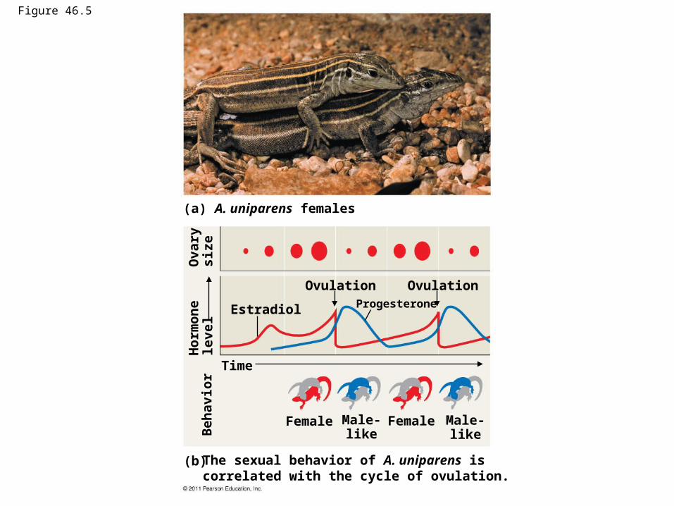
Figure 46.5
(a) A. uniparens females
(b) The sexual behavior of A. uniparens is correlated with the cycle of ovulation.
Estradiol
Ovulation OvulationProgesterone
Time
Female FemaleMale-like
Male-likeB
eha
vio
rH
orm
on
ele
vel
Ova
rysi
ze

Figure 46.5a
(a) A. uniparens females

Figure 46.5b
(b) The sexual behavior of A. uniparens is correlated with the cycle of ovulation.
Estradiol
Ovulation OvulationProgesterone
Time
Female FemaleMale-like
Male-likeB
ehav
ior
Ho
rmo
ne
leve
lO
vary
size

Figure 46.6

Figure 46.7

Figure 46.8
(a) Male fruit fly (b) Female fruit fly
Accessorygland
Ejaculatory duct
Penis andclaspers
Testis
Vasdeferens
Seminalvesicle
Ovary
Accessorygland
Oviduct
Spermatheca
Uterus
Vulva

Figure 46.8a
(a) Male fruit fly
Accessorygland
Ejaculatory duct
Penis andclaspers
Testis
VasdeferensSeminalvesicle

Figure 46.8b
(b) Female fruit fly
Ovary
Accessorygland
OviductSpermatheca
Uterus
Vulva

Figure 46.9
RESULTS
Control;not
remated
Remated towild-type
males
Remated to“no-sperm”
males
Remated to“no-ejaculate”
males
30
20
10
0
Per
cen
tag
e o
f fe
mal
esla
ckin
g s
per
m in
sp
erm
ath
eca

Figure 46.10
(Rectum)CervixVagina
Major vestibular(Bartholin’s) gland
Vaginal opening
Oviduct
OvaryUterus
(Urinary bladder)
(Pubic bone)
Urethra
BodyGlansPrepuce
Clitoris
Labia minoraLabia majora
Ovaries Oviduct
Follicles
Corpus luteumUterine wallEndometrium
Vagina
Uterus
Cervix

Figure 46.10a
(Rectum)CervixVagina
Major vestibular(Bartholin’s) gland
Vaginal opening
Oviduct
OvaryUterus
(Urinary bladder)
(Pubic bone)
Urethra
BodyGlansPrepuce
Clitoris
Labia minoraLabia majora

Figure 46.10b
Ovaries Oviduct
Follicles
Corpus luteumUterine wallEndometrium
Vagina
Uterus
Cervix

Figure 46.11
Seminalvesicle(behindbladder)
Urethra
Scrotum
(Urinary bladder)
Prostate gland
Bulbourethral gland
Erectile tissue of penis
Vas deferensEpididymis
Testis
Seminal vesicle
(Rectum)Vas deferens
Ejaculatory duct
Prostate gland
Bulbourethral gland Vas deferensEpididymisTestisScrotum
(Urinary bladder)
(Urinary duct)
(Pubic bone)
Erectiletissue
Urethra
Glans
Prepuce
Penis

Figure 46.11a
Seminalvesicle(behindbladder)
Urethra
Scrotum
(Urinary bladder)
Prostate gland
Bulbourethral gland
Erectile tissue of penis
Vas deferensEpididymisTestis

Figure 46.11b
Seminal vesicle
(Rectum)Vas deferens
Ejaculatory ductProstate glandBulbourethral gland Vas deferens
EpididymisTestisScrotum
(Urinary bladder)
(Urinary duct)
(Pubic bone)
Erectiletissue
Urethra
Glans
Prepuce
Penis

Figure 46.12aEpididymis
Seminiferous tubule
Testis
Cross section ofseminiferous tubule
Sertoli cellnucleus
Lumen ofseminiferous tubule
Plasmamembrane
Tail
Neck
Midpiece Head
Mitochondria
NucleusAcrosome
Primordial germ cell in embryo
Mitotic divisions
Spermatogonialstem cell
Spermatogonium
Mitotic divisions
Mitotic divisions
Primary spermatocyte
Meiosis I
Meiosis II
Spermatids (two stages)
Secondary spermatocyte
Earlyspermatid
Sperm cell
Differentiation(Sertoli cellsprovide nutrients)
2n
2n
2n
n n
n n n n
n n n n

Figure 46.12aa
Epididymis Seminiferous tubule
Testis
Cross section ofseminiferous tubule
Spermato-gonium
Primary spermatocyte
Spermatids (two stages)
Secondary spermatocyte
Sperm cell
Sertoli cellnucleus
Lumen ofseminiferous tubule

Figure 46.12abPrimordial germ cell in embryo
Mitotic divisions
Spermatogonial stem cell
Spermatogonium
Mitotic divisions
Mitotic divisions
Primary spermatocyte
Meiosis I
Meiosis II
Secondary spermatocyte
Earlyspermatid
Sperm cell
Differentiation(Sertoli cellsprovide nutrients)
2n
2n
2n
n n
n n n n
n n n n

Figure 46.12ac
Plasmamembrane
Tail
Neck
Midpiece Head
Mitochondria
NucleusAcrosome

Figure 46.12b
2n
2n
nn
n
n
Ovary
Primordial germ cell
Mitotic divisions
Mitotic divisions
Oogonium
In embryo
Primary oocyte(present at birth), arrestedin prophase of meiosis I
Completion of meiosis Iand onset of meiosis II
Secondary oocyte,arrested at metaphase ofmeiosis II
Firstpolarbody
Ovulation, sperm entry
Completion of meiosis IISecondpolarbody
Primary oocytewithinfollicle
Growingfollicle
Mature follicle
Rupturedfollicle
Ovulatedsecondaryoocyte
Corpus luteum
Degeneratingcorpus luteum
Fertilized egg

Figure 46.12ba
Ovary
Primary oocytewithinfollicle
Growingfollicle
Mature follicle
Rupturedfollicle
Ovulatedsecondaryoocyte
Corpus luteum
Degeneratingcorpus luteum

Figure 46.12bb
2n
2n
nn
n
n
Primordial germ cellMitotic divisions
Mitotic divisions
Oogonium
In embryo
Primary oocyte(present at birth), arrestedin prophase of meiosis I
Completion of meiosis Iand onset of meiosis II
Secondary oocyte,arrested at metaphase ofmeiosis II
Firstpolarbody
Ovulation, sperm entry
Completion of meiosis IISecondpolarbody
Fertilized egg

Figure 46.13Control by hypothalamus(a)
Hypothalamus
GnRH
Anterior pituitary
FSH LH
Inhibited by combination ofestradiol and progesterone
Stimulated by high levelsof estradiol
Inhibited by low levels ofestradiol
109
87
65
4
3
2
1
(b) Pituitary gonadotropinsin blood
LH
FSH
FSH and LH stimulatefollicle to grow
6
LH surge triggers ovulation
Ovarian cycle
Growing follicle Maturing follicle
Corpusluteum
Degeneratingcorpus luteum
Follicular phase Ovulation Luteal phase
Estradiol secretedby growing follicle inincreasing amounts
(c)
Progesterone andestradiol secretedby corpus luteum
(d) Ovarian hormonesin blood
Estradiol
Peak causes LH surge(see )
Progesterone
Progesterone and estra-diol promote thickeningof endometrium
Estradiol levelvery low
(e) Uterine (menstrual) cycle
Endometrium
Menstrual flow phase Proliferative phase Secretory phase
Day
s
28252015141050

Figure 46.13a
Control by hypothalamus(a)
Hypothalamus
GnRH
Anterior pituitary
FSH LH
Inhibited by combination ofestradiol and progesteroneStimulated by high levelsof estradiol
Inhibited by low levels ofestradiol
2
1
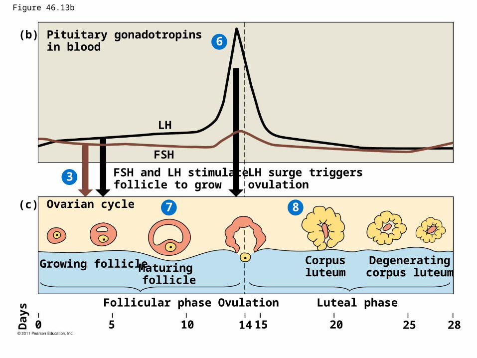
Figure 46.13b
(b) Pituitary gonadotropinsin blood
LH
FSH
FSH and LH stimulatefollicle to grow
LH surge triggers ovulation
Ovarian cycle
Growing follicle Maturing follicle
Corpusluteum
Degeneratingcorpus luteum
Follicular phase Ovulation Luteal phase
(c)
28252015141050Day
s
3
6
7 8

Figure 46.13c
Estradiol secretedby growing follicle inincreasing amounts
Progesterone andestradiol secretedby corpus luteum
(d) Ovarian hormonesin blood
Estradiol
Peak causes LH surge(see )
Progesterone
Progesterone and estra-diol promote thickeningof endometrium
Estradiol levelvery low
(e)
Uterine (menstrual) cycle
Endometrium
Menstrual flow phase Proliferative phase Secretory phase
Da
ys
28252015141050
4
5 6
910
(e)

Figure 46.14
Hypothalamus
GnRH
Anterior pituitary
FSH LH
Sertoli cells Leydig cells
Inhibin Spermatogenesis Testosterone
Testis
Neg
ativ
e fe
edb
ack
Neg
ativ
e fe
edb
ack

Figure 46.15
5
4
1
2
3 Cleavage
Fertilization
Ovary
Ovulation
Uterus
Cleavage continues
Implantation
Endometrium(a) From ovulation to implantation
(b) Implantation of blastocyst
Endometrium
Cavity
Inner cellmass
Blastocyst Trophoblast

Figure 46.16
Placenta
Uterus
Umbilicalcord
Chorionicvillus,containingfetalcapillaries
Maternalblood pool
Maternalarteries
Maternalveins
Maternalportion ofplacenta
Fetalportion ofplacenta(chorion)
Umbilicalarteries
Umbilical veinUmbilical cord
Fetal venuleFetal arteriole

Figure 46.17
(a) 5 weeks (b) 14 weeks (c) 20 weeks

Figure 46.17a
(a) 5 weeks

Figure 46.17b
(b) 14 weeks

Figure 46.17c
(c) 20 weeks

Figure 46.18
Estradiol
from ovaries
Activates oxytocinreceptors on uterus
Oxytocin
from fetusand mother’s posterior pituitary
Prostaglandins
Po
siti
ve f
eed
bac
k
Stimulates uterusto contract
Stimulatesplacenta to make
Stimulate morecontractions
of uterus

Figure 46.19
Dilation of the cervix
2
1
3
PlacentaUmbilical cord
Uterus
Cervix
Expulsion: delivery of the infant
Delivery of the placenta
Uterus
Umbilical cord
Placenta (detaching)

Figure 46.19a
Dilation of the cervix1
PlacentaUmbilical cord
Uterus
Cervix

Figure 46.19b
Expulsion: delivery of the infant2

Figure 46.19c
Delivery of the placenta
Uterus
Umbilical cord
Placenta (detaching)
3

Figure 46.20Male Female
Method MethodEvent Event
Vasectomy
Abstinence
CondomCoitusinterruptus(very highfailure rate)
Productionof sperm
Production ofprimary oocytes
Sperm transportdown male
duct system
Oocytedevelopmentand ovulation
Combinationbirth controlpill (or injection,patch, or vaginal ring)
AbstinenceFemale condom
Spermdepositedin vagina
Capture of the oocyte by the
oviduct
Spermmovement
through femalereproductive
tract
Transportof oocyte in
oviduct
Tubal ligation
Spermicides;diaphragm;progestin alone(as minipillor injection)
Meeting of sperm and oocytein oviduct
Union of sperm and egg
Implantation of blastocystin endometrium
Morning-afterpill; intrauterinedevice (IUD)

Figure 46.20a
Male FemaleMethod MethodEvent Event
Vasectomy
AbstinenceCondomCoitusinterruptus(very highfailure rate)
Productionof sperm
Production ofprimary oocytes
Sperm transportdown male
duct system
Oocytedevelopmentand ovulation
Combinationbirth controlpill (or injection,patch, or vaginal ring)
AbstinenceFemale condom
Spermdepositedin vagina
Capture of the oocyte by the
oviduct
Tubal ligation
Spermicides;diaphragm;progestin alone(as minipillor injection)

Figure 46.20b
Spermmovement
through femalereproductive
tract
Transportof oocyte in
oviduct
Meeting of sperm and oocytein oviduct
Union of sperm and egg
Implantation of blastocystin endometrium
Morning-afterpill; intrauterinedevice (IUD)
Male FemaleMethod MethodEvent Event

Figure 46.UN01Human gametogenesis
Spermatogenesis Oogenesis
Primaryspermatocyte
Primaryoocyte
Secondaryspermatocytes
Secondaryoocyte
Polar body
Spermatids
Sperm
Polar body
Fertilized egg
2n 2n
n
n
n
n
n n
n n n n
n n n n

Figure 46.UN02