behavioral responses to chemical …jeb.biologists.org/content/jexbio/162/1/209.full.pdfbehavioral...
TRANSCRIPT

J. exp. Biol. 162, 209-229 (1992) 2 0 9Printed in Great Britain © The Company of Biologists Limited 1992
BEHAVIORAL RESPONSES TO CHEMICAL STIMULATIONOF THE OLFACTORY ORGAN IN THE SQUID LOLIGO
OPALESCENS
BY WM F. GILLY AND MARY T. LUCERO
Hopkins Marine Station, Stanford University, Pacific Grove, CA 93950, USA
Accepted 19 August 1991
Summary
Behavioral experiments were carried out on restrained, but otherwise fullyactive, squid to test the chemoreceptive capabilities of the olfactory organ.Specific chemical substances stimulated high-pressure jet escape responses whenejected from a small pipette into the area immediately around the olfactory organ.These included squid ink and L-Dopa (3,4 dihydroxyphenylalanine) as well asagents that block voltage-dependent potassium channels, such as quaternaryammonium ions and 4-aminopyridine. Experiments designed to map chemosensit-ivity spatially identified the olfactory organ as the receptive site. Unilateralapplication of a topical local anesthetic to an olfactory organ selectively andreversibly abolished responsiveness on the treated side only. The olfactory organcan thus mediate detection of water-borne chemicals. This detection, in turn, islinked to motor control pathways involved in initiating escape-jetting behavior.
Introduction
Squids, cuttlefishes and octopuses (the coleoid cephalopods) are intelligent,highly mobile predators. Although they are largely visually oriented (Packard,1972), these animals are also well equipped with complex vestibular (Budelmann,1990), auditory (Hanlon and Budelmann, 1987; Budelmann and Bleckmann, 1988)and tactile (Wells, 1964; Wells and Young, 1975) capabilities. Sensory pathwaysfor these modalities converge in the central nervous system and regulate importantbehavioral outputs, such as escape jetting, chromatophore display, mating,homing and long-distance migration (Messenger, 1983; Boyle, 1986).
Chemoreception is another sense that might be expected to be important tothese animals, many of which are largely nocturnal, demersal or benthic.Although a rich literature on the chemosensory competence of many marineinvertebrates exists (Grant and Mackie, 1974; Atema etal. 1988), comparativelylittle is known about chemoreception or chemotaxis in cephalopods. In Octopusvulgaris (Lamarck), chemoreceptors have been described on the sucker cups ofthe arms (Graziadei, 1962) and are thought to constitute the basis for thechemotactile sense that is evident in behavioral and learning experiments (Wells,
Key words: cephalopod, squid, chemoreception, olfaction, escape response, Loligo opalescens.

210 W. F. GlLLY AND M. T. LUCERO
1963; Wells etal. 1985). Morphologically similar receptors occur in the buccallobes surrounding the beak ('lips') of squids (Emery, 1975) and cuttlefishes(Graziadei, 1965) and it has been proposed that they are involved in the initiationof feeding behavior when food touches the lips (LaRoe, 1971; Emery, 1975).
Evidence for detection of water-borne molecules arising from a distant sourcevia a 'distance'-chemoreceptive sense, as opposed to a 'contact'-based chemotac-tile one, is more scarce in these animals, apparently being limited to two reports onOctopus (Boyle, 1983; Chase and Wells, 1986). Essentially nothing is known aboutthe chemoreceptive capabilities of other cephalopods (Boyle, 1986). Morphologi-cal studies have revealed putative chemosensory cells in an elaborate structurecalled the 'olfactory organ' (Messenger, 1967; Woodhams and Messenger, 1974)that is present in all coleoids, and most prominently in squids. This organ is the siteof a sensory epithelium containing several types of primary sensory neurons withdistinctive morphologies (Emery, 1975; Wildenburg and Fioroni, 1990). In nocase, however, has a function for this organ been demonstrated throughbehavioral or physiological experiments (Messenger, 1967) or have functionalproperties of the receptor cells been determined.
In addition to these studies on the olfactory organ itself, anatomical work onsquid has focused on tracing the central projections of sensory axons. Afferentfiber tracts lead to the 'olfactory lobes' as well as to motor centers involved in thecontrol of swimming and jetting, and relatively direct connections to the giant fiberpathway may occur in the magnocellular lobe (Messenger, 1979; Young, 1939).These findings suggest that sensory inputs from the olfactory organ may be acomponent of the neural control of escape jetting (Young, 1938; Wilson, 1960; Otisand Gilly, 1990). The optic gland, a region of the brain thought to be involved inthe control of sexual maturation and reproduction, also receives major inputs fromthe olfactory lobes and from olfactory nerve axons (Messenger, 1979).
This paper is the first of two in which we address the function of the squidolfactory organ. Here we provide behavioral evidence that squids can detectwater-borne chemical stimuli by means of this organ. These experiments alsodemonstrate that olfactory organ projections in the central nervous system doinfluence the motor centers that control escape jetting. The second paper (Luceroetal. 1992) describes the electrical characteristics of receptor neurons in thesensory epithelium of the olfactory organ and the manner in which chemicalstimuli that produce behavioral responses in vivo act to modulate receptorexcitability.
Materials and methods
Behavioral experiments and chemical stimulation
All experiments were carried out on Loligo opalescens Berry collected locally inMonterey Bay. Techniques for restraining the living animals were similar to thosedescribed by Otis and Gilly (1990), but in the present study recordings of neuralactivity were not carried out. Briefly, the dorsal mantle surface was attached to a

Olfactory organ function in squid 211
plastic support platform with cyanoacrylate cement, and the restrained animal wassuspended in oxygenated sea water at 13-15 °C. The experimental tank had avolume of approximately 301, and a slow flow (2-31 min"1) of fresh sea water wasmaintained during experiments. Experimenters were shielded from the animal'sview, and the animal was observed by means of a video camera. Great care wastaken to avoid any sudden noises that could startle the animal and lead to aberrantescape responses. If an animal was deemed to be hypersensitive to spuriousstimulation, the experiment was terminated and the associated results werediscarded.
Chemical stimuli (test substances added to sea water) were delivered bypressure ejection at low rates, usually one stimulus every 1-2 min. Fasterstimulation rates, at least with some substances, can lead to a habituation-likephenomenon that is characterized by intermittent, or even complete, loss ofresponsiveness (Long etal. 1989). In experiments designed to test the efficacy ofdifferent substances, 75-300 ms duration pressure pulses delivered fluid at a rate ofapproximately l/xlms"1 from a port 0.65 mm in diameter positioned a fewmillimeters anterior and lateral to the olfactory organ. The ejection pipette wasaimed posteriorly and oriented at an angle of approximately 45-60° with respect tothe body axis. Generally, each test stimulus was bracketed by seawater controls.Test and control fluids were extruded only during pressure pulses; at other timesthe lines were closed to prevent leakage out of or siphoning back into the supplyline. For some experiments, the stimulating probe also carried a pair of platinumwires and a fiber optic guide to transmit electrical and photic stimuli. Experimentsdesigned to map sensitivity around the olfactory organ employed ejection pipettesof small diameter (0.2mm), fluid delivery rates of approximately 2iulms~1 andpulse durations of 50-75 ms.
Escape responses were detected as intramantle pressure transients (Otis andGilly, 1990). The pressure transducer was not sensitive enough to record pressuresassociated with respiration or weak swimming. Only escape responses that wereaccompanied by measurable pressure transients occurring with a delay of less than20 s following a chemical stimulus were scored as positive responses in our analysisof behavioral data. Jetting (and other) behavior was also documented byvideotaping. Individual video frames were captured from the recorded tapes witha Sony videoprinter.
All chemicals were obtained from Sigma (St Louis, MO), with the exception ofblue food coloring and Brilliant Blue FCF (McCormick, Inc., Baltimore, MD).Squid ink was prepared by mincing 1-4 ink sacs in 1 ml of sea water, brieflyvortexing and diluting the sample into 20 ml of sea water. Other natural extractswere prepared by crude homogenization and brief centrifugation.
Electron microscopy
For scanning electron microscopy, the olfactory organ and surrounding tissuewere dissected and pinned out for fixation in 2 % glutaraldehyde in artificial seawater containing 0.1 moll"1 Hepes (pH7.8). Samples were post-fixed in 1 % OsO4

212 W. F. GlLLY AND M. T. LUCERO
plus 0.1 mol I"1 Hepes and dehydrated in graded ethanol followed by critical pointdrying. Gold-coated specimens were examined in a Hitachi S-450 microscope.
Olfactory organ/nerve preparations were fixed for transmission microscopy in1.5% glutaraldehyde plus lOmmolP1 CoCl2, lOmmolP1 MgCl2, 25mmoir 1
sodium cacodylate and sucrose to attain an osmolality of QSOmosmolkg"1 H2O.Specimens were post-fixed with 0.5% OsO4, 0.8% K3Fe(CN)6, 0.1 moll"1
sodium cacodylate and sucrose, followed by 0.15% tannic acid in 25mmoll~1
sodium cacodylate plus sucrose, and finally stained en bloc with 4 % uranyl acetatein deionized water. Thin sections were examined in a Phillips 201 microscopeoperating at 80 kV.
ResultsGeneral anatomy of the olfactory organ
The location and overall form of the olfactory organ in Loligo opalescens issimilar to that in Lolliguncula brevis (Emery, 1975). The organ is situated ventrallyon the anterior aspect of a ridge of tissue that runs dorso-ventrally just posterior tothe orbit (Fig. 1A). The sensory organ proper is a small knob-like structurelocated in a funnel-shaped cavity composed of muscular tissue (Fig. IB). Thisknob is covered by a sensory epithelium, composed of several types of receptorcells, all of which project their axons into a well-defined 'olfactory' nerve leadingto the brain, and by support cells bearing motile cilia (Emery, 1975; Wildenburgand Fioroni, 1990). The motile cilia are longer and more numerous than thespecialized apical processes of the receptor cells, and their high density accountsfor the distinctive appearance of the olfactory knob (Fig. 1C,D).
Secretory cells are also present in and around the olfactory knob (Barber andWright, 1969). Examination of living material (an excised olfactory organ andsurrounding tissues) under water-immersion optics reveals a robust flow of mucousover the sensory epithelium. This flow moves upwards and out of the funnel-shaped cavity and probably serves to cleanse the sensory epithelium continuouslyand to prevent fouling with particulate matter.
The sensory nerve that emanates from directly beneath the olfactory knobcontains numerous small axons, 0.1-1.5 im in diameter, that are wrapped tightlyin fascicles by Schwann cell elements and densely staining extracellular material(Fig. 2A). Shortly after exiting from the base of the organ, this nerve is joined by asmaller nerve branch that emerges from the surrounding muscular tissue. Axons inthis latter process are larger, 1.5-25.0(xm in diameter, and most are wrappedindividually by Schwann cells (see Fig. 2B). The two nerves then run in parallel,though distinctly separated by connective tissues, for a few millimeters ventrallyand slightly anteriorly where they enter the cartilaginous 'skull' through a commonforamen. The sensory portion of the nerve then passes dorsally along the floor ofthe orbit to the region of the olfactory lobes of the brain (Messenger, 1979).
Demonstration of chemoreception and links to motor controlSmall volumes (typically 100-300^1) of sea water containing certain test

Olfactory organ function in squid 213
Fig. 1. Anatomy of the olfactory organ in Loligo opalescens. (A) The location of theolfactory organ in a living squid is indicated by the square posterior to the eye. (B) Ascanning electron micrograph of fixed material shows the flap of muscular tissue at thebase of which the olfactory knob lies. (C) An enlargement of the boxed area in Breveals the distinctive appearance of the sensory knob of the olfactory organ. (D) Anenlargement of the boxed in area in C shows the dense mat of motile cilia that arisefrom supporting epithelial cells. Apical processes of receptor cells are buried beneaththis mat and are not visible. Scale bars: A, lcm; B, 250^m; C, 50 jun; D, 5/an.
substances routinely elicit escape responses when ejected from a pipette into theregion of the olfactory organ of a living, restrained squid. Control tests with seawater are ineffective in experiments of this type (see also below). An example of atypical series of high-pressure escape jets in response to a 300 ms pulse of diluted(1:500) blue food coloring is illustrated in Fig. 3. As demonstrated below (see

214 W. F. GiLLY AND M. T. LUCERO
• > • •

Olfactory organ function in squid 215
Fig. 2. Transmission electron micrographs of transverse sections of the nerve leadingfrom the olfactory organ to the brain. (A) The major portion of the olfactory nerve,presumably sensory in function, contains many fine axons grouped into fascicles bySchwann cells. Very few axons are individually wrapped by a Schwann cell. (B) Adistinct portion of the nerve, separated from the sensory nerve by connective tissues,contains mostly large axons that are individually ensheathed by Schwann cells. Thisportion of the nerve probably contains motor as well as sensory fibers. Scale bar,
~u
lkPa
5s
Fig. 3. Intramantle pressure transients associated with escape jetting behavior pro-duced by chemical stimulation. Ordinary blue food coloring (1:500 dilution in seawater) was ejected from a pipette in the immediate vicinity of the olfactory organ, andfour cycles of escape jetting ensued. See text for additional details.
Fig. 4), blue food coloring contains a colorless compound, propyl paraben (4-hydroxybenzoic acid propyl ester), that is the specific behaviorally active com-ponent.
Delays to the onset of escape jetting in response to chemicals are highly variable(2-20 s) and substantially longer than the delays in response to electrical shocks tothe tentacles (0.2-2 s) or to visual (flash) stimulation (50-75 ms) (Otis and Gilly,1990). Several complex behaviors transpire during the latent period for jettingfollowing chemical stimulation. These include one or more cycles of mantlehyperinflation (Gosline et al. 1983), accompanied by a lifting and pointing of thearms anteriorly so that they roughly form a cone, and by aiming of the siphon.Chemical stimuli often also elicit strong, fin-driven swimming that commences5-10 s before the first hyperinflation episode and jet (W. F. Gilly and M. T.Lucero, unpublished observations).
Although behavior like arm-lifting or fin-driven swimming might be useful toindicate whether animals respond to specific stimuli, these actions are obscured bynormal respiratory-swimming behavior and are often relatively subtle. Becausestrong escape jetting is a more unambiguously defined behavioral output that iseasily monitored, our behavioral analyses were restricted to this aspect.
Blue food coloring was originally intended as an inert tracking dye, and its

216 W. F. GlLLY AND M. T. LUCERO
0.51
B BIG
100s
Fig. 4. Escape jets are produced by propyl paraben, the preservative in blue foodcoloring, but not by Brilliant Blue, the actual dye, or by Fast Green, a structurallyrelated dye. 100 ms pulses of the following chemicals were delivered as indicated by thedownwards marks: 50 /«mol 1"' propyl paraben (P), 100 /rniol 1~1 Brilliant Blue (B) and100/mioir1 Fast Green (G).
efficacy at stimulating escape responses was unexpected. This agent is a mixture ofcarrier (2moll"1 propylene glycol), dye (20mmol P 1 Brilliant Blue) and preserv-ative (lOmmolP1 propyl paraben), and efforts were made to identify the activecomponent. Fig. 4 shows the results of an experiment testing individual constitu-ents and a structurally related dye, Fast Green. Only propyl paraben elicitedescape responses. Tests with propylene glycol (20mmolP1 and lOOmmolP1)were also negative (not illustrated).
Experiments with blue food coloring were also carried out at different dilutions,and the results are summarized in Table 1. Although these dose-response data arelimited, they clearly indicate that the probability of a stimulus evoking an escapejet increases as the stimulus becomes more concentrated. 'Threshold' for activityappears to occur with a dilution of approximately 1:1000, corresponding to apropyl paraben concentration of 10/umolP1. Because of uncertainty concerningthe actual stimulus concentration delivered to the olfactory organ (owing tomixing, etc.) and because of the restrictive method for scoring positive behavioralreactions (high-pressure jetting only), this value represents an upper limit fordetectability. Similarly, dilution values cited in Table 1 must be taken asapproximations.
Results similar to those described for blue food coloring and propyl parabenwere obtained with a variety of other compounds, and these are discussed in thenext section. We also tested many organic compounds that failed to produceescape jetting reliably. Marginal efficacy was observed with 5 mmol P 1 ammoniumchloride, but results were inconsistent and may have been complicated by multipleeffects of ammonium ions, which are impermeant, compared with ammoniamolecules, which can penetrate cells. Substances that were non-effective included5 mmol P 1 sodium nitrate, 20 mmol P 1 methionine, 1 mmol P 1 proline, 1 mmol P 1

Olfactory organ function in squid 217
Table 1. Efficacy of blue food coloring at different dilutions in eliciting escape jetsin behavioral experiments when pressure-ejected onto the olfactory organ
(150-300 ms pulses)
Dilution
1:10001:5001:2001:40*
Concentrationof propylparaben
(^moir1)102050
250
Positivetest
responses(%)
146847
100
Trials(AO
722195
Positivecontrol
responses(%)
0000
Trials(AO
820174
Numberof
squid
1311
See text for additional experimental details. Responsiveness is given as the percentage of thetrials (total number=Ar) that resulted in escape jets recordable as a pressure transient with alatency of <20s.
Control stimuli consisted of sea water containing the following compounds that were notbehaviorally active: 20-100/imol l~l Fast Green dye for the 1:000, 1:500 and 1:200 trials,5mmoll~' betaine and 5mmolP' isethionate for a 1:500 trial and 5mmoll~' menthol for the1:40 trial.
* Green food coloring, rather than blue, was used for the 1:40 test.
taurine, 5mmoll 1 menthol, 20mmoll 1 trimethylamine N-oxide, and pH5 seawater.
Application of a specific, colorless chemical (propyl paraben) to the generalregion of the olfactory organ can thus lead to escape responses driven by high-pressure jetting, and behavioral responsiveness is graded with stimulus intensity.A more rigorous identification of the olfactory organ as the actual receptive site ismade below.
Stimulation of escape jetting by potassium (K+) channel blockers
Electrophysiological studies on receptor cells of the olfactory organ indicatedthat propyl paraben is an effective blocker of voltage-controlled potassiumchannels (Lucero et al. 1992). With this in mind, a series of behavioral experimentswas undertaken to assess the efficacy of other K+ channel blockers in elicitingescape responses when applied to the olfactory organ. Although these substancesmight not be expected to be naturally occurring stimulants, they serve as usefulprobes for olfactory organ (and receptor cell) function.
Quaternary ammonium ions (e.g. tetraethylammonium, TEA+) and aminopyri-dine compounds (e.g. 4-aminopyridine, 4-AP) are well-known K+ channelblockers, and these substances are also potent activators of escape jetting in vivo.Results of an experiment with tetrabutylammonium (TBA+) are shown inFig. 5A. Following eight control stimulations, none of which produced an escaperesponse, periodic application of a brief pulse of 20mmoir 1 TBA+ commencedfor a period of 30min. Every TBA+ stimulus produced a strong escape jet with a

218 W. F. GILLY AND M. T. LUCERO
lkPa
500s
0.25 kPa
500sTTX TTX
Fig. 5. Tetrabutylammonium (TBA+, 20mmoll 1), a potassium channel blocker,reliably elicits escape jets, whereas tetrodotoxin (TTX), a sodium channel blocker,does not. (A) TBA+ was applied during the time spanned by the arrows at eachdownward tick. Control stimuli (sea water) were delivered at the ticks before and afterthis period. (B) 20mmoir ' TBA+ was used as a positive control (star), sea water as anegative control (unlabeled ticks) and 1 mmol I"1 TTX as a test substance. TTX neitherproduces escape jets nor interferes with the ability of TBA+ to do so. See text fordetails.
mean latency of 15.1±3.8s ( I S . D . ) . Three controls immediately following thisseries were again negative. Thus, TBA+ is a potent stimulus that does not producehabituation at this frequency of stimulation ( lmin - 1 ) .
Table 2 summarizes results with TBA+ and other quaternary ammonium salts ata single, high concentration. TBA+ is more effective at triggering escaperesponses than is TEA"1", which in turn is much more effective than tetramethylam-monium (TMA+). This potency series mirrors the K+ channel blocking action ofthese ions in receptor cells from the olfactory organ (Lucero et al. 1992).
4-AP is another specific K+ channel blocker that is effective at producing escaperesponses at lower concentrations (see Table 2). Some form of habituation doesdevelop with this substance, however, and animals tend to become unresponsiveafter several applications. For this reason experiments with 4-AP were limited.

Olfactory organ function in squid 219
Table 2. Efficacy of K+ channel blocker in eliciting escape jets in behavioralexperiments when pressure-ejected onto the olfactory organ
Chemical
Tetrabutylammonium(TBA+)
Tetraethylammonium(TEA+)
Tetramethylammonium(TMA+)
4-Aminopyridine
Methadone
Concentration(mmoir1)
20
20
20
1051.00.5
Positivetest
responses(%)
92
63
21
100758373
Trials
38
57
19
5
86
30
Positivecontrol
responses(%)
7
1
0
3204
Trials(AO93
95
47
35411649
Numberof
squid
7
8
4
2413
See text for experimental details.Responsiveness is given as the percentage of the trials (total number=Ar) that resulted in
escape jets recordable as a pressure transient with a latency of <20 s for both test substances andseawater controls.
Each substance was tested several times in a number of squid (indicated in right-handcolumn).
Methadone, a third blocker of K+ channels (Horrigan, 1990) also reliablystimulates escape jetting at even lower concentrations. Methadone is intermediatebetween 4-AP and TBA+ in producing habituation.
A specific sodium channel blocker does not cause escape jetting
Because all of the above compounds both block K+ channels in isolatedreceptor cells (Lucero et al. 1992) and elicit vigorous escape jets in behavioralexperiments, the possibility emerges that K+ channel block is the receptor-leveltransduction mechanism for these substances. Not all of these compounds areselective K+ channel blockers, however, and other alternative or additionalpathways could be involved in their detection. Although the quaternary am-monium ions (TEA+ and TBA+) and 4-AP block only K+ channels in the receptorcells, propyl paraben and methadone also reversibly block sodium (Na+) channels(Lucero et al. 1992). Thus, changes in electrical activity in receptor cells due toNa+ channel block might also lead to escape jetting behavior.
To test this idea, behavioral experiments were carried out with lmmoll"1
tetrodotoxin (TTX), a highly specific Na+ channel blocker. Fig. 5B illustratesresults of such an experiment. The first stimulus in the record was 20mmoll~1
TBA+, intended to serve as a positive control, and a strong escape jet wasproduced. Three seawater controls and three subsequent TTX applications allfailed to produce a response, whereas a second TBA+ stimulation did. In theright-hand portion of Fig. 5B, obtained later in the experiment, two seawater

220 W. F. GlLLY AND M. T. LUCERO
controls were followed by a single TTX pulse and, 1 min later, by ten more pulsesdelivered in rapid succession. A single TBA+ pulse at the end of the TTX trainresulted in an escape jet. TTX evidently neither causes escape jetting nor preventsresponsiveness to TBA+.
Defining the location of chemosensitivity
Mapping
Two methods were used to map the spatial distribution of chemosensitivity in aliving squid. In one case, a potent stimulus was applied at different locations, andat each site the duration of the pressure pulse was varied in order to identify athreshold value (the shortest pulse needed to elicit a high-pressure jet). Becausepulse length is proportional to the amount of material ejected, the effective spatialspread of a stimulus will diminish as pulse duration decreases. We assume,therefore, that the stimulation site with the minimum threshold duration can beequated with the receptive site of highest chemosensitivity.
Fig. 6 shows results from an experiment in which the stimulant mixturecontained 20mmoll~1 TBA+ and a 1:25 dilution of blue food coloring. The insetindicates the two axes along which the position of the stimulating pipette wasvaried (distances given in millimeters) with respect to the approximate position ofthe olfactory organ. Fig. 6A plots the duration of every pulse tested against theposition along the horizontal axis, and the symbol type denotes whether a pulseproduced an escape jet or failed to do so. Threshold duration (indicated by thedotted line) is approximately 75 ms around the site of the olfactory organ (upwardsarrow on the abscissa) and increases dramatically in the anterior direction. Thus,pulses as long as 425 ms failed to produce any response when applied directly overthe eye.
Fig. 6B shows analogous results for vertical displacements. Threshold duration,approximated by the dotted line, passes through a minimum near the immediatesite of the olfactory organ and increases when the stimulating pipette is movedeither ventrally or dorsally. The pulse at +16 mm hit the external surface of themantle, missing the area of the olfactory organ altogether, and no responses couldbe obtained at this site. Similar mapping experiments carried out on five otheranimals were consistent with the idea that the olfactory organ is the site of highestchemosensitivity.
The second type of mapping experiment was designed to improve spatialresolution. A pipette containing a chemical stimulus (lOmmolT1 TBA+) and atracking dye (5 mmol I"1 Brilliant Blue) served to deliver stimuli of 50 ms duration,and a video camera was used to track the stimulus plume. In this way, the actualsite of impact for stimuli that produced escape jets, or failed to do so, could bedirectly defined in relation to the location of the olfactory knob.
Fig. 7A shows selected video frames (frame numbers indicated; 33 ms elapsedtime per frame) from such an experiment taken sequentially after the onset of thestimulus ejection during frame zero. The stimulus stream initially bypassed the

Olfactory organ function in squid 221
500—
100 -
/ o
-
500 —
-
-
100 -
B
o«I
o
\ A• " O
• 1
O V(UDOCD
f//
o
o
o
o
10
Horizontal position (mm)r
Vertical position (mm)
T10
Fig. 6. Mapping the location of high chemosensitivity to the olfactory organ. A potentchemical stimulus (20mmoir' TBA+ and 1:25 blue food coloring in sea water) wasdelivered at different positions along two axes intersecting at the olfactory knob (star),as indicated on the inset (distances in mm). Pulse duration for ejecting the stimulantwas varied in order to identify a threshold duration for each site. Filled symbolsindicate a positive behavioral response in an individual trial; open symbols correspondto failure of a given pulse to produce an escape response. Threshold is thus defined bythe transition zone (dotted line) between open and filled symbols at each location.(A) Varying the horizontal position of the stimulating pipette yielded low-thresholdvalues adjacent to the olfactory organ (indicated by an arrow on the abscissa) andfailed to produce responses over the eye (7 mm). (B) Changing the vertical position ofthe pipette reveals a minimum threshold near the site of the olfactory knob (arrow onthe abscissa).
olfactory organ and entered directly into the mantle cavity with the inhalantrespiratory current (Fig. 7Ai). During the exhalant phase of the respiratory cycle,the stimulus collected in a restricted area dorsal and posterior to the olfactoryorgan (Fig. 7Aii,iii) but did not actually reach it. The accompanying pressurerecord (Fig. 7C) indicates that no escape response occurred.
Stimuli that definitely reached the olfactory knob did elicit escape responses, asshown in Fig. 7B,D. In Fig. 7Bi the stimulus initially struck very close to theolfactory organ, and water currents then carried the plume directly over it

222 W. F. GlLLY AND M. T. LUCERO
"17
Aii
36
Aiii
D
10S
Fig. 7. Identification of the site of chemosensitivity with the olfactory organ. Achemical stimulus (lOmmoir1 TBA+) and a tracking dye (SmmolP1 Brilliant Blue)were delivered from a small pipette (stippled) during frame zero (not illustrated), andthe stimulus plumes were tracked on video to determine whether they impinged on theolfactory knob (star). The anterior-most border of the mantle is indicated by the solidcurve. Intramantle pressure was simultaneously recorded to indicate success or failureof the stimulus at producing an escape response. (A) Panels i-iii show numbered videoframes taken after the pulse. The stimulus never reached the olfactory organ and failedto produce an escape jet as shown in the pressure recording in C. (B) Analogous resultsfrom a trial in which the stimulus touched the olfactory knob and produced an escapejet. (C,D) Pressure recordings corresponding to A and B, respectively. Verticalcalibration was not recorded.

Olfactory organ function in squid 223
(Fig. 7Bii,iii). Fig. 7D demonstrates that an escape response occurred. Thislocalization of responsiveness was seen in all three animals tested in such mappingexperiments. Positive responses were not obtained when stimului unambiguouslymissed the olfactory knob, and definite hits reliably produced escape jets.
Chemical 'ablation'
Attempts were also made to localize chemosensitivity to the olfactory organ byspecifically interfering with the functional integrity of the organ and then testingfor loss of chemoreceptive ability in behavioral experiments. Surgical ablation ofthe olfactory organ in squid is not practicable because skin lesions do not heal andthis approach was not attempted (see Messenger, 1967). Instead, temporarychemical ablation of the olfactory organ was performed by treating this structurewith a potent local anesthetic to impair transduction in the receptor cells and/orafferent transmission in sensory axons of the olfactory nerve. Anesthetic treat-ment was performed on one side of the head only ('test' side); the contralateral,untreated organ served as the control.
Fig. 8 shows results of such an experiment. Before removal of the squid foranesthetic application at 9min, high-pressure escape jets were produced by everystimulation (150 ms pulse of lOO/zmolP1 propyl paraben) of both control and testorgans. After these trials, the squid was removed from the tank (still attached tothe restraining platform), the region posterior to the right eye was blotted dry
l . o - o
a.O
i i i i I i i r~Qp-B/i i
0 10 20 30 40
DibucaineTime (min)
Fig. 8. Reversible impairment of chemoreceptive ability by treatment of the olfactoryorgan with a local anesthetic. Propyl paraben served as a chemical stimulant to produceescape responses in either the left (control) or right (test) olfactory organ, and theamplitudes of the resultant pressure transients ( • , control; O, test) are plotted as afunction of time during the experiment. Before treatment of the left side with localanesthetic, both sides were responsive. Following application of anesthetic, the testside became unresponsive for approximately 20 min, after which time functionreturned. Stimuli delivered to the control side during the period of block remainedeffective at eliciting escape jets. See text for additional details.

224 W. F. GILLY AND M. T. LUCERO
(without touching the organ itself), and 10 fi\ of O.Smmoir1 dibucaine (in seawater) was applied to the olfactory knob on this side. The animal was thenremounted in the tank, and testing with propyl paraben resumed at 16min.Stimulation of the anesthetized organ consistently failed to produce an escaperesponse for the next 20min (except for a single, possibly spurious, response at29min in Fig. 8). During this period of impaired function, stimulation of theuntreated, control organ reliably produced escape jets. Complete recovery of thedibucaine-treated organ occurred by the end of the experiment.
Results consistent with those depicted in Fig. 8 were also obtained in a similarexperiment on a second animal. These experiments, along with the mappingstudies, support the idea that receptor cells of the olfactory organ mediatedetection of water-borne chemical information.
Detection of and responses to naturally occurring substances
Experiments described thus far have employed fairly potent K+ channelblockers as stimuli in behavioral studies. It is unclear, however, to what extent asquid in its natural environment would encounter such molecules. To explore thepotential biological relevance of the olfactory organ, attention was turned tonaturally occurring substances to which squid would be more likely to be exposed.
Crude extracts of tissues associated with reproduction were used as stimuli inbehavioral experiments, but none reliably led to escape jetting behavior (or anyother definite reaction). These extracts were prepared from spermatophores, eggjelly, nidamental and accessory nidamental glands, and gonads of both sexes.
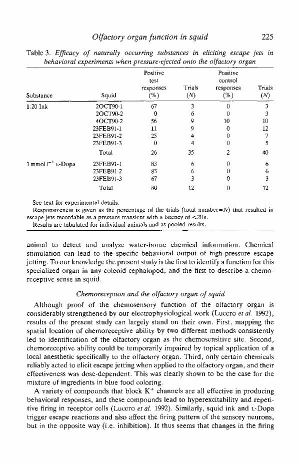
Positive behavioral results were obtained, however, with a simple preparationof diluted squid ink (see Materials and methods) and with l m m o l P 1 3,4dihydroxyphenylalanine (L-Dopa). L-Dopa is the precursor of melanin (Fox, 1976;Needham, 1974), the major pigment found in the ink sac of Loligo opalescens (Foxand Crane, 1942). Table 3 summarizes these results. Diluted ink was fairly variablein its effectiveness in different animals, and two of the six animals tested werecompletely unresponsive. L-Dopa was very effective in all three animals tested.
Squid thus appear to be capable of detecting the ink of members of their ownspecies as well as L-Dopa, a compound that is probably present in the ink sac(Jimbow et al. 1984). Squid react to these naturally occurring substances withescape responses that would serve to remove them from the source of stimulationin their environment.
Discussion
It has been recognized for over a century that the cephalopod olfactory organdisplays the anatomical attributes necessary for it to function in a chemosensorycapacity (Zernoff, 1869; Messenger, 1967). It is covered by a sensory epithelium,projects a large afferent nerve into the brain, and is situated directly in the streamof inhalant respiratory current. The main point of this paper is to provide evidencethat the olfactory organ in Loligo opalescens functions in a way that allows the
Olfactory organ function in squid 225
Table 3. Efficacy of naturally occurring substances in eliciting escape jets inbehavioral experiments when pressure-ejected onto the olfactory organ
Substance
1:20 Ink
lmmol l " ' L-Dopa
Squid
2OCT90-12OCT90-24OCT90-2
23FEB91-123FEB91-223FEB91-3
Total
23FEB91-123FEB91-223FEB91-3
Total
Positivetest
responses
(%)
670
561125
0
26
838367
80
Trials(TV)
369944
35
663
12
Positivecontrol
responses
(%)
00
10000
2
000
0
Trials(A/)
33
101275
40
663
12
See text for experimental details.Responsiveness is given as the percentage of the trials (total number=Ar) that resulted in
escape jets recordable as a pressure transient with a latency of <20s.Results are tabulated for individual animals and as pooled results.
animal to detect and analyze water-borne chemical information. Chemicalstimulation can lead to the specific behavioral output of high-pressure escapejetting. To our knowledge the present study is the first to identify a function for thisspecialized organ in any coleoid cephalopod, and the first to describe a chemo-receptive sense in squid.
Chemoreception and the olfactory organ of squid
Although proof of the chemosensory function of the olfactory organ isconsiderably strengthened by our electrophysiological work (Lucero etal. 1992),results of the present study can largely stand on their own. First, mapping thespatial location of chemoreceptive ability by two different methods consistentlyled to identification of the olfactory organ as the chemosensitive site. Second,chemoreceptive ability could be temporarily impaired by topical application of alocal anesthetic specifically to the olfactory organ. Third, only certain chemicalsreliably acted to elicit escape jetting when applied to the olfactory organ, and theireffectiveness was dose-dependent. This was clearly shown to be the case for themixture of ingredients in blue food coloring.
A variety of compounds that block K+ channels are all effective in producingbehavioral responses, and these compounds lead to hyperexcitability and repeti-tive firing in receptor cells (Lucero etal. 1992). Similarly, squid ink and L-Dopatrigger escape reactions and also affect the firing pattern of the sensory neurons,but in the opposite way (i.e. inhibition). It thus seems that changes in the firing

226 W. F. GILLY AND M. T. LUCERO
rate or pattern of the receptor cells are integrated in the central nervous system,and that these highly processed outputs ultimately influence the motor centers thatcontrol jetting. The long behavioral delays to chemical stimulation (2-20 s)evident in our studies are probably set by this processing, and in nature mightcorrespond to an important period of environmental assessment for the animal.
Tetrodotoxin, a specific Na+ channel blocker, was found to be ineffective atproducing escape responses in behavioral studies. This toxin blocks Na+ channelsin the receptor cells (Lucero et al. 1992), and it appears, therefore, that the densityof Na+ channels on the apical surface of receptor cells in the olfactory organ maybe low. Each receptor cell is surrounded by a desmosome-tight junction complex(Wildenburg and Fioroni, 1990) that presumably acts as a diffusional barrier andinterferes with solutes in sea water acting on the basolateral surface of the cell. Theobservation that quaternary ammonium ions produce escape responses whenapplied to the sea water bathing the apical surface of the receptor cells implies thatK+ channels susceptible to extracellular blockage by these ions are located on theapical surface.
One piece of evidence linking the olfactory organ and chemoreception that ismissing from the present paper concerns recording the sensory discharge in theolfactory nerve in response to chemical stimulants that elicit escape jetting in vivo.Such recordings were attempted with a dissected olfactory organ/nerve prep-aration using conventional suction and hook electrodes but were unsuccessful.Although electrical stimulation of the sensory nerve or olfactory knob producedcompound action potential waves that propagated slowly (<0.5ms~1; data notillustrated), the small size of the sensory axons and their extensive wrapping bySchwann cell and connective tissue elements (Fig. 2A) make single-unit record-ings very difficult. We were also unable to record a convincing summed sensorydischarge in response to chemical stimulation, probably because of the sametechnical problems. In the accompanying paper (Lucero et al. 1992), however, wedescribe successful intracellular recordings from isolated receptor cells that showappropriate receptor potentials and changes in firing pattern upon application ofthose chemicals that produce escape jets in living squid.
Biological relevance of the olfactory organ
The results described in this paper indicate that the squid olfactory organ canmediate detection of water-borne chemicals, but they also reveal that many agentstested, both potentially attractive (e.g. amino acids, egg jelly, extracts of gonads)and potentially repulsive (e.g. ammonium, nitrate, low pH), failed to producedefinite behavioral reactions. Many of these substances would be expected tostimulate other marine invertebrates, and their apparent failure to elicit responseswhen applied to the olfactory organ merits discussion.
Although negative results were obtained with many stimuli, our assay fordetection was specifically limited to escape jet production occurring within tens ofseconds following a stimulus. Attractive substances would not be expected to leadto escape jetting, and dramatic changes in ventilation rate which might indicate

Olfactory organ function in squid 227
'arousal' (Boyle, 1983) were not observed. Long-term effects on sexual maturationor encouragement of mating behavior would have been impossible to detect withour approach. Similarly, repulsive stimuli that failed to produce escape jetting didnot induce obvious changes in the respiratory rhythm, nor did they lead to unusualchromatophore diplays that might have indicated alarm (Long et al. 1989).
At present, we have no evidence whether substances that do not lead to escapejetting can be detected by receptors in the olfactory organ or whether theirdetection leads to other behavioral responses like those suggested above.Additional behavioral assays for detection of such substances would clearly bevaluable in further developing this work. Unfortunately, these assays might haveto be limited to restrained animals in a way similar to the approach described here.Our results on the detection of K+ channel blockers, squid ink and L-Dopa by theolfactory organ do not imply that receptors for these chemicals are restricted tothis structure. Responsive cells could also exist at other sites known to containchemosensory cells, e.g. around the lips or on the tentacles. Activation of theseadditional receptors could also lead to behaviors other than escape responses.Thus, application of stimuli to the sea water containing one or several free-swimming squid might be difficult to interpret.
Although the complete chemosensory competence of squid largely remains tobe explored and defined, this paper provides an indication of at least onebiologically important function mediated by the olfactory organ - the generationof escape responses. Our results lend credence to the idea, proposed originally byWatkinson (1909; cited by Messenger, 1967), that the olfactory organ serves tomonitor water quality, and that the sensory information passed to the brain leadsto avoidance or escape responses when the ambient water becomes tainted withnoxious substances. Whether the olfactory organ serves only this purpose remainsto be determined, but such a high degree of specialization would be surprising.
It is noteworthy that substances detected by the olfactory organ includebiologically relevant compounds that may be released by other squid. Squid ink,for example, ought to be avoided in the environment, because its presence in thewater would specifically indicate that another squid had been alarmed or attackedrecently and nearby. Although the color of ink could serve as a visual alarm signalunder conditions of sufficient illumination, chemical messengers such as L-Dopa,or an indole precursor to the melanin derived from it (Fox, 1976; Needham, 1974),would be more effective in the darkness of night or at great depths.
This work was funded by the US Office of Naval Research and the WhitehallFoundation. We acknowledge the technical assistance of Patricia Gosline andJenifer Levitt, who carried out and analyzed some of the behavioral experiments,and of Bruce Hopkins, who was responsible for the electron microscopy. We alsothank Dr George Mackie, for attempting recordings from the olfactory nerve, DrJanet Voight, for commenting on an earlier version of the manuscript, and DrRoger Hanlon, for comments on the final draft. We are grateful to Charles Baxterfor the loan of video equipment and valuable discussion.

228 W. F. GILLY AND M. T. LUCERO
ReferencesATEMA, J., FAY, R. R., POPPER, A. N. AND TAVOLGA, W. N. (eds) (1988). Sensory Biology of
Aquatic Animals. New York: Springer-Verlag. 936pp.BARBER, V. C. AND WRIGHT, D. E. (1969). The fine structure of the sense organs of the
cephalopod mollusc Nautilus. Z. Zellforsch. mikrosk. Anat. 102, 293-312.BOYLE, P. R. (1983). Ventilation rate and arousal in the octopus. J. exp. mar. Biol. Ecol. 69,
129-136.BOYLE, P. R. (1986). Neural control of cephalopod behavior. In The Mollusca, vol. 9, part 2 (ed.
A. O. D. Willows), pp. 1-99. New York: Academic Press.BUDELMANN, B. U. (1990). The statocysts of squid. In Squid as Experimental Animals (ed. D. L.
Gilbert, W. J. Adelman and J. M. Arnold), pp. 421-439. New York, London: Plenum Press.BUDELMANN, B. U. AND BLECKMANN, H. (1988). A lateral line analogue in cephalopods: water
waves generate microphonic potentials in the epidermal head lines of Sepia and Lolliguncula.J. comp. Physiol. A 164, 1-5.
CHASE, R. J. AND WELLS, M. J. (1986). Chemotactic behavior in Octopus. J. comp. Physiol. A158, 375-381.
EMERY, D. G. (1975). The histology and fine structure of the olfactory organ of the squidLolliguncula brevis. Tissue & Cell 7, 357-367.
Fox, D. L. (1976). Animal Biochromes and Structural Colors, pp. 215-240. Berkeley: Universityof California Press.
Fox, D. L. AND CRANE, S. C. (1942). Concerning the pigments of the two-spotted octopus andthe opalescent squid. Biol Bull. Mar. biol. Lab., Woods Hole 82, 284-291.
GOSLINE, J. M., STEEVES, J. D., HARMAN, A. D. AND DEMON, M. E. (1983). Patterns of circularand radial mantle muscle activity in respiration and jetting of the squid, Loligo opalescens.J. exp. Biol. 104, 97-109.
GRANT, P. T. AND MACKIE, A. M. (eds) (1974). Chemoreception in Marine Organisms. NewYork: Academic Press. 295pp.
GRAZIADEI, P. (1962). Receptors in the sucker of Octopus. Nature 203, 57-59.GRAZIADEI, P. (1965). Sensory receptor cells and related neurons in cephalopods. Cold Spring
Harb. Symp. quant. Biol. 30, 45-57.HANLON, R. T. AND BUDELMANN, B.-U. (1987). Why cephalopods are probably not 'deaf. Am.
Nat. 129, 312-317.HORRIGAN, F. T. (1990). Methadone block of neuronal K current. Biophys. J. 57, 515a.JlMBOW, K., MlYAKE, Y., HOMMA, K., YASUDA, K., 1ZUM1, Y., TSUTSUMI, A. AND ITO, S. (1984).
Characterization of melanogenesis and morphogenesis of melanosomes by physicochemicalproperties of melanin and melanosomes in malignant melanoma. Cancer Res. 44,1128-1134.
LAROE, E. T. (1971). The culture and maintenance of the loliginid squids Sepioteuthis sepioideaand Doryteuthis plei. Mar. Biol. 9, 9-25.
LONG, T. M., HANLON, R. T., MAAT, A. T. AND PINSKER, H. M. (1989). Non-associative
learning in the squid Lolliguncula brevis (Mollusca, Cephalopoda). Mar. Behav. Physiol. 16,1-9.
LUCERO, M. T., HORRIGAN, F. T. AND GILLY, W. F. (1992). Electrical responses to chemicalstimulation of squid olfactory receptor cells. J. exp. Biol. 162, 231-249.
MESSENGER, J. B. (1967). The peduncle lobe: a visuo-motor centre in Octopus. Proc. R. Soc.Lond. B 167, 225-251.
MESSENGER, J. B. (1979). The nervous system of Loligo. IV. The peduncle and olfactory lobes.Phil. Trans. R. Soc. Lond. B 285, 275-309.
MESSENGER, J. B. (1983). Multimodal convergence and the regulation of motor programs incephalopods. Fortschr. Zool. 28,11-91.
NEEDHAM, A. E. (1974). The Significance of Zoochromes. Zoophysiology and Ecology, vol. 3.429pp. New York: Springer-Verlag.
OTIS, T. AND GILLY, W. F. (1990). Jet-propelled escape in the squid Loligo opalescens:concerted control by giant and non-giant motor axon pathways. Proc. natn. Acad. Sci. U.S.A.87, 2911-2915.
PACKARD, A. (1972). Cephalopods and fish: the limits of convergence. Biol. Rev. 47, 241-307.

Olfactory organ function in squid 229
WATKINSON, G. B. (1909). Untersuchungen uber die Sog. Geruchsorgane der Cephalopoden.Jena Z. Nautrw. 44, 353-414.
WELLS, M. J. (1963). Taste by touch: some experiments with Octopus. J. exp. Biol. 40,187-193.WELLS, M. J. (1964). Tactile discrimination of surface curvature and shape by octopuses. /. exp.
Biol. 41,435-445.WELLS, M. J., FREEMAN AND ASHBURNER (1985). Some experiments on the chemotactile sense of
octopuses. J. exp. Biol. 43, 553-563.WELLS, M. J. AND YOUNG, J. Z. (1975). The subfrontal lobe and touch learning in the octopus.
Brain Res. 92, 103-121.WILDENBURG, G. AND FIORONI, P. (1990). Ultrastructure of the olfactory organ during
embryonic development and at the hatching stage of Loligo vulgaris Lam. (Cephalopoda).J. Ceph. Biol. 1, 56-70.
WILSON, D. M. (1960). Nervous control of movement in cephalopods. J. exp. Biol. 37, 57-72.WOODHAMS, P. L. AND MESSENGER, J. B. (1974). A note on the ultrastructure of the Octopus
olfactory organ. Cell Tissue Res. 152, 253-258.YOUNG, J. Z. (1938). The functioning of the giant nerve fibres of the squid. J. exp. Biol. 15,
170-185.YOUNG, J. Z. (1939). Fused neurons and synaptic contacts in the giant nerve fibres of
cephalopods. Phil. Trans. R. Soc. Lond. B 229, 465-503.ZERNOFF, D. (1869). Uber das Geruschsorgane der Cephalopoden. Bull. Soc. Imp. Nat.
Moscow 42, 71-90.
