behrmann (2003) hemispatial neglect and visual search. …wexler.free.fr/library/files/behrmann...
TRANSCRIPT

HEMISPATIAL NEGLECT AND VISUAL SEARCH: A LARGE SCALE ANALYSIS
Marlene Behrmann1, Patricia Ebert2,3 and Sandra E. Black2,3
(1Dept. of Psychology, Carnegie Mellon University, Pittsburgh, PA, USA; 2CognitiveNeurology Unit, Department of Medicine and the Ontario Heart and Stroke FoundationCentre for Stroke Recovery, Sunnybrook and Women’s College Health Sciences Centre,
University of Toronto; 3Institute of Medical Sciences, University of Toronto, Toronto, ON,Canada)
ABSTRACT
Visual search tasks have standardly been divided into two categories: those in whichthe target is detected through a serial, attention-driven search and those in which the targetis detected rapidly in parallel and, apparently, without attentional processing. Several studieshave examined this distinction in patients with hemispatial neglect with the clear predictionthat the former, but not the latter, should be impaired. These studies, however, have provedinconclusive. We have addressed this issue in a large sample of patients with unilateralhemispheric infarcts to the left or right hemisphere. In addition to measuring the patients1performance on both types of visual search tasks, we documented the presence and severityof neglect and of visual field defects in these same individuals. Patients with brain-damagewith or without accompanying neglect were impaired at searching for the contralateraltarget on both forms of visual search, relative to normal control subjects, although thisdeficit was magnified in individuals with neglect and was also exacerbated by the presenceof hemianopia. This pattern was also more pronounced in individuals with right- than withleft-hemisphere lesions. The findings not only clarify the contradictory neuropsychologicaldata but also provide clear evidence for the involvement of attentional processing in allforms of visual search.
Key words: hemispatial neglect, attentional deficit, visual search
SPECIAL NOTE
The authors pay special tribute to Dr Eduardo Bisiach who has been one of the primaryresearchers in the study of hemispatial neglect and whose seminal contribution is widelyrecognized. Two of the three authors of this paper (MB and SEB) met Dr Bisiach for thefirst time in 1986 when he visited the University of Toronto. His visit had a significantimpact on us and our future work. In addition, over the years, Dr Bisiach has commentedon some of our papers and has been very helpful on several other occasions. His input andcollegiality are much appreciated.
INTRODUCTION
Visual search paradigms, in which individuals search for a pre-defined targetin a display containing multiple items, have been used extensively over the lastdecade or so in an attempt to characterize the neurobehavioral disorder termed“hemispatial neglect” (or “neglect” for short). Neglect is a disorder in whichindividuals, following an acquired brain lesion, fail to notice or report
Cortex, (2003) 39, 000-000

information on the side of space opposite the lesion, despite intact sensory andmotor processes (Bisiach and Vallar, 2000; Bartolomeo and Chokron, 2001).Thus, for example, patients with a right hemisphere lesion fail to copy featureson the right of a display while incorporating the corresponding features on theipsilesional left. The same individual may eat from only the right side of theirplate or dress only the right side of their body. The deficit may affect all sensorymodalities, including contralateral visual, auditory, somatosensory and olfactoryinputs. The presence of neglect may also adversely affect manual andoculomotor behavior in that these patients are often impaired at directing theireyes and/or hand to the contralateral side, even in the absence of visual input(Behrmann et al., 2001; Gore et al., 2001/2002; Hornak, 1992; Mattingley et al.,1998). Finally, neglect can affect the contralateral side of an internalrepresentation in the absence of sensory input, and can be reflected in mentalimagery, as so elegantly demonstrated in the seminal work by Bisiach andLuzzatti (1978).
The deficit that gives rise to hemispatial neglect is often attributed to thefailure to construct an appropriate representation of space as a consequence ofan attentional bias, which favors the processing of ipsilesional stimuli.Interestingly, patients with neglect may orient to highly salient contralesionalstimuli but, left to their own devices, do not volitionally direct their attention tothat side of space (Làdavas et al., 1994). Given that visual search tasks havebeen used extensively over the last several decades to examine patterns of visualattention in normal subjects (Bricolo et al., 2002; Neisser, 1964; Treisman andGelade, 1980; Wolfe, 1998), the use of such measures may be particularly usefulin elucidating the nature of the attentional biases in patients with hemispatialneglect. Despite the robustness of this experimental approach in normal subjects,the findings from visual search studies with neglect patients to date remaincontroversial. We start by describing briefly the paradigms employed in visualsearch studies with normal subjects, pointing out the central assumptions andmajor results. We then review the existing data obtained in individuals withhemispatial neglect. Following this, we report the findings we have obtainedusing a well-established visual search paradigm in a very large group of patients,who have sustained a unilateral hemispheric stroke to either the left or righthemisphere, and we indicate ways in which these data can shed light on themechanisms giving rise to the neglect deficit.
Visual Search as an Experimental Paradigm in Normal Subjects
Visual search studies are well-suited as a proxy for real-world attentionalrequirements as features of the natural environment such as object clutter arecaptured while a controlled stimulus environment is maintained. A particularlyprolific subset of these studies focuses on the conditions under which thereaction time (RT) and accuracy to locate the target is affected by the number ofdistractors appearing in the display (Geng and Behrmann, 2002b; Behrmann andHaimson, 1999; Treisman, 1999; Yantis, 2000). Cases in which the time todetect a target is largely unaffected by increasing the number of distractors (e.g.,5 msec/distractor item) are labeled as “feature search” or “disjunctive”, whereas
2 Marlene Behrmann and Others

cases in which detection time is significantly slowed by the increasing numberof distractors (e.g., 50msec/item) are labeled “conjunctive”. These differentsearch functions have also been referred to as “parallel” vs. “serial” or “simple”vs. “difficult”. The critical distinction is that visual search for targetsdistinguished by a single feature is scarcely affected by the number of distractorspresent whereas targets distinguished by feature conjunctions appear to beaffected linearly by the number of distractors present. The interpretation of thisdistinction is that feature search can be executed effortlessly and preattentively(without attention); because search can be conducted in parallel across the entiredisplay and the target “pops out” in this form of search, there is no increase intarget detection time with increasing number of items in the display. In contrast,in the conjunctive search task, each item must be sequentially examined todetermine whether it is a target. This process requires the allocation of attention,and the serial search results in the monotonic increase in detection time as afunction of display size (see, for example, Bricolo et al., 2002). The effect ofdisplay size is a critical indicator and is taken to be the primary assay for theinvolvement of attention (Bundesen, 1990; Duncan and Humphreys, 1989).
Visual Search Tasks in Patients with Neglect
The assumptions derived from the visual search studies with normal subjectslead to a number of critical predictions with regard to neglect. If unilateralneglect does arise from a deficit of attention, then, in the feature search(preattentive) task, performance in individuals with hemispatial neglect shouldnot differ from that of normal individuals and should be unaffected by the sizeof the display. In addition, feature search should be identical for targets on thecontralateral and ipsilateral sides. In contrast, performance should be impaired,relative to normal controls, for conjunction search when the target appears onthe contralateral side and this should be exaggerated as the display sizeincreases. Whether search for an ipsilateral target should be normal is notentirely clear but patients should be differentially impaired for contralateralversus ipsilateral targets in conjunction search. Unfortunately, despite theabundance of studies, there is no clear consensus on the visual searchperformance of individuals with neglect, as will be apparent from the review ofthe literature below, and many questions remain unanswered. In addition to thislack of agreement, there are a number of other outstanding and controversialissues which affect the existing findings and we return to these after we havelaid out the major studies and their results.
In one of the earliest studies examining visual search with neglect patients,Riddoch and Humphreys (1987) presented a series of cards with displays tothree patients with left-sided neglect. The patients were required to search for atarget, which was present on half the trials, and accuracy and reaction time (RT)were recorded. In the feature search task, the display contained a red circleamong green circle distractors whereas, in the conjunction search task, thedisplay contained an inverted “T” among upright “T” distractors. In the featuresearch task, RT was unaffected by the number of distractors even when thetarget appeared on the contralateral side, consistent with parallel search. Note,
Visual search in hemispatial neglect 3

however, that, even in this condition, there was a high error rate for contralateraltargets, suggesting that feature search was not totally intact in these patients. Asexpected, detection was poor both in accuracy and RT for targets on thecontralateral side in the conjunction search task.
A subsequent study by Eglin and colleagues (Eglin et al., 1989), using a reddot among blue and yellow dots (feature search) or a red dot among split blueand intact red dots (conjunction search) and varying array size, distractornumber, and location of stimuli, confirmed the impairment in contralateralfeature search in six patients with right hemisphere damage (RHD) and in onepatient with left hemisphere damage (LHD). In contrast with the control subjectswho showed only a linear slope in the conjunctive search task, there was asignificant increase in the time taken to detect contralateral targets for neglectpatients in both the feature and conjunction search task. Consistent with this isthe finding from a related study by the same authors in which patients wererequired to point to a target (Eglin et al., 1994; Eglin et al., 1991). Here, asbefore, search rates in patients were also slower than those of controlparticipants for feature as well as conjunction search (for other consistentconfirmatory evidence, see (Rapcsak et al., 1989)).
Finally, in a recent study, Pavlovskaya et al. (2002) compared theperformance of four RHD and one LHD patients with neglect, sustainedfollowing rather extensive cortical damage, and six healthy control subjects on atask involving search for an oriented line element. In the feature search, thetarget was an oblique line embedded among vertical lines and in the conjunctionsearch the target was an oblique yellow line embedded among blue lines of ashared orientation and yellow lines of a differing orientation. Consistent with thedata reviewed above, all patients were impaired in both the feature andconjunction versions of these tasks and their performance deteriorated as thetarget appeared further contralaterally.
In direct contrast to the studies described above, however, several otherstudies have argued for preservation of feature search in neglect patients. Forexample, three patients with neglect and cortical lesions tested by Esterman andcolleagues (Esterman et al., 2000) revealed normal preattentive search. A fourthpatient with neglect following a subcortical lesion did not show normal featuresearch and exhibited an effect of array size on search time. Note, however, thattwo additional patients with neglect and hemianopia also showed impairedcontralateral feature search. All patients were impaired on the conjunctive searchtask with contralesional targets, leading the authors to conclude that only serial,effortful search is affected in hemispatial neglect but that the ability to extractlow-level featural information across the field in parallel is preserved.
Consistent with the Esterman et al. study, Aglioti and his colleagues (Agliotiet al., 1997) examined the search performance of a very large group ofindividuals, consisting of 75 participants with left hemisphere damage (LHD) orright hemisphere damage (RHD). Both groups included individuals with andwithout neglect. Subjects performed a task using two different visual textures inwhich, in one case, the target was easily segregated and detected and, in theother case, was difficult to detect. The critical finding was that contralateralerrors were disproportionately higher on the latter task as opposed to the former,
4 Marlene Behrmann and Others
indicating that neglect only impaired the more effortful search performance. It isof note here that because the number of items was not manipulated in thesedisplays, it is difficult to know whether these tasks map directly onto thepreattentive versus attentive distinction made previously.
In a similar vein, Arguin et al. (1993) investigated eight left hemisphere-damaged (LHD) participants both with and without visual attention deficits onfeature (orientation or colour as the distinctive feature) and conjunction searchtasks (orientation and colour conjoined). The patients with visual attentiondeficits performed similarly to controls in contralateral hemispace on the featuresearch task, but had longer reaction times for contralateral targets on theconjunction task. The authors concluded from this finding that feature searchperformance was preserved in participants with visual attention impairments.
The preservation of feature search performance in neglect patients is alsoconsistent with findings using experimental paradigms that do not necessarilyrequire visual search. For example, several studies have reported that patientswith neglect are still able to extract low-level information and derive primitiveshape descriptions from information appearing on the contralateral side. Forexample, when the contralesional item of a display could be grouped with theipsilesional information on the basis of Gestalt factors such as similarity (Ward etal., 1994), symmetry (Driver et al., 1992), colour and proximity (Driver andHalligan, 1991), or brightness or collinear edges (Gilchrist et al., 1996; Rorden etal., 1997), report of the left-sided stimulus was better than when the left sidedinformation could not be grouped with a simultaneously-presented right sidedstimulus. This was also the case when the left-sided information could begrouped with the right-sided information by Œgoodness1 of an object such as aglobal outline (Farah et al., 1993), illusory contour (Kanizsa-type figure)(Mattingley et al., 1997) or of any well-configured object or whole (Boutsen andHumphreys, 2000). The benefit attributed to the contralesional information underthese conditions is thought to arise from the fact that low-level visual informationcan be extracted preattentively and this enables the grouping of the contralateraland ipsilateral information. It has also been suggested that the extraction ofpreattentive contralateral information may suffice for deriving detailedinformation to allow access to lexical and semantic processing (Esterman et al.,2000; Kumada and Humphreys, 2001; Humphreys, 2003; but see Behrmann etal., 1990 for an alternative explanation of how these effects might arise).
Finally, the preservation of feature search is consistent with the findings of arecent study using evoked response potential (ERP) and functional magneticresonance imaging (fMRI) in patients with neglect and extinction. Note that invisual search tasks, aside from trials with a single item, there are alwaysmultiple items in a display, and, as such, this resembles double simultaneousstimulation trials on which extinction is elicited. In this study, Vuilleumier et al.(2000), using combined ERP and event-related fMRI, showed that even stimulithat were not explicitly reported (i.e., suffered extinction) gave rise to activationin right V1 and inferior temporal cortex and elicited a nonsignificantly reducedN1 evoked potential. These findings suggest that visual information may beprocessed by posterior and early parts of the visual system and that this mightcorrespond to preattentive processing. However, in the absence of coupling with
Visual search in hemispatial neglect 5

dorsal frontal and parietal areas (perhaps mediating attentional processing),conscious awareness is precluded and this may be consistent with attentiveprocessing (but see Marzi et al., 2000, for a different result).
As is evident from this overview, there are clearly a number of discrepantfindings especially with regard to the preservation of contralateral feature searchin neglect and the extent to which contralateral information can be processed bythese patients. This lack of agreement may arise for several different reasons.One obvious possibility is that the methods adopted in the different studies varyquite substantially, including the number of subjects tested (with very smallnumbers in some cases), the nature of the search task (colour discrimination,letter detection, pointing or cancellation), and the reliance on a single or onmultiple dependent measures (accuracy and/or RT). Of course, the qualitativeand quantitative differences between the different subject samples can alsocontribute to the different outcomes; this heterogeneity in lesion size and site isexacerbated when the subject sample is small and, indeed, there is well-documented variability across patients.
In addition to these obvious reasons, a number of other factors couldpotentially complicate the results and these confounding factors are notnecessarily addressed or controlled in the various studies. Firstly, we do notknow whether any apparent deficit that is observed in the neglect patients is afunction of a hemispheric lesion per se or whether the deficit is solely aconsequence of hemispatial neglect. Because many studies compare theperformance of the neglect patients to the performance of a group of normal,healthy control subjects and do not include a group of brain-damaged individualswithout neglect, it is not possible to know whether the deficit is attributable toneglect per se or to brain damage more generally. Secondly, related to this, wedo not know whether the apparent deficit is correlated with the severity ofneglect, as one might predict if the deficit is truly attentional in nature. Becausethe number of subjects is small in some studies, or in those studies in whichthere are a large number of subjects, patients are simply assigned to apresence/absence of neglect dichotomy, it has not been possible to examine thecorrelation between severity of neglect and visual search behavior in detail.Thirdly, we do not know to what extent the presence of a visual field defectaffects performance. This has recently become a rather substantial issue inunderstanding hemispatial neglect; whereas Doricchi and Angelelli (1999) andToth and Kirk (2002) have shown that neglect patients with hemianopias makegreater ipsilesional bisection errors in line bisection tasks, Ferber and Karnath(1999) have found otherwise. To the extent that the presence of a field defecthas been taken into account in studies of visual search, the results have provencontradictory. As mentioned above, Esterman et al. (2000) find that only patientswith hemispatial neglect accompanied by a field defect are impaired at featuresearch whereas Aglioti et al. (1997) report no difference as a function of thepresence/absence of field defects in reaction time (RT). Note that Aglioti et al.(1997) do find an increased number of errors but this is so in all individualswith field defects and is not restricted to those patients with neglect.
The final issue concerns differences between individuals with left hemispherelesions and those with right hemisphere lesions. Neglect is notoriously associated
6 Marlene Behrmann and Others

with RHD more often than with LHD, although the extent of this relationship isalso somewhat controversial (Ogden, 1987). Studies using transcranial magneticstimulation and functional magnetic resonance imaging have pointed todifferences between the two hemispheres in their relative involvement inattentional processing (Ashbridge et al., 1997; Corbetta et al., 1995; Corbetta etal., 1998) and suggest greater involvement of the right hemisphere in attentionaltasks such as conjunction search. To the extent that this issue has beenconsidered, the data remain contradictory. Gainotti et al. (1986), for example,have argued for no difference between left and right brain damaged patients invisual search tasks whereas others (for example, Halligan et al., 1992; Weintrauband Mesulam, 1987) do find differences between these groups. Because thenumber of subjects in these studies is typically small, and because patients withLHD with neglect tend to have milder forms of neglect than their RHDcounterparts, a clear comparison between hemispheric groups with severity ofneglect and presence of hemianopia equated, is very difficult. It remains to bedetermined, therefore, whether there are hemispheric differences in visual searchin patients with neglect when these other factors are taken into account.
In light of the controversial findings and the many remaining outstandingquestions, we have undertaken a study of the visual search performance of alarge group of patients, consecutively admitted to a university teaching hospitalstroke care unit, who suffered a stroke to either the left or right hemisphere. Wehave included not only individuals with no neurological deficits to serve ascontrols but also individuals who have suffered a hemispheric lesion but who donot exhibit neglect to serve as an additional control group. In addition tocompleting a bedside battery used to diagnose neglect and to document itsseverity, we had subjects complete a computerized version of feature andconjunction search performance for targets presented to the contralateral oripsilateral side and we measured accuracy and RT. Subjects also underwentclinical visual field testing, and the extent of a visual field defect wasdocumented. Finally, because of the large patient sample, we have been able tocompare RHD and LHD with neglect where the severity of the neglect (andpresence of hemianopia) is equated in the two groups. With this data set, we willfirst attempt to replicate the finding of impaired contralateral search in patientswith neglect. We will then examine detection of ipsilateral targets in conjunctionsearch. But perhaps most relevant is that we will determine whether featuresearch is normal in individuals with neglect. Lastly, we will explore whether thevisual search performance of the patients is influenced by the presence and/orseverity of neglect, the side of the lesion and/or the presence of hemianopia.
MATERIALS AND METHODS
Subjects
Individuals with and without brain damage consented to participate in thisstudy. The non-brain-damaged control group consisted of volunteers, age- andeducation-matched to the patients, living independently in the community served
Visual search in hemispatial neglect 7
by Sunnybrook and Women1s College Health Sciences Centre. These controlsubjects were screened for neurological and serious medical illness and wereexcluded if such diseases were present.
The brain-damaged group was recruited from a consecutive series of patientswith stroke admitted to the Sunnybrook and Women1s Stroke Care Unit. Allbrain-damaged participants were right-handed with corrected visual acuity of atleast 20/40. All patients met the following criteria: age between 20-85 years,clinical and radiological evidence of a single, unilateral lesion, no otherneurological/mental illness (i.e., dementia, epilepsy, previous stroke), no otherserious concomitant illness (e.g., cancer), and no history of substance abuse. Allstroke patients were tested within three months of stroke onset. They wererecruited as soon after the stroke as they were able to sit up and undergocomputerized testing. Those who could not understand test directions because ofsevere aphasia were excluded (n = 4). Although an attempt was made to performthe computerized visual search task and neglect testing on the same day, thiswas not always possible for logistical reasons. Patients tested within the first 2weeks post-onset were allowed a maximum of 4 days between differentcomponents of the testing since neglect can improve substantially in the first 2weeks after a stroke. If the patient was unable to be tested in the first twoweeks, an interval of 9 days was allowed. If performance on the neglect batterywas within normal limits, a longer testing interval was allowed if the patientremained neurologically stable, since it was unlikely that performance wouldchange in that interval.
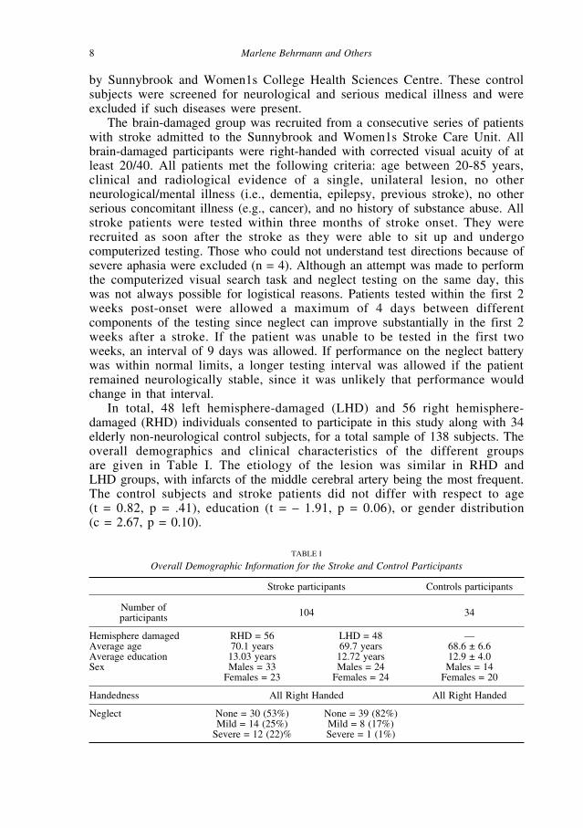
In total, 48 left hemisphere-damaged (LHD) and 56 right hemisphere-damaged (RHD) individuals consented to participate in this study along with 34elderly non-neurological control subjects, for a total sample of 138 subjects. Theoverall demographics and clinical characteristics of the different groups are given in Table I. The etiology of the lesion was similar in RHD and LHD groups, with infarcts of the middle cerebral artery being the most frequent.The control subjects and stroke patients did not differ with respect to age (t = 0.82, p = .41), education (t = – 1.91, p = 0.06), or gender distribution (c = 2.67, p = 0.10).
8 Marlene Behrmann and Others
TABLE I
Overall Demographic Information for the Stroke and Control Participants
Stroke participants Controls participants
Number of 104 34participants
Hemisphere damaged RHD = 56 LHD = 48 —Average age 70.1 years 69.7 years 68.6 ± 6.6Average education 13.03 years 12.72 years 12.9 ± 4.0Sex Males = 33 Males = 24 Males = 14
Females = 23 Females = 24 Females = 20
Handedness All Right Handed All Right Handed
Neglect None = 30 (53%) None = 39 (82%) Mild = 14 (25%) Mild = 8 (17%)
Severe = 12 (22)% Severe = 1 (1%)
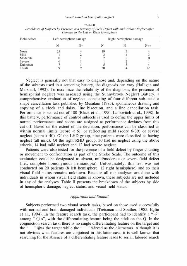
Neglect is generally not that easy to diagnose and, depending on the natureof the subtests used in a screening battery, the diagnosis can vary (Halligan andMarshall, 1992). To maximize the reliability of the diagnosis, the presence ofhemispatial neglect was assessed using the Sunnybrook Neglect Battery, acomprehensive evaluation of neglect, consisting of four different sub-tests: ashape cancellation task published by Mesulam (1985), spontaneous drawing andcopying of a clock and daisy, line bisection, and a line cancellation task.Performance is scored out of 100 (Black et al., 1990; Leibovitch et al., 1998). Inthis battery, performance of control subjects is used to define the upper limits ofnormal performance, and scores are assigned as performance deviates from thiscut-off. Based on the extent of the deviation, performance can be classified aswithin normal limits (score < 6), or reflecting mild (score 6-39) or severeneglect (score > 40). Of the LHD group, nine patients were classified as havingneglect (all mild). Of the right RHD group, 30 had no neglect using the abovecriteria, 14 had mild neglect and 12 had severe neglect.
Patients were also tested for the presence of a field defect by finger countingor movement to confrontation as part of the Stroke Scale. The outcome of thisevaluation could be designated as absent, mild/moderate or severe field defect(i.e., complete homonymous hemianopia). Unfortunately, this test was notconducted on 20 patients (8 left hemisphere, 12 right hemisphere) and so theirvisual field status remains unknown. Because all our analyses are done withindividuals in whom visual field status is known, these subjects are not includedin any of the analyses. Table II presents the breakdown of the subjects by sideof hemispheric damage, neglect status, and visual field status.
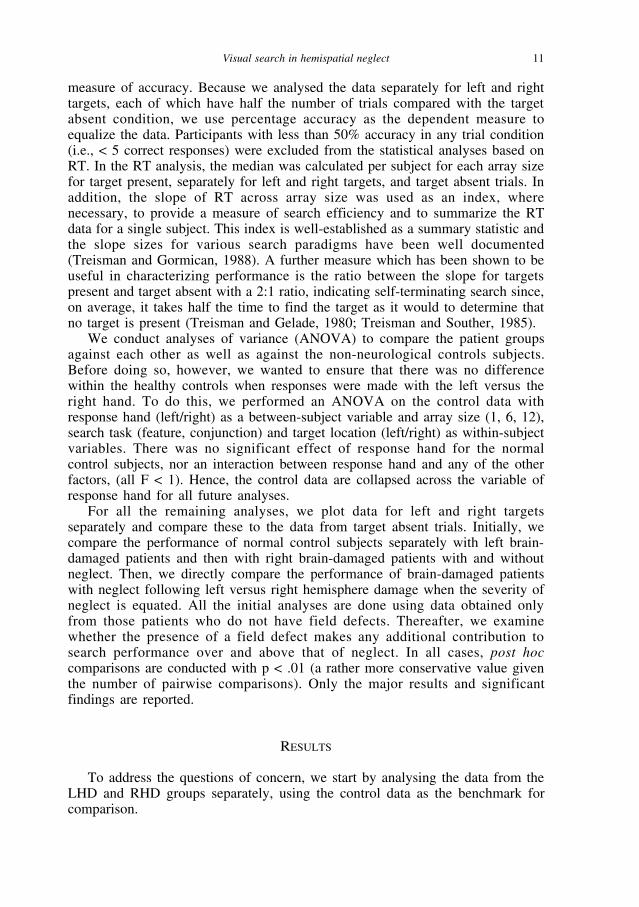
Apparatus and Stimuli
Subjects performed two visual search tasks, based on those used successfullywith normal and brain-damaged individuals (Treisman and Souther, 1985; Eglinet al., 1994). In the feature search task, the participant had to identify a “ ”among “ s”, with the differentiating feature being the stick on the Q. In theconjunction search task, there is no single differentiating feature on the target andthe “ ” was the target while the “ ” served as the distractors. Although it isnot obvious what features are conjoined in this latter case, it is well known thatsearching for the absence of a differentiating feature leads to serial, labored search
Visual search in hemispatial neglect 9
TABLE II
Breakdown of Subjects by Presence and Severity of Field Defect with and without Neglect afterDamage to the Left or Right Hemisphere
Field defect Left hemisphere damage Right hemisphere damage
N– N+ N– N– N++
None 25 4 19 3 4Mild 1 2 3 4 3Moderate 0 1 2 2 0Severe 6 1 1 2 1Unknown 7 1 5 3 4Totals 39 9 30 14 12

in the same way that conjoining two features does (Treisman and Gelade, 1980),and hence we refer to this task as conjunction search to remain consistent with theliterature. In half of the trials, the target was present and, in the other half, it wasabsent. The array size varied from 1, 6, and 12 items with an equal number oftrials for each array size. The position of the target, when present, varied across12 different points (6 left-sided and 6 right-sided) on the screen with an equalsampling of all these positions. The target was never located directly along thevertical midline of the array. There were a total of 120 trials in each search task.
The tasks were administered using a Mac Plus computer and PsychLabSoftware (Bub and Gum, 1991). Subjects responded using a button box, placedalong the midsagittal plane. To avoid the complications of stimulus-responsecompatibility, subjects used the top and bottom buttons, with the upper buttonindicating target present and the lower indicating target absent. Stroke patientsused their ipsilesional hand to perform the task. To account for the fact that halfthe patients used their left hand and the other half used their right, handednesswas manipulated in the control group so that half of the controls responded withtheir right hand and the other half responded with their left hand. Each trial waspreceded by a fixation point (a large dot), which was presented in the center ofthe screen 150 msec before each trial. Immediately thereafter, the array appearedand remained on the screen until the subject responded. Before each test session,there was a 9 trial practice session, which could be repeated once, if necessary.If a subject was unable to perform the task after 2 practice blocks, s/he wasexcluded from the study.
Data Analysis
Two dependent variables were used in the analyses: accuracy and medianreaction time (RT). The number of correctly identified targets provided the
10 Marlene Behrmann and Others
Fig. 1 – The pop-out and serial visual search tasks. The search tasks are similar to thosepublished by (Treisman and Souther, 1985). In the pop-out task, the target is defined by the presenceof a vertical line on the circle. In the serial search task, the target lacked the vertical line present onthe distractors.
measure of accuracy. Because we analysed the data separately for left and righttargets, each of which have half the number of trials compared with the targetabsent condition, we use percentage accuracy as the dependent measure toequalize the data. Participants with less than 50% accuracy in any trial condition(i.e., < 5 correct responses) were excluded from the statistical analyses based onRT. In the RT analysis, the median was calculated per subject for each array sizefor target present, separately for left and right targets, and target absent trials. Inaddition, the slope of RT across array size was used as an index, wherenecessary, to provide a measure of search efficiency and to summarize the RTdata for a single subject. This index is well-established as a summary statistic andthe slope sizes for various search paradigms have been well documented(Treisman and Gormican, 1988). A further measure which has been shown to beuseful in characterizing performance is the ratio between the slope for targetspresent and target absent with a 2:1 ratio, indicating self-terminating search since,on average, it takes half the time to find the target as it would to determine thatno target is present (Treisman and Gelade, 1980; Treisman and Souther, 1985).
We conduct analyses of variance (ANOVA) to compare the patient groupsagainst each other as well as against the non-neurological controls subjects.Before doing so, however, we wanted to ensure that there was no differencewithin the healthy controls when responses were made with the left versus theright hand. To do this, we performed an ANOVA on the control data withresponse hand (left/right) as a between-subject variable and array size (1, 6, 12),search task (feature, conjunction) and target location (left/right) as within-subjectvariables. There was no significant effect of response hand for the normalcontrol subjects, nor an interaction between response hand and any of the otherfactors, (all F < 1). Hence, the control data are collapsed across the variable ofresponse hand for all future analyses.
For all the remaining analyses, we plot data for left and right targetsseparately and compare these to the data from target absent trials. Initially, wecompare the performance of normal control subjects separately with left brain-damaged patients and then with right brain-damaged patients with and withoutneglect. Then, we directly compare the performance of brain-damaged patientswith neglect following left versus right hemisphere damage when the severity ofneglect is equated. All the initial analyses are done using data obtained onlyfrom those patients who do not have field defects. Thereafter, we examinewhether the presence of a field defect makes any additional contribution tosearch performance over and above that of neglect. In all cases, post hoccomparisons are conducted with p < .01 (a rather more conservative value giventhe number of pairwise comparisons). Only the major results and significantfindings are reported.
RESULTS
To address the questions of concern, we start by analysing the data from theLHD and RHD groups separately, using the control data as the benchmark forcomparison.
Visual search in hemispatial neglect 11

Left Hemisphere Damage
To examine whether there is a deficit in visual search in patients with LHDand whether this differs depending on the presence of neglect, we separated thebrain-damaged patients without field defects into those with neglect (LHD N+;N = 4) and those without neglect (LHD N–; N = 25). In addition to thisbetween-subjects variable, we included three within-subjects variables: searchtype (feature, conjunction), side of target (left, right, target absent) and displaysize (1, 6, 12). In the RT analysis, all main effects and interactions weresignificant. Many of these effects are also present in the analyses of accuracy.Because detection time is the metric used most often for visual search (andindeed, in our paradigm, accuracy is a rather limited dependent measure giventhe relatively restricted number of trials per cell and the unlimited exposureduration of a trial), we focus more specifically on RT and only make some briefstatements about accuracy of performance.
The RT data are plotted separately for feature (left hand panels) andconjunction (right hand panels) search and for each of the three subgroups inFigure 2. Note that we maintain the same y-axis for all subgroups in this figureand we use the same axis in subsequent figures for ease of comparison. Thenormal control subjects detect targets significantly faster than either the LHD N– or LHD N+ group by about 400-500 ms, F (2, 60) = 17.8, p < .001, whereasthe latter two subgroups do not differ from one another in overall RT. Feature search is 319 ms faster than conjunction search, F (1, 60) = 185.4, p < .001, although this difference varies across the three subgroups, F (2, 60) =12.2, p < .0001 (222 for controls, 382 for LHD N–, 581 for LHD N+). RTincreases as a function of display size, F (2, 120) = 35.9, p < .0001, and this toovaries across subgroups, F (4, 120) = 8.2, p < .0001, with slopes of 22.6, 35.8 and 42.4 for the controls, LHD N– and LHD N+ subgroups, respectively.The increase in RT with display size is greater for conjunction than featuresearch, as expected, F (2, 120) = 46, p < .0001. This too is qualified by aninteraction with subgroup, F (4, 120) = 4.2, p < .0001; the slopes for featuresearch were 6.9 ms, 8.1 ms and 12.4 ms for the controls, LHD N– and LHD N+subgroups whereas those for conjunction search were 38.2 ms , 71.8 ms and64.3 ms respectively.
Of particular interest, however, is whether search differs as a function of theside of the target: search for left targets is 76 ms faster than for right targets andtarget absent trials are slowest, with an increment of 283 ms over right trials, F (2, 120) = 81.4, p < .0001. There is an interaction of side of target with searchtype, F (2, 120) = 103.1, p < .0001, and with display size, F (4, 240) = 17.5, p < .0001, and a three-way interaction of search type × display size × side, F (4, 240) = 27.8, p < .0001. When we examine how these factors affect thedifferent subgroups, we observe a two-way interaction of side of target ×subgroup, F (4, 120) = 6.3, p < .0001, but this is qualified in a three-wayinteraction with search type, F (4, 120) = 6.5, p < .001. The three-wayinteraction reflects the finding that there is an asymmetry in search for leftversus right targets (slower on right) in the patients but not the control subjects,which is exaggerated in conjunction over feature search. This asymmetry is a
12 Marlene Behrmann and Others

Visual search in hemispatial neglect 13
Fig. 2 – Mean of median reaction time for (A) non-neurological control subjects, (B) patientswith LHD but no neglect and (C) patients with LHD and accompanying neglect on feature andconjunction search as a function of display size and presence (left/right) or absence of target.

little more evident in the LHD N+ than LHD N– subgroup, who do not show anasymmetry in the feature search task.
There is also a three-way interaction of display size × side of target ×subgroup, F (8, 240) = 2.8, p < .01. This three-way interaction can beinterpreted using the data from Table III, which presents the slopes (in ms)across the display sizes, reflecting the increment in RT per item for targets onthe left and right and for target absent trials. The essential finding is that,relative to the control subjects, both brain-damaged groups show steeper slopesin visual search for contralateral targets. Although there is no statisticallysignificant difference between the two patient groups on this measure, the LHDN+ patients show numerically faster search for ipsilateral targets than do theLHD N–, who do not differ from the control subjects on this measure. Thisrelative facilitation for ipsilateral targets, primarily evident in the conjunctionsearch, and the slowing for contralateral targets is well-documented in theneglect literature (see below for further illustration of this pattern) (Behrmann etal., 1998; Cate and Behrmann, 2002; Làdavas et al., 1990) and is often attributedto competitive effects between more ipsilateral versus contralateral stimuli.Importantly and crucially, the four-way interaction of all the variables is notsignificant, F (8, 240) = 1.6, p > .05, suggesting that the impairment for searchis equivalent across feature and conjunction search. These findings support theidea that the patients, particularly those with neglect, perform more poorly thanthe control subjects in conjunction search, as expected, but also in feature searchfor contralateral targets.
The accuracy data are largely compatible with the RT data. As in the RT,there is no significant four-way interaction but there is a three-way interaction ofsearch type × display size × subgroup, F (4, 120) = 5.4, p < .001, reflecting theincrease in error rate in both subgroups, again slightly greater in LHD N+ thanLHD N–, in conjunction over feature search with increasing display size. Thereis no additional effect of side of target and so the asymmetry revealed in RT isnot observed here. Because accuracy is reasonably high given the nature of theparadigm, this dependent measure is not as revealing as RT and we do not dwellon it further.
In conclusion, the critical finding is that both left hemisphere brain-damagedgroups are impaired compared with the normal control subjects in the detectionof contralateral right versus ipsilateral left targets (and absent targets) and this isso to a greater extent as display size increases. This disadvantage for right overleft trials especially with increasing display size occurs in both types of searchtasks although it is magnified in conjunction over feature search. The two brain-
14 Marlene Behrmann and Others
TABLE III
Slope of Search (ms) Across Display Size for the Three Subgroups for Left, Right and Absent Targets,Collapsed Across Search Type
Target left Target right Target absent
Normals 17.4 26.8 25.6LHD N– 23.3 47.8 71.5LHD N+ 13.1 56 53
damaged groups show roughly similar patterns across all factors, although theasymmetry of side seen in RT and exaggerated in conjunction search, is a bitmore prominent in the LHD N+ than in the LHD N– subgroup. The more severeneglect group also show a trend towards a complementary facilitation indetecting ipsilateral targets.
Right Hemisphere Damage
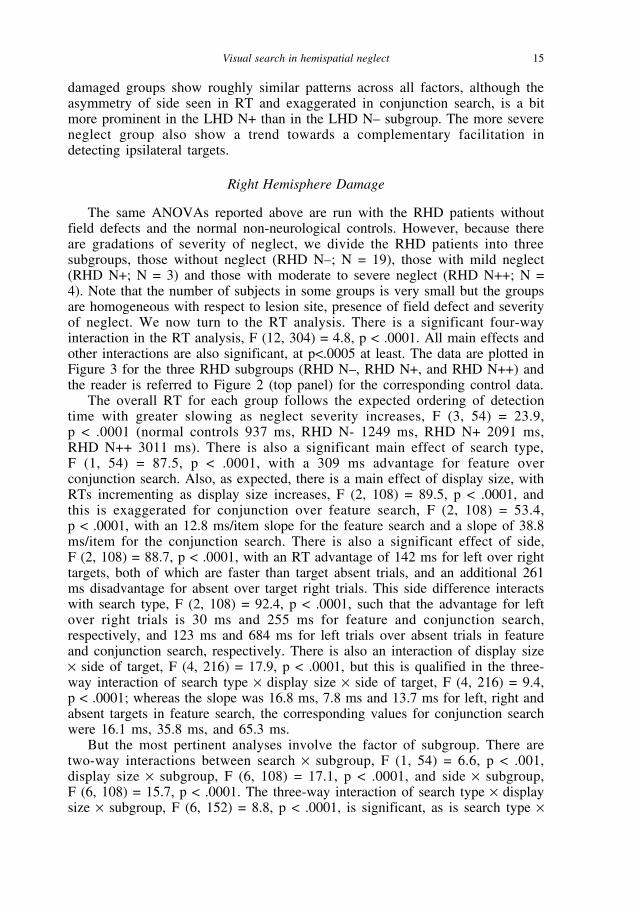
The same ANOVAs reported above are run with the RHD patients withoutfield defects and the normal non-neurological controls. However, because thereare gradations of severity of neglect, we divide the RHD patients into threesubgroups, those without neglect (RHD N–; N = 19), those with mild neglect(RHD N+; N = 3) and those with moderate to severe neglect (RHD N++; N =4). Note that the number of subjects in some groups is very small but the groupsare homogeneous with respect to lesion site, presence of field defect and severityof neglect. We now turn to the RT analysis. There is a significant four-wayinteraction in the RT analysis, F (12, 304) = 4.8, p < .0001. All main effects andother interactions are also significant, at p<.0005 at least. The data are plotted inFigure 3 for the three RHD subgroups (RHD N–, RHD N+, and RHD N++) andthe reader is referred to Figure 2 (top panel) for the corresponding control data.
The overall RT for each group follows the expected ordering of detectiontime with greater slowing as neglect severity increases, F (3, 54) = 23.9, p < .0001 (normal controls 937 ms, RHD N- 1249 ms, RHD N+ 2091 ms, RHD N++ 3011 ms). There is also a significant main effect of search type, F (1, 54) = 87.5, p < .0001, with a 309 ms advantage for feature overconjunction search. Also, as expected, there is a main effect of display size, withRTs incrementing as display size increases, F (2, 108) = 89.5, p < .0001, andthis is exaggerated for conjunction over feature search, F (2, 108) = 53.4, p < .0001, with an 12.8 ms/item slope for the feature search and a slope of 38.8ms/item for the conjunction search. There is also a significant effect of side, F (2, 108) = 88.7, p < .0001, with an RT advantage of 142 ms for left over righttargets, both of which are faster than target absent trials, and an additional 261ms disadvantage for absent over target right trials. This side difference interactswith search type, F (2, 108) = 92.4, p < .0001, such that the advantage for leftover right trials is 30 ms and 255 ms for feature and conjunction search,respectively, and 123 ms and 684 ms for left trials over absent trials in featureand conjunction search, respectively. There is also an interaction of display size× side of target, F (4, 216) = 17.9, p < .0001, but this is qualified in the three-way interaction of search type × display size × side of target, F (4, 216) = 9.4,p < .0001; whereas the slope was 16.8 ms, 7.8 ms and 13.7 ms for left, right andabsent targets in feature search, the corresponding values for conjunction searchwere 16.1 ms, 35.8 ms, and 65.3 ms.
But the most pertinent analyses involve the factor of subgroup. There aretwo-way interactions between search × subgroup, F (1, 54) = 6.6, p < .001,display size × subgroup, F (6, 108) = 17.1, p < .0001, and side × subgroup, F (6, 108) = 15.7, p < .0001. The three-way interaction of search type × displaysize × subgroup, F (6, 152) = 8.8, p < .0001, is significant, as is search type ×
Visual search in hemispatial neglect 15

16 Marlene Behrmann and Others
Fig. 3 – Mean of median reaction time for (A) patients with RHD and no neglect (B) patientswith RHD and mild neglect and (C) patients with RHD and more severe neglect on feature andconjunction search as a function of display size and presence (left/right) or absence of target.

side × subgroup, F (6, 108) = 12.5, p < .0001, and display size × side of target× subgroup, F (12, 216) = 11.9, p < .0001. To facilitate comparisons across thesubgroups, Table IV contains the slope of the RT function in ms, calculated overdisplay size, for the two types of search task for each of the four subgroupsseparately for left, right and absent target trials. We should note at the outsetthat there is one strange data point in the feature search, display size 1, targetabsent trials in both the RHD N+ and RHD N++ subgroups (see Figure 3 middleand bottom left panels; also to some extent in RHD N++ conjunction search).Target absent on search size 1 is notoriously complicated (Treisman andGormican, 1988). Also, given that we have rather few subjects in these cells, thevariability on this point is high. Aside from this oddity, the remaining datafollow a relatively clear pattern.
Relative to the non-neurological control subjects, all three RHD subgroupsshow steeper slopes in the feature search task and the slope increments overpresence and severity of neglect. Moreover, this increase in slope is greater forcontralateral left over ipsilateral right targets, as seen in Table IV. A similarpattern is observed in the conjunction search task in which, relative to the non-neurological controls, all brain-damaged subgroups are impaired, including theRHD N– patients, in detecting targets on the left. However, the patients withneglect all search more slowly on the left than those without neglect but there isno difference between RHD N+ and RHD N++. With regard to ipsilateral righttargets, there is a trend towards a reverse effect in that the most severe group,RHD N++, shows somewhat faster search and shallower slopes across displaysize (29.6 ms), relative to the other groups, and no difference between ipsilateraltargets in feature and conjunction search. Also, in the most severe neglect group,the slopes for the target absent trials do not necessarily reflect the self-terminating 2:1 ratio, suggesting that search may be terminated early or thatwhen a target is present on the contralateral side, the patients may continue tosearch for a long time until they acquire it, reducing the difference betweentarget present and target absent search times. Support for this latter claim comesfrom the finding that patients with right parietal lesions and neglect showabnormally long RTs to the absence of a target (Mijovic-Prelec et al., 1998);because of their lowered confidence, they spend an inordinately long timeverifying the absence of the target.
The same analysis using accuracy as a dependent measure yields almostidentical results. The four-way interaction is significant, F (12, 224) = 2.3, p < .01. All main effects are significant and there are a number of significant
Visual search in hemispatial neglect 17
TABLE IV
Slope of RT (in ms) for Feature and Conjunction Tasks for Four Subgroups of Participants
Group Feature search Conjunction search
Target left Target right No target Target left Target right No target
Normals 5.3 8.9 17.3 22.9 30.4 34RHD N– 18.6 13.7 210.3 59.3 33.5 118.2RHD N+ 64.2 23.7 31.8 146 46.3 181 RHD N++ 135.9 34.2 – 120.3 155 29.6 56.2

two-way and three-way interactions. The pattern yielded by the interaction isroughly the same as that of the RT data. Some small differences do emerge,likely because accuracy is not as telling a dependent measure as RT in thisparadigm, but, for the most part, the findings support those obtained from RT.
Taken together, the results are fairly clear. All patients, including those withRHD but no neglect are impaired relative to the control subjects for targets onthe left to a greater degree than targets on the right, and this asymmetry isexaggerated in the conjunction search in comparison with the feature search task.Patients with neglect, however, show steeper slopes than those without neglect.Importantly, this increase in slope for contralateral targets is evident in bothforms of search and to a greater degree on the left than right. There is also ascaling of the deficit such that the increased contralateral slope is less evident insubjects with no neglect (RHD N–) compared with those with neglect (RHD N+and N++). The two neglect groups differ from each other in two ways: oncontralateral targets, the RHD N++ group is more severely affected by displaysize than the RHD N+ groups, but mostly this is true in feature search, and, foripsilateral targets, the RHD N++ group is faster than the RHD N+ group (andRHD N–) in conjunction search. The apparent improvement in search for righttargets in the most severe neglect group in the conjunction task is consistentwith the observation that there is a Œmagnetic appeal1 for targets on the rightand this may, in a competitive fashion, give rise to the facilitation of right-sidedtarget detection even over that of normal subjects (Cate and Behrmann, 2002;Làdavas, Petronio et al., 1990; Behrmann, Barton et al., 1997). A similar resultwas noted above for the left brain-damaged group with neglect.
Comparisons of Left and Right Hemisphere Damage
Having shown that each of the brain-damaged groups is impaired relative tonormal control subjects, we now compare directly the performance of thepatients with left versus right hemisphere lesions. As above, these analyses areperformed using the data only from those patients without a field defect. Theanalysis is conducted with side of lesion (left, right) and presence/absence ofneglect as between-subjects factors and search type, display size and target (left,right, none) as within-subject factors. We excluded the RHD N++ group so as toequate neglect severity across the two sides of hemispheric lesion groups withthe result that only 4 subgroups were included (LHD N–, LHD N+, RHD N–,RHD N+).
The most critical finding is of a five-way interaction between all the factors.Despite the fact that this is a high-order interaction, the pattern is rather simpleand can be inferred from Table V. This table incorporates the relevant data fromTables III and IV but “left” and “right” have been replaced with contralateraland ipsilateral to facilitate comparison across the two hemisphere groups.Because our interest here is on side of lesion and presence/absence of neglect,we focus only on these aspects of the analysis.
RHD patients are 58 ms slower in RT than LHD patients, F (1, 46) = 14.2,p < .0005, but this differs depending on the presence of neglect, F (1, 46) =22.6, p < .0001; although LHD N– patients are 97 ms slower than RHD N–,
18 Marlene Behrmann and Others

LHD N+ patients are 1570 ms slower than RHD N– patients. But thesehemispheric × neglect differences are exaggerated in RHD N+ patients forcontralateral over ipsilateral targets especially for conjunction search and asdisplay size increases. Whereas there is no difference between LHD N– andRHD N– in either form of search, LHD N+ and RHD N+ differ on both searchtasks. On both tasks, RHD N+ shows a much steeper slope for both contralateraland ipsilateral targets than LHD N+.
In sum, there are several central results from this analysis: although, patientswith RHD detect conjunction targets more slowly overall than LHD patientsespecially as display size increases and this is particularly so for contralateraltargets, this is qualified by the presence of neglect. The difference between RHDpatients with and without neglect is greater than the difference between LHDpatients with and without neglect. It is also the case that LHD and RHD patientswithout neglect do not differ from each other and it is primarily the presence ofneglect that differentiates between these two hemispheric lesioned groups. Wenote, however, that, over and above this, the presence of neglect in the RHD N+group affects both forms of search to a greater degree than the LHD N+ group.
Comparisons of Patients with and without Field Defects
As alluded to previously, there is an ongoing controversy regarding theinfluence of a hemianopia on neglect performance. Given our large sample, wewere able to evaluate this claim by comparing search times for patients with andwithout field defects (although by the time we classify patients by hemisphere,presence/absence of neglect and presence/absence of hemianopia, the cells arenot that large any more). Table II reflects the co-occurrence of field defects (andseverity of field defect) with hemispheric damage with and without neglect. Asis apparent from these numbers, there is no obvious correlation between fielddefect and neglect; strikingly, six individuals with severe field defects fall in theLHD N– subgroup whereas four individuals from RHD N++ do not show a fielddefect.
To examine the effect of a field defect on search performance moresystematically, we analysed the RT of the subjects, categorized bypresence/absence of field defect, on the two search tasks using a three-factorbetween-subject ANOVA (side of hemispheric damage, presence/absence ofneglect, status of field defect) and the three within-subject variables of search
Visual search in hemispatial neglect 19
TABLE V
Slope of RT (in ms) for Feature and Conjunction Tasks for LHD and RHD Patients ClassifiedAccording to Severity of Neglect
Group Feature search Conjunction search
Contra Ipsi No target Contra Ipsi No target
LHD N– 27 21 41 68.4 26.1 102LHD N+ 19.1 3.9 12.1 65.9 16.2 94.9RHD N– 18.6 13.7 20.3 59.3 33.5 118.2RHD N+ 64.2 23.7 31.8 146 46.3 181
type, side of target and display size. Note that patients are classified in a binaryfashion for presence/absence of field defect rather than by severity in order to beable to cross this factor with side of damage and presence of neglect, and tomaintain enough subjects in each cell for the ANOVA. We do not includesubjects for whom we do not have definitive information about the status oftheir visual fields.
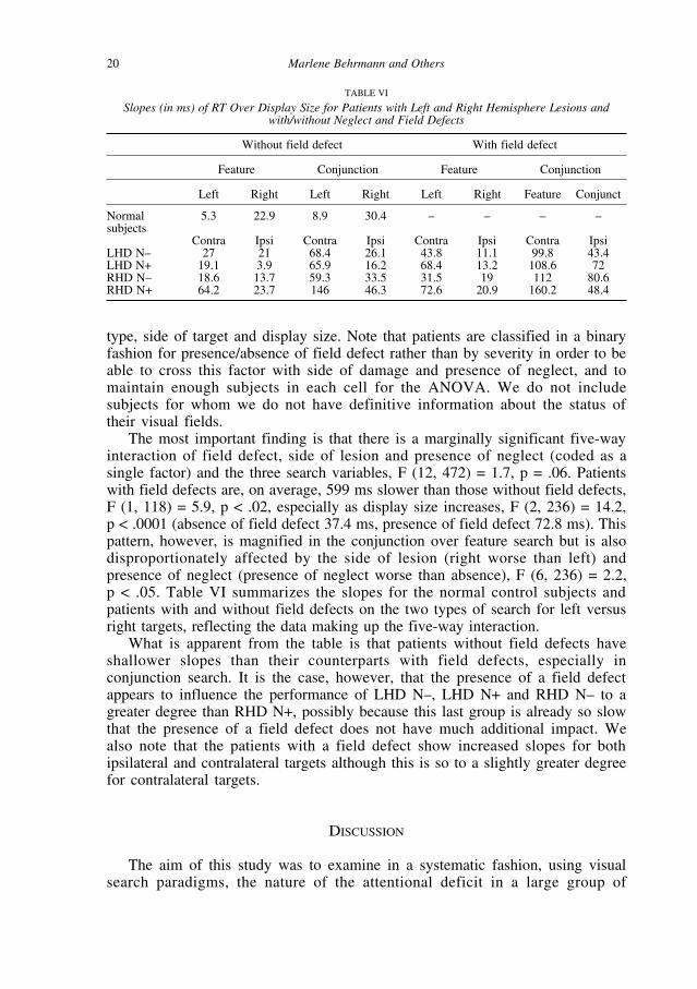
The most important finding is that there is a marginally significant five-wayinteraction of field defect, side of lesion and presence of neglect (coded as asingle factor) and the three search variables, F (12, 472) = 1.7, p = .06. Patientswith field defects are, on average, 599 ms slower than those without field defects,F (1, 118) = 5.9, p < .02, especially as display size increases, F (2, 236) = 14.2, p < .0001 (absence of field defect 37.4 ms, presence of field defect 72.8 ms). Thispattern, however, is magnified in the conjunction over feature search but is alsodisproportionately affected by the side of lesion (right worse than left) andpresence of neglect (presence of neglect worse than absence), F (6, 236) = 2.2, p < .05. Table VI summarizes the slopes for the normal control subjects andpatients with and without field defects on the two types of search for left versusright targets, reflecting the data making up the five-way interaction.
What is apparent from the table is that patients without field defects haveshallower slopes than their counterparts with field defects, especially inconjunction search. It is the case, however, that the presence of a field defectappears to influence the performance of LHD N–, LHD N+ and RHD N– to agreater degree than RHD N+, possibly because this last group is already so slowthat the presence of a field defect does not have much additional impact. Wealso note that the patients with a field defect show increased slopes for bothipsilateral and contralateral targets although this is so to a slightly greater degreefor contralateral targets.
DISCUSSION
The aim of this study was to examine in a systematic fashion, using visualsearch paradigms, the nature of the attentional deficit in a large group of
20 Marlene Behrmann and Others
TABLE VI
Slopes (in ms) of RT Over Display Size for Patients with Left and Right Hemisphere Lesions andwith/without Neglect and Field Defects
Without field defect With field defect
Feature Conjunction Feature Conjunction
Left Right Left Right Left Right Feature Conjunct
Normal 5.3 22.9 8.9 30.4 – – – –subjects
Contra Ipsi Contra Ipsi Contra Ipsi Contra IpsiLHD N– 27 21 68.4 26.1 43.8 11.1 99.8 43.4LHD N+ 19.1 3.9 65.9 16.2 68.4 13.2 108.6 72RHD N– 18.6 13.7 59.3 33.5 31.5 19 112 80.6RHD N+ 64.2 23.7 146 46.3 72.6 20.9 160.2 48.4

individuals who had sustained unilateral hemispheric lesions following cerebralinfarction. After confirming the decrement in performance for contralateral overipsilateral targets in conjunction search, we were interested in measuring thedetection time for ipsilateral targets in patients relative to control subjects. Butperhaps, of more relevance is the status of the feature search performance of thepatients compared with the control subjects. These analyses took into accountthe hemisphere affected (left or right), the presence of neglect and its severityand the presence of hemianopia.
To address these issues, we obtained data from 48 left-hemisphere damagedpatients, 56 right-hemisphere damaged patients and a group of non-neurologicalcontrol subjects on a standard neglect battery. We also documented the presenceof a visual field defect in the patients on confrontation testing. Finally, all subjectscompleted a computerized experiment consisting of a well-established visualsearch task with two components; the first component involved search for a targetwhich is typically acquired rapidly and independently of the number of distractorsby normal subjects (feature search) and the second component involved search fora target which is acquired more slowly and is significantly affected by the numberof distractors in the array (conjunction search). Although studies of this type havebeen conducted in the past, the findings remain controversial.
The first major result is that following a lesion to either hemisphere,individuals without neglect are impaired relative to intact normal subjects.However, individuals with a lesion and with concurrent neglect are even moreimpaired than their non-neglect counterparts. Interestingly, both patient groups,with and without neglect, show incrementally slower search in both feature andconjunction searches as display size increases and this is so to a greater extentfor contralateral than ipsilateral targets. Because of the graded severity of neglectamong the RHD group, we could also evaluate whether this pattern was affectedby the extent of the neglect. There was no significant difference as a function ofseverity of neglect and RHD N+ and RHD N++ show roughly similarmagnitudes of deficit on contralateral search. Interestingly, the more severegroup showed a shallower slope for ipsilateral targets in the conjunction task,reflecting the competition between items on the left and right and the advantagefor the ipsilateral items. A hint of this competitive advantage for ipsilateraltargets was also seen after LHD in individuals with neglect.
A comparison of the patients1 performance as a function of side ofhemispheric lesion shows that, even when we equated for the presence andextent of neglect in the two groups, RHD patients are more impaired atcontralateral conjunction search as display size increases than their LHDcounterparts. This difference is magnified by the presence of neglect so that thegreater impairment for RHD N– than LHD N– is magnified when comparingRHD N+ and their LHD N+ counterparts.
The presence of a field defect also contributes to the impairment in searchperformance although, once the difference between presence/absence of neglectis taken into account, the additional contribution of a field defect is not thatlarge. Also, to the extent that it exists, the presence of hemianopia does notappear to be specific and seems to slow search down for both ipsilateral andcontralateral targets.
Visual search in hemispatial neglect 21

These findings have provided some clear answers. Our results are consistentwith the other studies that find a deficit in contralateral feature search (Riddochand Humphreys, 1983; Eglin et al., 1989; Pavlovskaya et al., 2002).Interestingly, the magnitude of the deficit we have observed is also comparableto that obtained in these other studies. In light of this, our data challenge thosestudies, which claim that feature search is preserved in hemispatial neglect. Aslaid out above, a number of possible factors can account for the discrepantfindings across the different studies. In our case, given the large patient sampleand the heterogeneity of our subjects, we have been able to control a range ofvariables, which might have influenced the previous results.
In addition to addressing the controversial findings and to exploring thefactors that might have confounded these previous studies, our data have alsohighlighted some other interesting effects that have not received much discussionto date. Patients with severe neglect, counterintuitively, show a speed up onipsilateral conjunction search. In contrast, patients with no or mild neglect areslow on conjunction search for ipsilateral and contralateral search. Theseobservations are compatible with a view in which there is competition forattention. If the attentional bias is strong ipsilesionally, targets will be quicklydetected but this might have adverse consequences for contralateral detection. If,however, the ipsilesional bias is less strong, search might be slow for bothipsilesional targets and for contralesional targets. Some studies have alsoreported poorer ipsilesional performance for patients compared with controls(Eglin et al., 1989; Eglin et al., 1996; Geng and Behrmann, 2002a) and othershave found that the search patterns of neglect patients are equally poor in thecontralesional and ipsilesional visual field (Chatterjee et al., 1992; Halligan etal., 1992). The claim that there are no hemifield differences also finds support instudies that do not use visual search; for example, using partial and whole reportprocedures, Duncan and colleagues document the presence of poor visualprocessing in both hemifields in neglect patients (Duncan et al., 1999). Onepossible explanation for the range of results concerns the severity of neglect. Weonly see an ipsilesional advantage for the severe neglect patients whereas forthose with less severe neglect (and even for those with no neglect), we finddecrements in performance in both hemifields but somewhat greater in thecontralateral than ipsilateral space.
Before going on to examine the theoretical implications of these findings, weneed to rule out an alternative interpretation of our results. One possible reasonthat we find poorer feature search in the neglect patients, relative to the controls,might be that the patients have additional brain damage to earlier visual areasand it is this damage, rather than the neglect per se, that might contribute toimpaired parallel search and preattentive processing. In a detailed analysis of thelesion sites of all patients, done by identification of the lesion location/s on theCT scans in reference to the 24 best fitting slices from the Talairach andTournox stereotactical anatomical reference system (Talairach and Tournox,1998), only 8 of the RHD patients (16%) and only 8 of the LHD patients (18%)have damage to occipital cortex (Ebert, 1998). There are several obvious reasonsfrom the data to indicate that the impaired feature search is not solelyattributable to this additional damage to early visual areas. Firstly, only a small
22 Marlene Behrmann and Others
proportion of the groups have patients with damage to this area and this samplesize is not large enough to carry the statistical effects of the whole group.Secondly, because some of these patients have field defects, and make manycontralateral errors, their data are not included in the RTs and so, the slopes arecalculated without their data. Finally, we note that when we examine theperformance of individuals with versus those without field defects, we stillobserve an impairment in feature search in those subjects who do not have fielddefects. Taken together, these findings suggest that the increased slope in RTsfor feature search as a function of array size is not simply attributable to theadditional presence of occipital lobe damage.
In addition to clarifying the neuropsychological data on hemispatial neglect,our findings have implications for claims about the fundamental nature ofattentional processing. Although the distinction between preattentive (feature)search and attentive (conjunction) search is well engrained in cognitivepsychology, its validity has been challenged from numerous perspectives. Ourdata also challenge this dichotomy in that the supposed preattentive task does notsurvive hemispatial neglect. Consistent with our result, it has been suggested thateven preattentive search requires some amount of attention; when a concurrenttask is performed, interference is seen even though this should not be the case,according to a view of a capacity-free parallel preattentive search mechanism(Joseph et al., 1997). Indeed, even in the original empirical visual search studiesin normal subjects, feature search did not have a completely flat slope, whichwould truly indicate attention-free processing, and, instead, a serial search patternwas defined as one in which the slope is greater than 10-20 milliseconds per item(Treisman and Gelade, 1980). Our findings are concordant with the notion thatall forms of visual search engage some form of attentional processing.
It seems clear then that the binary distinction between preattentive/featuraland attentive/conjunction processing does not obviously hold and, indeed, therehave been recent attempts to articulate a theoretical perspective that does notrely on this dichotomy. On these more integrated accounts, there is no obviousdistinction between preattentive and attentive processing. Instead these viewsrely on the principles of competition and cooperation between features andobjects to resolve the constraints of visual attention and to determine theefficiency of attentional selection. Feature search is hypothesized to be fast andaccurate because competition between targets and distractors is resolved quickly.In contrast, conjunctive search is slower and more prone to error because target-distractor similarity or distractor-distractor heterogeneity produces greatercompetition between items and therefore takes longer to resolve (Duncan andHumphreys, 1989).
One theoretical perspective that eschews this dichotomy and that has gainedconsiderable popularity lately is the Biased Competition and the IntegratedCompetition accounts (Desimone and Duncan, 1995; Duncan and Humphreys,1989; Duncan et al., 1997). This view suggests that attention is an emergentproperty of competition between representations of stimuli within the nervoussystem and that processing is qualitatively similar regardless of whether a targetstimulus in visual search is distinguished from distractors by a single feature orby a conjunction of features. The obvious prediction from this type of account is
Visual search in hemispatial neglect 23

that in patients with neglect, search will be affected for both featural andconjunction targets but that the latter will be more impaired than the former.This view also makes allowance for competition between ipsilateral andcontralateral items, and we observe such competition in individuals with moresevere neglect who search ipsilesionally a little better. Our data fit well with thiscompetitive view and support an interactive account of attention and selection.
The conceptual divide between feature and conjunction search, like thedivide between “primary” and “high level” deficits (Halligan and Marshall,2002), is also not obviously supported by recent functional imaging studies. Therelevant finding from a large number of recent studies is that activation in earlyvisual areas can be affected by activation in parietal and frontal cortices andthese top-down influences can modulate activation in striate and extrastriateareas (for example, Noesselt et al., 2002; Somers et al., 1999) and in the lateralgeniculate nucleus (O’Connor et al., 2002), even in the absence of visualstimulation (Kastner et al., 1999) . Given the bi-directional reciprocalinteractions between earlier and later visual areas, there is no clear dichotomy orseparation onto which feature and conjunction search might be mapped. Instead,the dynamic feedforward and feedback interactions are more compatible with asingle, integrated account of visual search, as outlined above.
In conclusion, we have clearly demonstrated that patients with neglect areimpaired at feature and, as expected, on conjunction search on the contralateralside. This result has implications for models of attentional selection andchallenges the idea that feature search can be done in parallel in the absence ofattention. It is the case, however, that not all forms of contralateral search areimpaired to the same extent in individuals with neglect. In a recent series ofstudies, Humphreys and Riddoch (2001; 2001/2002) had a patient with leftneglect search for a target (cup) defined by an action “find an object you candrink from” or by a name “find the cup”. Interestingly, search was moreefficient in the former than latter case and this benefit was enhanced as displaysize increased. The advantage for action-defined search is attributed to theexistence of action-defined templates, which are activated by affordances ofobjects. These results are provocative and suggest that not all forms of searchare created equal and not all forms of search suffer equally following brain-damage. Understanding the distinctions and boundaries between these differentforms of search remains a future challenge.
Acknowledgements. This work was supported by grants from the National Institutes ofMental Health (NIMH 54246) to Marlene Behrmann, from the Ontario Heart and StrokeFoundation and from the Ontario Mental Health Foundation to Sandra E. Black and by aUniversity of Toronto Open Scholarship to Patricia Ebert. The authors thank Joy Geng forher constructive comments on this work, and Nancy Blair, Jane Collins and Jay Bondar fortheir help with data collection and patient testing. We also thank Dr D. Stuss and Dr R.McIntosh who served as members of the thesis committee for PE. This research wascompleted in partial fulfillment of PE1s Master’s Thesis at the University of Toronto.
REFERENCES
AGLIOTI S, SMANIA N, BARBIERI C and CORBETTA M. Influence of stimulus salience and attentionaldemands on visual search patterns in hemispatial neglect. Brain and Cognition, 34: 388-403, 1997.
24 Marlene Behrmann and Others
ARGUIN M, JOANETTE Y and CAVANAGH P. Visual search for feature and conjunction targets with anattention deficit. Journal of Cognitive Neuroscience, 5: 436-452, 1993.
ASHBRIDGE E, WALSH V and COWEY A. Temporal aspects of visual search studies by transcranialmagnetic stimulation. Neuropsychologia, 35: 1121-1131, 1997.
BARTOLOMEO P and CHOKRON S. Levels of impairment in unilateral neglect. In F Boller and J Grafman(Eds), Handbook of neuropsychology. North-Holland: Elsevier Science, 2001, pp. 67-98.
BEHRMANN M, BARTON JJS, WATT S and BLACK SE. Impaired visual search in patients with unilateralneglect: An oculographic analysis. Neuropsychologia, 35: 1445-1458, 1997.
BEHRMANN M, GHISELLI-CRIPPA T and DIMATTEO I. Impaired initiation but not execution of leftwardsaccades to left targets in hemispatial neglect. Behavioral Neurology, 13: 39-60, 2001.
BEHRMANN M and HAIMSON C. The cognitive neuroscience of visual attention. Current Opinion inNeurobiology, 9: 158-163, 1999.
BEHRMANN M, MOSCOVITCH M, BLACK SE and MOZER MC. Perceptual and conceptual factors in neglectdyslexia: Two contrasting case studies. Brain 113, 4: 1163-1883.
BISIACH E and LUZZATTI C. Unilateral neglect of representational space. Cortex, 14: 129-133, 1978.BISIACH E and VALLAR G. Unilateral neglect in humans. In F Boller and J Grafman (Eds), Handbook of
neuropsychology. North-Holland, Amsterdam: Elsevier Science, 2000, pp. 459-502.BLACK SE, VU B, MARTIN D and SZALAI JP. Evaluation of a bedside battery for hemispatial neglect in
acute stroke. Journal of Clinical and Experimental Neuropsychology, 12: 102 (abstract), 1990.BOUTSEN L and HUMPHREYS GW. Axis-based grouping reduces visual extinction. Neuropsychologia, 38:
896-905, 2000.BRICOLO E, GIANESINI T, FANINI A, BUNDESEN C and CHELAZZI L. Serial attention mechanisms in
visual search: A direct behavioral demonstration. Journal of Cognitive Neuroscience, 14: 980-993,2002.
BUB D and GUM T. Psychlab. McGill University: Neurolinguistics Department, 1991.BUNDESEN C. A theory of visual attention. Psychological Review, 97: 523-547, 1990.CATE A and BEHRMANN M. Spatial and temporal influences on extinction in parietal patients.
Neuropsychologia, 40: 2206-2225, 2002.CHATTERJEE A, MENNEMEIER M and HEILMAN KM. A stimulus-response relationship in unilateral
neglect: The power function. Neuropsychologia, 30: 1101-1108, 1992.CORBETTA M, AKBUDAK E, CONTURO TE, SNYDER AZ, OLLINGER JM, DRURY HA, LINENWEBER MR,
PETERSEN SE, RAICHLE ME, VAN ESSEN DC and SHULMAN GL. A common network of functionalareas for attention and eye movements. Neuron, 21: 761-773, 1998.
CORBETTA M, SHULMAN GL, MIEZEN FM and PETERSEN SE. Superior parietal cortex activation duringspatial attention shifts and visual feature conjunction. Science, 270: 802-805, 1995.
DESIMONE R and DUNCAN J. Neural mechanisms of selective visual attention. Annual Review ofNeuroscience, 18: 193-222, 1995.
DORICCHI F and ANGELELLI P. Misrepresentation of horizontal space in left unilateral neglect: Role ofhemianopia. Neurology, 52: 1845-1852, 1999.
DRIVER J, BAYLIS GC and RAFAL RD. Preserved figure-ground segregation and symmetry perception invisual neglect. Nature, 360: 73-75, 1992.
DRIVER J and HALLIGAN PW. Can visual neglect operate in object-centered coordinates: An affirmativestudy. Cognitive Neuropsychology, 8: 475-496, 1991.
DUNCAN J, BUNDESEN C, OLSON A, HUMPHREYS GW, CHAVDA S and SHIBUYA H. Systematic analysis ofdeficits in visual attention. Journal of Experimental Psychology: General, 128: 450-478, 1999.
DUNCAN J, HUMPHREYS G and WARD R. Competitive brain activity in visual attention. Current Opinionin Neurobiology, 7: 255-261, 1997.
DUNCAN J and HUMPHREYS GW. Visual search and stimulus similarity. Psychological Review, 96: 433-458, 1989.
EGLIN M, ROBERTSON L, KNIGHT RT and BRUGGER P. Search deficits in neglect patients are dependenton size of the visual scene. Neuropsychologia, 8: 451-463, 1996.
EGLIN M, ROBERTSON LC and KNIGHT RT. Visual search performance in the neglect syndrome. Journalof Cognitive Neuroscience, 1: 372-385, 1989.
EGLIN M, ROBERTSON LC and KNIGHT RT. Cortical substrates supporting visual search in humans.Cerebral Cortex, 1: 262-272, 1991.
EGLIN M, ROBERTSON LC, KNIGHT RT and BRUGGER P. Search deficits in neglect patients are dependenton size of the visual scene. Neuropsychology, 8: 451-463, 1994.
ESTERMAN M, MCGLINCHEY-BERROTH R and MILBERG W. Preattentive and attentive visual search inindividuals with hemispatial neglect. Neuropsychology, 14: 599-611, 2000.
FARAH MJ, WALLACE M and VECERA SP. “What’’ and “Where’’ in visual attention: Evidence from theneglect syndrome. In IH Robertson and JC Marshall (Eds), Unilateral neglect: Clinical andexperimental studies. Hove, UK: Lawrence Erlbaum Associates, 1993, pp. 123-138.
FERBER S and KARNATH HO. Parietal and occipital contributions to perception of straight aheadorientation. Journal of Neurology, Neurosurgery and Psychiatry, 67: 572-578, 1999.
Visual search in hemispatial neglect 25

GAINOTTI G, D’ERME P, MONTELEONE D and SILVERI MC. Mechanisms of unilateral neglect in relationto laterality of cerebral lesions. Brain, 109: 599-612, 1986.
GENG JJ and BEHRMANN M. Probability cueing of target location facilitates visual search implicitly innormal participants and patients with hemispatial neglect. Psychological Science, 13: 520-525,2002a.
GENG JJ and BEHRMANN M. Selective visual attention and visual search: Behavioral and neuralmechanisms. In B Ross and D Irwin (Eds), The psychology of learning and motivation. New York:Academic Press, 2002b.
GILCHRIST ID, HUMPHREYS GW and RIDDOCH MJ. Grouping and extinction: Evidence for low-levelmodulation of visual selection. Cognitive Neuropsychology, 13: 1223-1249, 1996.
GORE CL, RODRIGUEZ DP and BAYLIS GC. Deficits of motor intention following parietal lesions.Behavioral Neurology, 13: 29-37, 2001/2002.
HALLIGAN PW, BURN JP, MARSHALL JC and WADE DT. Visuo-spatial neglect: Qualitative differencesand laterality of cerebral lesion. Journal of Neurology, Neurosurgery and Psychiatry, 55: 1060-1068, 1992.
HALLIGAN PW and MARSHALL JC. Left visuospatial neglect: A meaningless entity? Cortex, 28: 525-535.HALLIGAN PW and MARSHALL JC. Primary sensory deficits after right brain-damage an attentional
disorder by any other name? In HO Karnath, AD Milner and G Vallar (Eds), The cognitive andneural bases of spatial neglect. Oxford, UK: Oxford University Press, 2002, pp. 327-340.
HORNAK J. Ocular exploration in the dark by patients with visual neglect. Neuropsychologia, 30: 547-552, 1992.
HUMPHREYS GW. Binding in vision as a multi-stage process. In R Kimchi, M Behrmann and C Olson.(Eds) Perceptual organization: Behavioral and neural perspectives. Hillsdale, NJ: ErlbaumAssociates, 2003.
HUMPHREYS GW and RIDDOCH MJ. Detection by action: Neuropsychological evidence for action-definedtemplates in search. Nature Neuroscience, 4: 84-88, 2001.
HUMPHREYS GW and RIDDOCH MJ. Knowing what you need but not what you want: Affordances andaction-defined templates in neglect. Behavioral Neurology, 13: 75-87, 2001/2002.
JOSEPH JS, CHUN M and NAKAYAMA K. Attentional requirements in a ‘preattentive’ feature search task.Nature, 387: 805-807, 1997.
KASTNER S, PINSK MA, DE WEERED P, DESIMONE R and UNGERLEIDER LG. Increased activity in humanvisual cortex during directed attention in the absence of visual stimulation. Neuron, 22: 751-761.
KUMADA T and HUMPHREYS GW. Lexical recovery from extinction: Interactions between visual form andstored knowledge modulate visual selection. Cognitive Neuropsychology, 18: 465-478, 2001.
LÀDAVAS E, CARLETTI M and GORI G. Automatic and voluntary orienting of attention in patients withvisual neglect: Horizontal and vertical dimensions. Neuropsychologia, 32: 1195-1208, 1994.
LÀDAVAS E, PETRONIO A and UMILTÀ C. The deployment of visual attention in the intact field ofhemineglect patients. Cortex, 26: 307-317, 1990.
LEIBOVITCH FS, BLACK SE, CALDWELL CB, EBERT PL, EHRLICH LE and SZALAI JP. Brain-behaviorcorrelations in left hemispatial neglect using ct and spect imaging: The Sunnybrook stroke study.Neurology, 50: 901-908, 1998.
MARZI, C, GIRELLI M, MIMUSSI C, SMANIA N and MARAVITA A. Electrophysiological correlates ofconscious vision: Evidence from unilateral extinction. Journal of Cognitive Neuroscience, 12: 869-877, 2000.
MATTINGLEY JB, DAVID G and DRIVER J. Preattentive filling in of visual surfaces in parietal extinction.Science, 275: 671-674, 1997.
MATTINGLEY JB, HUSAIN M, RORDEN C, KENNARD C and DRIVER J. Motor role of human inferior parietallobe revealed in unilateral neglect patients. Nature, 392: 179-182, 1998.
MESULAM MM. Principles of behavioral neurology. USA: F. A. Davis, 1985.MIJOVIC-PRELEC D, CHABRIS CF, SHIN LM, KOSSLYN SM and WRAY SH. The judgment of absence in
neglect. Neuropsychologia, 36: 797-802, 1998.NEISSER U. Visual search. Scientific American, 210: 94-102, 1964.NOESSELT T, HILLYARD SA, WOLDORFF MG, SCHOENFELD A, HAGNER T, JÄNCKE L, TEMPELMANN C,
HINRICHS H and Heinze HJ. Delayed striate cortical activation during spatial attention. Neuron, 35:575-587.
O’CONNOR DH, FUKUI MM, PINSK MA and KASTNER S. Attention modulates responses in the humanlateral geniculate nucleus. Nature Neuroscience, 5: 1203-1209.
OGDEN J. The “neglected” left hemisphere and its contribution to visuospatial neglect. In M Jeannerod(Ed), Neurophysiological and neuropsychological aspects of spatial neglect. Amsterdam: NorthHolland, 1987.
PAVLOVSKAYA M, RING H, GROSSWASSER Z and HOCHSTEIN S. Searching with unilateral neglect. Journalof Cognitive Neuroscience, 14: 745-756, 2002.
RAPCSAK SZ, VERFAELLIE M, FLEET S and HEILMAN KM. Selective attention in hemispatial neglect.Archives of Neurology, 46: 178-182, 1989.
26 Marlene Behrmann and Others

RIDDOCH MJ and HUMPHREYS GW. The effect of cueing on unilateral neglect. Neuropsychologia, 21:589-599, 1983.
RIDDOCH MJ and HUMPHREYS GW. Perceptual and action systems in unilateral visual neglect. In MJeannerod (Ed), Neurophysiological and neuropsychological aspects of spatial neglect. New York:Elsevier, 1987, pp. 151-181.
RORDEN C, MATTINGLEY JB, KARNATH H-O and DRIVER J. Visual extinction and prior entry: Impairedperception of temporal order with intact motion perception after unilateral parietal damage.Neuropsychologia, 35: 421-433, 1997.
SOMERS DC, DALE AM, SEIFFERT AE and TOOTELL RB. Functional MRI reveals spatially specificattentional modulation in human primary visual cortex. Proceedings of the National Academy ofSciences of the United States of America, 96: 1663-1668.
TOTH C and KIRK A. Representational bias does not affect bisection of lines with a pictorially orsemantically defined top by patients with left hemispatial neglect. Brain and Cognition, 50: 161-177, 2002.
TREISMAN A. Feature binding, attention and object perception. In GW Humphreys, J Duncan and ATreisman. (Eds), Attention, space and action. Oxford: Oxford University Press, 1999, pp. 91-111.
TREISMAN A and GELADE G. A feature-integration theory of attention. Cognitive Psychology, 12: 97-136,1980.
TREISMAN A and GORMICAN S. Feature analysis in early vision: Evidence from search asymmetries.Psychological Review, 95: 15-48, 1988.
TREISMAN A and SOUTHER J. Search asymmetry: A diagnostic for preattentive processing of separablefeatures. Journal of Experimental Psychology: General, 114: 285-310, 1985.
VUILLEUMIER P, SAGIV N, HAZELTINE E, POLDRACK RA, SWICK D and GABRIELI JDE. Neural fate of seenand unseen faces in visuospatial neglect; A combined event-related functional MRI and event-related potential study. Proceedings of the National Academy of Sciences, 98: 3495-3500, 2000.
WARD R, GOODRICH S and DRIVER J. Grouping reduces visual extinction: Neuropsychological evidencefor weight-linkage in visual selection. Visual Cognition, 1: 101-129, 1994.
WEINTRAUB S and MESULAM MM. Right cerebral dominance in spatial attention: Further evidence basedon ipsilateral neglect. Archives of Neurology, 44: 621-625, 1987.
WOLFE JM. What can 1 million trials tell us about visual search? Psychological Science, 9: 33-39, 1998.YANTIS S. Goal-directed and stimulus-driven determinants of attentional control. In S Monsell and J
Driver (Eds), Attention and Performance XVIII. Cambridge, MA: MIT Press, 2000, pp. 73-103.
??????
Visual search in hemispatial neglect 27