bell’s palsy amy stinson ent pgy-2 affinity medical center
TRANSCRIPT

Bell’s Palsy
Amy Stinson
ENT PGY-2
Affinity Medical Center

Outline Anatomy Definition Differential Exam Electrophysiology Treatment Outcome

Anatomy: The Facial Nerve Motor and Sensory SVA fibers: taste ant 2/3 tongue
Lingual & Chorda geniculate nervous intermedius solitary nucleus
SVE fibers: muscles of facial expression Facial motor nucleus stylomastoid foramen
GVA fibers: parasympathetics lacrimal, palatine, parotid, submandibular, sublingual glands Sup salivatory nucleus GSPN/Sphenopalatine, lesser
petrosal/otic, chorda/submandibular Sensory – concha and post auricular

Anatomy: The Facial Nerve Intracranial
lateral for 12-14mm with CN8 to IAC Meatal
8-10mm ant/sup of IAC to meatal foramen Diameter changes from 1.2 mm to 0.68 mm
Labyrinthine 2-4 mm to geniculate ganglion (GSPN exits)
Tympanic First genu 11mm post/inf to 2nd genu
Mastoid 12-14 mm inf (vertical seg) to SMF (chorda exits)
Peripheral Pes anserus 20 mm then 5 terminal branches (upper and lower seg)

Anatomy: The Facial Nerve Favorite mnemonics
Some Say Marry Money, But My Brother Says Big Breasts Matter More
To Zanzibar By Motor Car Ten Zebras Bit My Crotch Ten Zebras Beat My Cock Today Zoe Broke My Car


Anatomy

Anatomy

Bell’s Palsy Sir Charles Bell (1774-1842)
Studied facial anatomy extensively during Battle of Waterloo
Concluded that facial nerve controlled facial expression
“Respiratory nerve of the Face”

Bell’s Palsy Idiopathic Facial Paralysis DIAGNOSIS OF EXCLUSION MC Diagnosis given >60% Unilateral Rapid Onset <48hrs Not progressive!

Bell’s Palsy 30/100,000 M = F 3.3x greater incidence in pregnancy 4-5x increased risk with DM Fam Hx 10% Recurrence rate 10%

Bell’s Palsy - etiology
Exact etiology unknown Viral infection
Herpes Simplex
Vascular ischemia Autoimmune disorder Hereditary
Exact etiology unknown Viral infection
Herpes Simplex
Vascular ischemia Autoimmune disorder Hereditary


Bell’s Palsy
Reduced Stapedial reflex 71% Complete palsy @ presentation69% Tear flow 67% Post-auricular pain 52% Dysgeusia 34% Hyperacusis 14%
Reduced Stapedial reflex 71% Complete palsy @ presentation69% Tear flow 67% Post-auricular pain 52% Dysgeusia 34% Hyperacusis 14%

Bell’s Palsy
Peitersen E. Acta Otolaryngol 2002;549:4–30.
Complete Remission & Age

Bell’s Palsy
Peitersen E. Am. J. Otology. 1982
Bell’s Palsy
DIAGNOSIS OF EXCLUSION

Differential Diagnosis Infection
Herpes Zoster Oticus (Ramsey Hunt Syndrome)
Lyme disease Acute Otitis media +/- mastoiditis Malignant otitis externa TB AIDS Mono
Congenital Treacher Collins syndrome Mobius syndrome Compression injury
Trauma Temporal Bone fracture Barotrauma Penetration wounds, laceration, and
contusions
Metabolic
DiabetesHypothyroidism SarcoidGullian BarreAutoimmune disorders
VascularBenign intracranial
hypertension Neoplasm
Facial neuromaAcoustic neuromaCholesteatomaMenigiomaLeukemiaMetestatic
ToxicThalidomide
Iatrogenic

Differential Diagnosis If nerve function had not returned or has
gotten worse at the 6 month mark – You MUST revisit the previous list!

History Onset
Sudden, delayed, gradual
Degree of paralysis Complete, incomplete
Associated symptoms Numbness, otalgia, hyperacusis, diminished tearing,
altered taste Intense ear pain and vesicles Sensorineural hearing loss, vertigo

Exam Quick and dirty facial nerve exam
Raise eyebrows Tightly close eyes Wrinkle the nose Smile Pucker Grimace


Exam Complete Head and Neck exam
Special attention to otoscopy and CNs Progressive segmental paralysis w/lesion Laceration, battle sign, hemotympanum Multiple CN deficits
Compare motor function w/opposite side Bell phenomenon: visible vertical rotation of globe on
closing affected eye Audiometry CT/MRI

Pathophysiology HSV viral reactivation leading to damage of
facial nerve Neuropraxia– no axonal discontinuity Axonotmesis
Wallerian degeneration (distal to lesion) Axoplasmic disruption, endoneural sheaths intact
Neurotmesis Wallerian degeneration (distal to lesion) Axon disrupted, loss of tubules, support cells destroyed

Electrophysiology Sunderland’s Classification
Neurapraxia
Axonotmesis
Neurotmesis

Electrophysiology

Electrophysiology Nerve Excitation test (NET) Maximal Stimulation test (MST) Electroneurography (ENoG) Electromyography (EMG)
Measure amounts of neural degeneration occurred distal to injury by measuring muscle response to electrical stimulus
Able to differentiate nerve fibers undergoing Wallerian degeneration
Nerve Excitation test (NET) Maximal Stimulation test (MST) Electroneurography (ENoG) Electromyography (EMG)
Measure amounts of neural degeneration occurred distal to injury by measuring muscle response to electrical stimulus
Able to differentiate nerve fibers undergoing Wallerian degeneration

Treatment Observation Medical Treatment
Steroid Anti-viral agents
Surgery Decompression Dynamic vs. static reanimation
Facial Rehabilitation
Observation Medical Treatment
Steroid Anti-viral agents
Surgery Decompression Dynamic vs. static reanimation
Facial Rehabilitation

Treatment
Double-blind RCT 99 Bell’s palsy patients
53 treated with acyclovir- prednisone 46 with placebo – prednisone Prednisone dose 400 mg five times daily x 10 days
Combined therapy is better in terms of: Return of muscle motion Prevention of partial nerve degeneration
Double-blind RCT 99 Bell’s palsy patients
53 treated with acyclovir- prednisone 46 with placebo – prednisone Prednisone dose 400 mg five times daily x 10 days
Combined therapy is better in terms of: Return of muscle motion Prevention of partial nerve degeneration
Steroid vs. Steroid + AcyclovirSteroid vs. Steroid + Acyclovir
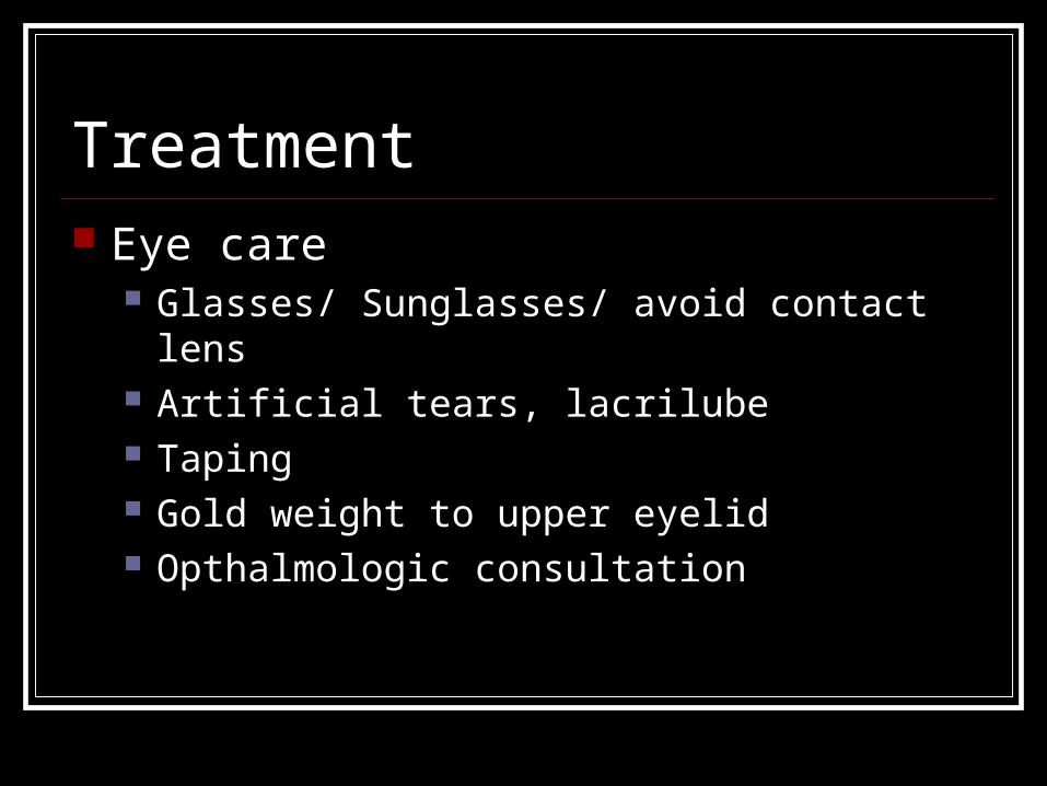
Treatment Eye care
Glasses/ Sunglasses/ avoid contact lens Artificial tears, lacrilube Taping Gold weight to upper eyelid Opthalmologic consultation

Treatment Surgical Decompression
Middle Fossa Transmastoid Translabyrinthine Retrolabyrinthine Retrosigmoid


Outcome Complete Remission & Age

Outcome Return of Muscular function
85 %