berries and their polyphenols as a potential therapy for
TRANSCRIPT
International Journal of
Molecular Sciences
Review
Berries and Their Polyphenols as a Potential Therapy forCoronary Microvascular Dysfunction: A Mini-Review
Rami S. Najjar 1 , Arielle M. Schwartz 2, Brett J. Wong 3 , Puja K. Mehta 4,5,* and Rafaela G. Feresin 1,*
�����������������
Citation: Najjar, R.S.; Schwartz,
A.M.; Wong, B.J.; Mehta, P.K.; Feresin,
R.G. Berries and Their Polyphenols as
a Potential Therapy for Coronary
Microvascular Dysfunction: A Mini-
Review. Int. J. Mol. Sci. 2021, 22, 3373.
https://doi.org/10.3390/
ijms22073373
Academic Editor: Yun Jung Lee
Received: 24 February 2021
Accepted: 23 March 2021
Published: 25 March 2021
Publisher’s Note: MDPI stays neutral
with regard to jurisdictional claims in
published maps and institutional affil-
iations.
Copyright: © 2021 by the authors.
Licensee MDPI, Basel, Switzerland.
This article is an open access article
distributed under the terms and
conditions of the Creative Commons
Attribution (CC BY) license (https://
creativecommons.org/licenses/by/
4.0/).
1 Department of Nutrition, Georgia State University, Atlanta, GA 30302, USA; [email protected] J. Willis Hurst Internal Medicine Residency Program, Emory University, Atlanta, GA 30322, USA;
[email protected] Department of Kinesiology & Health, Georgia State University, Atlanta, GA 30302, USA; [email protected] Division of Cardiology, Emory Women’s Heart Center, Emory University School of Medicine,
Atlanta, GA 30322, USA5 Division of Cardiology, Emory Clinical Cardiovascular Research Institute, Emory University School of
Medicine, Atlanta, GA 30322, USA* Correspondence: [email protected] (P.K.M.); [email protected] (R.G.F.)
Abstract: Ischemia with no obstructive coronary artery disease (INOCA) is a common diagnosis witha higher prevalence in women compared to men. Despite the absence of obstructive coronary arterydisease and no structural heart disease, INOCA is associated with major adverse cardiovascularoutcomes as well a significant contributor to angina and related disability. A major feature of INOCAis coronary microvascular dysfunction (CMD), which can be detected by non-invasive imaging andinvasive coronary physiology assessments in humans. CMD is associated with epicardial endothelial-dependent and -independent dysfunction, diffuse atherosclerosis, and left-ventricular hypertrophy,all of which lead to insufficient blood flow to the myocardium. Inflammatory and oxidative stresssignaling, upregulation of the renin-angiotensin-aldosterone system and adrenergic receptor signalingare major drivers of CMD. Treatment of CMD centers around addressing cardiovascular risk factors;however, there are limited treatment options for those who do not respond to traditional anti-anginaltherapies. In this review, we highlight the ability of berry-derived polyphenols to modulate thosepathways. The evidence supports the need for future clinical trials to investigate the effectiveness ofberries and their polyphenols in the treatment of CMD in INOCA patients.
Keywords: ischemic heart disease; endothelial dysfunction; microvascular; berries; polyphenols;inflammation; oxidative stress; angiotensin; adrenergic
1. Introduction
A large proportion of patients with signs and symptoms of myocardial ischemia,do not have obstructive coronary artery disease (CAD) on coronary angiography [1–3].Over the last 20 years, ischemia with no obstructive coronary artery disease (INOCA)has been increasingly recognized for its role in contributing to adverse cardiovascularoutcomes [1,2,4]. Approximately two-thirds of women who undergo coronary angiographyhave INOCA [3]. Despite the absence of an arterial blockage, the morbidity and mortalityin patients with INOCA is high and includes cardiovascular death, myocardial infarction,stroke, heart failure [3,5]. Indeed, women with INOCA are four times more likely to bere-hospitalized within 180 days of discharge compared to men [3]. A major driver ofINOCA is coronary microvascular dysfunction (CMD) [6]. In CMD, the microvasculatureis unable to meet myocardial oxygen demands, despite having a structurally normal heartand normal appearing epicardial coronary arteries [7]. In this review, we highlight thepathophysiologic mechanisms contributing to CMD in patients with INOCA. We alsodiscuss the therapeutic potential of berries and their polyphenols in the management ofCMD in this patient population.
Int. J. Mol. Sci. 2021, 22, 3373. https://doi.org/10.3390/ijms22073373 https://www.mdpi.com/journal/ijms

Int. J. Mol. Sci. 2021, 22, 3373 2 of 20
2. Coronary Microvascular Dysfunction
CMD can be detected by invasive coronary functional angiography or by non-invasiveimaging modalities [8]. Invasive techniques include measuring coronary flow reserve(CFR) by a doppler flow wire in the epicardial coronary arteries. Flow velocity changesare measured in response to adenosine. CFR is a ratio of coronary flow velocity at peakhyperemia to resting flow velocity, and a CFR < 2.5 is diagnostic of CMD [8,9]. Abnormalmyocardial flow reserve can also be detected non-invasively by cardiac positron emissiontomography (PET) imaging, which enables global myocardial blood flow quantificationat rest versus stress, in addition to flow reserve in individual coronary territories [10].CMD can also be detected by stress cardiac magnetic resonance imaging at specializedcenters. To test endothelial-dependent epicardial and microvascular function, intracoronaryacetylcholine is used during invasive functional coronary angiography [11]. While anormal response to acetylcholine is vasodilation, in patients with underlying endothelialdysfunction or atherosclerosis, there is paradoxical vasoconstriction with acetylcholinein patients with CMD [12]. Women seem to be more susceptible to abnormal coronaryvasoreactivity, and both abnormal responses to adenosine and acetylcholine are associatedwith adverse outcomes in women [8].
Pre-arteriole (100–400 µm) and arteriole (40–100 µm) vessels of the epicardium char-acterize the primary sites of vascular resistance in cardiac microcirculation, representing20% and 60% of total resistance, respectively [7]. Myocardial oxygen needs are dependentupon this microcirculatory network [13]. Thus, pathological perturbations, as seen in CMD,can result in ischemic conditions. CMD can occur in the presence of obstructive CAD, andalso occurs due to myocardial causes such as hypertrophic obstructive cardiomyopathy,infiltrative or dilated cardiomyopathy, or pressure-induced left ventricular hypertrophy(LVH) [14,15]. However, in INOCA patients, CMD is present despite overt structuralheart disease. One should note that in a majority of patients with CMD, diffuse coronaryatherosclerosis is present (detected by intravascular ultrasound) [16,17]. Both endothelium-dependent and -independent mechanisms contribute to abnormal myocardial blood flowregulation in CMD [18]. When these pathways are impaired, the microvasculature isnot able to augment flow appropriately, creating supply–demand mismatch, which ismanifested as ischemia and angina [19].
It is interesting to note that women who present with ischemic heart disease are far lesslikely to have focal atherosclerotic obstruction and more likely to have diffuse atherosclero-sis, which partially explains the observed sex-differences in INOCA morbidity [20]. Thisdifference in phenotype may be due to smaller epicardial arteries in women [21] coupledwith significantly higher coronary blood flow at both rest and during stress [22] facilitatingconditions of inherently higher shear stress. Under atherosclerotic conditions, higher shearstress facilitates diffuse atherosclerosis compared with lower shear stress conditions, whichare more conducive to focal atherosclerosis [23].
3. Mechanisms of CMD
Mechanisms underlying CMD are not fully understood [24,25]. Ischemic heart dis-ease risk factors such as hypertension, diabetes, and estrogen deficiency have all beenimplicated [26–28]; however, CMD is poorly predicted by traditional risk factors [29].
3.1. Oxidative Stress and Inflammation
Underlying inflammation and oxidative stress are key pathophysiologic mechanismsimplicated in CMD as both oxidative stress and inflammation are known to impair vascularendothelial function [30–33]. Increased vascular oxidative stress, characterized by an in-crease in reactive oxygen species (ROS), is caused by an imbalance between ROS-producingand -degrading enzyme systems. An increase in ROS levels can lead to thiol oxidation,which has been shown to be higher in ischemic heart disease patients [34,35]. Further,increased ROS can contribute to activation of redox-dependent inflammatory mediatorsexacerbating inflammation. Patients with CMD express higher systemic concentrations

Int. J. Mol. Sci. 2021, 22, 3373 3 of 20
of tumor necrosis factor (TNF)-α [36,37]. In fact, TNF-α inhibition in patients with CMDsignificantly improves CFR [38]. Further, in patients with non-obstructive coronary arterydisease, high sensitivity C-reactive protein (hs-CRP) correlates with both angina as well asmarkers of ischemia [39].
3.2. Endothelial-Dependent and -Independent Dysfunction
Endothelial cell (EC)- and vascular smooth muscle cell (VSMC)-derived oxidativestress are primarily mediated via NADPH oxidases (NOX), which abstract electrons fromNAPH to O2 to yield ROS, i.e., superoxide (O2
•−) and hydrogen peroxide (H2O2) [40]. Sig-nificant crosstalk occurs between the endothelium and VSMCs. ROS facilitates endothelialdysfunction via diminished production and bioavailability of nitric oxide (NO), a potentvasodilator. NO facilitates vasodilation in VSMCs via interaction with soluble guanylatecyclase (sGC) and a subsequent increase in cGMP, cGMP kinases and a consequent decreasein intracellular Ca2+, decreasing myosin-dependent contraction [41]. ROS antagonizes NOsignaling by increasing VSMC intracellular Ca2+ while also directly interacting with NO toform peroxynitrite (ONOO−), a reactive nitrogen species (RNS) [42]. Further, ROS facili-tates uncoupling of endothelial NO synthase (eNOS) due to impaired GTP-cyclohydrolaseactivity and, subsequently, reduced tetrahydrobiopterin (BH4) synthesis, a key substratefor eNOS [43]. With reduced BH4 bioavailability, eNOS produces O2
•− at the expense ofNO. NOX facilitates endothelial-independent dysfunction in VSMCs by increasing sar-coplasmic release of Ca2+ [44]. Thus, oxidative stress affects both endothelial-dependentand -independent pathways.
Further exacerbating microvascular dysfunction, inflammatory cytokines, such asTNF-α, can downregulate eNOS and activate NOX independently [45]. Interestingly,acetylcholine acts as a vasodilator in healthy vessels in an endothelial-dependent mannerby increasing NO. However, in endothelial dysfunction, acetylcholine cannot act on ECsdue to impaired eNOS and reduced NO, and instead acts on VSMCs facilitating intracellularCa2+ flux and subsequent vasoconstriction [11]. Thus, oxidative stress and inflammationplay important roles in mediating endothelial function (Figure 1).
3.3. Pathological Remodeling
Both diffuse atherosclerosis of the epicardial microvasculature and LVH severely im-pede arteriole and pre-arteriole diameter by means of reduced luminal space and abnormalvessel architecture, which results in increased vascular resistance [46]. Atherosclerosisand LVH are driven by inflammation and oxidative stress, with NOX enzymes playing acentral role. In atherosclerosis, ROS can trigger downstream activation of nuclear factorkappa-light-chain-enhancer of activated B cells (NF-κB), an inflammatory nuclear tran-scription factor [47]. Upon NF-κB phosphorylation and subsequent binding to the IκBregion of DNA, inflammatory cytokines and adhesion molecules are expressed on theendothelium, leading to leukocyte recruitment and infiltration into the sub-endothelialspace [48]. Further, circulating low-density lipoproteins (LDL) become trapped in the sub-endothelium in these pathological conditions and undergoes oxidation by NOX, leading tomacrophage ingestion and foam cell formation [49]. In LVH, inflammatory signaling viamitogen activated protein kinases (MAPK) is activated upstream by ROS and facilitatesthe transcription of cardiomyocyte fetal genes: β-myosin heavy chain, α-skeletal muscleand α-smooth muscle actin. This increases cardiomyocyte cross-sectional area leading toLVH [50]. Thus, targeting inflammatory and oxidative stress pathways in CMD is of majortherapeutic significance in both functional and structural aspects (Figure 1).

Int. J. Mol. Sci. 2021, 22, 3373 4 of 20Int. J. Mol. Sci. 2021, 22, x FOR PEER REVIEW 4 of 20
Figure 1. Cellular Mechanisms Contributing to Coronary Microvascular Dysfunction (CMD). In the microvessels of the epicardium, endothelial function is regulated by interactions between endothelial cells (ECs) and vascular smooth muscle cells (VSMCs). (A) In normal endothelial function, bradykinin receptor B2 (BK2) and nicotinic acetylcholine receptor (nA-ChR) activation triggers Ca2+-dependent endothelial nitric oxide synthase (eNOS) activation. Additionally, angiotensin II type 2 receptor (AT2R) activation, as well as detection of shear stress from cell membrane caveolae, lead to phosphorylation of protein kinase B (Akt) and subsequent phosphorylation of eNOS at Ser1177. Nitric oxide (NO) produced from eNOS diffuses into VSMCs, leading to eventual cGMP-kinase activation and sarcoplasmic reticulum Ca2+ uptake, allowing vas-odilation. (B) Clinical risk factors of CMD include hypertension, diabetes, hyperlipidemia and smoking. Microvascular dysfunction is induced by hyperglycemia, components of the renin-angiotensin-aldosterone system (RAAS) as well as catecholamines and inflammatory cytokines. These stimuli have selectivity for a number of receptors, including angioten-sin II type 1 receptor (AT1R), tumor necrosis factor receptor (TNFR), Toll-like receptor (TLR)-4, mineralocorticoid receptor (MR), and β-adrenergic receptor (βAR), which induce NADPH-oxidase (NOX) activation as well as nuclear translocation of nuclear factor kappa-light-chain-enhancer of activated B cells (NF-κB). Nox activation and ROS production exacerbates the cellular inflammatory response by enhancing NF-κB activation via upstream redox sensitive kinases. Upon NF-κB nuclear translocation, inflammatory cytokines and chemokines are expressed, facilitating endothelium permeability and macrophage recruitment and infiltration. Trapped low-density lipoproteins (LDL) are oxidized by NOX and ingested by macrophages leading to foam cell formation in the sub-endothelial space and diffuse atherosclerosis, impeding normal vascular tone. In INOCA diffuse atherosclerosis is present, despite no obstructive stenotic lesion. TNFR activation de-creases eNOS expression, while NOX facilitates eNOS uncoupling, leading to superoxide (O2•−) synthesis at the expense of NO. Further, ROS produced from NOX interacts with NO to form peroxynitrite (ONOO-). Reduced NO prevents cGMP-kinase activation in VSMCs and increased ROS; α-adrenergic receptor (αAR) and nAChR activation in VSMCs facilitates aberrant intracellular Ca2+ fluctuations leading to vasoconstriction. Excessive ROS produced by NOX facilitates mitogen activated protein kinase (MAPK) signaling in cardiomyocytes, leading to fetal gene transcription of β-myosin heavy chain, α-skeletal muscle and α-smooth muscle actin causing cellular hypertrophy and narrowing of pre-arteriole and arteriole luminal space. Created with Biorender.com, accessed on 24 February 2021.
Figure 1. Cellular Mechanisms Contributing to Coronary Microvascular Dysfunction (CMD). In the microvessels of theepicardium, endothelial function is regulated by interactions between endothelial cells (ECs) and vascular smooth musclecells (VSMCs). (A) In normal endothelial function, bradykinin receptor B2 (BK2) and nicotinic acetylcholine receptor(nAChR) activation triggers Ca2+-dependent endothelial nitric oxide synthase (eNOS) activation. Additionally, angiotensinII type 2 receptor (AT2R) activation, as well as detection of shear stress from cell membrane caveolae, lead to phosphorylationof protein kinase B (Akt) and subsequent phosphorylation of eNOS at Ser1177. Nitric oxide (NO) produced from eNOSdiffuses into VSMCs, leading to eventual cGMP-kinase activation and sarcoplasmic reticulum Ca2+ uptake, allowingvasodilation. (B) Clinical risk factors of CMD include hypertension, diabetes, hyperlipidemia and smoking. Microvasculardysfunction is induced by hyperglycemia, components of the renin-angiotensin-aldosterone system (RAAS) as well ascatecholamines and inflammatory cytokines. These stimuli have selectivity for a number of receptors, including angiotensinII type 1 receptor (AT1R), tumor necrosis factor receptor (TNFR), Toll-like receptor (TLR)-4, mineralocorticoid receptor(MR), and β-adrenergic receptor (βAR), which induce NADPH-oxidase (NOX) activation as well as nuclear translocation ofnuclear factor kappa-light-chain-enhancer of activated B cells (NF-κB). Nox activation and ROS production exacerbatesthe cellular inflammatory response by enhancing NF-κB activation via upstream redox sensitive kinases. Upon NF-κBnuclear translocation, inflammatory cytokines and chemokines are expressed, facilitating endothelium permeability andmacrophage recruitment and infiltration. Trapped low-density lipoproteins (LDL) are oxidized by NOX and ingested bymacrophages leading to foam cell formation in the sub-endothelial space and diffuse atherosclerosis, impeding normalvascular tone. In INOCA diffuse atherosclerosis is present, despite no obstructive stenotic lesion. TNFR activation decreaseseNOS expression, while NOX facilitates eNOS uncoupling, leading to superoxide (O2
•−) synthesis at the expense of NO.Further, ROS produced from NOX interacts with NO to form peroxynitrite (ONOO−). Reduced NO prevents cGMP-kinaseactivation in VSMCs and increased ROS; α-adrenergic receptor (αAR) and nAChR activation in VSMCs facilitates aberrantintracellular Ca2+ fluctuations leading to vasoconstriction. Excessive ROS produced by NOX facilitates mitogen activatedprotein kinase (MAPK) signaling in cardiomyocytes, leading to fetal gene transcription of β-myosin heavy chain, α-skeletalmuscle and α-smooth muscle actin causing cellular hypertrophy and narrowing of pre-arteriole and arteriole luminal space.Created with Biorender.com, accessed on 24 February 2021.

Int. J. Mol. Sci. 2021, 22, 3373 5 of 20
3.4. Renin-Angiotensin System
The renin-angiotensin-aldosterone system (RAAS) is a primary mediator of bloodpressure and fluid homeostasis [51]. Classically under conditions of low blood pressure,angiotensinogen (AGT) is produced by the liver and is cleaved by renin, produced by thekidneys to yield angiotensin (Ang) I. Angiotensin converting enzyme (ACE) is produced bylung and further cleaves Ang I to produce Ang II. Ang II is a ligand for angiotensin II type 1and type 2 receptors (AT1R and AT2R, respectively) which have differing effects on varioustissue. While Ang II-AT1R binding typically elicits a pathological response, Ang II-AT2Rbiding mitigates these effects via negative feedback [52]. AT1R signaling in the proximaltubules of the kidneys increases Na+-H+ exchange, which increases the osmolarity of theblood, leading to increased blood volume [52]. Kidneys also respond to aldosterone and an-tidiuretic hormone (ADH), which are secreted by the adrenal cortex and posterior pituitary,respectively, in response to Ang II signaling [53,54]. Aldosterone secretion increases sodiumreabsorption, while ADH promotes fluid resorption, increasing blood volume leading toincreased blood pressure. In VSMCs, AT1R signaling promotes vasoconstriction, whereasin ECs, this is potentiated by increased NOX expression, reducing NO bioavailability dueto direct NO interaction with ROS forming ONOO−, an RNS [55] (Figure 1). However,excessive RAAS activation due to inter-organ low-grade inflammation [56,57] can cause hy-pertension, endothelial dysfunction, cardiac oxidative stress, and hypertrophy, all of whichdrive the pathogenesis of CMD (Figure 2). Indeed, targeting RAAS with ACE inhibitorsimprove CFR in women with INOCA [58] and regresses arteriolar wall hypertrophy inhypertension-induced cardiovascular disease (CVD) [59]. Increased blood pressure inhypertension increases microvascular resistance, further exacerbating blood flow withinthe microcirculatory network [60]. Thus, targeting RAAS is a major therapeutic target.
3.5. Adrenergic Receptors in CMD
A major therapeutic target in CVD are the adrenergic receptors. β-adrenergic receptor(βAR) is a G-protein coupled receptor with isoform 1 (β1AR) being expressed primar-ily in the heart, while isoform 2 (β2AR) is found throughout the vasculature and theheart [61]. The α-adrenergic receptor (αAR) is expressed to a lesser extent in the heart butis highly expressed in the vasculature [62] with isoform 1 (α1AR) being the predominantisoform of clinical interest. Adrenergic receptors are overstimulated by epinephrine andnorepinephrine during times of decreased cardiac output, a compensatory mechanism toincrease cardiomyocyte excitation and contraction primarily via β1AR [63] as well as anincrease in blood pressure via α1AR-mediated VSMC contraction [64] (Figure 1). Indeed, asubset of patients with INOCA may have abnormal cardiac sympathetic activity [65,66]and in the context of CMD, β1AR blockers such as carvedilol improve CFR [67]. Theseeffects may be attributed to partial co-inhibition of αARs, leading to improvements invasodilation [68]. While αAR blockers do reduce BP [69], their use in heart failure (HF)has led to a worsening of HF and increased mortality [70]. β1AR blockers can improveejection fraction, reduce arrhythmias, and reduce mortality from HF [63]. Cardiac β1ARoverstimulation can cause cardiomyocyte toxicity, and transgenic animal models in whichβ1AR is increased 15-fold has exhibited progressive decline in heart function, reducingejection fraction to 20% causing overt HF [71].

Int. J. Mol. Sci. 2021, 22, 3373 6 of 20Int. J. Mol. Sci. 2021, 22, x FOR PEER REVIEW 6 of 20
Figure 2. Renin-angiotensin-aldosterone system (RAAS) in CMD. The RAAS is upregulated under inflammatory condi-tions, leading to upregulated synthesis of angiotensin (Ang) II. Ang II production can occur both systemically and at the organ level and can affect the epicardial microvasculature by promoting vessel vasoconstriction and cardiomyocyte hy-pertrophy via the Ang II type 1 receptor (AT1R). Additionally, aldosterone increases sodium resorption in the kidneys, promoting increased blood volume and high blood pressure. Aldosterone also increases mineralocorticoid receptor (MR) activity on the endothelium, exacerbating endothelial dysfunction. This increases microcirculatory resistance. Cumula-tively, these effects result in coronary microvascular dysfunction. Created with Biorender.com, accessed on 24 February 2021.
3.5. Adrenergic Receptors in CMD A major therapeutic target in CVD are the adrenergic receptors. β-adrenergic recep-
tor (βAR) is a G-protein coupled receptor with isoform 1 (β1AR) being expressed primar-ily in the heart, while isoform 2 (β2AR) is found throughout the vasculature and the heart [61]. The α-adrenergic receptor (αAR) is expressed to a lesser extent in the heart but is highly expressed in the vasculature [62] with isoform 1 (α1AR) being the predominant isoform of clinical interest. Adrenergic receptors are overstimulated by epinephrine and norepinephrine during times of decreased cardiac output, a compensatory mechanism to increase cardiomyocyte excitation and contraction primarily via β1AR [63] as well as an increase in blood pressure via α1AR-mediated VSMC contraction [64] (Figure 1). Indeed, a subset of patients with INOCA may have abnormal cardiac sympathetic activity [65,66]
Figure 2. Renin-angiotensin-aldosterone system (RAAS) in CMD. The RAAS is upregulated under inflammatory conditions,leading to upregulated synthesis of angiotensin (Ang) II. Ang II production can occur both systemically and at the organlevel and can affect the epicardial microvasculature by promoting vessel vasoconstriction and cardiomyocyte hypertrophyvia the Ang II type 1 receptor (AT1R). Additionally, aldosterone increases sodium resorption in the kidneys, promotingincreased blood volume and high blood pressure. Aldosterone also increases mineralocorticoid receptor (MR) activity onthe endothelium, exacerbating endothelial dysfunction. This increases microcirculatory resistance. Cumulatively, theseeffects result in coronary microvascular dysfunction. Created with Biorender.com, accessed on 24 February 2021.
4. Therapeutics in CMD
Treatment of symptomatic patients with CMD can be challenging and therapeuticstrategies are under-developed. Management revolves around using anti-anginal, anti-ischemic, and anti-atherosclerotic medications, although large randomized controlled trialsare needed [7]. Treatment of modifiable cardiac risk factors such as hypertension anddiabetes are the cornerstone of CMD management. Given underlying endothelial dysfunc-tion and diffuse atherosclerosis, statins and angiotensin converting enzyme inhibitors arereasonable [72]. Indeed, intense risk factor modification with medications can dramaticallyimprove myocardial ischemia [73]. Additionally, beta-blockers, calcium channel block-ers, nitrates, and ranolazine are used to manage these patients who often have persistentsymptoms, impacting their quality of life [74]. However, these drugs are not free of side

Int. J. Mol. Sci. 2021, 22, 3373 7 of 20
effects and potential interactions. Additionally, considering comorbidities compoundedwith other medications to manage CMD, patient drug burden can be high. Thus, an adjunctstrategy to treat CMD and reduce patient drug burden is needed. Lifestyle recommen-dations, including weight loss and aerobic exercise can independently improve CMD byimproving CFR [75]. However, dietary interventions, particularly the consumption ofpolyphenol-rich plant-based foods, may also be a viable strategy to treat CMD in a targetedmanner.
4.1. Polyphenols
Polyphenols are secondary metabolites of plants with numerous bioactive proper-ties (antioxidant, anti-inflammatory, anti-hypertensive, among others) and molecular tar-gets [76]. Four major polyphenols classes exist, including flavonoids, phenolic acids,lignans and stilbenes (see Del Rio et al. [77] for an extensive review of polyphenol classes,bioavailability, structure, and metabolism). Berries are a particularly rich source of polyphe-nols compared to other fruits [78], and primarily contain phenolic acids as well as flavonoidincluding flavanols, flavonols and anthocyanins [79]. Table S1 highlights the polyphenolicprofile of blueberries, strawberries, blackberries, red raspberries and cranberries, which arecommonly consumed in the United States and Table S2 indicates their total polyphenolcontent [80]. Consumption of polyphenols, especially flavonoids, are associated withreduced CVD mortality [81–83]. To our knowledge, no human or animal studies currentlyexist which assess the effects of berries or their polyphenols in treating CMD. However,considering the high polyphenolic concentration of berries, it is likely that berries can beused to manage CMD in a pharmacological fashion by targeting the following pathways:(1) oxidative stress and inflammation, (2) RAAS and (3) β-adrenergic signaling. Whilethese targets are of relevance across differing CVDs, they represent the primary sites ofpharmacological intervention in CMD. Thus, their attenuation is of significant clinicalrelevance. This review focuses on commonly consumed berries: blueberries, strawberries,blackberries, cranberries and red and black raspberries, and their polyphenols (Table S1).
4.1.1. Berry Polyphenols in Oxidative Stress and Inflammation
To counteract the detrimental effects of oxidative stress and inflammation, berrypolyphenols may be of major therapeutic relevance in treating INOCA and CMD in anumber of relevant preclinical models. For example, in spontaneously hypertensive rats(SHRs), gallic acid, a berry-derived phenolic acid, was provided in drinking water (1%concentration) for 16 weeks [84]. Cardiac protein expression of NOX2 was significantlyreduced compared to SHR control animals, and this corresponded with decreased bloodpressure. Further, hypertrophic fetal gene activation was significantly reduced with gallicacid, and cardiomyocyte hypertrophy was also significantly reduced. In a hyperglycemia-induced endothelial dysfunction model, isolated rat aorta were treated with 30 mM ofglucose for 24 h in the presence or absence of 20 µM ellagic acid [85], a phenolic acidfound in high concentrations in raspberries, strawberries and blackberries [86,87]. Ellagicacid was able to improve acetylcholine-induced vasodilation compared to hyperglycemiccontrol aorta. Ellagic acid-treated aortas also had substantially reduced ROS production,NOX4 expression and reduced extracellular signal-regulated kinase (ERK)1/2 expression,a MAPK.
In addition to NOX-mediated reductions in ROS, berry polyphenols can directlyupregulate nuclear factor erythroid 2–related factor 2 (NRF2), a primary regulator ofcellular antioxidant defenses [88]. In an ischemia-reperfusion model of cardiac injury,urolithin B, a metabolite of raspberry and strawberry [89,90], was provided to rats at aconcentration of 0.7 mg/kg of body weight 48 and 24 h prior to ischemia-reperfusioninjury [91]. Cardiac hemodynamics were substantially increased in the hearts of animalsprovided urolithin B, as was cardiac NRF2. Cardiac ROS production was also reduced andsuperoxide dismutase (SOD) was increased. While NRF2 genetic knockout blunted thebeneficial effects of urolithin B, this is not true across berry polyphenols. For example, the

Int. J. Mol. Sci. 2021, 22, 3373 8 of 20
blueberry and cranberry polyphenol myricetin [92] was provided to mice that underwentpressure overload-induced heart failure for six weeks at a dose of 200 mg/kg/d. Myricetinsignificantly reduced LVH as well as hemodynamic parameters of mouse hearts [93].Myricetin also significantly increased cardiac NRF2 protein expression and decreasedNF-κB activation. Interestingly, even with genetic NRF2 knockout, LVH was still reducedand hemodynamics were partially preserved. It was found that transforming growth factorbeta-activated kinase 1 (TAK1), an upstream kinase of MAPK and NF-κB was inhibitedwith myricetin. Indeed, activation of NF-κB, as well as MAPKs: p38 and c-Jun N-terminalkinase, were significantly reduced. It can he hypothesized that significant oxidative stresswas still present despite these changes, as siNrf2 transfected neonatal rat cardiomyocytestreated for 12 h with 50µM phenylephrine, an inducer of hypertrophy, and 20µM myricetinresulted in a significant increase in ROS as indicated by reduced SOD, catalase (catalyzesthe conversion of H2O2 to H2O) and increased 4-hydroxynonenal (HNE; indicator of lipidperoxidation) protein expression despite reduced hypertrophy and fetal gene activationcompared with phenylephrine alone [93]. Thus, berry polyphenols likely target every stageof oxidative stress and the inflammatory process. Of note, the investigations presentedabove are by no means exhaustive as berry polyphenols appear to mitigate inflammatorysignaling and oxidative stress in a number of CVD models [94–101].
4.1.2. Regulation of RAAS by Berry Polyphenols
Berry polyphenols likely possess pharmacological effects in a similar manner to medi-cations which inhibit RAAS-related enzymes. For example, in human embryonic kidney(HEK) 293 cells, pretreatment with 10 µM dexamethasone significantly increased the ac-tivity of ACE; however, 100 µM of the flavonol quercetin and anthocyanins cyanidin anddelphinidin, significantly decreased ACE activity regardless of dexamethasone pretreat-ment [102]. Although Captopril exerted these ACE inhibitory effects to a greater extent thanthese polyphenols, protein expression of ACE was not significantly decreased with eitherCaptopril or quercetin, but it was decreased with cyanidin and delphinidin. While thisin vitro study suggests that quercetin may be inhibiting the activity of ACE without chang-ing its expression, these effects may not translate in vivo. Rats that received 10 mg/kg/dayquercetin intraperitoneally for 14 days prior to receiving a single intravenous bolus of AngI or Ang II in doses ranging from 0.03 to 10 µg/kg did not experience any reduction inblood pressure compared to control animals with Ang I or Ang II alone [103]. Plasma ACEactivity was also not decreased due to quercetin supplementation.
Components of RAAS are expressed throughout the cardiovascular system, includingAGT, renin and ACE [104], all of which are potential targets of berry polyphenols. Whilelittle work has evaluated the effect of polyphenols on AGT expression, it is likely thatreductions in inflammation would attenuate AGT expression in both the liver and kid-ney [105–107]. However, this effect is not clear in the heart, as cardiac-restricted TNF-αoverexpression resulted in reduced cardiac AGT, but overexpression of ACE compared towild-type littermates [108]. Regardless, cardiac Ang II was significantly increased in thistransgenic line, suggesting that cardiac ACE and systemic components of RAAS may beof greater relevance than cardiac AGT, and that inflammation may drive their expression.In ECs, pretreatment with blueberry anthocyanins, malvidin, malvidin-3-glucoside, andmalvidin-3-galactoside (5 µg/mL), for 4 h followed by 24 h of high-glucose conditions(30 mM) significantly decreased ACE protein expression compared with high-glucosealone [109]. Likewise, in vivo, consumption of a 3% blueberry diet by SHRs for twoweeks resulted in a significant reduction in serum ACE activity compared to untreatedanimals [110]. Further, gallic acid-supplementation (1% in drinking water) for 24 weeksresulted in a significant reduction in both cardiac and aortic ACE and AT1R, which cor-responded with decreased blood pressure and reduced aortic wall thickness comparedto SHR controls [84]. Additionally, AT1R is a likely target of polyphenols, as AT1R wasalso reduced in this model [84]. An additional mechanism of decreased AT1R is mediatedby sirtuin 1 (SIRT1) upregulation as observed in VSMCs treated with resveratrol, a SIRT1

Int. J. Mol. Sci. 2021, 22, 3373 9 of 20
activator [111]. While SIRT1 is classically known as a positive mediator of SOD2, NRF2and eNOS [112], its expression is upregulated by a number of berry-derived polyphenols,including quercetin [113], myricetin [114] and delphinidin-3-glucoside [115].
Aldosterone may also be a target of berry polyphenols. For example, in adrenal glandsisolated from SHRs, incubation of 500 µM quercetin or chlorogenic acid for 24 h resulted ina ~36% and ~33% reduction in aldosterone production compared to untreated glands [116].Further, quercetin may also reduce aldosterone activity, as aldosterone-induced renalepithelial sodium channel activity was reduced in the presence of 100 µM of quercetinfor 24 h [117]. Aldosterone also impacts ECs and may dimish endothelial function viathe mineralocorticoid receptor (MR) by increasing NOX expression independent of bloodpressure [118]. Preliminary evidence suggests that polyphenols, such as kaempferol, mayalso inhibit MR in human umbilical vein endothelial cells (HUVECs), resulting in thereduction of ROS and inflammatory signaling [119,120]. Thus, berries may target RAASprimarily via reduced ACE activity and decreased AT1R but may also inhibit the productionand action of aldosterone.
4.1.3. Berry Polyphenols as Adrenergic Receptor Inhibitors
Berries and their polyphenols may be an efficacious alternative to classical pharmaceu-tical adrenergic receptor blockers. For example, rats consumed an 8% blueberry diet for 13weeks and VSMC-dependent aortic constriction was assessed with endothelium-denudedaortic rings and whole intact rings [121]. Phenylephrine-induced vasoconstriction wassignificantly blunted in whole intact aortic rings from animal which consumed the blue-berry diet. However, the vasorelaxation effects of blueberry in denuded aortic rings werenot apparent, suggesting that blueberries mediate their α1AR inhibitory effects primarilyvia the endothelium. This is further evidenced in aortic rings isolated from SHRs fed an8% blueberry diet for eight weeks [122]. While blueberry reduced phenylephrine-inducedvasoconstriction as predicted compared to control, the addition of l-NG-monomethyl argi-nine (L-NMMA), a NOS inhibitor, significantly blunted these effects in the blueberry group,suggesting a clear role of the endothelium in mediating the adrenergic response. Thesefindings are consistent across other preclinical models, including obesity- and high-fatdiet-induced vasoconstriction [123,124].
In the heart, berries and their polyphenols may also function as adrenergic recep-tor blockers. In rat cardiomyocytes pretreated with 6.55 µg/mL of blueberry-derivedflavonoids and anthocyanins, 0.25 µM norepinephrine stimulation for 24 h, an adrenergicreceptor agonist, resulted in a significant attenuation of aberrant contractility comparedto norepinephrine alone [125]. Further, norepinephrine resulted in a significant increasein cardiomyocyte hypertrophy, apoptosis, and ROS, all of which were attenuated withblueberry polyphenols. In vivo, rats were pretreated with 200 mg/kg/d of malvidin-3-glucoside, a berry anthocyanin, for 21 days followed by HF induction with isoproterenol(85 mg/kg/d for the last 2 days), a βAR agonist. Malvidin-3-glucoside significantly at-tenuated cardiac injury as evidenced by dramatically reduced lactate dehydrogenase andcreatine kinase, products of cardiomyocyte lysis. Further, isoproterenol alone resulted in asignificant increase in cardiac NF-κB signaling, pro-inflammatory cytokine release, and adramatic decline in catalase, SOD, and reduced glutathione, all of which were reversedby malvidin-3-glucoside [126]. These βAR-antagonistic effects are consistent with otherpolyphenols in isoproterenol-induced HF, as observed with gallic acid [127], quercetin [128]and resveratrol [129].
5. Clinical Effects of Berries: Implications in CMD
Combined, the effects of berries and their polyphenols in reducing inflammation,oxidative stress, RAAS, and adrenergic signaling in preclinical trials suggests that berriesmay be efficacious in treating CMD (Table 1). However, no human studies currently existwhich assess this hypothesis. Nonetheless, a number of human studies exist utilizingberries in the treatment of CVDs or CMD-relevant physiological and biochemical risk

Int. J. Mol. Sci. 2021, 22, 3373 10 of 20
factors. Discussed below are a sampling of clinical studies which assess the efficacy ofcommonly consumed berries, namely blueberries, strawberries, cranberries, red and blackraspberries, in CMD-relevant measures. Blackberries were not discussed below due to apaucity of clinical data.
Table 1. Potential Therapeutic Targets of Polyphenols in Coronary Microvascular Dysfunction.
Cellular Target Berry/Polyphenol(s) Cellular Effect and Physiological Consequence
αAR ↓[121–124] Blueberries
Decreased phenylephrine-induced αAR signaling in the vasculaturewith reduced VSMC-mediated vasoconstriction. Thus, epicardialmicrovascular blood flow can potentially be improved in the presenceof classical αAR agonists, epinephrine, and norepinephrine.
βAR ↓[125–129]
Blueberries, malvidin, gallic acid,quercetin, resveratrol and
syringic acid.
Decreased isoproterenol-, epinephrine- and norepinephrine-inducedβAR signaling in the heart, reducing LVH, aberrant Ca2+ handling, andcardiac ROS. These cumulative effects preserve myocardialarchitecture, thus, maintaining adequate microvascular flow.
ACE ↓[84,109,110]
Gallic acid andblueberries
Decreased ACE expression throughout the cardiovascular system,reducing the cleavage of Ang I to Ang II and reducing hypertension.ACE inhibitors are classically used in CMD treatment; thus, berrypolyphenols may target ACE in a pharmacological fashion.
Aldosterone ↓[116,117,119,120]
Caffeic acid,quercetin and
kaempferol
May decrease the synthesis of aldosterone in the adrenal cortex anddecrease the action of aldosterone in the kidneys, resulting in reducedblood pressure, decreasing microcirculatory resistance. Additionally,polyphenols may decrease MR activity in the endothelium, resulting inreduced endothelial dysfunction.
AT1R ↓[84,111]
Gallic acid andresveratrol
Decreased AT1R expression in the heart and endothelium, reducingdownstream AT1R signaling, potentially preserving endothelialfunction, and reducing LVH. Angiotensin receptor blockers arecommon medications prescribed to patients with CMD, thus,polyphenols may act in a similar pharmacological fashion.
NF-κB ↓[93–99]
Myricetin, resveratrol,cyandin-3-glucoside, quercetin
and catechin
Decreased NF-κB phosphorylation and nuclear translocation leading toa decrease in inflammatory cytokine expression in ECs, VSMCs, andcardiomyocytes. In the endothelium, this leads to a decrease inleukocyte infiltration, decreasing LDL phagocytosis and diffuseatherosclerosis in the sub endothelial space.
MAPK ↓[85,93,98,100]
Blackberry, raspberryand black raspberrypolyphenol extracts,
ellagic acid, myricetin, quercetinand catechin
Polyphenols decrease cardiomyocyte MAPK signaling, reducing fetalgene activation and a subsequent attenuation of cardiomyocytehypertrophic growth. Decreased myocardial hypertrophy prevents animpediment of lumenal space of arteriole and pre-arterioles ofepicardial microvasculature.
NOX ↓[84,85,100,101]
Blackberry, raspberryand black raspberrypolyphenol extracts,
gallic acid, cyanidin-3-glucoside
Decreased NOX protein expression in ECs, VSMCs, andcardiomyocytes, which reduces ROS, thereby reducing redox sensitivekinases upstream from MAPK and NF-κB. In ECs, reduced ROS fromNOX prevents eNOS uncoupling and increases NO bioavailability. InVSMCs, reduced NOX-derived ROS prevent aberrant intracellular Ca2+
fluctuations, thus, endothelial-independent and -dependentdysfunction is attenuated.
NRF2 ↑[91,93,95,96]
Urolithin, B, myricetin andcyanidin-3-glucoside
Increased nuclear translocation of NRF2, increasing transcription ofantioxidant enzymes, leading to the neutralization of ROS. Thus,upregulated NRF2 can lead to quenching of excessive ROS producedfrom NOX and other potential ROS sources.
Abbreviations: αAR, α-adrenergic receptor; βAR, β-adrenergic receptor; ACE, angiotensin converting enzyme; AT1R, angiotensin II type 1receptor; Ca2+, calcium; ECs, endothelial cells; LVH, left ventricular hypertrophy; LDL, low density lipoprotein; MAPK, mitogen-activatedprotein kinase; MR, mineralocorticoid receptor; NOX, NADPH-oxidase; NRF2, nuclear factor erythroid 2-related factor 2; NF-κB, nuclearfactor kappa-light-chain-enhancer of activated B cells; NO, nitric oxide; ROS, reactive oxygen species; VSMCs, vascular smooth muscle cells.
Int. J. Mol. Sci. 2021, 22, 3373 11 of 20
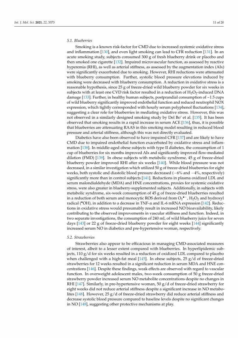
5.1. Blueberries
Smoking is a known risk-factor for CMD due to increased systemic oxidative stressand inflammation [130], and even light smoking can lead to CFR reduction [131]. In anacute smoking study, subjects consumed 300 g of fresh blueberry drink or placebo andthen smoked one cigarette [132]. Impaired microvascular function, as assessed by reactivehyperemia (RHI), as well as arterial stiffness, as assessed by the augmentation index (AIx)were significantly exacerbated due to smoking. However, RHI reductions were attenuatedwith blueberry consumption. Further, systolic blood pressure elevations induced bysmoking were decreased with blueberry consumption. A reduction in oxidative stress is areasonable hypothesis, since 25 g of freeze-dried wild blueberry powder for six weeks insubjects with at least one CVD risk factor resulted in a reduction of H2O2-induced DNAdamage [133]. Further, in healthy human subjects, postprandial consumption of ~1.5 cupsof wild blueberry significantly improved endothelial function and reduced neutrophil NOXexpression, which tightly corresponded with hourly serum polyphenol fluctuations [134],suggesting a clear role for blueberries in mediating oxidative stress. However, this wasnot observed in a similarly designed smoking study by Del Bo’ et al. [135]. It has beenobserved that smoking results in a rapid increase in serum ACE [136], thus, it is possiblethat blueberries are attenuating RAAS in this smoking model resulting in reduced bloodpressure and arterial stiffness, although this was not directly evaluated.
Diabetics have also been observed to have impaired CFR [137] and are likely to haveCMD due to impaired endothelial function exacerbated by oxidative stress and inflam-mation [138]. In middle-aged obese subjects with type II diabetes, the consumption of 1cup of blueberries for six months improved AIx and significantly improved flow-mediateddilation (FMD) [139]. In obese subjects with metabolic syndrome, 45 g of freeze-driedblueberry powder improved RHI after six weeks [140]. While blood pressure was notdecreased, in a similar investigation which utilized 50 g of freeze-dried blueberries for eightweeks, both systolic and diastolic blood pressure decreased (−6% and −4%, respectively)significantly more than in control subjects [141]. Reductions in plasma oxidized LDL andserum malondialdehyde (MDA) and HNE concentrations, proxies for systemic oxidativestress, were also greater in blueberry-supplemented subjects. Additionally, in subjects withmetabolic syndrome, six-week consumption of 45 g of freeze-dried blueberries resultedin a reduction of both serum and monocytic ROS derived from O2
•−, H2O2 and hydroxylradical (•OH), in addition to a decrease in TNF-α and IL-6 mRNA expression [142]. Reduc-tions in oxidative stress would presumably result in increased NO bioavailability, likelycontributing to the observed improvements in vascular stiffness and function. Indeed, intwo separate investigations, the consumption of 240 mL of wild blueberry juice for sevendays [143] or 22 g of freeze-dried blueberry powder for eight weeks [144] significantlyincreased serum NO in diabetics and pre-hypertensive woman, respectively.
5.2. Strawberries
Strawberries also appear to be efficacious in managing CMD-associated measuresof interest, albeit to a lesser extent compared with blueberries. In hyperlipidemic sub-jects, 110 g/d for six weeks resulted in a reduction of oxidized LDL compared to placebowhen challenged with a high-fat meal [145]. In obese subjects, 25 g/d of freeze-driedstrawberries for 12 weeks resulted in a significant reduction in serum MDA and HNE con-centrations [146]. Despite these findings, weak effects are observed with regard to vascularfunction. In overweight adolescent males, two-week consumption of 50 g freeze-driedstrawberry powder increased serum NO metabolite concentrations despite no changes inRHI [147]. Similarly, in pre-hypertensive woman, 50 g/d of freeze-dried strawberry foreight weeks did not reduce arterial stiffness despite a significant increase in NO metabo-lites [148]. However, 25 g/d of freeze-dried strawberry did reduce arterial stiffness anddecrease systolic blood pressure compared to baseline levels despite no significant changesin NO [148], suggesting other protective mechanisms at play.

Int. J. Mol. Sci. 2021, 22, 3373 12 of 20
5.3. Cranberries
A number of clinical investigations have demonstrated that cranberries are quite effica-cious in improving vascular function. For example, young healthy males consumed 450 mLof either 25, 48, 76, 94, or 117% concentrated cranberry juice equivalent to 409, 787, 1238,1534, and 1910 mg of total polyphenols, respectively [149]. While all doses of cranberrywere effective in improving FMD postprandially, 76% cranberry juice appeared most effica-cious. Interestingly, time-dependent changes were observed, with peak FMD at 4 h whichtightly corresponded with changes in polyphenols in serum. However, 117% cranberryjuice concentrate demonstrated the greatest changes in polyphenols despite less dramaticchanges in FMD compared with the 76% cranberry juice concentrate. Acute changes inFMD have also been observed with 450 mL of 54% juice in patients with coronary arterydisease [150]. However, chronic supplementation appears less effective, as two weeks of450 mL/d of 54% cranberry juice did not result in improvements in FMD [150], which wassimilar to observations in overweight men consuming 500 mL/d of 27% cranberry juicefor four weeks, in which AIx was unchanged [151]. Nonetheless, chronic supplementationwith 480 mL/d of 27% cranberry juice for eight weeks in women with metabolic syndromeresulted in a significant increase in plasma antioxidant capacity and decreased oxidizedLDL as well as MDA and HNE despite no changes in inflammatory biomarkers [152]. Inoverweight, middle aged subjects, 480 mL of ocean spray cranberry juice (% cranberryunspecified) for eight weeks significantly reduced diastolic blood pressure and CRP, ameasure of systemic inflammation [153]. Eight weeks of 500 mL/d of cranberry juiceconsumption also decreased oxidized LDL and endothelial inflammatory biomarkers, in-tracellular adhesion molecule-1 (ICAM-1) and vascular cell adhesion molecule-1 (VCAM-1)in overweight, middle-aged subjects [154]. Thus, cranberries appear to have protectivevascular effects and may decrease oxidative stress and inflammation.
5.4. Red and Black Raspberries
As with blueberries and cranberries, red raspberries appear to have potent postpran-dial effects on FMD. For example, 200 and 400 g of fresh red raspberries in healthy subjectsresulted in a significant increase in FMD at both 2 and 24 h postprandially, which corre-sponded with plasma changes in ellagic acid and urolithin metabolites, respectively [90]. Intype II diabetics, the consumption of a high-fat meal with 250 g of freeze-dried raspberriesresulted in significantly lower postprandial concentration of TNF-α and IL-6 comparedto control [155]. These protective effects were similar after four weeks of consuming250 g/d of fresh raspberries, in which fasting TNF-α and IL-6 were lower than control,as was systolic blood pressure. Interestingly, in overweight and obese subjects at risk formetabolic syndrome, 280 g/d of frozen red raspberry for eight weeks resulted in transcrip-tional changes of cells from whole blood, with downregulation of genes associated withinflammatory cytokine production [156].
Black raspberries also have protective vascular effects. For example, 750 mg ofdried black raspberry powder were provided to subjects with metabolic syndrome for12 weeks [157]. Significant reductions in TNF-α, IL-6 and ICAM-1 were observed com-pared to placebo, which corresponded with a significant reduction in AIx. Additionally,in patients with metabolic syndrome, 750 mg/d of black raspberry extract powder for12 weeks significantly improved FMD compared to placebo [158]. Lastly, in subject withpre-hypertension, 1500 or 2500 mg/d of black raspberry extract powder for eight weekssignificantly reduced 24 h systolic blood pressure, as well as serum IL-6 and TNF-α in adose-dependent manner [159]. Interestingly, serum renin and ACE appeared to decrease insubjects consuming the 2500 mg/d of the black raspberry powder; however, this differencewas not statistically significant between groups.
6. Limitations
Available clinical evidence suggests that berries, particularly blueberries, cranber-ries, red raspberries and black raspberries, elicit protective effects on vascular function,

Int. J. Mol. Sci. 2021, 22, 3373 13 of 20
potentially improving CMD. However, a major limitation of this conclusion is that noclinical studies exist utilizing berries or their polyphenols to treat CMD. Rather, clinical andpre-clinical evidence suggests that the polyphenols found in berries target the underlyingdrivers of CMD in a more comprehensive fashion compared to current pharmacologics.Specific recommendations regarding which berry to consume for CMD or which polyphe-nol is most efficacious cannot be appropriately made, and any such recommendation wouldbe unfounded based on the current state of the literature. Investigations utilizing berriesand their polyphenols in treating CMD are needed to determine efficacy and dosage.
7. Conclusions
INOCA is a clinically significant problem and is associated with morbidity and mortal-ity that is comparable to obstructive CAD. CMD is a major contributor of adverse outcomesin INOCA. The underlying drivers of CMD, namely, oxidative stress and inflammation,upregulated RAAS, and adrenergic signaling, represent major therapeutic targets. Whilemedications attempt to address these pathways, polypharmacy is typically not desirableand burdens patients with side effects and potential drug interactions. Compelling preclin-ical evidence suggests that berry-derived polyphenols can directly inhibit αAR, βAR, ACE,aldosterone, AT1R, NF-κB, MAPK, NOX and increase NRF2, thus targeting major pathwaysof interest in CMD that pharmaceuticals either partially address or do not address at all.Further, a number of clinical studies demonstrate the efficacy of berries in improving macro-and microvascular function, with blueberries, cranberries, and red and black raspberriesbeing particularly efficacious, with less compelling evidence for strawberries. Becauseno clinical studies utilizing berries currently exist that have aimed to treat CMD, futureclinical trials should utilize berries clinically as an adjunct treatment of this disease.
Supplementary Materials: Supplementary materials can be found at https://www.mdpi.com/1422-0067/22/7/3373/s1.
Author Contributions: Conceptualization: R.S.N., B.J.W., P.K.M. and R.G.F.; investigation: R.S.N.,A.M.S., B.J.W., P.K.M. and R.G.F.; resources: R.G.F.; writing—original draft preparation: R.S.N. andA.M.S.; writing—review and editing: R.S.N., A.M.S., B.J.W., P.K.M. and R.G.F.; supervision: B.J.W.,P.K.M. and R.G.F.; funding acquisition: P.K.M. and R.G.F. All authors have read and agreed to thepublished version of the manuscript.
Funding: This work is supported in part by the Agriculture and Food Research Initiative [grant no.2019-67017-29257/project accession no. 1018642] from the USDA National Institute of Food andAgriculture (R.G.F.) and by a developmental grant on Specialized Center of Research Excellencein Sex Differences (SCORE) from the NIH (1U54AG062334-01), K23HL105787, and Marcia Taylor(P.K.M.).
Conflicts of Interest: The authors declare no conflict of interest.
References1. Bugiardini, R.; Bairey Merz, C.N. Angina with “normal” coronary arteries: A changing philosophy. JAMA 2005, 293, 477–484.
[CrossRef]2. Jespersen, L.; Hvelplund, A.; Abildstrøm, S.Z.; Pedersen, F.; Galatius, S.; Madsen, J.K.; Jørgensen, E.; Kelbaek, H.; Prescott, E. Stable
angina pectoris with no obstructive coronary artery disease is associated with increased risks of major adverse cardiovascularevents. Eur. Heart J. 2011, 33, 734–744. [CrossRef] [PubMed]
3. Bairey Merz, C.N.; Pepine, C.J.; Walsh, M.N.; Fleg, J.L. Ischemia and No Obstructive Coronary Artery Disease (INOCA):Developing Evidence-Based Therapies and Research Agenda for the Next Decade. Circulation 2017, 135, 1075–1092. [CrossRef]
4. Gulati, M.; Cooper-DeHoff, R.M.; McClure, C.; Johnson, B.D.; Shaw, L.J.; Handberg, E.M.; Zineh, I.; Kelsey, S.F.; Arnsdorf, M.F.;Black, H.R.; et al. Adverse cardiovascular outcomes in women with nonobstructive coronary artery disease: A report from theWomen’s Ischemia Syndrome Evaluation Study and the St James Women Take Heart Project. Arch. Intern. Med. 2009, 169, 843–850.[CrossRef]
5. Sara, J.D.; Widmer, R.J.; Matsuzawa, Y.; Lennon, R.J.; Lerman, L.O.; Lerman, A. Prevalence of Coronary Microvascular DysfunctionAmong Patients with Chest Pain and Nonobstructive Coronary Artery Disease. JACC Cardiovasc. Interv. 2015, 8, 1445–1453.[CrossRef] [PubMed]

Int. J. Mol. Sci. 2021, 22, 3373 14 of 20
6. Mehta, P.K.; Bess, C.; Elias-Smale, S.; Vaccarino, V.; Quyyumi, A.; Pepine, C.J.; Merz, C.N.B. Gender in cardiovascular medicine:Chest pain and coronary artery disease. Eur. Heart J. 2019, 40, 3819–3826. [CrossRef] [PubMed]
7. Taqueti, V.R.; Di Carli, M.F. Coronary Microvascular Disease Pathogenic Mechanisms and Therapeutic Options: JACC State-of-the-Art Review. J. Am. Coll. Cardiol. 2018, 72, 2625–2641. [CrossRef] [PubMed]
8. Ong, P.; Camici, P.G.; Beltrame, J.F.; Crea, F.; Shimokawa, H.; Sechtem, U.; Kaski, J.C.; Merz, C.N.B. International standardizationof diagnostic criteria for microvascular angina. Int. J. Cardiol. 2018, 250, 16–20. [CrossRef]
9. Rahman, H.; Demir, O.M.; Ryan, M.; McConkey, H.; Scannell, C.; Ellis, H.; Webb, A.; Chiribiri, A.; Perera, D. Optimal Use ofVasodilators for Diagnosis of Microvascular Angina in the Cardiac Catheterization Laboratory. Circ. Cardiovasc. Interv. 2020, 13,009019. [CrossRef]
10. Bravo, P.E.; Di Carli, M.F.; Dorbala, S. Role of PET to evaluate coronary microvascular dysfunction in non-ischemic cardiomy-opathies. Heart Fail. Rev. 2017, 22, 455–464. [CrossRef]
11. Schwartz, B.G.; Economides, C.; Mayeda, G.S.; Burstein, S.; Kloner, R.A. The endothelial cell in health and disease: Its function,dysfunction, measurement and therapy. Int. J. Impot. Res. 2009, 22, 77–90. [CrossRef]
12. Ludmer, P.L.; Selwyn, A.P.; Shook, T.L.; Wayne, R.R.; Mudge, G.H.; Alexander, R.W.; Ganz, P. Paradoxical VasoconstrictionInduced by Acetylcholine in Atherosclerotic Coronary Arteries. N. Engl. J. Med. 1986, 315, 1046–1051. [CrossRef] [PubMed]
13. Pries, A.R.; Badimon, L.; Bugiardini, R.; Camici, P.G.; Dorobantu, M.; Duncker, D.J.; Escaned, J.; Koller, A.; Piek, J.J.; De Wit, C.Coronary vascular regulation, remodelling, and collateralization: Mechanisms and clinical implications on behalf of the workinggroup on coronary pathophysiology and microcirculation. Eur. Heart J. 2015, 36, 3134–3146. [CrossRef] [PubMed]
14. Bravo, P.E.; Zimmerman, S.L.; Luo, H.-C.; Pozios, I.; Rajaram, M.; Pinheiro, A.; Steenbergen, C.; Kamel, I.R.; Wahl, R.L.; Bluemke,D.A.; et al. Relationship of Delayed Enhancement by Magnetic Resonance to Myocardial Perfusion by Positron EmissionTomography in Hypertrophic Cardiomyopathy. Circ. Cardiovasc. Imaging 2013, 6, 210–217. [CrossRef] [PubMed]
15. Cecchi, F.; Olivotto, I.; Gistri, R.; Lorenzoni, R.; Chiriatti, G.; Camici, P.G. Coronary Microvascular Dysfunction and Prognosis inHypertrophic Cardiomyopathy. N. Engl. J. Med. 2003, 349, 1027–1035. [CrossRef] [PubMed]
16. Khuddus, M.A.; Pepine, C.J.; Handberg, E.M.; Bairey Merz, C.N.; Sopko, G.; Bavry, A.A.; Denardo, S.J.; McGorray, S.P.; Smith,K.M.; Sharaf, B.L.; et al. An intravascular ultrasound analysis in women experiencing chest pain in the absence of obstructivecoronary artery disease: A substudy from the National Heart, Lung and Blood Institute-Sponsored Women’s Ischemia SyndromeEvaluation (WISE). J. Interv. Cardiol. 2010, 23, 511–519. [CrossRef] [PubMed]
17. Albadri, A.; Eshtehardi, P.; Hung, O.Y.; Bouchi, Y.; Khawaja, S.; Mercado, K.; Corban, M.T.; Mehta, P.K.; Shaw, L.J.; Samady,H. Coronary Microvascular Dysfunction Is Associated with Significant Plaque Burden and Diffuse Epicardial AtheroscleroticDisease. JACC Cardiovasc. Interv. 2019, 12, 1519–1520. [CrossRef]
18. Yang, J.H.; Obokata, M.; Reddy, Y.N.; Redfield, M.M.; Lerman, A.; Borlaug, B.A. Endothelium-dependent and independentcoronary microvascular dysfunction in patients with heart failure with preserved ejection fraction. Eur. J. Heart Fail. 2020, 22,432–441. [CrossRef]
19. Löffler, A.I.; Bourque, J.M. Coronary Microvascular Dysfunction, Microvascular Angina, and Management. Curr. Cardiol. Rep.2016, 18, 1–7. [CrossRef]
20. Taqueti, V.R.; Shaw, L.J.; Cook, N.R.; Murthy, V.L.; Shah, N.R.; Foster, C.R.; Hainer, J.; Blankstein, R.; Dorbala, S.; Di Carli, M.F.Excess Cardiovascular Risk in Women Relative to Men Referred for Coronary Angiography Is Associated with Severely ImpairedCoronary Flow Reserve, Not Obstructive Disease. Circulation 2017, 135, 566–577. [CrossRef] [PubMed]
21. Hiteshi, A.K.; Li, N.; Gao, Y.; Chen, A.; Flores, F.; Mao, S.S.; Budoff, M.J. Gender Differences in Coronary Artery Diameter AreNot Related to Body Habitus or Left Ventricular Mass. Clin. Cardiol. 2014, 37, 605–609. [CrossRef]
22. Murthy, V.L.; Naya, M.; Taqueti, V.R.; Foster, C.R.; Gaber, M.; Hainer, J.; Dorbala, S.; Blankstein, R.; Rimoldi, O.; Camici, P.G.; et al.Effects of Sex on Coronary Microvascular Dysfunction and Cardiac Outcomes. Circulation 2014, 129, 2518–2527. [CrossRef]
23. Chatzizisis, Y.S.; Coskun, A.U.; Jonas, M.; Edelman, E.R.; Feldman, C.L.; Stone, P.H. Role of endothelial shear stress in the naturalhistory of coronary atherosclerosis and vascular remodeling: Molecular, cellular, and vascular behavior. J. Am. Coll. Cardiol. 2007,49, 2379–2393. [CrossRef]
24. Panza, A.J.; Laurienzo, J.M.; Curiel, R.V.; Unger, E.F.; Quyyumi, A.A.; Dilsizian, V.; Cannon, R.O. Investigation of the Mecha-nism of Chest Pain in Patients with Angiographically Normal Coronary Arteries Using Transesophageal Dobutamine StressEchocardiography. J. Am. Coll. Cardiol. 1997, 29, 293–301. [CrossRef]
25. Quyyumi, A.A.; Cannon, R.O.; Panza, J.A.; Diodati, J.G.; Epstein, S.E. Endothelial dysfunction in patients with chest pain andnormal coronary arteries. Circulation 1992, 86, 1864–1871. [CrossRef] [PubMed]
26. Kip, K.E.; Marroquin, O.C.; Shaw, L.J.; Arant, C.B.; Wessel, T.R.; Olson, M.B.; Johnson, B.D.; Mulukutla, S.; Sopko, G.; Merz, C.N.;et al. Global inflammation predicts cardiovascular risk in women: A report from the Women’s Ischemia Syndrome Evaluation(WISE) study. Am. Heart J. 2005, 150, 900–906. [CrossRef] [PubMed]
27. Marroquin, O.C.; Kip, K.E.; Mulukutla, S.R.; Ridker, P.M.; Pepine, C.J.; Tjandrawan, T.; Kelsey, S.F.; Mankad, S.; Rogers, W.J.;Merz, C.N.B.; et al. Inflammation, endothelial cell activation, and coronary microvascular dysfunction in women with chest painand no obstructive coronary artery disease. Am. Heart J. 2005, 150, 109–115. [CrossRef] [PubMed]
28. Shaw, L.J.; Bugiardini, R.; Merz, C.N. Women and ischemic heart disease: Evolving knowledge. J. Am. Coll. Cardiol. 2009, 54,1561–1575. [CrossRef]

Int. J. Mol. Sci. 2021, 22, 3373 15 of 20
29. Wessel, T.R.; Arant, C.B.; McGorray, S.P.; Sharaf, B.L.; Reis, S.E.; Kerensky, R.A.; von Mering, G.O.; Smith, K.M.; Pauly, D.F.;Handberg, E.M.; et al. Coronary microvascular reactivity is only partially predicted by atherosclerosis risk factors or coronaryartery disease in women evaluated for suspected ischemia: Results from the NHLBI Women’s Ischemia Syndrome Evaluation(WISE). Clin. Cardiol. 2007, 30, 69–74. [CrossRef] [PubMed]
30. Daiber, A.; Chlopicki, S. Revisiting pharmacology of oxidative stress and endothelial dysfunction in cardiovascular disease:Evidence for redox-based therapies. Free Radic. Biol. Med. 2020, 157, 15–37. [CrossRef]
31. Lanza, G.A.; Crea, F. Response to Letter Regarding Article, Primary Coronary Microvascular Dysfunction: Clinical Presentation,Pathophysiology, and Management. Circulation 2011, 123, 2317–2325. [CrossRef]
32. Higashi, Y.; Maruhashi, T.; Noma, K.; Kihara, Y. Oxidative stress and endothelial dysfunction: Clinical evidence and therapeuticimplications. Trends Cardiovasc. Med. 2014, 24, 165–169. [CrossRef] [PubMed]
33. Faccini, A.; Kaski, J.C.; Camici, P.G. Coronary microvascular dysfunction in chronic inflammatory rheumatoid diseases. Eur.Heart J. 2016, 37, 1799–1806. [CrossRef]
34. Altıparmak, I.H.; Erkus, M.E.; Sezen, H.; Demirbag, R.; Gunebakmaz, O.; Kaya, Z.; Sezen, Y.; Asoglu, R.; Dedeoglu, I.H.;Neselioglu, S.; et al. The relation of serum thiol levels and thiol/disulphide homeostasis with the severity of coronary arterydisease. Kardiol. Polska 2016, 74, 1346–1353. [CrossRef]
35. Bilir, B.; Akkoyun, D.C.; Aydın, M.; Ozkaramanli-Gur, D.; Degirmenci, H.; Albayrak, N.; Akyuz, A.; Alpsoy, S.; Koca, C.; Erel, Ö.Association of coronary artery disease severity and disulphide/native thiol ratio. Electron. J. Gen. Med. 2017, 14, 30–33. [CrossRef]
36. Schroder, J.; Mygind, N.D.; Frestad, D.; Michelsen, M.; Suhrs, H.E.; Bove, K.B.; Gustafsson, I.; Kastrup, J.; Prescott, E. Pro-inflammatory biomarkers in women with non-obstructive angina pectoris and coronary microvascular dysfunction. IJC HeartVasc. 2019, 24, 100370. [CrossRef]
37. Tona, F.; Serra, R.; Di Ascenzo, L.; Osto, E.; Scarda, A.; Fabris, R.; Montisci, R.; Famoso, G.; Tellatin, S.; Foletto, M.; et al. Systemicinflammation is related to coronary microvascular dysfunction in obese patients without obstructive coronary disease. Nutr.Metab. Cardiovasc. Dis. 2014, 24, 447–453. [CrossRef]
38. Piaserico, S.; Osto, E.; Famoso, G.; Zanetti, I.; Gregori, D.; Poretto, A.; Iliceto, S.; Peserico, A.; Tona, F. Treatment with tumornecrosis factor inhibitors restores coronary microvascular function in young patients with severe psoriasis. Atherosclerosis 2016,251, 25–30. [CrossRef] [PubMed]
39. Cosín-Sales, J.; Pizzi, C.; Brown, S.; Kaski, J.C. C-reactive protein, clinical presentation, and ischemic activity in patients withchest pain and normal coronary angiograms. J. Am. Coll. Cardiol. 2003, 41, 1468–1474. [CrossRef]
40. Bedard, K.; Krause, K.H. The NOX Family of ROS-Generating NADPH Oxidases: Physiology and Pathophysiology. Physiol. Rev.2007, 87, 245–313. [CrossRef] [PubMed]
41. Santillo, M.; Colantuoni, A.; Mondola, P.; Guida, B.; Damiano, S. NOX signaling in molecular cardiovascular mechanismsinvolved in the blood pressure homeostasis. Front. Physiol. 2015, 6, 194. [CrossRef]
42. Trebak, M.; Ginnan, R.; Singer, H.A.; Jourd’Heuil, D. Interplay between Calcium and Reactive Oxygen/Nitrogen Species: AnEssential Paradigm for Vascular Smooth Muscle Signaling. Antioxid. Redox. Signal. 2010, 12, 657–674. [CrossRef] [PubMed]
43. Montezano, A.C.; Touyz, R.M. Reactive Oxygen Species and Endothelial Function—Role of Nitric Oxide Synthase Uncouplingand Nox Family Nicotinamide Adenine Dinucleotide Phosphate Oxidases. Basic Clin. Pharmacol. Toxicol. 2011, 110, 87–94.[CrossRef] [PubMed]
44. Touyz, R.M.; Alves-Lopes, R.; Rios, F.J.; Camargo, L.L.; Anagnostopoulou, A.; Arner, A.; Montezano, A.C. Vascular smoothmuscle contraction in hypertension. Cardiovasc. Res. 2018, 114, 529–539. [CrossRef]
45. Zhang, H.; Park, Y.; Wu, J.; Chen, X.P.; Lee, S.; Yang, J.; Dellsperger, K.C.; Zhang, C. Role of TNF-α in vascular dysfunction. Clin.Sci. 2009, 116, 219–230. [CrossRef]
46. Camici, P.G.; Olivotto, I.; Rimoldi, O.E. The coronary circulation and blood flow in left ventricular hypertrophy. J. Mol. Cell.Cardiol. 2012, 52, 857–864. [CrossRef]
47. Perrotta, I.; Aquila, S. The Role of Oxidative Stress and Autophagy in Atherosclerosis. Oxidative Med. Cell. Longev. 2015, 2015,1–10. [CrossRef] [PubMed]
48. Hansson, G.K. Inflammation, Atherosclerosis, and Coronary Artery Disease. N. Engl. J. Med. 2005, 352, 1685–1695. [CrossRef]49. Aviram, M.; Rosenblat, M.; Etzioni, A.; Lévy, R. Activation of NADPH oxidase is required for macrophage-mediated oxidation of
low-density lipoprotein. Metabolism 1996, 45, 1069–1079. [CrossRef]50. Schaub, M.C.; Hefti, M.A.; Harder, B.A.; Eppenberger, H.M. Various hypertrophic stimuli induce distinct phenotypes in
cardiomyocytes. J. Mol. Med. 1997, 75, 901–920. [CrossRef]51. Wu, C.-H.; Mohammadmoradi, S.; Chen, J.Z.; Sawada, H.; Daugherty, A.; Lu, H.S. Renin-Angiotensin System and Cardiovascular
Functions. Arter. Thromb. Vasc. Biol. 2018, 38, e108–e116. [CrossRef] [PubMed]52. Carey, R.M.; Wang, Z.-Q.; Siragy, H.M. Role of the Angiotensin Type 2 Receptor in the Regulation of Blood Pressure and Renal
Function. Hypertension 2000, 35, 155–163. [CrossRef] [PubMed]53. Freel, E.M.; Connell, J.M. Mechanisms of hypertension: The expanding role of aldosterone. J. Am. Soc. Nephrol. 2004, 15,
1993–2001. [CrossRef] [PubMed]54. Usberti, M.; Federico, S.; Di Minno, G.; Ungaro, B.; Ardillo, G.; Pecoraro, C.; Cianciaruso, B.; Cerbone, A.M.; Cirillo, F.; Pannain,
M.; et al. Effects of angiotensin II on plasma ADH, prostaglandin synthesis, and water excretion in normal humans. Am. J. Physiol.Physiol. 1985, 248, F254–F259. [CrossRef] [PubMed]

Int. J. Mol. Sci. 2021, 22, 3373 16 of 20
55. Nakashima, H.; Suzuki, H.; Ohtsu, H.; Chao, J.Y.; Utsunomiya, H.; Frank, G.D.; Eguchi, S. Angiotensin II Regulates Vascular andEndothelial Dysfunction: Recent Topics of Angiotensin II Type-1 Receptor Signaling in the Vasculature. Curr. Vasc. Pharmacol.2006, 4, 67–78. [CrossRef]
56. Satou, R.; Penrose, H.; Navar, L.G. Inflammation as a Regulator of the Renin-Angiotensin System and Blood Pressure. Curr.Hypertens. Rep. 2018, 20, 1–9. [CrossRef]
57. Hanafy, S.; Tavasoli, M.; Jamali, F. Inflammation Alters Angiotensin Converting Enzymes (ACE and ACE-2) Balance in Rat Heart.Inflammation 2010, 34, 609–613. [CrossRef]
58. Pauly, D.F.; Johnson, B.D.; Anderson, R.D.; Handberg, E.M.; Smith, K.M.; Cooper-DeHoff, R.M.; Sopko, G.; Sharaf, B.M.; Kelsey,S.F.; Merz, C.N.B.; et al. In women with symptoms of cardiac ischemia, nonobstructive coronary arteries, and microvasculardysfunction, angiotensin-converting enzyme inhibition is associated with improved microvascular function: A double-blindrandomized study from the National Heart, Lung and Blood Institute Women’s Ischemia Syndrome Evaluation (WISE). Am.Heart J. 2011, 162, 678–684.
59. Schwartzkopff, B.; Brehm, M.; Mundhenke, M.; Strauer, B.E. Repair of Coronary Arterioles After Treatment with Perindopril inHypertensive Heart Disease. Hypertension 2000, 36, 220–225. [CrossRef]
60. Serné, E.H.; de Jongh, R.T.; Eringa, E.C.; IJzerman, R.G.; Stehouwer, C.D. Microvascular dysfunction: A potential pathophysiolog-ical role in the metabolic syndrome. Hypertension 2007, 50, 204–211. [CrossRef]
61. Woo, A.Y.; Xiao, R.P. beta-Adrenergic receptor subtype signaling in heart: From bench to bedside. Acta Pharmacol. Sin. 2012, 33,335–341. [CrossRef] [PubMed]
62. Michelotti, A.G.; Price, D.T.; Schwinn, A.D. Alpha 1-adrenergic receptor regulation: Basic science and clinical implications.Pharmacol. Ther. 2000, 88, 281–309. [CrossRef]
63. Pinilla-Vera, M.; Hahn, V.S.; Kass, D.A. Leveraging Signaling Pathways to Treat Heart Failure with Reduced Ejection Fraction.Circ. Res. 2019, 124, 1618–1632. [CrossRef]
64. Gericke, A.; Martinka, P.; Nazarenko, I.; Persson, P.B.; Patzak, A. Impact of α1-adrenoceptor expression on contractile propertiesof vascular smooth muscle cells. Am. J. Physiol. Integr. Comp. Physiol. 2007, 293, R1215–R1221. [CrossRef] [PubMed]
65. Mehta, P.K.; Nelson, M.; Thomson, L.; Friedman, J.; Hayes, S.; Hermel, D.; Slomka, P.; Swift, A.; Wei, J.; Cook-Wiens, G.; et al.Abnormal cardiac sympathetic activity detected by 123-I-meta-iodobenzylguanidine imaging in women with signs and symptomsof ischemia and no obstructive coronary artery disease. J. Am. Coll. Cardiol. 2016, 67 (Suppl. 13), 1619. [CrossRef]
66. Lanza, G.A.; Giordano, A.; Pristipino, C.; Calcagni, M.L.; Meduri, G.; Trani, C.; Franceschini, R.; Crea, F.; Troncone, L.; Maseri, A.Abnormal cardiac adrenergic nerve function in patients with syndrome X detected by [123I]metaiodobenzylguanidine myocardialscintigraphy. Circulation 1997, 96, 821–826. [CrossRef] [PubMed]
67. Galderisi, M.; D’Errico, A. Beta-blockers and coronary flow reserve: The importance of a vasodilatory action. Drugs 2008, 68,579–590. [CrossRef]
68. Pedersen, M.E.; Cockcroft, J.R. The vasodilatory beta-blockers. Curr. Hypertens. Rep. 2007, 9, 269–277. [CrossRef]69. Cohn, J.; Archibald, D.; Ziesche, S.; Franciosa, J.; Harston, W.; Tristani, F.; Dunkman, W.; Jacobs, W.; Francis, G.; Flohr, K.; et al.
Effect of Vasodilator Therapy on Mortality in Chronic Congestive Heart Failure. Results of a Veterans Administration CooperativeStudy. Surv. Anesthesiol. 1987, 31, 53. [CrossRef]
70. Lymperopoulos, A.; Rengo, G.; Koch, W.J. Adrenergic nervous system in heart failure: Pathophysiology and therapy. Circ. Res.2013, 113, 739–753. [CrossRef]
71. Engelhardt, S.; Hein, L.; Wiesmann, F.; Lohse, M.J. Progressive hypertrophy and heart failure in beta1-adrenergic receptortransgenic mice. Proc. Natl. Acad. Sci. USA 1999, 96, 7059–7064. [CrossRef]
72. Task Force, M.; Montalescot, G.; Sechtem, U.; Achenbach, S.; Andreotti, F.; Arden, C.; Budaj, A.; Bugiardini, R.; Crea, F.; Cuisset,T.; et al. 2013 ESC guidelines on the management of stable coronary artery disease: The Task Force on the management of stablecoronary artery disease of the European Society of Cardiology. Eur. Heart J. 2013, 34, 2949–3003.
73. Shaw, L.J.; Veledar, E.; Berman, D.S.; Hayes, S.W.; Friedman, J.; Slomka, P.; Germano, G.; Maron, D.J.; Mancini, G.B.J.; Hartigan,P.M.; et al. Response to Letters Regarding Article, “Optimal Medical Therapy with or Without Percutaneous Coronary Interventionto Reduce Ischemic Burden: Results from the Clinical Outcomes Utilizing Revascularization and Aggressive Drug Evaluation(COURAGE) Trial Nuclear Substudy”. Circulation 2008, 118, 1283–1291.
74. Merz, C.N.B.; Pepine, C.J.; Shimokawa, H.; Berry, C. Treatment of coronary microvascular dysfunction. Cardiovasc. Res. 2020, 116,856–870. [CrossRef] [PubMed]
75. Olsen, R.H.; Pedersen, L.R.; Jürs, A.; Snoer, M.; Haugaard, S.B.; Prescott, E. A randomised trial comparing the effect of exercisetraining and weight loss on microvascular function in coronary artery disease. Int. J. Cardiol. 2015, 185, 229–235. [CrossRef][PubMed]
76. Najjar, R.; Turner, C.; Wong, B.; Feresin, R. Berry-Derived Polyphenols in Cardiovascular Pathologies: Mechanisms of Disease andthe Role of Diet and Sex. Nutrients 2021, 13, 387. [CrossRef] [PubMed]
77. Del Rio, D.; Rodriguez-Mateos, A.; Spencer, J.P.; Tognolini, M.; Borges, G.; Crozier, A. Dietary (Poly)phenolics in Human Health:Structures, Bioavailability, and Evidence of Protective Effects Against Chronic Diseases. Antioxid. Redox. Signal. 2013, 18,1818–1892. [CrossRef] [PubMed]
78. Perezjimenez, J.; Neveu, V.; Vos, F.; Scalbert, A. Identification of the 100 richest dietary sources of polyphenols: An application ofthe Phenol-Explorer database. Eur. J. Clin. Nutr. 2010, 64 (Suppl. 3), S112–S120. [CrossRef]

Int. J. Mol. Sci. 2021, 22, 3373 17 of 20
79. Olas, B. Berry Phenolic Antioxidants—Implications for Human Health? Front. Pharmacol. 2018, 9, 78. [CrossRef] [PubMed]80. Rothwell, J.A.; Perez-Jimenez, J.; Neveu, V.; Medina-Remón, A.; M’Hiri, N.; García-Lobato, P.; Manach, C.; Knox, C.; Eisner, R.;
Wishart, D.S.; et al. Phenol-Explorer 3.0: A major update of the Phenol-Explorer database to incorporate data on the effects offood processing on polyphenol content. Database 2013, 2013, bat070. [CrossRef] [PubMed]
81. Kimble, R.; Keane, K.M.; Lodge, J.K.; Howatson, G. Dietary intake of anthocyanins and risk of cardiovascular disease: Asystematic review and meta-analysis of prospective cohort studies. Crit. Rev. Food Sci. Nutr. 2018, 59, 3032–3043. [CrossRef][PubMed]
82. Zamora-Ros, R.; Rabassa, M.; Cherubini, A.; Urpi-Sarda, M.; Bandinelli, S.; Ferrucci, L.; Andres-Lacueva, C. High Concentrationsof a Urinary Biomarker of Polyphenol Intake Are Associated with Decreased Mortality in Older Adults. J. Nutr. 2013, 143,1445–1450. [CrossRef] [PubMed]
83. Ivey, K.L.; Hodgson, J.M.; Croft, K.D.; Lewis, J.R.; Prince, R.L. Flavonoid intake and all-cause mortality. Am. J. Clin. Nutr. 2015,101, 1012–1020. [CrossRef] [PubMed]
84. Jin, L.; Piao, Z.H.; Sun, S.; Liu, B.; Kim, G.R.; Seok, Y.M.; Lin, M.Q.; Ryu, Y.; Choi, S.Y.; Kee, H.J.; et al. Gallic Acid Reduces BloodPressure and Attenuates Oxidative Stress and Cardiac Hypertrophy in Spontaneously Hypertensive Rats. Sci. Rep. 2017, 7, 15607.[CrossRef]
85. Rozentsvit, A.; Vinokur, K.; Samuel, S.; Li, Y.; Gerdes, A.M.; Carrillo-Sepulveda, M.A. Ellagic Acid Reduces High. Glucose-Induced Vascular Oxidative Stress Through ERK1/2/NOX4 Signaling Pathway. Cell Physiol. Biochem. 2017, 44, 1174–1187.[CrossRef] [PubMed]
86. Vrhovsek, U.; Giongo, L.; Mattivi, F.; Viola, R. A survey of ellagitannin content in raspberry and blackberry cultivars grown inTrentino (Italy). Eur. Food Res. Technol. 2008, 226, 817–824. [CrossRef]
87. Häkkinen, S.H.; Kärenlampi, S.O.; Mykkänen, H.M.; Heinonen, I.M.; Torronen, A. Ellagic acid content in berries: Influence ofdomestic processing and storage. Eur. Food Res. Technol. 2000, 212, 75–80. [CrossRef]
88. Tonelli, C.; Chio, I.I.C.; Tuveson, D.A. Transcriptional Regulation by Nrf2. Antioxid. Redox. Signal. 2018, 29, 1727–1745. [CrossRef]89. Truchado, P.; Larrosa, M.; García-Conesa, M.T.; Cerdá, B.; Vidal-Guevara, M.L.; Tomás-Barberán, F.A.; Espín, J.C. Strawberry
Processing Does Not Affect the Production and Urinary Excretion of Urolithins, Ellagic Acid Metabolites, in Humans. J. Agric.Food Chem. 2011, 60, 5749–5754. [CrossRef]
90. Istas, G.; Feliciano, R.P.; Weber, T.; Garcia-Villalba, R.; Tomas-Barberan, F.; Heiss, C.; Rodriguez-Mateos, A. Plasma urolithinmetabolites correlate with improvements in endothelial function after red raspberry consumption: A double-blind randomizedcontrolled trial. Arch. Biochem. Biophys. 2018, 651, 43–51. [CrossRef]
91. Zheng, D.; Liu, Z.; Zhou, Y.; Hou, N.; Yan, W.; Qin, Y.; Ye, Q.; Cheng, X.; Xiao, Q.; Bao, Y.; et al. Urolithin B, a gut microbiotametabolite, protects against myocardial ischemia/reperfusion injury via p62/Keap1/Nrf2 signaling pathway. Pharmacol. Res.2020, 153, 104655. [CrossRef] [PubMed]
92. Häkkinen, S.H.; Kärenlampi, S.O.; Heinonen, I.M.; Mykkänen, H.M.; Törrönen, A.R. Content of the Flavonols Quercetin,Myricetin, and Kaempferol in 25 Edible Berries. J. Agric. Food Chem. 1999, 47, 2274–2279. [CrossRef]
93. Liao, H.H.; Zhang, N.; Meng, Y.Y.; Feng, H.; Yang, J.J.; Li, W.J.; Chen, S.; Wu, H.M.; Deng, W.; Tang, Q.Z. Myricetin AlleviatesPathological Cardiac Hypertrophy via TRAF6/TAK1/MAPK and Nrf2 Signaling Pathway. Oxid. Med. Cell Longev. 2019, 2019,6304058. [CrossRef]
94. Zhang, C.; Lin, G.; Wan, W.; Li, X.; Zeng, B.; Yang, B.; Huang, C. Resveratrol, a polyphenol phytoalexin, protects cardiomyocytesagainst anoxia/reoxygenation injury via the TLR4/NF-kappaB signaling pathway. Int. J. Mol. Med. 2012, 29, 557–563. [CrossRef]
95. Pantan, R.; Tocharus, J.; Suksamrarn, A.; Tocharus, C. Synergistic effect of atorvastatin and Cyanidin-3-glucoside on angiotensinII-induced inflammation in vascular smooth muscle cells. Exp. Cell Res. 2016, 342, 104–112. [CrossRef] [PubMed]
96. Sivasinprasasn, S.; Pantan, R.; Thummayot, S.; Tocharus, J.; Suksamrarn, A.; Tocharus, C. Cyanidin-3-glucoside attenuatesangiotensin II-induced oxidative stress and inflammation in vascular endothelial cells. Chem. Interact. 2016, 260, 67–74. [CrossRef]
97. Zhang, M.; Xue, Y.; Chen, H.; Meng, L.; Chen, B.; Gong, H.; Zhao, Y.; Qi, R. Resveratrol Inhibits MMP3 and MMP9 Expressionand Secretion by Suppressing TLR4/NF-kappaB/STAT3 Activation in Ox-LDL-Treated HUVECs. Oxid. Med. Cell Longev. 2019,2019, 9013169.
98. Li, T.; Li, F.; Liu, X.; Liu, J.; Li, D. Synergistic anti-inflammatory effects of quercetin and catechin via inhibiting activation ofTLR4-MyD88-mediated NF-kappaB and MAPK signaling pathways. Phytother. Res. 2019, 33, 756–767. [CrossRef] [PubMed]
99. Bhaskar, S.; Sudhakaran, P.R.; Helen, A. Quercetin attenuates atherosclerotic inflammation and adhesion molecule expression bymodulating TLR-NF-kappaB signaling pathway. Cell Immunol. 2016, 310, 131–140. [CrossRef] [PubMed]
100. Feresin, R.G.; Huang, J.; Klarich, D.S.; Zhao, Y.; Pourafshar, S.; Arjmandi, B.H.; Salazar, G. Blackberry, raspberry and blackraspberry polyphenol extracts attenuate angiotensin II-induced senescence in vascular smooth muscle cells. Food Funct. 2016, 7,4175–4187. [CrossRef]
101. Luo, X.; Fang, S.; Xiao, Y.; Song, F.; Zou, T.; Wang, M.; Xia, M.; Ling, W. Cyanidin-3-glucoside suppresses TNF-α-inducedcell proliferation through the repression of Nox activator 1 in mouse vascular smooth muscle cells: Involvement of the STAT3signaling. Mol. Cell. Biochem. 2011, 362, 211–218. [CrossRef]
102. Parichatikanond, W.; Pinthong, D.; Mangmool, S. Blockade of the Renin-Angiotensin System with Delphinidin, Cyanin, andQuercetin. Planta Med. 2012, 78, 1626–1632. [CrossRef]

Int. J. Mol. Sci. 2021, 22, 3373 18 of 20
103. Neto-Neves, E.M.; Montenegro, M.F.; Dias-Junior, C.A.; Spiller, F.; Kanashiro, A.; Tanus-Santos, J.E. Chronic Treatment withQuercetin does not Inhibit Angiotensin-Converting Enzyme In Vivo or In Vitro. Basic Clin. Pharmacol. Toxicol. 2010, 107, 825–829.[CrossRef]
104. Paul, M.; Wagner, J.; Dzau, V.J. Gene expression of the renin-angiotensin system in human tissues. Quantitative analysis by thepolymerase chain reaction. J. Clin. Investig. 1993, 91, 2058–2064. [CrossRef]
105. Jain, S.; Li, Y.; Patil, S.; Kumar, A. HNF-1α plays an important role in IL-6-induced expression of the human angiotensinogengene. Am. J. Physiol. Physiol. 2007, 293, C401–C410. [CrossRef]
106. Brasier, A.R.; Ron, D.; Tate, J.E.; Habener, J.F. A family of constitutive C/EBP-like DNA binding proteins attenuate the IL-1 alphainduced, NF kappa B mediated trans-activation of the angiotensinogen gene acute-phase response element. EMBO J. 1990, 9,3933–3944. [CrossRef] [PubMed]
107. O’Leary, R.; Penrose, H.; Miyata, K.; Satou, R. Macrophage-derived IL-6 contributes to ANG II-mediated angiotensinogenstimulation in renal proximal tubular cells. Am. J. Physiol. Renal Physiol. 2016, 310, F1000–F1007. [CrossRef] [PubMed]
108. Flesch, M.; Höper, A.; Italia, L.D.; Evans, K.; Bond, R.; Peshock, R.; Diwan, A.; Brinsa, T.A.; Wei, C.-C.; Sivasubramanian, N.;et al. Activation and Functional Significance of the Renin-Angiotensin System in Mice with Cardiac Restricted Overexpression ofTumor Necrosis Factor. Circulation 2003, 108, 598–604. [CrossRef] [PubMed]
109. Huang, W.; Hutabarat, R.P.; Chai, Z.; Zheng, T.; Zhang, W.; Li, D. Antioxidant Blueberry Anthocyanins Induce Vasodilation viaPI3K/Akt Signaling Pathway in High-Glucose-Induced Human Umbilical Vein Endothelial Cells. Int. J. Mol. Sci. 2020, 21, 1575.[CrossRef] [PubMed]
110. Wiseman, W.; Egan, J.M.; Slemmer, J.E.; Shaughnessy, K.S.; Ballem, K.; Gottschall-Pass, K.T.; Sweeney, M.I. Feeding blueberrydiets inhibits angiotensin II-converting enzyme (ACE) activity in spontaneously hypertensive stroke-prone rats. Can. J. Physiol.Pharmacol. 2011, 89, 67–71. [CrossRef]
111. Miyazaki, R.; Ichiki, T.; Hashimoto, T.; Inanaga, K.; Imayama, I.; Sadoshima, J.; Sunagawa, K. SIRT1, a Longevity Gene,Downregulates Angiotensin II Type 1 Receptor Expression in Vascular Smooth Muscle Cells. Arter. Thromb. Vasc. Biol. 2008, 28,1263–1269. [CrossRef]
112. D’Onofrio, N.; Servillo, L.; Balestrieri, M.L. SIRT1 and SIRT6 Signaling Pathways in Cardiovascular Disease Protection. Antioxid.Redox. Signal. 2018, 28, 711–732. [CrossRef]
113. Hung, C.-H.; Chan, S.-H.; Chu, P.-M.; Tsai, K.-L. Quercetin is a potent anti-atherosclerotic compound by activation of SIRT1signaling under oxLDL stimulation. Mol. Nutr. Food Res. 2015, 59, 1905–1917. [CrossRef]
114. de Boer, V.C.; de Goffau, M.C.; Arts, I.C.; Hollman, P.C.; Keijer, J. SIRT1 stimulation by polyphenols is affected by their stabilityand metabolism. Mech. Ageing Dev. 2006, 127, 618–627. [CrossRef]
115. Jin, X.; Chen, M.; Yi, L.; Chang, H.; Zhang, T.; Wang, L.; Ma, W.; Peng, X.; Zhou, Y.; Mi, M. Delphinidin-3-glucoside protectshuman umbilical vein endothelial cells against oxidized low-density lipoprotein-induced injury by autophagy upregulation viathe AMPK/SIRT1 signaling pathway. Mol. Nutr. Food Res. 2014, 58, 1941–1951. [CrossRef] [PubMed]
116. Bhullar, K.S.; Lassalle-Claux, G.; Touaibia, M.; Rupasinghe, H.V. Antihypertensive effect of caffeic acid and its analogs throughdual renin–angiotensin–aldosterone system inhibition. Eur. J. Pharmacol. 2014, 730, 125–132. [CrossRef]
117. Fujimoto, S.-I.; Niisato, N.; Sugimoto, T.; Marunaka, Y. Quercetin and NPPB-induced diminution of aldosterone action on Na+absorption and ENaC expression in renal epithelium. Biochem. Biophys. Res. Commun. 2005, 336, 401–407. [CrossRef] [PubMed]
118. Maron, B.A.; Leopold, J.A. Mineralocorticoid receptor antagonists and endothelial function. Curr. Opin. Investig. Drugs 2008, 9,963–969. [PubMed]
119. Xiao, H.B.; Lu, X.Y.; Liu, Z.K.; Luo, Z.F. Kaempferol inhibits the production of ROS to modulate OPN-alphavbeta3 integrinpathway in HUVECs. J. Physiol. Biochem. 2016, 72, 303–313. [CrossRef]
120. Liu, Z.-K.; Xiao, H.-B.; Fang, J. Anti-inflammatory properties of kaempferol via its inhibition of aldosterone signaling andaldosterone-induced gene expression. Can. J. Physiol. Pharmacol. 2014, 92, 117–123. [CrossRef]
121. Norton, C.; Kalea, A.Z.; Harris, P.D.; Klimis-Zacas, D.J. Wild Blueberry-Rich Diets Affect the Contractile Machinery of the VascularSmooth Muscle in the Sprague–Dawley Rat. J. Med. Food 2005, 8, 8–13. [CrossRef] [PubMed]
122. Kristo, A.S.; Kalea, A.Z.; Schuschke, D.A.; Klimis-Zacas, D.J. A Wild Blueberry-Enriched Diet (Vaccinium angustifolium) ImprovesVascular Tone in the Adult Spontaneously Hypertensive Rat. J. Agric. Food Chem. 2010, 58, 11600–11605. [CrossRef]
123. Rodriguez-Mateos, A.; Ishisaka, A.; Mawatari, K.; Vidal-Diez, A.; Spencer, J.P.E.; Terao, J. Blueberry intervention improvesvascular reactivity and lowers blood pressure in high-fat-, high-cholesterol-fed rats. Br. J. Nutr. 2013, 109, 1746–1754. [CrossRef][PubMed]
124. Vendrame, S.; Kristo, A.; Schuschke, D.A.; Klimis-Zacas, D. Wild blueberry consumption affects aortic vascular function in theobese Zucker rat. Appl. Physiol. Nutr. Metab. 2014, 39, 255–261. [CrossRef] [PubMed]
125. Louis, X.L.; Thandapilly, S.J.; Kalt, W.; Vinqvist-Tymchuk, M.; Aloud, B.M.; Raj, P.; Yu, L.; Le, H.; Netticadan, T. Blueberrypolyphenols prevent cardiomyocyte death by preventing calpain activation and oxidative stress. Food Funct. 2014, 5, 1785–1794.[CrossRef]
126. Wei, H.; Li, H.; Wan, S.-P.; Zeng, Q.-T.; Cheng, L.-X.; Jiang, L.-L.; Peng, Y.-D. Cardioprotective Effects of Malvidin AgainstIsoproterenol-Induced Myocardial Infarction in Rats: A Mechanistic Study. Med. Sci. Monit. 2017, 23, 2007–2016. [CrossRef]

Int. J. Mol. Sci. 2021, 22, 3373 19 of 20
127. Ryu, Y.; Jin, L.; Kee, H.J.; Piao, Z.H.; Cho, J.Y.; Kim, G.R.; Choi, S.Y.; Lin, M.Q.; Jeong, M.H. Gallic acid prevents isoproterenol-induced cardiac hypertrophy and fibrosis through regulation of JNK2 signaling and Smad3 binding activity. Sci. Rep. 2016, 6,34790. [CrossRef]
128. De Lacerda Alexandre, J.V.D.L.; Viana, Y.I.P.; David, C.E.B.; Cunha, P.L.O.; Albuquerque, A.C.; Varela, A.L.N.; Kowaltowski,A.J.; Facundo, H.T. Quercetin treatment increases H2O2 removal by restoration of endogenous antioxidant activity and blocksisoproterenol-induced cardiac hypertrophy. Naunyn-Schmiedeberg’s Arch. Pharmacol. 2021, 394, 217–226. [CrossRef]
129. Manjunatha, S.; Shaik, A.H.; E., M.P.; Al Omar, S.Y.; Mohammad, A.; Kodidhela, L.D. Combined cardio-protective ability ofsyringic acid and resveratrol against isoproterenol induced cardio-toxicity in rats via attenuating NF-kB and TNF-alpha pathways.Sci. Rep. 2020, 10, 3426.
130. Van Der Vaart, H.; Postma, D.S.; Timens, W.; Hacken, N.H.T.T. Acute effects of cigarette smoke on inflammation and oxidativestress: A review. Thorax 2004, 59, 713–721. [CrossRef]
131. Gullu, H.; Caliskan, M.; Ciftci, O.; Erdogan, D.; Topcu, S.; Yildirim, E.; Yildirir, A.; Muderrisoglu, H. Light cigarette smokingimpairs coronary microvascular functions as severely as smoking regular cigarettes. Heart 2007, 93, 1274–1277. [CrossRef]
132. Del Bo’, C.; Porrini, M.; Fracassetti, D.; Campolo, J.; Klimis-Zacas, D.; Riso, P. A single serving of blueberry (V. corymbosum)modulates peripheral arterial dysfunction induced by acute cigarette smoking in young volunteers: A randomized-controlledtrial. Food Funct. 2014, 5, 3107–3116. [CrossRef]
133. Riso, P.; Klimis-Zacas, D.; Del Bo’, C.; Martini, D.; Campolo, J.; Vendrame, S.; Møller, P.; Loft, S.; De Maria, R.; Porrini, M. Effect ofa wild blueberry (Vaccinium angustifolium) drink intervention on markers of oxidative stress, inflammation and endothelialfunction in humans with cardiovascular risk factors. Eur. J. Nutr. 2012, 52, 949–961. [CrossRef] [PubMed]
134. Rodrigues-Mateos, A.M.R.; Rendeiro, C.; Bergillos-Meca, T.; Tabatabaee, S.; George, T.W.; Heiss, C.; Spencer, J.P. Intake andtime dependence of blueberry flavonoid–induced improvements in vascular function: A randomized, controlled, double-blind,crossover intervention study with mechanistic insights into biological activity. Am. J. Clin. Nutr. 2013, 98, 1179–1191. [CrossRef][PubMed]
135. Del Bo’, C.; Porrini, M.; Campolo, J.; Parolini, M.; Lanti, C.; Klimis-Zacas, D.; Riso, P. A single blueberry (Vaccinium corymbosum)portion does not affect markers of antioxidant defence and oxidative stress in healthy volunteers following cigarette smoking.Mutagenesis 2016, 31, 215–224. [CrossRef] [PubMed]
136. Mizuno, K.; Yaginuma, K.; Hashimoto, S.; Toki, T.; Nakamura, I.; Fukuchi, S. Acute effect of cigarette smoking on serumangiotensin-converting enzyme activity in normal man. Tohoku J. Exp. Med. 1982, 137, 113–114. [CrossRef]
137. von Scholten, B.J.; Hasbak, P.; Christensen, T.E.; Ghotbi, A.A.; Kjaer, A.; Rossing, P.; Hansen, T.W. Cardiac (82)Rb PET/CT for fastand non-invasive assessment of microvascular function and structure in asymptomatic patients with type 2 diabetes. Diabetologia2016, 59, 371–378. [CrossRef]
138. Picchi, A.; Capobianco, S.; Qiu, T.; Focardi, M.; Zou, X.; Cao, J.-M.; Zhang, C. Coronary microvascular dysfunction in diabetesmellitus: A review. World J. Cardiol. 2010, 2, 377–390. [CrossRef] [PubMed]
139. Curtis, P.J.; Van Der Velpen, V.; Berends, L.; Jennings, A.; Feelisch, M.; Umpleby, A.M.; Evans, M.; Fernandez, B.O.; Meiss, M.S.;Minnion, M.; et al. Blueberries improve biomarkers of cardiometabolic function in participants with metabolic syndrome—resultsfrom a 6-month, double-blind, randomized controlled trial. Am. J. Clin. Nutr. 2019, 109, 1535–1545. [CrossRef] [PubMed]
140. Stull, A.J.; Cash, K.C.; Champagne, C.M.; Gupta, A.K.; Boston, R.; Beyl, R.A.; Johnson, W.D.; Cefalu, W.T. Blueberries ImproveEndothelial Function, but Not Blood Pressure, in Adults with Metabolic Syndrome: A Randomized, Double-Blind, Placebo-Controlled Clinical Trial. Nutrients 2015, 7, 4107–4123. [CrossRef]
141. Basu, A.; Du, M.; Leyva, M.J.; Sanchez, K.; Betts, N.M.; Wu, M.; Aston, C.E.; Lyons, T.J. Blueberries Decrease Cardiovascular RiskFactors in Obese Men and Women with Metabolic Syndrome. J. Nutr. 2010, 140, 1582–1587. [CrossRef]
142. Nair, A.R.; Mariappan, N.; Stull, A.J.; Francis, J. Blueberry supplementation attenuates oxidative stress within monocytes andmodulates immune cell levels in adults with metabolic syndrome: A randomized, double-blind, placebo-controlled trial. FoodFunct. 2017, 8, 4118–4128. [CrossRef]
143. Stote, K.S.; Sweeney, M.I.; Kean, T.; Baer, D.J.; Novotny, J.A.; Shakerley, N.L.; Chandrasekaran, A.; Carrico, P.M.; Melendez,J.A.; Gottschall-Pass, K.T. The effects of 100% wild blueberry (Vaccinium angustifolium) juice consumption on cardiometablicbiomarkers: A randomized, placebo-controlled, crossover trial in adults with increased risk for type 2 diabetes. BMC Nutr. 2017,3, 1–11. [CrossRef]
144. Johnson, S.A.; Figueroa, A.; Navaei, N.; Wong, A.; Kalfon, R.; Ormsbee, L.T.; Feresin, R.G.; Elam, M.L.; Hooshmand, S.; Payton,M.E.; et al. Daily Blueberry Consumption Improves Blood Pressure and Arterial Stiffness in Postmenopausal Women with Pre-and Stage 1-Hypertension: A Randomized, Double-Blind, Placebo-Controlled Clinical Trial. J. Acad. Nutr. Diet. 2015, 115, 369–377.[CrossRef]
145. Burton-Freeman, B.; Linares, A.; Hyson, D.; Kappagoda, T. Strawberry Modulates LDL Oxidation and Postprandial Lipemia inResponse to High-Fat Meal in Overweight Hyperlipidemic Men and Women. J. Am. Coll. Nutr. 2010, 29, 46–54. [CrossRef]
146. Basu, A.; Betts, N.M.; Nguyen, A.; Newman, E.D.; Fu, D.; Lyons, T.J. Freeze-Dried Strawberries Lower Serum Cholesterol andLipid Peroxidation in Adults with Abdominal Adiposity and Elevated Serum Lipids. J. Nutr. 2014, 144, 830–837. [CrossRef][PubMed]
147. Djurica, D.; Holt, R.R.; Ren, J.; Shindel, A.W.; Hackman, R.M.; Keen, C.L. Effects of a dietary strawberry powder on parameters ofvascular health in adolescent males. Br. J. Nutr. 2016, 116, 639–647. [CrossRef] [PubMed]
Int. J. Mol. Sci. 2021, 22, 3373 20 of 20
148. Feresin, R.G.; Johnson, S.A.; Pourafshar, S.; Campbell, J.C.; Jaime, S.J.; Navaei, N.; Elam, M.L.; Akhavan, N.S.; Alvarez-Alvarado,S.; Tenenbaum, G.; et al. Impact of daily strawberry consumption on blood pressure and arterial stiffness in pre- and stage1-hypertensive postmenopausal women: A randomized controlled trial. Food Funct. 2017, 8, 4139–4149. [CrossRef]
149. Rodriguez-Mateos, A.; Feliciano, R.P.; Boeres, A.; Weber, T.; Dos Santos, C.N.; Ventura, M.R.; Heiss, C. Cranberry (poly)phenolmetabolites correlate with improvements in vascular function: A double-blind, randomized, controlled, dose-response, crossoverstudy. Mol. Nutr. Food Res. 2016, 60, 2130–2140. [CrossRef] [PubMed]
150. Dohadwala, M.M.; Holbrook, M.; Hamburg, N.M.; Shenouda, S.M.; Chung, W.B.; Titas, M.; Kluge, M.A.; Wang, N.; Palmisano, J.;Milbury, P.E.; et al. Effects of cranberry juice consumption on vascular function in patients with coronary artery disease. Am. J.Clin. Nutr. 2011, 93, 934–940. [CrossRef] [PubMed]
151. Ruel, G.; Lapointe, A.; Pomerleau, S.; Couture, P.; Lemieux, S.; Lamarche, B.; Couillard, C. Evidence that cranberry juice mayimprove augmentation index in overweight men. Nutr. Res. 2013, 33, 41–49. [CrossRef]
152. Basu, A.; Betts, N.M.; Ortiz, J.; Simmons, B.; Wu, M.; Lyons, T.J. Low-energy cranberry juice decreases lipid oxidation andincreases plasma antioxidant capacity in women with metabolic syndrome. Nutr. Res. 2011, 31, 190–196. [CrossRef]
153. Novotny, J.A.; Baer, D.J.; Khoo, C.; Gebauer, S.K.; Charron, C.S. Cranberry Juice Consumption Lowers Markers of CardiometabolicRisk, Including Blood Pressure and Circulating C-Reactive Protein, Triglyceride, and Glucose Concentrations in Adults. J. Nutr.2015, 145, 1185–1193. [CrossRef]
154. Ruel, G.; Pomerleau, S.; Couture, P.; Lemieux, S.; Lamarche, B.; Couillard, C. Low-calorie cranberry juice supplementation reducesplasma oxidized LDL and cell adhesion molecule concentrations in men. Br. J. Nutr. 2008, 99, 352–359. [CrossRef]
155. Schell, J.; Betts, N.M.; Lyons, T.J.; Basu, A. Raspberries Improve Postprandial Glucose and Acute and Chronic Inflammation inAdults with Type 2 Diabetes. Ann. Nutr. Metab. 2019, 74, 165–174. [CrossRef] [PubMed]
156. Franck, M.; De Toro-Martín, J.; Garneau, V.; Guay, V.; Kearney, M.; Pilon, G.; Roy, D.; Couture, P.; Couillard, C.; Marette, A.; et al.Effects of Daily Raspberry Consumption on Immune-Metabolic Health in Subjects at Risk of Metabolic Syndrome: A RandomizedControlled Trial. Nutrients 2020, 12, 3858. [CrossRef] [PubMed]
157. Jeong, H.S.; Kim, S.; Hong, S.J.; Choi, S.C.; Choi, J.-H.; Kim, J.-H.; Park, C.-Y.; Cho, J.Y.; Lee, T.-B.; Kwon, J.-W.; et al. BlackRaspberry Extract Increased Circulating Endothelial Progenitor Cells and Improved Arterial Stiffness in Patients with MetabolicSyndrome: A Randomized Controlled Trial. J. Med. Food 2016, 19, 346–352. [CrossRef]
158. Jeong, H.S.; Hong, S.J.; Lee, T.-B.; Kwon, J.-W.; Jeong, J.T.; Joo, H.J.; Park, J.H.; Ahn, C.-M.; Yu, C.W.; Lim, D.-S. Effects of BlackRaspberry on Lipid Profiles and Vascular Endothelial Function in Patients with Metabolic Syndrome. Phytother. Res. 2014, 28,1492–1498. [CrossRef] [PubMed]
159. Jeong, H.S.; Hong, S.J.; Cho, J.Y.; Lee, T.-B.; Kwon, J.-W.; Joo, H.J.; Park, J.H.; Yu, C.W.; Lim, D.-S. Effects of Rubus occidentalisextract on blood pressure in patients with prehypertension: Randomized, double-blinded, placebo-controlled clinical trial.Nutrients 2016, 32, 461–467. [CrossRef] [PubMed]