best practices for managing tube feeding - abbott … practices for managing tube feeding ......
TRANSCRIPT
NUTRIT ION
Provided as a service by Abbott Nutrition
Best Practices for Managing Tube FeedingA Nurse’s Pocket Manual
© 2015 Abbott LaboratoriesM4619-005/MAY 2015LITHO IN USA
Best Practices for Managing Tube Feeding:A Nurse’s Pocket Manual
Initially these guidelines were created based on the Best Practice Guidelines for Tube Feeding issued in 1977 by the Health Care Financing Administration (now called the Centers for Medicare & Medicaid Services). These interventions were revised in 2012 to reflect current science, recommendations, and guidelines.
Developed by: Sheila M. Campbell, PhD, RD, LD
Reviewed by: Bonnie F. Gahn, MSN, MA, RNC Abbott Nutrition
Kelly Strausbaugh, MS, RN Abbott Nutrition
These practice guidelines are intended to assist you in providing quality patient care that meets regulatory requirements. They do not alter, replace, eliminate, or dilute any of the existing federal procedures, guidelines, or regulations applicable to this topic or your facility’s protocol.
© 1997-2015 Abbott Nutrition
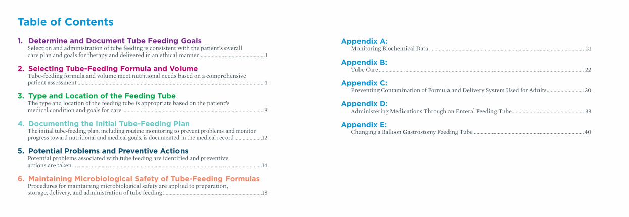
Appendix A:Monitoring Biochemical Data ................................................................................................................21
Appendix B: Tube Care ................................................................................................................................................... 22
Appendix C: Preventing Contamination of Formula and Delivery System Used for Adults ...........................30
Appendix D:Administering Medications Through an Enteral Feeding Tube.................................................... 33
Appendix E:Changing a Balloon Gastrostomy Feeding Tube ...............................................................................40
Table of Contents
1. Determine and Document Tube Feeding GoalsSelection and administration of tube feeding is consistent with the patient’s overall care plan and goals for therapy and delivered in an ethical manner ...............................................1
2. Selecting Tube-Feeding Formula and VolumeTube-feeding formula and volume meet nutritional needs based on a comprehensive patient assessment ..................................................................................................................................... 4
3. Type and Location of the Feeding TubeThe type and location of the feeding tube is appropriate based on the patient’s medical condition and goals for care ..................................................................................................... 8
4. Documenting the Initial Tube-Feeding PlanThe initial tube-feeding plan, including routine monitoring to prevent problems and monitor progress toward nutritional and medical goals, is documented in the medical record ....................12
5. Potential Problems and Preventive ActionsPotential problems associated with tube feeding are identified and preventive actions are taken ........................................................................................................................................14
6. Maintaining Microbiological Safety of Tube-Feeding FormulasProcedures for maintaining microbiological safety are applied to preparation, storage, delivery, and administration of tube feeding .......................................................................18

h Determine and Document Tube Feeding Goals 1
1. Determine and Document Tube Feeding GoalsTube feeding is consistent with the patient’s overall care plan and goals for therapy, and is delivered in an ethical manner.
Interventions 1. Incorporate the plan for tube feeding management in the patient’s overall care plan.
A. Document that the care provided meets the privacy and dignity needs of the patient.
B. Document patient and family wishes regarding enteral nutrition and hydration in the medical record. a. Record presence or absence of Advance Directive or Durable Power of Attorney
for Health Care, especially noting nutrition and hydration. b. If these documents are not in place, encourage the patient/family to
consider completing them. C. State the goals of tube feeding. Examples of tube feeding goals include,
but are not limited to:a. Maintaining nutritional status and/or body weight b. Improving nutritional status and/or body weight c. Improving quality of life d. Providing comfort e. Determining if tube feeding is beneficial for patient through time trial
with specific outcome criteria

2 Determine and Document Tube Feeding Goals 3
2. Transition the patient from tube feeding when the patient is able to consume 75% of their nutrient needs through an oral diet, when another feeding modality is used, or when tube feeding is no longer consistent with the patient’s management plan. A. Document daily intake from all sources in the medical record. B. Evaluate nutrient intake, as compared to estimated nutrient need, and document
in the medical record. C. When the health care team and family wish to have tube feedings given for a trial
period to determine the benefit to the patient, document goals, progress, and results in the medical record. If at the end of the trial it is determined that tube feeding is not appropriate for the patient, consider discontinuation of tube feeding.
D. Discussing the following questions can help the patient, family, significant others, and the health care team consider ethical issues1: a. Does the patient suffer from a condition that is likely to benefit from
tube feeding?b. Will nutritional support improve outcome and/or accelerate recovery?c. Does the patient suffer from an incurable disease, but one in which quality
of life and well-being is possible to maintain or improve by enteral nutrition?d. Does the anticipated benefit outweigh the potential risks?
1. Berner YN. Enteral nutrition in geriatric patients. Mediterr J Nutr Metab. 2009;1:141-144.
1
4 5Selecting Tube-Feeding Formula and Volume
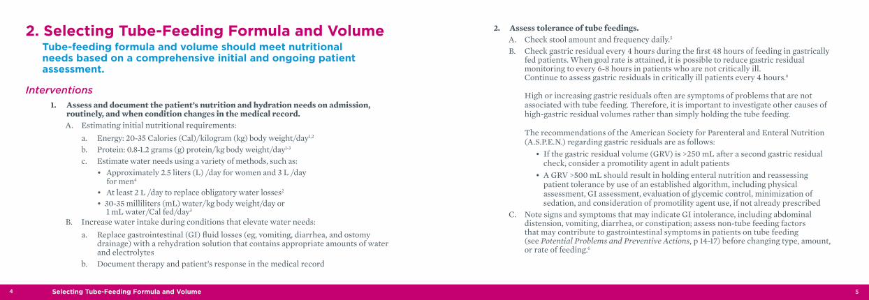
2. Assess tolerance of tube feedings. A. Check stool amount and frequency daily.5 B. Check gastric residual every 4 hours during the first 48 hours of feeding in gastrically
fed patients. When goal rate is attained, it is possible to reduce gastric residual monitoring to every 6-8 hours in patients who are not critically ill. Continue to assess gastric residuals in critically ill patients every 4 hours.6 High or increasing gastric residuals often are symptoms of problems that are not associated with tube feeding. Therefore, it is important to investigate other causes of high-gastric residual volumes rather than simply holding the tube feeding. The recommendations of the American Society for Parenteral and Enteral Nutrition (A.S.P.E.N.) regarding gastric residuals are as follows:
•Ifthegastricresidualvolume(GRV)is>250mLafterasecondgastricresidualcheck, consider a promotility agent in adult patients
•AGRV>500mLshouldresultinholdingenteralnutritionandreassessingpatient tolerance by use of an established algorithm, including physical assessment, GI assessment, evaluation of glycemic control, minimization of sedation, and consideration of promotility agent use, if not already prescribed
C. Note signs and symptoms that may indicate GI intolerance, including abdominal distension, vomiting, diarrhea, or constipation; assess non-tube feeding factors that may contribute to gastrointestinal symptoms in patients on tube feeding (see Potential Problems and Preventive Actions, p 14-17) before changing type, amount, or rate of feeding.6
2. Selecting Tube-Feeding Formula and VolumeTube-feeding formula and volume should meet nutritional needs based on a comprehensive initial and ongoing patient assessment.
Interventions1. Assess and document the patient’s nutrition and hydration needs on admission,
routinely, and when condition changes in the medical record. A. Estimating initial nutritional requirements:
a. Energy: 20-35 Calories (Cal)/kilogram (kg) body weight/day1,2 b. Protein: 0.8-1.2 grams (g) protein/kg body weight/day1-3 c. Estimate water needs using a variety of methods, such as: • Approximately 2.5 liters (L) /day for women and 3 L /day
for men4
• Atleast2L/daytoreplaceobligatorywaterlosses2
•30-35milliliters(mL)water/kgbodyweight/dayor 1 mL water/Cal fed/day3
B. Increase water intake during conditions that elevate water needs:a. Replace gastrointestinal (GI) fluid losses (eg, vomiting, diarrhea, and ostomy
drainage) with a rehydration solution that contains appropriate amounts of water and electrolytes
b. Document therapy and patient’s response in the medical record
6 7Selecting Tube-Feeding Formula and Volume
4. Each day check for factors that can affect hydration needs. Document the following conditions in the medical record, and notify the health care team regarding the need for additional fluids. A. Potential for water deficit:
a. Increased water needs (eg, increased room temperature, low room humidity, fever (every degree of fever increases water needs by 10%), unhumidified oxygen therapy, air-fluidized bed therapy, and diuretics
B. Potential for water and electrolyte deficit secondary to GI fluid losses (eg, vomiting, diarrhea, and wound and/or ostomy drainage)
1. Dorner B, Posthauer ME, Thomas D. The role of nutrition in pressure ulcer prevention and treatment: National Pressure Ulcer Advisory Panel White Paper. Adv Skin Wound Care. 2009;22:212-221.
2. McClave SA, Martindale RG, Vanek VW, et al. Guidelines for the Provision and Assessment of Nutrition Support Therapy in the Adult Critically Ill Patient: Society of Critical Care Medicine (SCCM) and American Society for Parenteral and Enteral Nutrition (A.S.P.E.N.). JPEN J Parenter Enteral Nutr. 2009;33:277-316.
3. Clark M, Schols JM, Benati G, et al. Pressure ulcers and nutrition: a new European guideline. J Wound Care. 2004;13:267-272.
4. Institute of Medicine. National Academy of Sciences: Dietary Reference Intakes for Water, Potassium, Sodium, Chloride, and Sulfate. Available at: http://www.iom.edu/?id=54343. Accessed November 12, 2009.
5. Russell MK. Monitoring complications of enteral feedings. In: Charney P, Malone A (eds). ADA Pocket Guide to Enteral Nutrition. Chicago, Ill: American Dietetic Association; 2006:155-192.
6. Bankhead R, Boullata J, Brantley S, et al. Enteral nutrition practice recommendations. JPEN J Parenter Enteral Nutr. 2009;33:122-167.
D. Check urinary output every 8 hours.5 E. Check skin turgor every 8 hours.5
F. Evaluate laboratory values (see Appendix B).G. Document findings in the medical record.
3. Choose the formula that best meets the patient’s nutritional needs and medical conditions. A. Most patients can tolerate a standard formula. B. Other patients may benefit from a specialized formula because of conditions,
such as the following:a. Chronic conditions (eg, cancer, diabetes, short bowel syndrome, inflammatory bowel
disease) b. Suboptimal nutritional status c. Catabolic hypermetabolic stress (eg, multiple fractures, poorly healing or
chronic wounds, burns)d. Organ system dysfunction (eg, GI surgery, malabsorption, acute and chronic kidney
dysfunction, acute respiratory distress syndrome, chronic obstructive pulmonary disease)
8 9Type and Location of the Feeding Tube
3b. Open systems:
• Formulaisdecantedfromoriginalcontainerintofeedingreservoir• Hangready-to-useformulafor8-12hours• Hangreconstitutedformulaorformulawithmodularcomponentsfor
up to 4 hours C. Select most appropriate method of delivery:
a. Bolus: • Relativelylargeformulavolume(≥250 mL) is given over 10-20 minutes)
4-6 times/day by syringe or gravity infusion • AssociatedwithsymptomsofGIintolerance(eg,abdominalpainandcramping,
nausea, and vomiting) • Bestusedforgastricfeeding,forambulatorypatients,andforpatients
who are stable and neurologically intact and able to protect their airwayb. Intermittent:
• Largevolumeofformula(250-500mL)isgivenover60-75minutes, five to eight times per day
• Mayusegravityorenteralfeedingpump• Indicatedforstablepatientswhoaregastricallyfed,withnormalgastric
function, ability to protect their own airway, and demonstrated tolerance to feeding
3. Type and Location of the Feeding TubeThe type and location of the feeding tube required is based on the patient’s medical condition and goals for care.
Interventions1,2
1. Assure that the most appropriate route, system, and method of tube-feeding delivery are provided. A. Select appropriate feeding tube:
a. For short-term feeding (<30 days), use a nasal feeding tube b. Forlong-termfeeding(>30days),useagastrostomyorjejunostomyfeeding
tube c. For patients at risk for aspiration of gastric contents, consider using a small-
bowel feeding tube d. Use feeding tubes made of polyurethane or silicone to reduce risk of allergies
related to the use of latex catheters B. Select most appropriate feeding system:
a. Closed systems: •Formulaisprefilledbymanufacturerintofeedingcontainer;reduced
handling decreases risk of microbial contamination
•Followmanufacturerrecommendations;mostclosedsystemscanhang for 24-48 hours when label instructions are followed
10 11Type and Location of the Feeding Tube
c. Cyclic: • Prescribedamountofformulavolumeisgivenoveraspecificperiodoftime
that is usually <24 hours (eg, 8-20 hours/day)• Usuallygivenbyenteralfeedingpump• Oftenusedtosupportambulation;whentransitioningtoanoraldiet,oras
a supplement to an adequate oral intake to support ambulation d. Continuous:
• Prescribedformulavolumeisgivencontinuouslyover16-24hours• Usuallygivenviaenteralfeedingpump;feedingpumpisrequiredforjejunal
feedings • Usedforpatientswhocannottolerateintermittentorbolusfeedings;
sometimes preferred for patients at risk for gastroesophageal reflux or with history of aspiration pneumonia, those who are medically unstable, andthosewhorequirejejunalfeedings
2. Document route, system, and method of tube-feeding delivery in the medical record.
1. Bankhead R, Boullata J, Brantley S, et al. Enteral nutrition practice recommendations. JPEN J Parenter Enteral Nutr. 2009;33:122-167.
2. Russell MK. Monitoring complications of enteral feedings. In: Charney P, Malone A (eds). ADA Pocket Guide to Enteral Nutrition. Chicago, Ill: American Dietetic Association; 2006:155-192.

12 13Documenting the Initial Tube-Feeding Plan
4. Documenting the Initial Tube-Feeding PlanThe initial tube-feeding plan, including routine monitoring to prevent problems and monitor progress toward nutritional and medical goals, is documented in the medical record.
Interventions1,2
1. Assure that medical orders include the following and that they are documented in the medical record:A. Patient informationB. Formula typeC. Administration method and rateD. Additional water volume and administration method
2. Document monitoring parameters in the medical record (see Appendix B for suggested biochemistries). Assess and document biochemistries, body weight, and clinical signs of tolerance of tube feeding in the medical record. Notify physician of any abnormalities or changes to items noted in A through D above.
1. Russell MK. Monitoring complications of enteral feedings. In: Charney P, Malone A (eds). ADA Pocket Guide to Enteral Nutrition. Chicago, Ill: American Dietetic Association; 2006:155-192.
2. Bankhead R, Boullata J, Brantley S, et al. Enteral nutrition practice recommendations. JPEN J Parenter Enteral Nutr. 2009;33:122-167.
14 15Potential Problems and Preventive Actions
5. Potential Problems and Preventive ActionsPotential problems associated with tube feeding are identified and preventive actions are taken.
Interventions1. Identify potential problems:
A. Metabolic: a. Fluid imbalances:
• Dehydration• Overhydration
b. Electrolyte abnormalities: • Hyponatremia• Hypernatremia• Hypokalemia• Hyperkalemia• Hypophosphatemia• Hyperphosphatemia
c. Glucose abnormalities: • Hypoglycemia• Hyperglycemia
B. Gastrointestinal: a. Nausea b. Vomitingc. Diarrhea or constipationd. Abdominal distension e. Abdominal pain
C. Mechanical: a. Pulmonary aspiration of gastric contents and associated pneumonia b. Tube clogging c. Tube migration d. Balloon rupture or inability to empty the gastrostomy tube balloon e. Skin irritation/breakdown from the feeding tube or drainage around the tube f. Inadvertent removal of the feeding tube g. Tube degradation or occlusion by yeast
2. Prevention of metabolic complications: A. Provide appropriate amount of formula and water B. Assess laboratory values (see Appendix B)C. Notify physician of any abnormalities D. Document any abnormalities and the interventions taken to correct them in the
medical record
16 17Potential Problems and Preventive Actions
3. Prevention of gastrointestinal complications: A. Provide appropriate type of formulaB. Provide formula at prescribed rate using appropriate delivery method C. Set up enteral delivery system using clean technique D. Assess medications and consult with a pharmacist E. Assess for infectious illnesses (eg, Clostridium difficile) or other conditions (eg,
malabsorption, acute illness) that may cause GI complications F. Notify physician of any abnormalities G. Document in the medical record any potential abnormalities and the interventions
taken to correct them
4. Prevention of mechanical complications: A. Keep head of bed (HOB) elevated 30-45 degrees during feeding and for 30-60 minutes
after feeding1
a. Use the reverse Trendelenburg position to elevate the HOB, unless contraindicated, when the patient cannot tolerate a backrest elevated position
b. If necessary to lower the HOB for a procedure or a medical contraindication, return the patient to an elevated HOB position as soon as feasible
B. Use appropriate tube size to enhance formula flow and provide patient comfort. C. Routinely flush feeding tube with 30 mL water2:
a. Every 4 hours during continuous feedingb. Before and after intermittent feedings
c. After residual volume measurementsd. Use sterile or purified water for immunocompromised or critically ill patients,
especially when the safety of tap water is questionable3
D. Tape/anchor nasal tubes properly. E. Usegastrostomytubeswithskindisksorjejunostomytubeswithskinanchors.F. Use enteral feeding pump with automatic flush feature to help prevent clogging of
feeding tube. G. Maintain proper skin disk placement. H. Check feeding tube placement. I. Use clean technique when caring for feeding tubes. J. Check balloon fill volume, according to manufacturer’s instructions. K. Clean around feeding tube daily. L. Donotusedressingswithgastrostomyorjejunostomyfeedingtubes,unlessordered.M. Notify physician of any abnormalities. N. Document in the medical record any abnormalities and the interventions taken to correct
them.
1. McClave SA, DeMeo MT, DeLegge MH, et al. North American Summit on Aspiration in the Critically Ill Patient: consensus statement. JPEN J Parenter Enteral Nutr. 2002;26(suppl 6):S80-S85.
2. Russell MK. Monitoring complications of enteral feedings. In: Charney P, Malone A (eds). ADA Pocket Guide to Enteral Nutrition. Chicago, Ill: American Dietetic Association; 2006:155-192.
3. Bankhead R, Boullata J, Brantley S, et al. Enteral nutrition practice recommendations. JPEN J Parenter Enteral Nutr. 2009;33:122-167.
18 19Maintaining Microbiological Safety of Tube-Feeding Formulas
6. Maintaining Microbiological Safety of Tube-Feeding FormulasProcedures for maintaining microbiological safety are required for preparation, storage, delivery, and administration of tube feeding.
Interventions1,2
1. Identify and take preventive measures against contamination hazards associated with the formula, delivery system, and patient.
2. Maintain proper temperature of formula during storage and delivery: A. Cover opened, unused formula, and store in refrigerator B. Discard opened, unused ready-to-feed formula after 48 hours (record date and time of
opening) C. Discard unused reconstituted formula after 24 hours (record date and time of mixing)
3. Do not hang the formula at the bedside for prolonged periods: A. Follow manufacturer’s recommendations for closed system hangtimes; most can
hang between 24-48 hours when label instructions are followed B. Hang ready-to-use formula that is decanted into a feeding reservoir between 8-12 hoursC. Hang reconstituted formula or formula with modular components up to 4 hours
4. Ensure that the work surface and equipment used for preparing tube feeding is clean.
5. Maintain clean technique when working with the formula and delivery system: A. Wash hands well before touching formula or delivery system B. Wear disposable, nonsterile gloves when handling feeding tube and as needed to prevent
contact with body secretions (eg, when checking residual)
6. Avoid adding water, colorants, medications, or other substances directly to the formula.
1. Campbell SM. Preventing Microbial Contamination of Enteral Formulas and Delivery Systems: Hazard Analysis Critical Control Point (HACCP) in the Clinical Setting. Columbus, Ohio: Ross Products Division, Abbott Laboratories; 2000.
2. Bankhead R, Boullata J, Brantley S, et al. Enteral nutrition practice recommendations. JPEN J Parenter Enteral Nutr. 2009;33:122-167.
Maintaining Microbiological Safety of Tube-Feeding Formulas

20 21Appendix
Appendix A: Monitoring Biochemical DataParameter At Baseline Routine Frequency
Serum sodium, potassium, chloride, bicarbonate, blood urea nitrogen (BUN), creatinine, calcium, phosphorus, magnesium
√Daily for the first 3 days, then reduce to 1-2 times weekly
Blood glucose At least every 6-8 hours
Stop blood glucose monitoring after blood glucose has stabilized at an appropriate level
Source: Russell MK. Monitoring complications of enteral feedings. In: Charney P, Malone A (eds). ADA Pocket Guide to Enteral Nutrition. Chicago, Ill: American Dietetic Association; 2006:155-192.
22 23Appendix
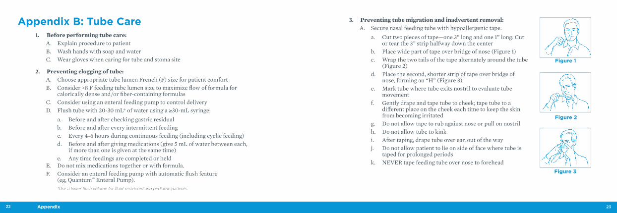
3. Preventing tube migration and inadvertent removal: A. Secure nasal feeding tube with hypoallergenic tape:
a. Cut two pieces of tape—one 3” long and one 1” long. Cut or tear the 3” strip halfway down the center
b. Place wide part of tape over bridge of nose (Figure 1) c. Wrap the two tails of the tape alternately around the tube
(Figure 2) d. Place the second, shorter strip of tape over bridge of
nose, forming an “H” (Figure 3) e. Mark tube where tube exits nostril to evaluate tube
movement f. Gently drape and tape tube to cheek; tape tube to a
different place on the cheek each time to keep the skin from becoming irritated
g. Do not allow tape to rub against nose or pull on nostrilh. Do not allow tube to kinki. After taping, drape tube over ear, out of the way j. Donotallowpatienttolieonsideoffacewheretubeis
taped for prolonged periods k. NEVERtapefeedingtubeovernosetoforehead
Appendix B: Tube Care 1. Before performing tube care:
A. Explain procedure to patient B. Wash hands with soap and water C. Wear gloves when caring for tube and stoma site
2. Preventing clogging of tube: A. Choose appropriate tube lumen French (F) size for patient comfort B. Consider>8Ffeedingtubelumensizetomaximizeflowofformulafor
calorically dense and/or fiber-containing formulas C. Consider using an enteral feeding pump to control delivery D. Flush tube with 20-30 mL* of water using a ≥30-mL syringe:
a. Before and after checking gastric residual b. Before and after every intermittent feedingc. Every 4-6 hours during continuous feeding (including cyclic feeding) d. Before and after giving medications (give 5 mL of water between each,
if more than one is given at the same time)e. Any time feedings are completed or held
E. Do not mix medications together or with formula. F. Consider an enteral feeding pump with automatic flush feature
(eg, Quantum™ Enteral Pump). *Use a lower flush volume for fluid-restricted and pediatric patients.
Figure 1
Figure 3
Figure 2
24 25Appendix
4. Preventing/managing G-tube balloon emptying problems:A. Follow institutional policy for handling balloon emptying problems. B. To prevent problems:
a. Wash hands thoroughly before touching the G-tube, and wear gloves to prevent contamination or infection
b. Flush the G-tube frequently to maintain tube patency (a blockage in the tube may cause narrowing or blockage of the balloon channel)
c. Use appropriate syringe for tube balloon valve (ie, Luer-tip or Luer-lock when emptying balloon)
d. Because small amounts of water are lost over time, check balloon volume every 7-10 days by removing water with a Luer-tip or Luer-lock syringe:
•ReplacewithcorrectvolumeofwaterC. If the prescribed procedures are followed and difficulty emptying the balloon still is
experienced, use the following methods for functional and nonfunctional G-tubes: a. UseaLuer-tiporLuer-locksyringetoinjectanadditional1-2mLofwater
into balloon valve to open balloon channel b. Remove the piston from the syringe, insert the syringe tip into the balloon valve,
and place the syringe on a clean towel at a level lower than the balloon for 5-10 minutes to allow for gravity drainage of water from the balloon; water may drain slowly
D. If efforts to empty the balloon are unsuccessful and it is necessary to remove or replace the nonfunctional G-tube, notify the physician.
B. Use skin disk on G-tubes and skin anchor on J-tubes to prevent inward or outward movement: a. Inward movement of G-tubes can cause pyloric obstruction b. Outward movement can dislodge tube into stoma tract or peritoneum
C. Use a G-tube with centimeter markings: a. Monitor marks daily to evaluate movement b. Notify physician and stop feeding if tube placement is in question
D. Maintain proper fill volume of balloon G-tube:a. Use a Luer-tip or Luer-lock syringe (choose syringe appropriate for tube
balloon valve) to fill balloon with water:• Filltheballoontothevolumerecommendedbythemanufacturer• Neveruseairorfluidsotherthanwatertofillballoon• Itisnotpossibletomaintainballoonvolumewithairorfluidsotherthan
water b. Check balloon volume every 7-10 days. Add water as needed to maintain
proper fill volume E. Secure tubing under clothing to prevent patient from pulling out the tube.
26 27Appendix
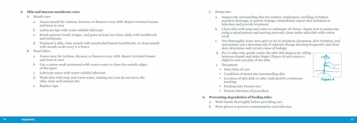
C. Stoma site: a. Inspect the surrounding skin for redness, tenderness, swelling, irritation,
purulent drainage, or gastric leakage; immediately report skin irritation or infection and provide treatment
b. Clean skin with soap and water or antiseptic of choice—begin next to stoma site, using a spiral pattern and moving outward; clean under skin disk with cotton swab
c. Dry thoroughly; leave area open to air to minimize dampness, skin irritation, and maceration; use a dressing only if ordered; change dressing frequently and clean skin; determine and correct cause of leakage
d. For G-tube only, gently rotate the tube 360 degrees by rolling between thumb and index finger (Figure 4) and ensure a slight in-and-out play of the tube.
e. Document:• Date/timeofcare• Conditionofstomasite/surroundingskin• Locationofskindiskontube,indicatedbycentimeter
marking • FeedingtubeFrenchsize• Patienttoleranceofprocedure
6. Preventing degradation of feeding tube: A. Wash hands thoroughly before providing care B. Wear gloves to prevent contamination and infection
5. Skin and mucous membrane care: A. Mouth care:
a. Assess mouth for redness, dryness, or fissures every shift. Report irritated tissues and treat at once
b. Lubricate lips with water-soluble lubricant c. Brush patient’s teeth, tongue, and gums at least two times daily with toothbrush
and toothpaste d. If patient is able, rinse mouth with nonalcohol-based mouthwash, or clean mouth
with mouth swab every 2-4 hours B. Nasal tubes:
a. Assess nose for redness, dryness, or fissures every shift. Report irritated tissues and treat at once
b. Use a cotton swab moistened with warm water to clean the outside edges of the nares
c. Lubricate nares with water-soluble lubricantd. Wash skin with soap and warm water, making sure you do not move the
tube; rinse well and pat dry e. Replace tape
Figure 4
28 29Appendix
7. Verifying proper tube placement: A. Verifypropertubeplacementbeforefeedingsandevery4-6hoursduringcontinuous
feedings B. Use a combination of methods to verify tube position according to your facility’s protocol C. Bedside verification techniques:
a. Auscultation: • Inject10-20mLofairusinga>30mLsyringe,whilelisteningforawhooshing
or gurgling sound in upper left quadrant of abdomen• Donotrelyonauscultationtodifferentiateamonggastric,smallbowel,orrespiratory
tube placementb. Aspiration:
• Usinga≥30 mL syringe, clear the tube with 20 mL of air • Withdrawgastriccontents,andevaluatethecoloroftheaspirate
c. pH testing: • Usinga≥30 mL syringe, clear the tube with 20 mL of air • Withdrawgastriccontents.TesttheaspiratewithpHpaperorapHmeter
(normal gastric pH ranges from 1.5-5.5) D. X-ray for tube placement verification any time proper placement is questioned. E. Monitor patient for clinical signs of misplaced tube—dyspnea (difficulty breathing),
tachypnea (increased respiratory rate), tachycardia (increased heart rate), pulmonary congestion, and abdominal distention/tenderness/hardness.
30 31Appendix
4. Maintain safe hangtime: A. Closed system:
a. Formulas in closed systems can safely hang for 24-48 hours Follow manufacturer’s recommendations and instructions for use
b. Record date/time container is hung c. Using a closed system container with a recessed spike is preferabled. When preparing tube feeding formula and during administration, use clean
techniquee. Change tubing according to manufacturer’s recommendations
B. Open system (formula decanted from original container to feeding reservoir): a. Hang ready-to-use formula 8-12 hours b. Hang reconstituted formula or formula with modular components up to 4 hours c. NEVERaddfreshformulatohangingformulad. Change container/tubing at least every 24 hours
5. Do not add substances to enteral formula: A. Use commercially prepared formula; avoid homemade formula B. Avoid adding water, colorants, or other substances directly to formula; do not add
medications to formula C. Reduce handling by using prefilled containers
Appendix C: Preventing Contamination of Formula and Delivery System Used for Adults
1. Wash hands thoroughly and apply gloves before handling formula, delivery system, or feeding tube.
2. Maintain clean work area, equipment, and delivery system: A. Wash area/equipment before and after formula preparation B. Do not touch any part of delivery system coming in contact with the formula
3. Maintain proper storage and handling of formula: A. Thoroughly clean the top of formula containers before opening B. Record date/time formula is opened C. Cover opened, unused formula in refrigerator D. Discard opened, unused ready-to-feed formula after 48 hours (record date and
time of opening) E. Discard unused reconstituted formula after 24 hours (record date and time of mixing) F. Maintain proper temperature where formula is stored G. Do not use after expiration date on container
32 33Appendix
Appendix D: Administering Medications Through an
Enteral Feeding Tube Note: These are general guidelines. Your facility’s policies and
procedures should always serve as your guide.
The medication administration recommendations below can help maintain tube patency, prevent drug-nutrient interactions, and assure proper medication absorption.
1. Evaluate medication and route of feeding—some medications may not absorb properly if they bypass the stomach.
2. Second, consider giving medications by an alternate route, such as transdermal, rectal, inhaled, intramuscular, subcutaneous, buccal, sublingual, or intravenous when available and clinically appropriate.
3. Never mix medications and formula together.
4. Prior to administering medication, stop the feeding and flush the feeding tube with 30 mL of water.1 Flush feeding tube between and after administering medications as well.
5. Use liquid medications when possible. Immediate-release tablets are acceptable substitutes for liquid medications. When it is necessary to crush a tablet, crush into a fine powder and mix well with water. Do not crush enteric-coated, extended-release, or effervescent medications.
6. Prevent retrograde contamination from patient into the feeding bag container: A. Keep head of bed elevated 30-45 degrees during feeding and for 30-60 minutes after
feeding: a. Use the reverse Trendelenburg position to elevate the HOB, unless
contraindicated, when the patient cannot tolerate a backrest elevated position b. If necessary to lower the HOB for a procedure or because of a medical
contraindication, return the patient to an elevated HOB position as soon as possible
B. Position feeding set higher than stomach C. Use gravity feeding set with physical barriers (eg, drip chamber) to prevent reflux
of formula into the feeding bag
1. Campbell SM. Preventing Microbial Contamination of Enteral Formulas and Delivery Systems. Hazard Analysis Critical Control Point (HACCP) in the Clinical Setting. Columbus, Ohio: Ross Products Division, Abbott Laboratories; 2000.
2. Bankhead R, Boullata J, Brantley S, et al. Enteral nutrition practice recommendations. JPEN J Parenter Enteral Nutr. 2009;33:122-167.
34 35Appendix
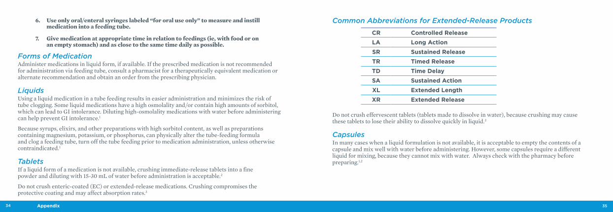
Common Abbreviations for Extended-Release Products
CR Controlled Release
LA Long Action
SR Sustained Release
TR Timed Release
TD Time Delay
SA Sustained Action
XL Extended Length
XR Extended Release
Do not crush effervescent tablets (tablets made to dissolve in water), because crushing may cause these tablets to lose their ability to dissolve quickly in liquid.2
CapsulesIn many cases when a liquid formulation is not available, it is acceptable to empty the contents of a capsule and mix well with water before administering. However, some capsules require a different liquid for mixing, because they cannot mix with water. Always check with the pharmacy before preparing.1,2
6. Use only oral/enteral syringes labeled “for oral use only” to measure and instill medication into a feeding tube.
7. Give medication at appropriate time in relation to feedings (ie, with food or on an empty stomach) and as close to the same time daily as possible.
Forms of Medication Administer medications in liquid form, if available. If the prescribed medication is not recommended for administration via feeding tube, consult a pharmacist for a therapeutically equivalent medication or alternate recommendation and obtain an order from the prescribing physician.
LiquidsUsing a liquid medication in a tube feeding results in easier administration and minimizes the risk of tube clogging. Some liquid medications have a high osmolality and/or contain high amounts of sorbitol, which can lead to GI intolerance. Diluting high-osmolality medications with water before administering can help prevent GI intolerance.1
Because syrups, elixirs, and other preparations with high sorbitol content, as well as preparations containing magnesium, potassium, or phosphorus, can physically alter the tube-feeding formula and clog a feeding tube, turn off the tube feeding prior to medication administration, unless otherwise contraindicated.1
TabletsIf a liquid form of a medication is not available, crushing immediate-release tablets into a fine powder and diluting with 15-30 mL of water before administration is acceptable.2
Do not crush enteric-coated (EC) or extended-release medications. Crushing compromises the protective coating and may affect absorption rates.2

36 37Appendix
Commercially Available Product Average Osmolality
Acetaminophen elixir (Tylenol®) 325 mg/5 mL 5400
Extra Strength Tylenol® Adult Liquid 500 mg/15 mL 3058
Children’s Tylenol® Elixir 6040
Acetaminophen/Codeine (Tylenol No. 3) Elixir 4700
Amantadine HCl solution 3900
Aminophylline liquid 450
Amoxicillin suspension 125 mg/5 mL 1541
Amoxicillin suspension 250 mg/5 mL 2250
Ampicillin suspension 250 mg/5 mL 2250
Cephalexin suspension (Keflex®) 250 mg/5 mL 1950
Cimetidine solution (Tagamet®) 5550
Co-trimoxazole suspension 2200
Dexamethasone Intensol™ Solution 3100
Digoxin elixir 1350
Diphenhydramine HCl elixir (Benadryl®) 850
Diphenoxylate/atropine (Lomotil®) suspension 8800
Docusate sodium syrup (Colace®) 3900
Granular or microencapsulated medicationExamples of these types of medications include diltiazem (Cardizem® CD and SR formulations) and pancreatic enzymes (Creon® and Pancrease®). Avoid giving these medications via feeding tube, because they often clog the tube. If it is necessary to use a granular medication, consult your pharmacy. Administer immediately after dilution and flush right after the medication is given.1
Proton-pump inhibitors (PPI)Prilosec® (omeprazlole), Prevacid® (lansoprazole), and Nexium® (esomeprazole) are formulated as delayed-release capsules containing enteric-coated drug granules. Gastric acid dissolves the delayed-release capsule during transit of the dosage form. Crushing the enteric-coated granules results in tube clogging from the enteric coating.4 If the patient is on PPI, discuss administration options with the pharmacist.
Protonix® (pantoprazole) and Aciphex® (rabeprazole) are formulated as enteric-coated, delayed-release tablets. The coating dissolves in the stomach and the drug is absorbed in the intestine. Because these drugs are enteric-coated tablets, you cannot split, chew, or crush them. Therefore, you cannot administer these medications via feeding tubes.4
Sublingual and buccal medicationsDo not give these medications through a feeding tube. Administer only by placing them under the patient’s tongue or in the cheek pouch, because they will not absorb well in the stomach or small bowel.4
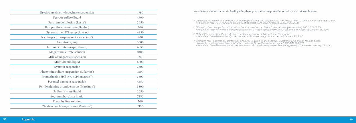
Medications: Average Osmolality1,3,4
Osmolality is defined as the number of molecules and ions of a solution per kilogram.
38 39Appendix
1. Dickerson RN, Melnik G. Osmolality of oral drug solutions and suspensions. Am J Hosp Pharm [serial online]. 1988;45:832-834. Available at: http://www.ajhp.org/cgi/content/abstract/45/4/832. Accessed January 25, 2010.
2. Mitchell J. Oral dosage forms that should not be crushed or chewed. Hosp Pharm [serial online].2002; 37:213-214. Available at: http://www.factsandcomparisons.com/assets/hospitalpharm/feb2002_oral.pdf. Accessed January 25, 2010.
3. McNeil Consumer Healthcare. A pharmacologic overview of Tylenol® (acetaminophen). Available at: http://www.tylenolprofessional.com/pharmacology.html. Accessed January 25, 2010.
4. Beckwith MC, Feddema SS, Barton RG, Graves C. A guide to drug therapy in patients with enteral feeding tubes: dosage form selection and administration methods. Hosp Pharm [serial online]. 2004;39:225-237. Available at: http://www.factsandcomparisons.com/assets/hospitalpharm/mar2004_peer1.pdf. Accessed January 25, 2010.
Erythromycin ethyl succinate suspension 1750
Ferrous sulfate liquid 4700
Furosemide solution (Lasix®) 2050
Haloperidol concentrate (Haldol®) 500
Hydroxyzine HCl syrup (Atarax) 4450
Kaolin-pectin suspension (Kaopectate®) 900
Lactulose syrup 3600
Lithium citrate syrup (lithium) 6850
Magnesium citrate solution 1000
Milk of magnesia suspension 1250
Multivitamin liquid 5700
Nystatin suspension 3300
Phenytoin sodium suspension (Dilantin®) 1500
Promethazine HCl syrup (Phenegran™) 3500
Pyrantel pamoate suspension 4350
Pyridostigmine bromide syrup (Mestinon®) 3800
Sodium citrate liquid 2050
Sodium phosphate liquid 7250
Theophylline solution 700
Thiabendazole suspension (Mintezol®) 2150
Note: Before administration via feeding tube, these preparations require dilution with 10-30 mL sterile water.

40 41Appendix
Appendix E: Changing a Balloon Gastrostomy Feeding Tube
SuppliesTube; water-soluble lubricant; Luer-tip or Luer-lock syringe; water; gloves; washcloth or 4”x4” gauze; soap and water or antiseptic.
Procedure
1. Explain procedure to patient.
2. Wash hands with soap and water.
3. Wear gloves when caring for tube and stoma site.
4. Choose appropriate French size replacement tube for stoma.
5. Pull back skin disk on new tube to facilitate tube insertion.
6. Lubricate stoma and tip only with water-soluble lubricant. Skin disk will slip if entire tube is lubricated. DO NOT USE PETROLEUM-BASED LUBRICANT.
7. Insert Luer-tip or Luer-lock syringe as appropriate for balloon valve into balloon port of existing tube and remove water from balloon.
8. With an upward twisting motion, gently remove existing tube and discard.
9. Clean stoma site with soap and water, or antiseptic as designated by facility policy. Dry thoroughly.
10. With a slight twisting motion, gently insert tip of tube into stoma and guide it through stoma tract into stomach. To avoid trauma, never force against resistance.
11. Using a Luer-tip or Luer-lock syringe, fill balloon with proper amount of water to keep tube securely in stomach, according to manufacturer’s recommendations (Figure 5). Never use air or fluids other than water to fill balloon. It is not possible to maintain balloon volume with air, and using fluids other than water to fill balloon may cause problems with maintaining proper balloon volume.
12. Gently snug balloon up against gastric mucosa, and slide skin disk down against skin. Slide disk back about 1 centimeter (cm) allowing for slight in-and-out play, which minimizes pressure-related complications, such as necrosis.
13. Confirm proper tube placement, function, and patency before proceeding with feeding.
Figure 5
42 43Appendix
14. Avoid applying dressings to stoma because dressings may lead to skin irritation/infection. When excess moisture or stoma leakage occurs under the skin disk, use a thin gauze dressing (only one) and change promptly when moist.
15. Document: A. Date/time of procedure B. Condition of stoma site/surrounding skin C. French size of tube placedD. Amount of water in balloon E. Location of skin disk on tube, indicated by centimeter marking F. Patient tolerance of procedure
Notes:
___________________________________________________________________________________________________
___________________________________________________________________________________________________
___________________________________________________________________________________________________
___________________________________________________________________________________________________
___________________________________________________________________________________________________
___________________________________________________________________________________________________
___________________________________________________________________________________________________
___________________________________________________________________________________________________
___________________________________________________________________________________________________
___________________________________________________________________________________________________
___________________________________________________________________________________________________
___________________________________________________________________________________________________
___________________________________________________________________________________________________
___________________________________________________________________________________________________