beta-caryophyllene protects against alcoholic ... protects against.pdf · zoltan v. varga1, csaba...
TRANSCRIPT
Varga et al.
This article has been accepted for publication and undergone full peer review but has not been through the copyediting, typesetting, pagination and proofreading process which may lead to differences between this version and the Version of Record. Please cite this article as doi: 10.1111/bph.13722
This article is protected by copyright. All rights reserved.
Beta-caryophyllene protects against alcoholic steatohepatitis by attenuating
inflammation and metabolic dysregulation in mice
Running Title: Beta-caryophyllene is hepatoprotective
Zoltan V. Varga1, Csaba Matyas
1, Katalin Erdelyi
1, Resat Cinar
2, Daniela Nieri
3, Andrea
Chicca3, Balazs Tamas Nemeth
1, Janos Paloczi
1, Tamas Lajtos
1, Lukas Corey
1, Gyorgy
Hasko4, Bin Gao
5, George Kunos
2, Jürg Gertsch
3 and Pal Pacher
1
Affiliations:
1Laboratory of Cardiovascular Physiology and Tissue Injury, National Institutes of
Health/NIAAA, Bethesda, MD 20852, USA.
2Laboratory of Physiologic Studies, National Institutes of Health/NIAAA, Bethesda, MD
20852, USA.
3Institute of Biochemistry and Molecular Medicine, National Center of Competence in
Research TransCure, University of Bern, CH-3012 Bern, Switzerland.
4Departments of Surgery, Rutgers New Jersey Medical School, Newark, NJ 07103, USA.
5Laboratory of Liver Diseases, National Institutes of Health/NIAAA, Bethesda, MD 20852,
USA.
Contact:
Pal Pacher, MD, PhD, FAHA, FACC
Laboratory of Cardiovascular Physiology and Tissue Injury
5625 Fishers Lane, Room 2N-17; Bethesda, MD 20892-9413
Phone: (301)443-4830 Email: [email protected]
Conflict of interest disclosure: All Authors declare no conflict of interest.
Varga et al.
This article is protected by copyright. All rights reserved.
Tables of links
TARGETS
GPCRsb
cannabinoid 1 receptor
cannabinoid 2 receptor
Nuclear hormone receptorsc
peroxisome proliferator-activated receptor-α
Enzymese
3-hydroxy-3-methylglutaryl-CoA synthase 2
arginase I
cyclooxygenase 2
fatty acid amide hydrolase
sirtuin-1
LIGANDS
2-arachidonoylglycerol
anandamide
arachidonic acid
chemokine (C-C motif) ligand 2
chemokine (C-C motif) ligand 4
chemokine (C-X-C motif) ligand 2
intercellular adhesion molecule 1
interleukin 1 beta
interleukin 10
interleukin-6
N-oleoylethanolamide
tumor necrosis factor alpha
Varga et al.
This article is protected by copyright. All rights reserved.
Abbreviations
4-HNE: 4-hydroxynonenal
ALT: alanine aminotransferase
ARG1: arginase 1
BCP: beta-caryophyllene
CB1: cannabinoid receptor type 1
CB2: cannabinoid receptor type 2
CCL2: monocyte chemoattractant protein - MCP1
CCL4: Chemokine (C-C motif) ligand 4 - Macrophage inflammatory protein-1β - MIP-1β
CD11b: cluster of differentiation molecule 11B
CD32: cluster of differentiation 32
CD34: cluster of differentiation 34
CD117: cluster of differentiation 117
CD163: cluster of differentiation 163
CD68: cluster of differentiation 68
CLEC7A: C-type lectin domain family 7 member A
COX-2: cyclooxygenase-2 or prostaglandin-endoperoxide synthase 2
CXCL2: macrophage inflammatory protein 2-alpha - MIP2-alpha
DMSO: dimethyl sulfoxide
ECL: enhanced chemiluminescence
Varga et al.
This article is protected by copyright. All rights reserved.
EDTA: ethylenediaminetetraacetic acid
F4/80: EGF-like module-containing mucin-like hormone receptor-like 1
FAAH: fatty acid amide hydrolase
FDA: Food and Drug Administration
FFPE: formalin-fixed, paraffin-embedded tissue samples
FGF21: fibroblast growth factor 21
FOXO1: forkhead box protein O1
FOXO3: forkhead box O3
G6PC: glucose-6-phosphatase
GC/MS: gas chromatography / mass spectrometry
H2O2: hydrogen peroxide
HMGB1: high-mobility group protein 1
HMGCS2: 3-hydroxy-3-methylglutaryl-CoA synthase 2
IBA-1: ionized calcium-binding adapter molecule 1
ICAM-1: intercellular adhesion molecule 1
IL10: interleukin 10
IL1ß: interleukin 1 beta
IL-6: interleukin 6
LY6G: lymphocyte antigen 6 complex locus G6D
MGL1/CD301: macrophage galactose-type C-type lectin 1
Varga et al.
This article is protected by copyright. All rights reserved.
MRC2: mannose receptor C type 2
NAD+: nicotinamide adenine dinucleotide
NIAAA: National Institute on Alcohol Abuse and Alcoholism
PBS: phosphate-buffered saline
PCK1: phosphoenolpyruvate carboxykinase 1
PFK: phosphofructokinase
PGC1ɑ : peroxisome proliferator-activated receptor gamma coactivator 1- alpha
PGC-1ß: peroxisome proliferator-activated receptor gamma coactivator 1- beta
PPAR-ɑ : peroxisome proliferator-activated receptor alpha
RIPA: radioimmunoprecipitation assay buffer
SIRT-1: NAD-dependent deacetylase sirtuin-1
SREBP1c: sterol regulatory element-binding protein 1
TBS-T: Tris-buffered saline with Tween 20
Varga et al.
This article is protected by copyright. All rights reserved.
Abstract
Background and aims: Beta-caryophyllene (BCP) is a plant-derived FDA approved food
additive with anti-inflammatory properties. Some of its beneficial effects in vivo reported to
involve activation of cannabinoid 2 receptors (CB2) that are predominantly expressed in
immune cells. Herein, we evaluated the translational potential of BCP using a well-
established model of chronic and binge alcohol-induced liver injury.
Methods: In this study we investigated the effects of BCP on liver injury induced by chronic
plus binge alcohol feeding in mice in vivo by using biochemical assays, real-time PCR and
histology analyses. Serum and hepatic BCP levels were also determined by GC/MS.
Results: Chronic treatment with BCP attenuated the chronic and binge alcohol-induced liver
injury and inflammation by attenuating the pro-inflammatory phenotypic `M1` switch of
Kupffer cells and by decreasing the expression of vascular adhesion molecules ICAM-1, E-
Selectin and P-Selectin, as well as the neutrophil infiltration. It also beneficially influenced
hepatic metabolic dysregulation (steatosis, protein hyperacetylation, and PPAR-ɑ signaling).
The above mentioned protective effects of BCP against alcohol-induced liver injury were
attenuated in CB2 knockout mice, indicating that the beneficial effects of this natural product
in liver injury involve CB2 receptor activation. Following acute or chronic administration
BCP was detectable both in the serum and liver tissue homogenates but not in the brain.
Conclusions: Given the safety of BCP in humans this food additive has a high translational
potential in treating or preventing hepatic injury associated with oxidative stress,
inflammation and steatosis.
Varga et al.
This article is protected by copyright. All rights reserved.
Introduction
Beta-caryophyllene (BCP) is a bicyclic sesquiterpene found in larger amounts in
numerous essential oils of food plants from cloves, basil, and black pepper. Moreover, BCP is
found in copaiba (Copaifera spp.) and marijuana/hemp (Cannabis spp.), which have been
used in traditional medicine for centuries due to their anti-inflammatory and analgesic effects
(Gertsch et al., 2010). Due to its favorable taste and scent and apparent lack of toxicity, BCP
is approved by the FDA as a food additive for flavoring. BCP, which is devoid of
psychoactive effects, has been demonstrated to activate cannabinoid 2 receptors (CB2),
which are primarily expressed in immune and immune-derived cells. This makes BCP a
promising food-derived agent that may be exploited therapeutically to treat various
inflammatory diseases (Gertsch et al., 2008). BCP has been reported to exert protective
effects in experimental animal models of inflammatory pain (Gertsch et al., 2008), kidney
injury (Horvath et al., 2012b), ischemic stroke (Choi et al., 2013), Parkinson`s disease (Ojha
et al., 2016), toxic hepatitis (D-galactosamine- and endotoxin-induced) (Cho et al., 2015),
experimental liver fibrosis (Mahmoud et al., 2014), and colitis (Bento et al., 2011). BCP has
been also proposed recently, to exert anti-addictive potential (Al Mansouri et al., 2014). BCP
has been reported to act on targets other than CB2, involving SIRT-1 (Zheng et al., 2013),
PPAR-ɑ (Wu et al., 2014), FAAH, or COX-2 (Chicca et al., 2014).
Endocannabinoids and cannabinoid receptor signaling play a central role in the
development of liver diseases by influencing pivotal inflammatory and metabolic pathways
(Silvestri et al., 2013; Tam et al., 2011; Teixeira-Clerc et al., 2010). Activation of hepatic
cannabinoid 1 receptor (CB1) by endocannabinoids or synthetic ligands promotes alcoholic-
(Jeong et al., 2008) and non-alcoholic steatohepatitis (Osei-Hyiaman et al., 2005; Tam et al.,
2012), liver injury (Cao et al., 2013; Horvath et al., 2012a) and fibrosis (Teixeira-Clerc et al.,
2006). In contrast, CB2 receptor activation has tissue protective, anti-inflammatory and
antifibrotic effects in preclinical models of liver injury, inflammation and fibrosis (Batkai et
al., 2007; Cao et al., 2013; Horvath et al., 2012a; Louvet et al., 2011; Teixeira-Clerc et al.,
2010). However, despite the promise of selective CB2 receptor agonists in liver disease based
on preclinical studies, no CB2 agonists are available suitable for human testing in liver
disease to date. Unlike the potent synthetic CB2 receptor agonists currently used in animal
models, the phytochemical BCP could be more readily tested in humans as it is a FDA
approved food additive, thus having immediate translational potential.
Varga et al.
This article is protected by copyright. All rights reserved.
Inflammation plays a crucial role in the development and progression of alcoholic
liver disease. In this study, we investigated if BCP treatment exerts beneficial effects against
liver injury and inflammation induced by chronic plus binge ethanol feeding, and whether
these effects were mediated via CB2 receptors.
Materials and Methods
Animals and Chemicals
All the animal protocols conformed to the National Institutes of Health (NIH)
guidelines and were approved by the Institutional Animal Care and use Committee of the
National Institute on Alcohol Abuse and Alcoholism (Bethesda, MD). The experiments were
complied with BJP Policy on reporting experiments involving animals and with the principles
of ARRIVE guidelines. 10-week-old male C57BL/6J mice were obtained from the Jackson
Laboratory (Bar Harbor, ME). Male CB2−/−
mice on C57BL/6J background and their wild-
type controls (CB2+/+
) were used in the study (termed CB2−/−
and CB2+/+
mice).
(E)-beta-caryophyllene (BCP) was obtained as previously described (Gertsch et al.,
2008). Analytical measurements by GC-MS showed that it was 95% pure with beta-
caryophylelne oxide and alpha-humulene as the major impurities.
Alcoholic Steatohepatitis Model
Male C57BL/6J mice, weighing more than 20 g were used for ad libitum ethanol
feeding, as described as the chronic plus binge alcohol feeding-induced steatohepatitis model
used and developed at the NIAAA (Bertola et al., 2013a). Lieber-DeCarli ‘82 Shake and
Pour control liquid diet (Bio-Serv, product no. F1259SP) and Lieber-DeCarli ‘82 Shake and
Pour ethanol liquid diet (Bio-Serv, product no. F1258SP) were used for diet preparation.
Mice were fed liquid control diet (Bio-Serv, Frenchtown, NJ) for 5 days, and from day 5
mice were switched either to a liquid diet containing 5% ethanol for 10 days, or were pair-fed
a control diet for 10 days. BCP (10mg/kg dose dissolved in DMSO-Tween-Saline in a ratio of
1:1:18) or vehicle were administered intraperitoneally every day. At day 11, mice in the
ethanol groups were gavaged with a single dose of ethanol (5 g/kg body weight, 30%
ethanol), whereas the mice in the control groups were gavaged with isocaloric maltodextrin
solution. All mice were sacrificed 9 h after gavage.
Varga et al.
This article is protected by copyright. All rights reserved.
Determination of BCP Pharmacokinetics In Vivo
We studied the pharmacokinetic properties of BCP, in two set of experiments. To test
the effect of chronic BCP treatment and the potential influence of ethanol feeding, in a
separate set of experiments, BCP (10mg/kg/day dose dissolved in DMSO-Tween-Saline in a
ratio of 1:1:18) or vehicle were administered intraperitoneally to male C57BL/6J mice for 10
days in accordance with our Lieber-DeCarli ethanol feeding protocol, described above. After
the administration of the last dose of BCP, liver, brain and serum samples were collected at
30, 60, 120, and 360 min time points.
In a separate set of experiment BCP (10mg/kg single dose dissolved in DMSO-
Tween-Saline in a ratio of 1:1:18) or vehicle were administered intraperitoneally or orally to
male C57BL/6J mice and serum samples were collected at 30, 60, 120, and 360 min after
drug administration.
BCP was quantified in liver, kidney, brain and serum. Snap-frozen tissues were weighted and
transferred into a 2 ml tube containing 3 chrome-steel beads and 0.1 M formic acid and
homogenized using a mini bead beater (except for the serum). An aliquot of the homogenized
tissues (100 μl) was rapidly transferred into plastic tube containing 90 μl of ethyl acetate and
10 μl of α-humulene (used as internal standard), strongly vortexed for 30 seconds and
sonicated in ice-cold bath for 5 min. Then, samples were centrifuged at max speed for 10 min
at 4°C and kept for 1 h at -20°C to facilitate the recovery of the upper organic phase. Samples
were analyzed by gas chromatography (GC)/electron ionization (EI)-mass spectrometry using
an Agilent 6890 N GC equipped with a 30 m HP-5MS column and a 5975 C EI-MS with
triple-axis detector. As carrier gas, helium was used at a constant flow rate of 1.0 ml/min with
splitless injection. Separation of BCP and its internal standard was achieved with the
following oven program: initial temp. of 100 °C followed by an increase to 120 °C at
15 °C/min, kept for 0.5 min before increasing to 180°C at 7 °C/min. Oven temperature was
finally increased to 310°C at 20 °C/min for a total time of 18.9 min. The following specific
ions were used for selected ion monitoring: m/z 93 for α-humulene and m/z 91 for BCP.
Determination of Liver Injury
After venous blood collection, serum was prepared (centrifugation for 10 min at 2500
G) immediately followed by the determination of the serum levels of alanine
aminotransferase (ALT) using a clinical chemistry analyzer - Idexx VetTest 8008 (Idexx
Laboratories, Westbrook, ME, USA).
Reverse Transcription and Real-Time PCR
Varga et al.
This article is protected by copyright. All rights reserved.
Hepatic tissues were homogenized in Trizol (Invitrogen, Carlsbad, CA, USA) and
total RNA was isolated with Direct-zol™ RNA MiniPrep Kit (Zymo Research, Irvine, CA,
USA). All RNA samples have been DNAse digested, and RNA concentration have been
measured with NanoDrop (Thermo Scientific, Waltham, MA USA). 2 µg RNA was, reverse-
transcribed (High-Capacity cDNA Reverse Transcription Kit, Applied Biosystems, Foster
City, CA, USA) and the target genes were amplified using the standard SyberGreen based
real-time PCR kit (SYBR® Select Master Mix, Applied Biosystems, Foster City, CA).
Primers sequences are provided in Supplementary Table 1.
Histology and Immunohistochemistry
After routine FFPE specimen processing, 5 µm thick liver sections were prepared and
stained with hematoxylin and eosin (H&E) for histological evaluation of liver injury.
For immunohistochemistry, deparaffinized sections underwent antigen retrieval (pH=6 citrate
buffer, at 95 0C for 10 min or Proteinase K (20µg/ml in Tris-EDTA buffer pH=8) digestion
for F4/80 staining, at 37 0C for 15 min followed by 10 min additional digestion at room
temperature). After blocking endogenous peroxidase activity (3% H2O2 solution in PBS), the
sections were blocked in appropriate sera (2.5% goat, or horse serum in PBS and 2% milk
powder or bovine serum albumin). Primary antibodies (4-HNE (Japan Institute for the
Control of Aging, Nikken SEIL Co., Fukuroi, Shizuoka, Japan), Iba-1 (Wako Pure Chemical
Industries, Chuo-Ku, Osaka, Japan), F4/80 (eBioscience, San Diego, CA, USA), Ly6-G
(Abcam, Cambridge, MA, USA)) were incubated with the sections overnight in diluted
blocking solution at 4 0C. After primary antibody incubations, the sections were washed three
times in PBS and incubated for an hour either with biotinylated secondary antibody
(Vectastain ABC kit, Vector Laboratories, Burlingame, CA, USA) or with an anti-rat
IgG/anti-rabbit IgG conjugated with a peroxidase polymer (ImmPress reagents, Vector
Laboratories, Burlingame, CA, USA). Secondary antibodies were washed 3 times for 10 min
and the specific signal was developed with diaminobenzidine (ImmPACT DAB EqV
Peroxidase (HRP) Substrate, Vector Laboratories, Burlingame, CA, USA). The specific
staining was visualized and images were acquired using BX-41 microscope (Olympus,
Tokyo, Japan).
For confocal imaging, the sections were incubated with a goat anti-rabbit IgG secondary
antibody, conjugated to Alexa Fluor® 594 or Alexa Fluor® 488 (Thermo Scientific,
Waltham, MA USA). Nuclei were stained with the far-red emitting DRAQ5 stain (Cell
Signaling Technology, Danvers, MA, USA) and visualized under a Zeiss LSM710 confocal
microscope (Jena, Germany).
Varga et al.
This article is protected by copyright. All rights reserved.
Liver samples embedded in optimal cutting temperature (OCT) compound were cut (10 µm)
and stained with Oil Red O (Sigma-Aldrich, St. Louis, MO, USA) dissolved in isopropanol to
evaluate hepatic lipid accumulation.
Determination of Hepatic Triglyceride Content
Triglyceride content was measured from frozen liver tissues by the Triglyceride
Quantification Colorimetric Kit (Biovision, San Francisco, CA, USA) according to the
manufacturer’s instructions.
Determination of Hepatic Endocannabinoid Content
Endocannabinoids were measured from frozen liver tissues by stable isotope dilution
liquid chromatography/tandem mass spectrometry (LC-MS/MS) as described previously
(Mukhopadhyay et al., 2011)
Western Blots
Frozen liver samples were homogenized in RIPA lysis buffer (150 mM NaCl, 50 mM
Tris, 1%NP-40). Protein concentrations were determined by means of bicinchoninic acid
method using bovine serum albumin as standard (Pierce, Rockford, USA). 20 μg of protein
was loaded from each sample onto 4-20% polyacrylamide gel. After separation by
electrophoresis, proteins were transferred (Trans-Blot Turbo Blotting System, BioRad,
Hercules, CA, USA) onto the PVDF membrane. Successful transfer was controlled by using
Ponceau dye. The membrane was blocked with 5% non-fat dry milk in 0.05% TBS-T for 1
hour at room temperature. After the blocking step, the membrane was incubated with a
primary antibody (dissolved in 1% non-fat dry milk–TBS-T, 1:1000 dilution) against either
SIRT-1 (Cell Signaling Technology, Danvers, MA, USA), PPAR-ɑ (Abcam, Cambridge,
MA, USA), or Acetyl-Lysine (Cell Signaling Technology, Danvers, MA, USA) for 2 h at
room temperature, followed by washing with 0.05% TBS-T (3 × 10 min). After washing, the
membrane was incubated with a secondary antibody (horseradish peroxidase-conjugated
affinity purified goat anti-rabbit, 1/5000 dilution) in 1% non-fat dry milk in TBS-T for 1 h at
room temperature. Then the membrane was washed again 3 times for 10 min. For detection
of the bands, the membrane was incubated with ECL reagent (SuperSignal West Pico
Substrate, Thermo Scientific - Pierce, Rockford, IL, USA) for 5 min and the signal was
recorded in a gel documentation system (Versadoc 4000MP, Bio-Rad Imaging System,
Hercules, USA). Band densities were evaluated by using Quantity One software (Bio-Rad
Imaging System, Hercules, USA). Loading control was done by determining the actin content
of each sample. Briefly, after stripping the membrane, it was probed with a primary antibody
directly conjugated with HRP that recognizes actin (1/10,000 dilution - Abcam, Cambridge,
Varga et al.
This article is protected by copyright. All rights reserved.
MA, USA) for 1 h at room temperature, followed by washing with TBS-T. Actin band
visualization and evaluation of band densities were done as described above. There was no
significant difference in actin between the groups.
Statistical Analysis
All the values are represented as mean ± SEM. Statistical analysis of the data was
performed by one or two-way analysis of variance (ANOVA) followed by Tukey's post-hoc
test for multiple comparisons, as appropriate. The analysis was conducted using GraphPad-
Prism4 software. P < 0.05 was considered statistically significant.
Varga et al.
This article is protected by copyright. All rights reserved.
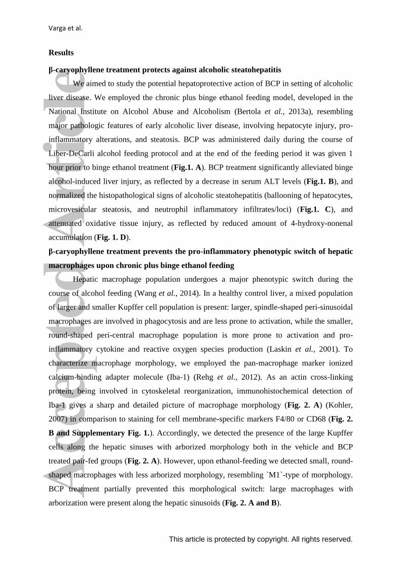
Results
β-caryophyllene treatment protects against alcoholic steatohepatitis
We aimed to study the potential hepatoprotective action of BCP in setting of alcoholic
liver disease. We employed the chronic plus binge ethanol feeding model, developed in the
National Institute on Alcohol Abuse and Alcoholism (Bertola et al., 2013a), resembling
major pathologic features of early alcoholic liver disease, involving hepatocyte injury, pro-
inflammatory alterations, and steatosis. BCP was administered daily during the course of
Liber-DeCarli alcohol feeding protocol and at the end of the feeding period it was given 1
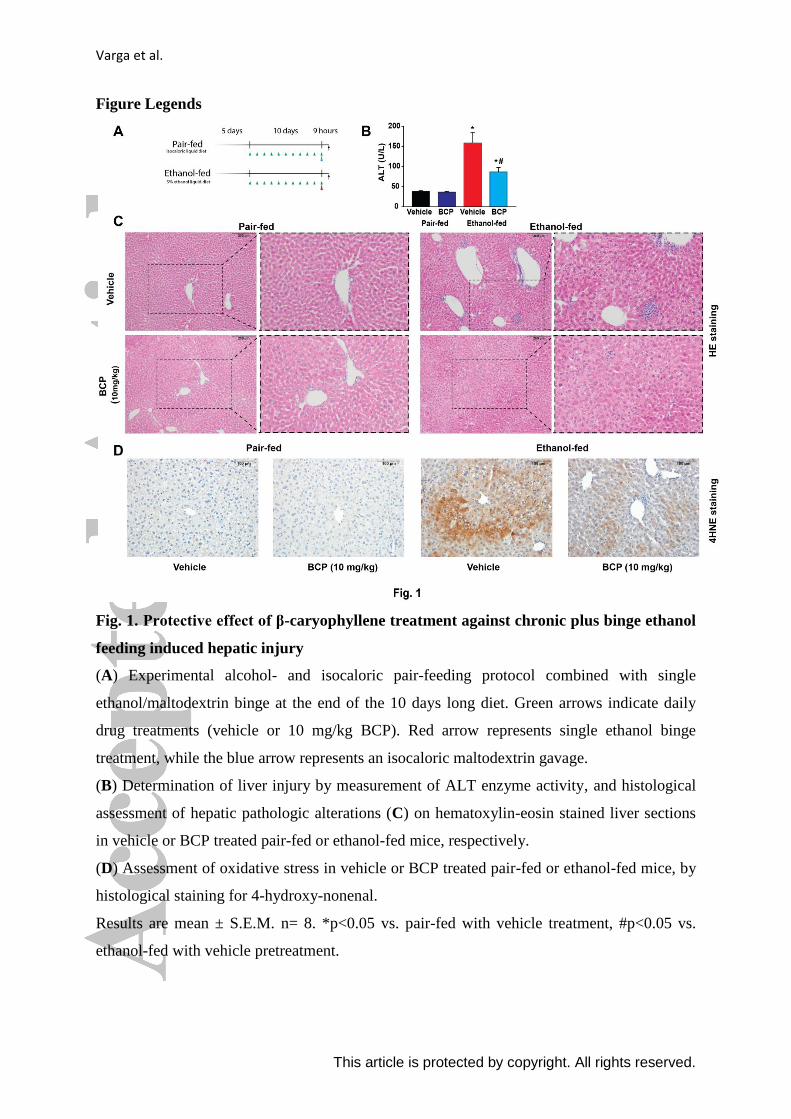
hour prior to binge ethanol treatment (Fig.1. A). BCP treatment significantly alleviated binge
alcohol-induced liver injury, as reflected by a decrease in serum ALT levels (Fig.1. B), and
normalized the histopathological signs of alcoholic steatohepatitis (ballooning of hepatocytes,
microvesicular steatosis, and neutrophil inflammatory infiltrates/loci) (Fig.1. C), and
attenuated oxidative tissue injury, as reflected by reduced amount of 4-hydroxy-nonenal
accumulation (Fig. 1. D).
β-caryophyllene treatment prevents the pro-inflammatory phenotypic switch of hepatic
macrophages upon chronic plus binge ethanol feeding
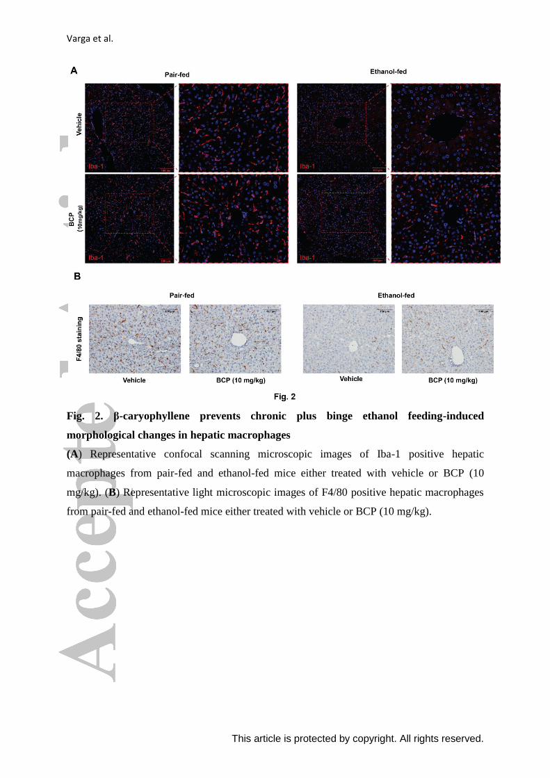
Hepatic macrophage population undergoes a major phenotypic switch during the
course of alcohol feeding (Wang et al., 2014). In a healthy control liver, a mixed population
of larger and smaller Kupffer cell population is present: larger, spindle-shaped peri-sinusoidal
macrophages are involved in phagocytosis and are less prone to activation, while the smaller,
round-shaped peri-central macrophage population is more prone to activation and pro-
inflammatory cytokine and reactive oxygen species production (Laskin et al., 2001). To
characterize macrophage morphology, we employed the pan-macrophage marker ionized
calcium-binding adapter molecule (Iba-1) (Rehg et al., 2012). As an actin cross-linking
protein, being involved in cytoskeletal reorganization, immunohistochemical detection of
Iba-1 gives a sharp and detailed picture of macrophage morphology (Fig. 2. A) (Kohler,
2007) in comparison to staining for cell membrane-specific markers F4/80 or CD68 (Fig. 2.
B and Supplementary Fig. 1.). Accordingly, we detected the presence of the large Kupffer
cells along the hepatic sinuses with arborized morphology both in the vehicle and BCP
treated pair-fed groups (Fig. 2. A). However, upon ethanol-feeding we detected small, round-
shaped macrophages with less arborized morphology, resembling `M1`-type of morphology.
BCP treatment partially prevented this morphological switch: large macrophages with
arborization were present along the hepatic sinusoids (Fig. 2. A and B).
Varga et al.
This article is protected by copyright. All rights reserved.
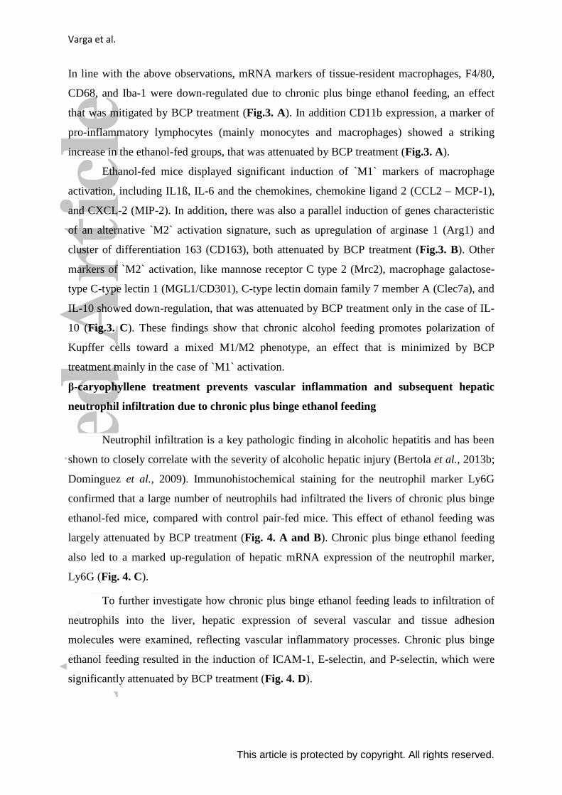
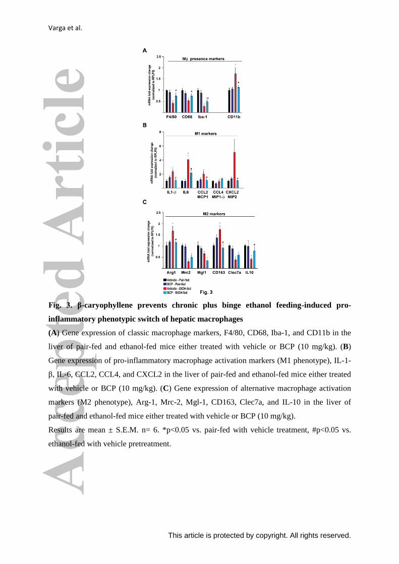
In line with the above observations, mRNA markers of tissue-resident macrophages, F4/80,
CD68, and Iba-1 were down-regulated due to chronic plus binge ethanol feeding, an effect
that was mitigated by BCP treatment (Fig.3. A). In addition CD11b expression, a marker of
pro-inflammatory lymphocytes (mainly monocytes and macrophages) showed a striking
increase in the ethanol-fed groups, that was attenuated by BCP treatment (Fig.3. A).
Ethanol-fed mice displayed significant induction of `M1` markers of macrophage
activation, including IL1ß, IL-6 and the chemokines, chemokine ligand 2 (CCL2 – MCP-1),
and CXCL-2 (MIP-2). In addition, there was also a parallel induction of genes characteristic
of an alternative `M2` activation signature, such as upregulation of arginase 1 (Arg1) and
cluster of differentiation 163 (CD163), both attenuated by BCP treatment (Fig.3. B). Other
markers of `M2` activation, like mannose receptor C type 2 (Mrc2), macrophage galactose-
type C-type lectin 1 (MGL1/CD301), C-type lectin domain family 7 member A (Clec7a), and
IL-10 showed down-regulation, that was attenuated by BCP treatment only in the case of IL-
10 (Fig.3. C). These findings show that chronic alcohol feeding promotes polarization of
Kupffer cells toward a mixed M1/M2 phenotype, an effect that is minimized by BCP
treatment mainly in the case of `M1` activation.
β-caryophyllene treatment prevents vascular inflammation and subsequent hepatic
neutrophil infiltration due to chronic plus binge ethanol feeding
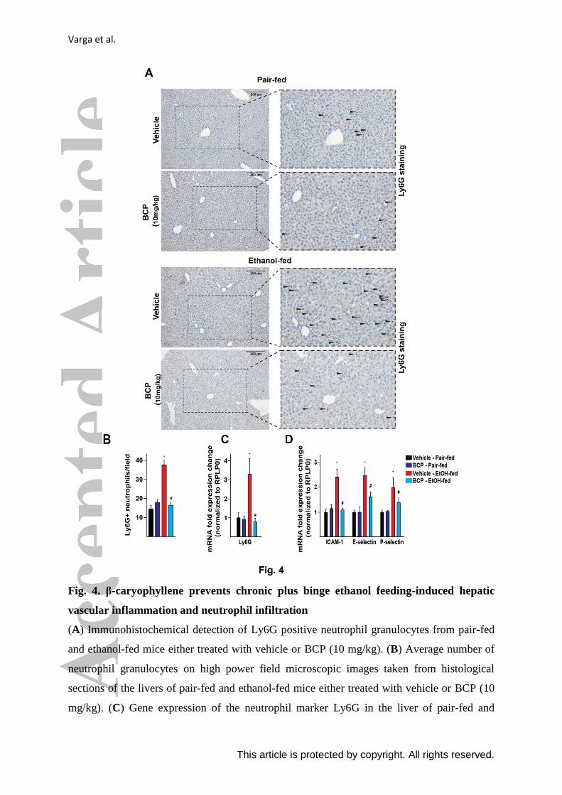
Neutrophil infiltration is a key pathologic finding in alcoholic hepatitis and has been
shown to closely correlate with the severity of alcoholic hepatic injury (Bertola et al., 2013b;
Dominguez et al., 2009). Immunohistochemical staining for the neutrophil marker Ly6G
confirmed that a large number of neutrophils had infiltrated the livers of chronic plus binge
ethanol-fed mice, compared with control pair-fed mice. This effect of ethanol feeding was
largely attenuated by BCP treatment (Fig. 4. A and B). Chronic plus binge ethanol feeding
also led to a marked up-regulation of hepatic mRNA expression of the neutrophil marker,
Ly6G (Fig. 4. C).
To further investigate how chronic plus binge ethanol feeding leads to infiltration of
neutrophils into the liver, hepatic expression of several vascular and tissue adhesion
molecules were examined, reflecting vascular inflammatory processes. Chronic plus binge
ethanol feeding resulted in the induction of ICAM-1, E-selectin, and P-selectin, which were
significantly attenuated by BCP treatment (Fig. 4. D).
Varga et al.
This article is protected by copyright. All rights reserved.
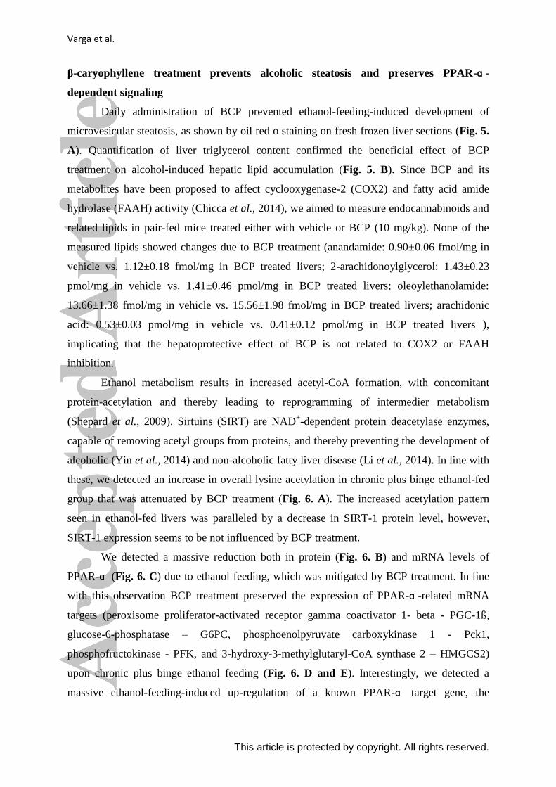
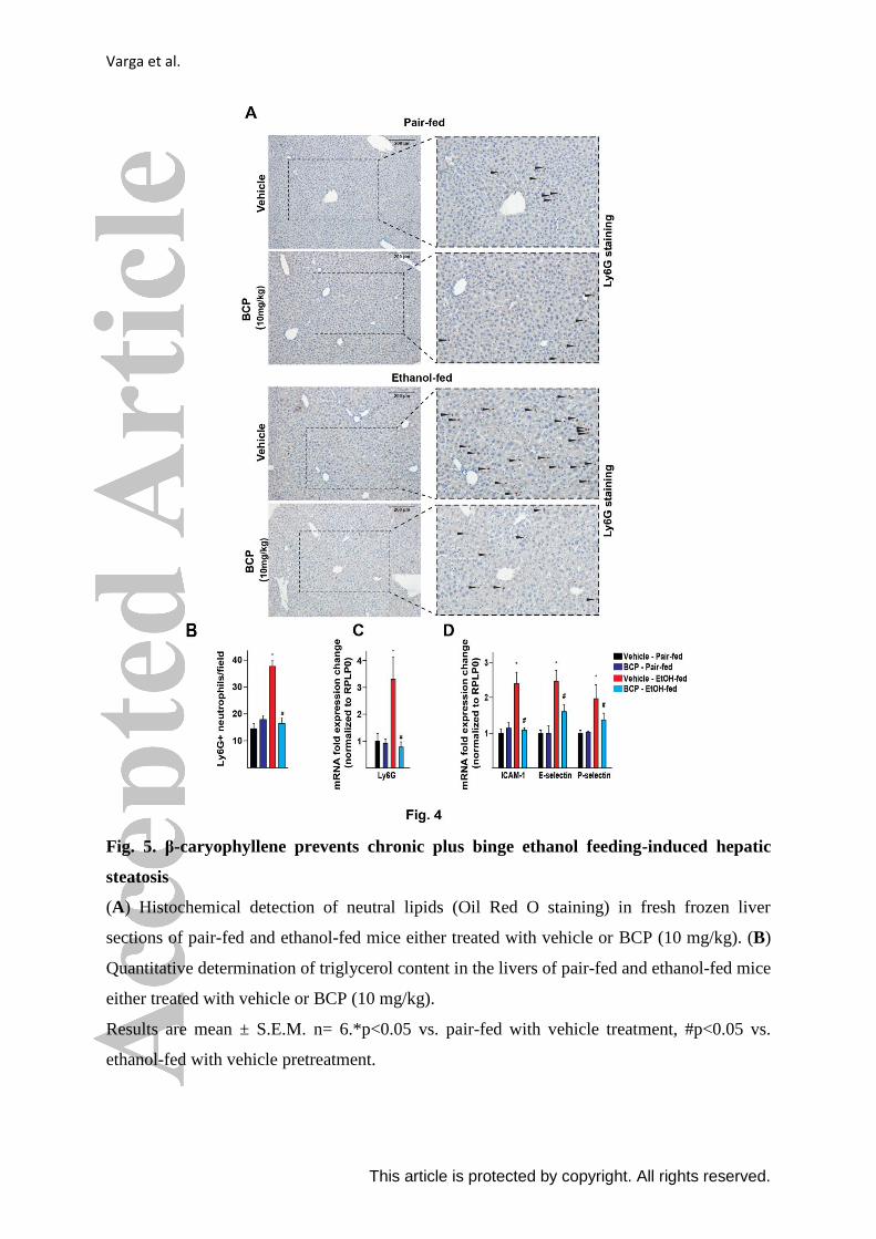
β-caryophyllene treatment prevents alcoholic steatosis and preserves PPAR-ɑ -
dependent signaling
Daily administration of BCP prevented ethanol-feeding-induced development of
microvesicular steatosis, as shown by oil red o staining on fresh frozen liver sections (Fig. 5.
A). Quantification of liver triglycerol content confirmed the beneficial effect of BCP
treatment on alcohol-induced hepatic lipid accumulation (Fig. 5. B). Since BCP and its
metabolites have been proposed to affect cyclooxygenase-2 (COX2) and fatty acid amide
hydrolase (FAAH) activity (Chicca et al., 2014), we aimed to measure endocannabinoids and
related lipids in pair-fed mice treated either with vehicle or BCP (10 mg/kg). None of the
measured lipids showed changes due to BCP treatment (anandamide: 0.90±0.06 fmol/mg in
vehicle vs. 1.12±0.18 fmol/mg in BCP treated livers; 2-arachidonoylglycerol: 1.43±0.23
pmol/mg in vehicle vs. 1.41±0.46 pmol/mg in BCP treated livers; oleoylethanolamide:
13.66±1.38 fmol/mg in vehicle vs. 15.56±1.98 fmol/mg in BCP treated livers; arachidonic
acid: 0.53±0.03 pmol/mg in vehicle vs. 0.41±0.12 pmol/mg in BCP treated livers ),
implicating that the hepatoprotective effect of BCP is not related to COX2 or FAAH
inhibition.
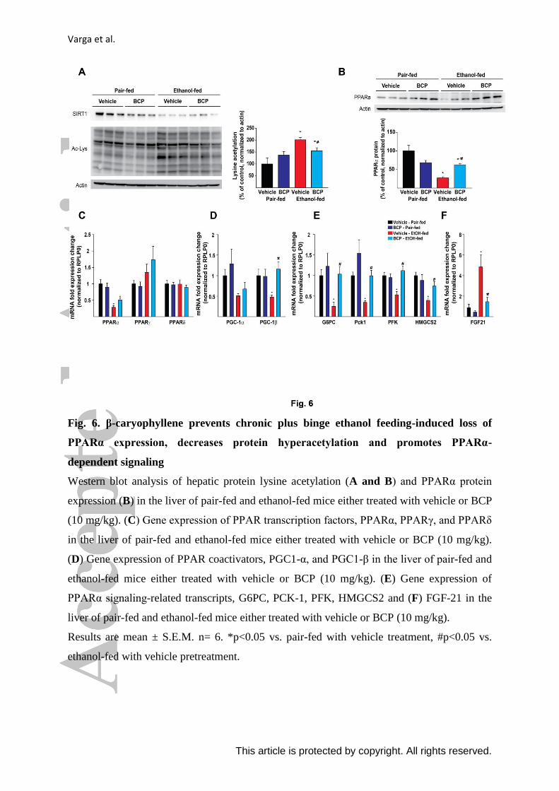
Ethanol metabolism results in increased acetyl-CoA formation, with concomitant
protein-acetylation and thereby leading to reprogramming of intermedier metabolism
(Shepard et al., 2009). Sirtuins (SIRT) are NAD+-dependent protein deacetylase enzymes,
capable of removing acetyl groups from proteins, and thereby preventing the development of
alcoholic (Yin et al., 2014) and non-alcoholic fatty liver disease (Li et al., 2014). In line with
these, we detected an increase in overall lysine acetylation in chronic plus binge ethanol-fed
group that was attenuated by BCP treatment (Fig. 6. A). The increased acetylation pattern
seen in ethanol-fed livers was paralleled by a decrease in SIRT-1 protein level, however,
SIRT-1 expression seems to be not influenced by BCP treatment.
We detected a massive reduction both in protein (Fig. 6. B) and mRNA levels of
PPAR-ɑ (Fig. 6. C) due to ethanol feeding, which was mitigated by BCP treatment. In line
with this observation BCP treatment preserved the expression of PPAR-ɑ -related mRNA
targets (peroxisome proliferator-activated receptor gamma coactivator 1- beta - PGC-1ß,
glucose-6-phosphatase – G6PC, phosphoenolpyruvate carboxykinase 1 - Pck1,
phosphofructokinase - PFK, and 3-hydroxy-3-methylglutaryl-CoA synthase 2 – HMGCS2)
upon chronic plus binge ethanol feeding (Fig. 6. D and E). Interestingly, we detected a
massive ethanol-feeding-induced up-regulation of a known PPAR-ɑ target gene, the
Varga et al.
This article is protected by copyright. All rights reserved.
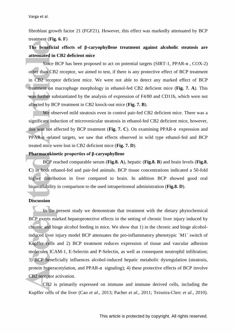
fibroblast growth factor 21 (FGF21). However, this effect was markedly attenuated by BCP
treatment (Fig. 6. F)
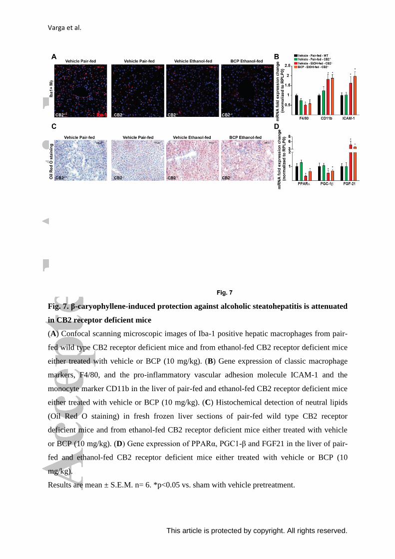
The beneficial effects of β-caryophyllene treatment against alcoholic steatosis are
attenuated in CB2 deficient mice
Since BCP has been proposed to act on potential targets (SIRT-1, PPAR-ɑ , COX-2)
other than CB2 receptor, we aimed to test, if there is any protective effect of BCP treatment
in CB2 receptor deficient mice. We were not able to detect any marked effect of BCP
treatment on macrophage morphology in ethanol-fed CB2 deficient mice (Fig. 7. A). This
was further substantiated by the analysis of expression of F4/80 and CD11b, which were not
affected by BCP treatment in CB2 knock-out mice (Fig. 7. B).
We observed mild steatosis even in control pair-fed CB2 deficient mice. There was a
significant induction of microvesicular steatosis in ethanol-fed CB2 deficient mice, however,
this was not affected by BCP treatment (Fig. 7. C). On examining PPAR-ɑ expression and
PPAR-ɑ -related targets, we saw that effects observed in wild type ethanol-fed and BCP
treated mice were lost in CB2 deficient mice (Fig. 7. D).
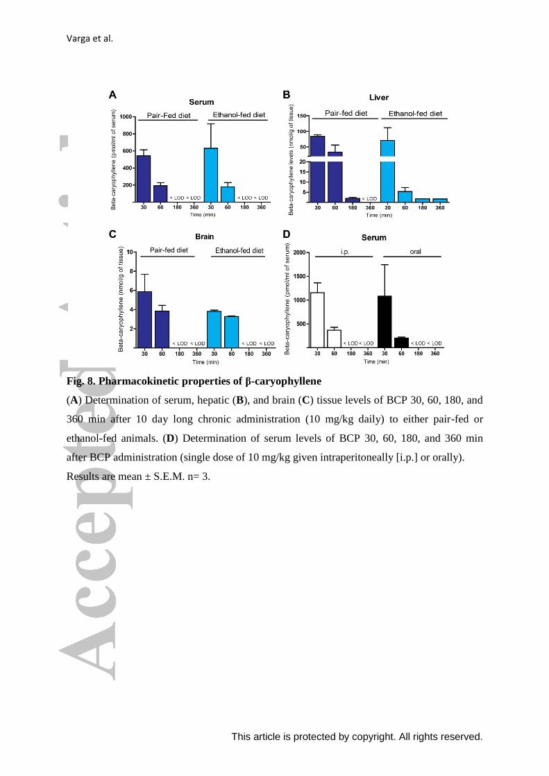
Pharmacokinetic properties of β-caryophyllene
BCP reached comparable serum (Fig.8. A), hepatic (Fig.8. B) and brain levels (Fig.8.
C) in both ethanol-fed and pair-fed animals. BCP tissue concentrations indicated a 50-fold
higher distribution in liver compared to brain. In addition BCP showed good oral
bioavailability in comparison to the used intraperitoneal administration (Fig.8. D).
Discussion
In the present study we demonstrate that treatment with the dietary phytochemical
BCP exerts marked hepatoprotective effects in the setting of chronic liver injury induced by
chronic and binge alcohol feeding in mice. We show that 1) in the chronic and binge alcohol-
induced liver injury model BCP attenuates the pro-inflammatory phenotypic `M1` switch of
Kupffer cells and 2) BCP treatment reduces expression of tissue and vascular adhesion
molecules ICAM-1, E-Selectin and P-Selectin, as well as consequent neutrophil infiltration;
3) BCP beneficially influences alcohol-induced hepatic metabolic dysregulation (steatosis,
protein hyperacetylation, and PPAR-ɑ signaling); 4) these protective effects of BCP involve
CB2 receptor activation.
CB2 is primarily expressed on immune and immune derived cells, including the
Kupffer cells of the liver (Cao et al., 2013; Pacher et al., 2011; Teixeira-Clerc et al., 2010).
Varga et al.
This article is protected by copyright. All rights reserved.
Activation of CB2 receptors mediate anti-inflammatory actions, limiting inflammatory and
subsequent oxidative/nitrative tissue injury and organ damage (Pacher et al., 2011).
Resident macrophages of the liver (Kupffer cells), account approximately for 10–15%
of all liver cells. These cells are localized along the sinusoidal space of Disse anchored to
endothelial cells by long cytoplasmic processes, being involved in maintaining tissue
homeostasis and providing immunosurveillance. In addition, they also play a protective role
in promoting liver regeneration (Meijer et al., 2000). Kupffer cells may become activated by
a variety of stimuli that trigger innate immunity, involving endogenous molecules released
during hepatocyte injury (e.g. hyaluronic acid, heparin sulfate, high-mobility group box 1
(HMGB1), or heat shock proteins) (Tsung et al., 2005). In addition, Kupffer cell activation
might be triggered by environmental toxins, as in the case of alcoholic steatohepatitis by
ethanol and its metabolites or by endotoxin, absorbed due to increased gastrointestinal
permeability. Pathologically over-activated macrophages, as seen in alcoholic-, and in non-
alcoholic fatty liver disease, may further aggravate tissue injury and inflammation, leading to
liver failure. Since BCP has been proposed to protect the colonic mucosa against dextran
sulfate-induced colitis (Bento et al., 2011; Cho et al., 2007), it is possible that BCP exert
similar protection against ethanol-induced mucosal damage and thereby also attenuates
endotoxin translocation from the gut to the portal circulation (Bode et al., 1987).
Kupffer cells consist of three major different subsets; the CD11b+ cells, and CD68
+
cells overlapping with CD32+ cells, representing precursors of CD68+ cells (expressing stem
cell markers, such as c-kit (CD117) and CD34 (Kinoshita et al., 2010). Detailed flow
cytometric and immunohistochemical studies also revealed that the CD68+ cells are large and
spindle-shaped, while the CD11b+ cells are small and round or oval-shaped (Ikarashi et al.,
2013). While large macrophages are more phagocytic and generate increased quantities of
lysosomal enzymes, the smaller macrophages release more reactive oxygen species, and
appear to be more susceptible to `M1`-type of activation and cytokine production.
Interestingly, we found that chronic and binge alcohol feeding resulted in pro-inflammatory
phenotypic switch in liver Kupffer cells, which was attenuated by BCP treatment.
Experimental CB2 agonists (e.g.: JWH-133, HU910, HU308) have been shown to
favorably affect overall hepatic macrophage activation upon chronic alcohol exposure
(Louvet et al., 2011) and hepatocyte injury (Batkai et al., 2007; Horvath et al., 2012a; Rajesh
et al., 2008). However, not a single CB2 agonist is approved for clinical testing in liver
disease yet. In this study, we demonstrate that the FDA approved food additive BCP, which is
present in various food and medicinal plants, as well as in cannabis essential oil, exerts
Varga et al.
This article is protected by copyright. All rights reserved.
hepatoprotective effects in a model of alcohol-induced chronic liver injury. The observed
protective effects were attenuated in CB2 knockout mice indicating involvement of CB2. To
increase the translational potential of our study, we also tested the pharmacokinetic properties
of oral and intraperitoneal BCP administration both in healthy and in ethanol-fed mice. BCP
was orally bioavailable, and ethanol-feeding did not interfere with its kinetics (e.g.
absorption, or metabolism). The BCP dose used in our study in mice (10 mg/kg) is
convertible to a human equivalent dose of approximately 0.8-1 mg/kg/day for adult human
subjects (Nair et al., 2016). Based on the mouse pharmacokinetics data, repeated oral dosing
of BCP might be desirable, the benefit of which remains to be tested.
Our results also suggest that BCP has beneficial properties on intermediary
metabolism in ethanol-fed animals, potentially by decreasing pro-inflammatory cytokine
expression in a CB2-dependent manner and thereby preserving PPAR-ɑ -related signaling
that is characteristic for the healthy liver.
An intriguing field of investigation is how pro-inflammatory alterations influence
hepatic intermediary metabolism. It is now well established that pro-inflammatory cytokines
(IL-1ß or TNFɑ ) may profoundly affect hepatic lipid and glucose metabolism in a paracrine
manner (Louvet et al., 2011; Stienstra et al., 2010). Stienstra et al. has shown that IL-1ß is
capable to suppress human and mouse PPAR-ɑ promoter activity and in parallel interferes
with the ability of PPAR-ɑ to activate transcription of target genes (Stienstra et al., 2010).
We found that alcohol markedly impaired PPAR-ɑ signaling and increased lipid
accumulation in the liver, which were significantly attenuated by BCP treatment, however,
the effects seem to relate to BCP-induced CB2 activation rather than to a direct PPAR-ɑ
mediated effect.
During hepatic ethanol metabolism, liver mitochondria convert acetate into acetyl-
CoA that is further processed in the citric acid cycle. However, due to ethanol metabolism,
there is an increased level of NADH that inhibits further metabolism of the acetyl-CoA by the
citric acid cycle. In addition excess NADH inhibits gluconeogenesis by preventing the
oxidation of lactate to pyruvate, leading to accumulation of lactate (lactic acidosis), and
causing hypoglycemia (Tsai et al., 2015). Further consequences of hepatic acetyl-CoA
accumulation involves increased protein acetylation (histones, transcription factors –
SREBP1c, FOXO1, FOXO3, PGC1ɑ - (Shepard et al., 2009)), ketone body formation,
increased fatty acid synthesis and fat storage, and buildup of acetaldehyde. Increased
production of acetaldehyde forms covalent bonds with many important functional groups in
proteins, impairing protein function. It may also react with phospholipids and arachidonic
Varga et al.
This article is protected by copyright. All rights reserved.
acid, which triggers lipid peroxidation reactions promoting hepatocyte cell death.
Consistently with the above, we found increased protein acetylation and lipid peroxidation
(4HNE formation) in livers of ethanol-fed groups, which were attenuated by BCP treatment.
Collectively, our results demonstrate that BCP treatment exerts beneficial effects against liver
injury induced by chronic plus binge ethanol feeding by attenuating the Kupffer cell-
mediated pro-inflammatory response (activation and/or pro-inflammatory phenotypic `M1`
switch), neutrophil-mediated oxidative/nitrative stress/injury, vascular inflammation
(expression of vascular adhesion molecules), and hepatic metabolic dysregulation (steatosis,
protein hyperacetylation, and PPAR-ɑ signaling). Our results also indicate that these in vivo
protective effects of BCP against alcohol-induced hepatic injury may involve, at least in part,
CB2 mediated mechanisms. Our study may have immediate translational potential in liver
disease since BCP is an FDA approved food additive in humans.
Author Contributions
Z.V.V., P.P. conception and design of research; Z.V.V., C.M., K.E., R.C., D.N., A.C.,
B.T.N., J.P., T.L., L.C. performed experiments; Z.V.V., C.M., A.C., J.P., and B.T.N.
analyzed data; Z.V.V., R.C., G.H., A.C., B.G., G.K., J.G., and P.P. interpreted results of
experiments; Z.V.V. and P.P. prepared figures; Z.V.V and P.P. drafted manuscript; Z.V.V.,
A.C., G.H., B.G.,G.K., J.G., and P.P. edited and revised manuscript; Z.V.V., C.M., K.E.,
R.C., D.N., A.C., B.T.N., J.P., T.L., L.C., G.H., B.G.,G.K., J.G., and P.P. approved final
version of manuscript.
Acknowledgments
The recent work was supported by the Intramural Research Program of NIAAA/NIH (to P.
Pacher). C. Matyas was supported by the scholarship of the Hungarian-American Enterprise
Scholarship Fund/Council on International Educational Exchange. Z. V. Varga was supported
by the Rosztoczy Foundation.
Varga et al.
This article is protected by copyright. All rights reserved.
References
Al Mansouri S, Ojha S, Al Maamari E, Al Ameri M, Nurulain SM, Bahi A (2014). The cannabinoid receptor 2 agonist, beta-caryophyllene, reduced voluntary alcohol intake and attenuated ethanol-induced place preference and sensitivity in mice. Pharmacology, biochemistry, and behavior 124: 260-268. Batkai S, Osei-Hyiaman D, Pan H, El-Assal O, Rajesh M, Mukhopadhyay P, et al. (2007). Cannabinoid-2 receptor mediates protection against hepatic ischemia/reperfusion injury. FASEB journal : official publication of the Federation of American Societies for Experimental Biology 21(8): 1788-1800. Bento AF, Marcon R, Dutra RC, Claudino RF, Cola M, Leite DF, et al. (2011). beta-Caryophyllene inhibits dextran sulfate sodium-induced colitis in mice through CB2 receptor activation and PPARgamma pathway. The American journal of pathology 178(3): 1153-1166. Bertola A, Mathews S, Ki SH, Wang H, Gao B (2013a). Mouse model of chronic and binge ethanol feeding (the NIAAA model). Nature protocols 8(3): 627-637. Bertola A, Park O, Gao B (2013b). Chronic plus binge ethanol feeding synergistically induces neutrophil infiltration and liver injury in mice: a critical role for E-selectin. Hepatology 58(5): 1814-1823. Bode C, Kugler V, Bode JC (1987). Endotoxemia in patients with alcoholic and non-alcoholic cirrhosis and in subjects with no evidence of chronic liver disease following acute alcohol excess. Journal of hepatology 4(1): 8-14. Cao Z, Mulvihill MM, Mukhopadhyay P, Xu H, Erdelyi K, Hao E, et al. (2013). Monoacylglycerol lipase controls endocannabinoid and eicosanoid signaling and hepatic injury in mice. Gastroenterology 144(4): 808-817 e815. Chicca A, Caprioglio D, Minassi A, Petrucci V, Appendino G, Taglialatela-Scafati O, et al. (2014). Functionalization of beta-caryophyllene generates novel polypharmacology in the endocannabinoid system. ACS chemical biology 9(7): 1499-1507. Cho HI, Hong JM, Choi JW, Choi HS, Kwak JH, Lee DU, et al. (2015). beta-Caryophyllene alleviates D-galactosamine and lipopolysaccharide-induced hepatic injury through suppression of the TLR4 and RAGE signaling pathways. European journal of pharmacology 764: 613-621. Cho JY, Chang HJ, Lee SK, Kim HJ, Hwang JK, Chun HS (2007). Amelioration of dextran sulfate sodium-induced colitis in mice by oral administration of beta-caryophyllene, a sesquiterpene. Life sciences 80(10): 932-939. Choi IY, Ju C, Anthony Jalin AM, Lee DI, Prather PL, Kim WK (2013). Activation of cannabinoid CB2 receptor-mediated AMPK/CREB pathway reduces cerebral ischemic injury. The American journal of pathology 182(3): 928-939. Dominguez M, Miquel R, Colmenero J, Moreno M, Garcia-Pagan JC, Bosch J, et al. (2009). Hepatic expression of CXC chemokines predicts portal hypertension and survival in patients with alcoholic hepatitis. Gastroenterology 136(5): 1639-1650.
Varga et al.
This article is protected by copyright. All rights reserved.
Gertsch J, Leonti M, Raduner S, Racz I, Chen JZ, Xie XQ, et al. (2008). Beta-caryophyllene is a dietary cannabinoid. Proceedings of the National Academy of Sciences of the United States of America 105(26): 9099-9104. Gertsch J, Pertwee RG, Di Marzo V (2010). Phytocannabinoids beyond the Cannabis plant - do they exist? British journal of pharmacology 160(3): 523-529. Horvath B, Magid L, Mukhopadhyay P, Batkai S, Rajesh M, Park O, et al. (2012a). A new cannabinoid CB2 receptor agonist HU-910 attenuates oxidative stress, inflammation and cell death associated with hepatic ischaemia/reperfusion injury. British journal of pharmacology 165(8): 2462-2478. Horvath B, Mukhopadhyay P, Kechrid M, Patel V, Tanchian G, Wink DA, et al. (2012b). beta-Caryophyllene ameliorates cisplatin-induced nephrotoxicity in a cannabinoid 2 receptor-dependent manner. Free radical biology & medicine 52(8): 1325-1333. Ikarashi M, Nakashima H, Kinoshita M, Sato A, Nakashima M, Miyazaki H, et al. (2013). Distinct development and functions of resident and recruited liver Kupffer cells/macrophages. Journal of leukocyte biology 94(6): 1325-1336. Jeong WI, Osei-Hyiaman D, Park O, Liu J, Batkai S, Mukhopadhyay P, et al. (2008). Paracrine activation of hepatic CB1 receptors by stellate cell-derived endocannabinoids mediates alcoholic fatty liver. Cell metabolism 7(3): 227-235. Kinoshita M, Uchida T, Sato A, Nakashima M, Nakashima H, Shono S, et al. (2010). Characterization of two F4/80-positive Kupffer cell subsets by their function and phenotype in mice. Journal of hepatology 53(5): 903-910. Kohler C (2007). Allograft inflammatory factor-1/Ionized calcium-binding adapter molecule 1 is specifically expressed by most subpopulations of macrophages and spermatids in testis. Cell and tissue research 330(2): 291-302. Laskin DL, Weinberger B, Laskin JD (2001). Functional heterogeneity in liver and lung macrophages. Journal of leukocyte biology 70(2): 163-170. Li Y, Wong K, Giles A, Jiang J, Lee JW, Adams AC, et al. (2014). Hepatic SIRT1 attenuates hepatic steatosis and controls energy balance in mice by inducing fibroblast growth factor 21. Gastroenterology 146(2): 539-549 e537. Louvet A, Teixeira-Clerc F, Chobert MN, Deveaux V, Pavoine C, Zimmer A, et al. (2011). Cannabinoid CB2 receptors protect against alcoholic liver disease by regulating Kupffer cell polarization in mice. Hepatology 54(4): 1217-1226. Mahmoud MF, Swefy SE, Hasan RA, Ibrahim A (2014). Role of cannabinoid receptors in hepatic fibrosis and apoptosis associated with bile duct ligation in rats. European journal of pharmacology 742: 118-124. Meijer C, Wiezer MJ, Diehl AM, Schouten HJ, Schouten HJ, Meijer S, et al. (2000). Kupffer cell depletion by CI2MDP-liposomes alters hepatic cytokine expression and delays liver regeneration after partial hepatectomy. Liver 20(1): 66-77.
Varga et al.
This article is protected by copyright. All rights reserved.
Mukhopadhyay B, Cinar R, Yin S, Liu J, Tam J, Godlewski G, et al. (2011). Hyperactivation of anandamide synthesis and regulation of cell-cycle progression via cannabinoid type 1 (CB1) receptors in the regenerating liver. Proceedings of the National Academy of Sciences of the United States of America 108(15): 6323-6328. Nair AB, Jacob S (2016). A simple practice guide for dose conversion between animals and human. Journal of basic and clinical pharmacy 7(2): 27-31. Ojha S, Javed H, Azimullah S, Haque ME (2016). beta-Caryophyllene, a phytocannabinoid attenuates oxidative stress, neuroinflammation, glial activation, and salvages dopaminergic neurons in a rat model of Parkinson disease. Molecular and cellular biochemistry 418(1-2): 59-70. Osei-Hyiaman D, DePetrillo M, Pacher P, Liu J, Radaeva S, Batkai S, et al. (2005). Endocannabinoid activation at hepatic CB1 receptors stimulates fatty acid synthesis and contributes to diet-induced obesity. The Journal of clinical investigation 115(5): 1298-1305. Pacher P, Mechoulam R (2011). Is lipid signaling through cannabinoid 2 receptors part of a protective system? Progress in lipid research 50(2): 193-211. Rajesh M, Mukhopadhyay P, Hasko G, Huffman JW, Mackie K, Pacher P (2008). CB2 cannabinoid receptor agonists attenuate TNF-alpha-induced human vascular smooth muscle cell proliferation and migration. British journal of pharmacology 153(2): 347-357. Rehg JE, Bush D, Ward JM (2012). The utility of immunohistochemistry for the identification of hematopoietic and lymphoid cells in normal tissues and interpretation of proliferative and inflammatory lesions of mice and rats. Toxicologic pathology 40(2): 345-374. Shepard BD, Tuma PL (2009). Alcohol-induced protein hyperacetylation: mechanisms and consequences. World journal of gastroenterology 15(10): 1219-1230. Silvestri C, Di Marzo V (2013). The endocannabinoid system in energy homeostasis and the etiopathology of metabolic disorders. Cell metabolism 17(4): 475-490. Stienstra R, Saudale F, Duval C, Keshtkar S, Groener JE, van Rooijen N, et al. (2010). Kupffer cells promote hepatic steatosis via interleukin-1beta-dependent suppression of peroxisome proliferator-activated receptor alpha activity. Hepatology 51(2): 511-522. Tam J, Cinar R, Liu J, Godlewski G, Wesley D, Jourdan T, et al. (2012). Peripheral cannabinoid-1 receptor inverse agonism reduces obesity by reversing leptin resistance. Cell metabolism 16(2): 167-179. Tam J, Liu J, Mukhopadhyay B, Cinar R, Godlewski G, Kunos G (2011). Endocannabinoids in liver disease. Hepatology 53(1): 346-355. Teixeira-Clerc F, Belot MP, Manin S, Deveaux V, Cadoudal T, Chobert MN, et al. (2010). Beneficial paracrine effects of cannabinoid receptor 2 on liver injury and regeneration. Hepatology 52(3): 1046-1059. Teixeira-Clerc F, Julien B, Grenard P, Tran Van Nhieu J, Deveaux V, Li L, et al. (2006). CB1 cannabinoid receptor antagonism: a new strategy for the treatment of liver fibrosis. Nature medicine 12(6): 671-676.
Varga et al.
This article is protected by copyright. All rights reserved.
Tsai WW, Matsumura S, Liu W, Phillips NG, Sonntag T, Hao E, et al. (2015). ATF3 mediates inhibitory effects of ethanol on hepatic gluconeogenesis. Proceedings of the National Academy of Sciences of the United States of America 112(9): 2699-2704. Tsung A, Hoffman RA, Izuishi K, Critchlow ND, Nakao A, Chan MH, et al. (2005). Hepatic ischemia/reperfusion injury involves functional TLR4 signaling in nonparenchymal cells. Journal of immunology 175(11): 7661-7668. Wang M, You Q, Lor K, Chen F, Gao B, Ju C (2014). Chronic alcohol ingestion modulates hepatic macrophage populations and functions in mice. Journal of leukocyte biology 96(4): 657-665. Wu C, Jia Y, Lee JH, Jun HJ, Lee HS, Hwang KY, et al. (2014). trans-Caryophyllene is a natural agonistic ligand for peroxisome proliferator-activated receptor-alpha. Bioorganic & medicinal chemistry letters 24(14): 3168-3174. Yin H, Hu M, Liang X, Ajmo JM, Li X, Bataller R, et al. (2014). Deletion of SIRT1 from hepatocytes in mice disrupts lipin-1 signaling and aggravates alcoholic fatty liver. Gastroenterology 146(3): 801-811. Zheng X, Sun T, Wang X (2013). Activation of type 2 cannabinoid receptors (CB2R) promotes fatty acid oxidation through the SIRT1/PGC-1alpha pathway. Biochemical and biophysical research communications 436(3): 377-381.
Varga et al.
This article is protected by copyright. All rights reserved.
Figure Legends
Fig. 1. Protective effect of β-caryophyllene treatment against chronic plus binge ethanol
feeding induced hepatic injury
(A) Experimental alcohol- and isocaloric pair-feeding protocol combined with single
ethanol/maltodextrin binge at the end of the 10 days long diet. Green arrows indicate daily
drug treatments (vehicle or 10 mg/kg BCP). Red arrow represents single ethanol binge
treatment, while the blue arrow represents an isocaloric maltodextrin gavage.
(B) Determination of liver injury by measurement of ALT enzyme activity, and histological
assessment of hepatic pathologic alterations (C) on hematoxylin-eosin stained liver sections
in vehicle or BCP treated pair-fed or ethanol-fed mice, respectively.
(D) Assessment of oxidative stress in vehicle or BCP treated pair-fed or ethanol-fed mice, by
histological staining for 4-hydroxy-nonenal.
Results are mean ± S.E.M. n= 8. *p<0.05 vs. pair-fed with vehicle treatment, #p<0.05 vs.
ethanol-fed with vehicle pretreatment.
Varga et al.
This article is protected by copyright. All rights reserved.
Fig. 2. β-caryophyllene prevents chronic plus binge ethanol feeding-induced
morphological changes in hepatic macrophages
(A) Representative confocal scanning microscopic images of Iba-1 positive hepatic
macrophages from pair-fed and ethanol-fed mice either treated with vehicle or BCP (10
mg/kg). (B) Representative light microscopic images of F4/80 positive hepatic macrophages
from pair-fed and ethanol-fed mice either treated with vehicle or BCP (10 mg/kg).
Varga et al.
This article is protected by copyright. All rights reserved.
Fig. 3. β-caryophyllene prevents chronic plus binge ethanol feeding-induced pro-
inflammatory phenotypic switch of hepatic macrophages
(A) Gene expression of classic macrophage markers, F4/80, CD68, Iba-1, and CD11b in the
liver of pair-fed and ethanol-fed mice either treated with vehicle or BCP (10 mg/kg). (B)
Gene expression of pro-inflammatory macrophage activation markers (M1 phenotype), IL-1-
β, IL-6, CCL2, CCL4, and CXCL2 in the liver of pair-fed and ethanol-fed mice either treated
with vehicle or BCP (10 mg/kg). (C) Gene expression of alternative macrophage activation
markers (M2 phenotype), Arg-1, Mrc-2, Mgl-1, CD163, Clec7a, and IL-10 in the liver of
pair-fed and ethanol-fed mice either treated with vehicle or BCP (10 mg/kg).
Results are mean ± S.E.M. n= 6. *p<0.05 vs. pair-fed with vehicle treatment, #p<0.05 vs.
ethanol-fed with vehicle pretreatment.
Varga et al.
This article is protected by copyright. All rights reserved.
Fig. 4. β-caryophyllene prevents chronic plus binge ethanol feeding-induced hepatic
vascular inflammation and neutrophil infiltration
(A) Immunohistochemical detection of Ly6G positive neutrophil granulocytes from pair-fed
and ethanol-fed mice either treated with vehicle or BCP (10 mg/kg). (B) Average number of
neutrophil granulocytes on high power field microscopic images taken from histological
sections of the livers of pair-fed and ethanol-fed mice either treated with vehicle or BCP (10
mg/kg). (C) Gene expression of the neutrophil marker Ly6G in the liver of pair-fed and

Varga et al.
This article is protected by copyright. All rights reserved.
ethanol-fed mice either treated with vehicle or BCP (10 mg/kg). (D) Gene expression of pro-
inflammatory vascular adhesion molecules, ICAM-1, E-selectin, and P-selectin in the liver of
pair-fed and ethanol-fed mice either treated with vehicle or BCP (10 mg/kg).
Results are mean ± S.E.M. n= 6. *p<0.05 vs. pair-fed with vehicle treatment, #p<0.05 vs.
ethanol-fed with vehicle pretreatment.
Varga et al.
This article is protected by copyright. All rights reserved.
Fig. 5. β-caryophyllene prevents chronic plus binge ethanol feeding-induced hepatic
steatosis
(A) Histochemical detection of neutral lipids (Oil Red O staining) in fresh frozen liver
sections of pair-fed and ethanol-fed mice either treated with vehicle or BCP (10 mg/kg). (B)
Quantitative determination of triglycerol content in the livers of pair-fed and ethanol-fed mice
either treated with vehicle or BCP (10 mg/kg).
Results are mean ± S.E.M. n= 6.*p<0.05 vs. pair-fed with vehicle treatment, #p<0.05 vs.
ethanol-fed with vehicle pretreatment.
Varga et al.
This article is protected by copyright. All rights reserved.
Fig. 6. β-caryophyllene prevents chronic plus binge ethanol feeding-induced loss of
PPARα expression, decreases protein hyperacetylation and promotes PPARα-
dependent signaling
Western blot analysis of hepatic protein lysine acetylation (A and B) and PPARα protein
expression (B) in the liver of pair-fed and ethanol-fed mice either treated with vehicle or BCP
(10 mg/kg). (C) Gene expression of PPAR transcription factors, PPARα, PPARγ, and PPARδ
in the liver of pair-fed and ethanol-fed mice either treated with vehicle or BCP (10 mg/kg).
(D) Gene expression of PPAR coactivators, PGC1-α, and PGC1-β in the liver of pair-fed and
ethanol-fed mice either treated with vehicle or BCP (10 mg/kg). (E) Gene expression of
PPARα signaling-related transcripts, G6PC, PCK-1, PFK, HMGCS2 and (F) FGF-21 in the
liver of pair-fed and ethanol-fed mice either treated with vehicle or BCP (10 mg/kg).
Results are mean ± S.E.M. n= 6. *p<0.05 vs. pair-fed with vehicle treatment, #p<0.05 vs.
ethanol-fed with vehicle pretreatment.
Varga et al.
This article is protected by copyright. All rights reserved.
Fig. 7. β-caryophyllene-induced protection against alcoholic steatohepatitis is attenuated
in CB2 receptor deficient mice
(A) Confocal scanning microscopic images of Iba-1 positive hepatic macrophages from pair-
fed wild type CB2 receptor deficient mice and from ethanol-fed CB2 receptor deficient mice
either treated with vehicle or BCP (10 mg/kg). (B) Gene expression of classic macrophage
markers, F4/80, and the pro-inflammatory vascular adhesion molecule ICAM-1 and the
monocyte marker CD11b in the liver of pair-fed and ethanol-fed CB2 receptor deficient mice
either treated with vehicle or BCP (10 mg/kg). (C) Histochemical detection of neutral lipids
(Oil Red O staining) in fresh frozen liver sections of pair-fed wild type CB2 receptor
deficient mice and from ethanol-fed CB2 receptor deficient mice either treated with vehicle
or BCP (10 mg/kg). (D) Gene expression of PPARα, PGC1-β and FGF21 in the liver of pair-
fed and ethanol-fed CB2 receptor deficient mice either treated with vehicle or BCP (10
mg/kg).
Results are mean ± S.E.M. n= 6. *p<0.05 vs. sham with vehicle pretreatment.
Varga et al.
This article is protected by copyright. All rights reserved.
Fig. 8. Pharmacokinetic properties of β-caryophyllene
(A) Determination of serum, hepatic (B), and brain (C) tissue levels of BCP 30, 60, 180, and
360 min after 10 day long chronic administration (10 mg/kg daily) to either pair-fed or
ethanol-fed animals. (D) Determination of serum levels of BCP 30, 60, 180, and 360 min
after BCP administration (single dose of 10 mg/kg given intraperitoneally [i.p.] or orally).
Results are mean ± S.E.M. n= 3.