bilateral distal femoral nailing in a rare symmetrical...
TRANSCRIPT

Case ReportBilateral Distal Femoral Nailing in a Rare SymmetricalPeriprosthetic Knee Fracture
Marcos Carvalho, Ruben Fonseca, Pedro Simões, André Bahute,António Mendonça, and Fernando Fonseca
Orthopaedics Department, Coimbra Hospital and University Center, Rua Fonseca Pinto, 3000-075 Coimbra, Portugal
Correspondence should be addressed to Marcos Carvalho; [email protected]
Received 3 November 2014; Accepted 3 December 2014; Published 14 December 2014
Academic Editor: Werner Kolb
Copyright © 2014 Marcos Carvalho et al. This is an open access article distributed under the Creative Commons AttributionLicense, which permits unrestricted use, distribution, and reproduction in any medium, provided the original work is properlycited.
The authors report a case of a 78-year-old polytrauma patient, with severe thoracic trauma and bilateral symmetrical periprostheticfemoral fractures after a violent car accident. After the primary survey, with the thoracic trauma stabilized, neurovascular lesionsexcluded, and provisional immobilization applied, both fractures were classified as OTA: 33-A3, Rorabeck Type II, and closedreduction and internal fixation with distal femoral nails were performed. At 5 months of follow-up, the patient was able to walkwith crutches and clear radiologic signs of fracture consolidation could be seen. At 24 months, the patient walked without anywalking aid and had recovered her previous functional status. This surgical option allowed the authors to achieve relative stabilityusing an intramedullary technique, preserving fracture hematoma in an osteopenic patient, and was found to be successful inrecovering the patient’s previous functional status and satisfaction after major trauma.
1. Introduction
Periprosthetic femoral fractures above total knee replacementare an uncommon condition (0,3–3%) [1–3] that is becomingmore frequent, in possible relation with the growing numberof knee arthroplasties. This type of fractures, commonly seenin older patients, is often caused by minor trauma such as afall from standing height and less frequently by high-energytrauma (road-traffic accidents, seizures, or forced manipula-tion of a stiff knee).
Risk factors for this condition include osteoporosis,rheumatoid arthritis, neurologic disorders, chronic steroidtherapy, anterior cortical notching of the femur, local oste-olysis, local infection, and revision knee arthroplasty [4–7].
This type of fracture requires meticulous classificationand clinical evaluation based on the location and stability ofthe prosthetic components. This information, supported byclinical examination and imaging results, is crucial to plan thesurgical approach, in order to manage the best option amongthe variety of implants, methods, and principles available.
Because it is possible to treat this type of fracturewith different reduction techniques, stability principles, and
arthroplasty options, we found it important to share our expe-rience and results with the use of relative stability with a distalintramedullary technique, in a rare pattern fracture, a OTA:33-A3.2 bilateral fracture. With this method of closed reduc-tion, wewere able to achieve indirect bone healing by preserv-ing fracture hematoma with its local osteogenic stem cells,inductive proteins, and chemicalmediators—whatwe refer toas the callus induction cocktail—and obtain good functionalresults at 2 years of follow-up in an osteopenic patient.
2. Case Presentation
A 78-year-old female, with history of bilateral total kneearthroplasty (TKA), presented to the emergency departmentafter a car accident. Clinical examination revealed a flail chest,respiratory distress and limb deformity around the knees,crepitus and abnormal mobility without signs of neurovascu-lar lesion, and a soft tissue lesion around the middle third ofher right leg. Plain film radiography showed five fracturedribs on the left hemithorax and two almost identical peripros-thetic femoral fractures, classified as OTA: 33-A3.2 and as
Hindawi Publishing CorporationCase Reports in OrthopedicsVolume 2014, Article ID 745083, 4 pageshttp://dx.doi.org/10.1155/2014/745083

2 Case Reports in Orthopedics
Figure 1: Preoperative AP X-rays showing bilateral symmetricalperiprosthetic femoral fracture above knee arthroplasties.
Figure 2: Immediate postoperative AP and lateral X-rays showinga right periprosthetic knee fracture, stabilized with retrogradeintramedullary nailing technique.
a type II in Rorabeck classification of periprosthetic fractures(Figure 1).
The patient was resuscitated, intubated, andmechanicallyventilated and a chest tube was implanted for drainage. Ourtrauma team conducted provisional alignment and splintimmobilization of both lower limbs and evaluation of theneurovascular status and soft tissue viability.
Six days after admission, the patient underwent surgicalclosed reduction and internal fixation of both femoral frac-tures with distal femoral nails (Figures 2 and 3). Postopera-tively, she developed pneumonia and worsening of her res-piratory function, which prolonged her stay in our intensivecare unit for three months. There were no surgical complica-tions observed.The patient was discharged 4months after thetraumatic event.
Five months after surgery (Figures 4 and 5), the patientwas able to walk with crutches and there were clear radiologicsigns of fracture consolidation. At 24 months, bone consol-idation was obvious (Figures 6 and 7); the patient walked
Figure 3: Immediate postoperative AP and lateral X-rays show-ing a left periprosthetic knee fracture, stabilized with retrogradeintramedullary nailing technique.
Figure 4: Five months of postoperative AP and lateral right kneeX-rays showing signs of bone healing.
Figure 5: Five months of postoperative AP and lateral left knee X-rays showing signs of bone healing.
Case Reports in Orthopedics 3
Figure 6: 24 months of postoperative AP and lateral right knee X-rays showing complete consolidation of the fracture.
Figure 7: 24months of postoperative AP and lateral left knee X-raysshowing complete consolidation of the fracture.
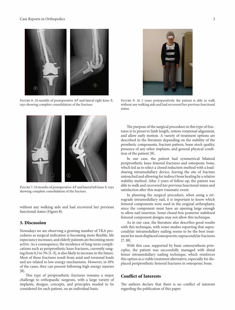
without any walking aids and had recovered her previousfunctional status (Figure 8).
3. Discussion
Nowadays we are observing a growing number of TKA pro-cedures as surgical indication is becoming more flexible, lifeexpectancy increases, and elderly patients are becomingmoreactive. As a consequence, the incidence of long-term compli-cations such as periprosthetic knee fractures, currently rang-ing from 0,3 to 3% [1–3], is also likely to increase in the future.Most of these fractures result from axial and torsional loadsand are related to low energy mechanisms. However, in 10%of the cases, they can present following high energy injuries[8].
This type of periprosthetic fractures remains a majorchallenge to orthopaedic surgeons, with a large variety ofimplants, designs, concepts, and principles needed to beconsidered for each patient, on an individual basis.
Figure 8: At 2 years postoperatively the patient is able to walkwithout any walking aids and had recovered her previous functionalstatus.
The purpose of the surgical procedure in this type of frac-tures is to preserve limb length, restore rotational alignment,and allow early motion. A variety of treatment options aredescribed in the literature depending on the stability of theprosthetic components, fracture pattern, bone stock quality,presence of any other implants, and general physical condi-tion of the patient [9].
In our case, the patient had symmetrical bilateralperiprosthetic knee femoral fractures and osteopenic bone,which led us to select a closed reduction method with a load-sharing intramedullary device, leaving the site of fractureuntouched and allowing for indirect bone healing by a relativestability method. After 2 years of follow-up, the patient wasable to walk and recovered her previous functional status andsatisfaction after this major traumatic event.
In planning the surgical procedure, when using a ret-rograde intramedullary nail, it is important to know whichfemoral components were used in the original arthroplasty,since the component must have an opening large enoughto allow nail insertion. Some closed-box posterior stabilizedfemoral component designs may not allow this technique.
As in our case, the literature also describes good resultswith this technique, with some studies reporting that supra-condylar intramedullary nailing seems to be the best treat-ment formost displaced osteoporotic supracondylar fractures[7, 10].
With this case, supported by basic osteosynthesis prin-ciples, the patient was successfully managed with distalfemur intramedullary nailing technique, which reinforcesthis option as a viable treatment alternative, especially for dis-placed periprosthetic femoral fractures in osteopenic bone.
Conflict of Interests
The authors declare that there is no conflict of interestsregarding the publication of this paper.

4 Case Reports in Orthopedics
References
[1] D. B. Bazylewicz and K. A. Egol, “Periprosthetic fractures aftertotal knee arthroplasty,”Orthopaedic Knowledge Online Journal,vol. 12, no. 2, 2014.
[2] P. L. Althausen, M. A. Lee, C. G. Finkemeier, J. P. Meehan, andJ. J. Rodrigo, “Operative stabilization of supracondylar femurfractures above total knee arthroplasty: a comparison of fourtreatment methods,” The Journal of Arthroplasty, vol. 18, no. 7,pp. 834–839, 2003.
[3] D. J. Berry, “Epidemiology: hip and knee,” Orthopedic Clinics ofNorth America, vol. 30, no. 2, pp. 183–190, 1999.
[4] K. D. Merkel and E. W. Johnson Jr., “Supracondylar fracture ofthe femur after total knee arthroplasty,” Journal of Bone and JointSurgery—Series A, vol. 68, no. 1, pp. 29–43, 1986.
[5] R. W. Culp, R. G. Schmidt, G. Hanks, A. Mak, J. L. EsterhaiJr., and R. B. Heppenstall, “Supracondylar fracture of the femurfollowing prosthetic knee arthroplasty,” Clinical Orthopaedicsand Related Research, no. 222, pp. 212–222, 1987.
[6] P. R. Cain, H. E. Rubash, H. A. Wissinger, and E. J. McClain,“Periprosthetic femoral fractures following total knee arthro-plasty,” Clinical Orthopaedics, vol. 208, pp. 205–214, 1986.
[7] E. T. Su, H. DeWal, and P. E. Di Cesare, “Periprosthetic femoralfractures above total knee replacements,” The Journal of theAmerican Academy of Orthopaedic Surgeons, vol. 12, no. 1, pp.12–20, 2004.
[8] D. A. Herrera, P. J. Kregor, P. A. Cole, B. A. Levy, A. Jonsson,and M. Zlowodzki, “Treatment of acute distal femur fracturesabove a total knee arthroplasty: systematic review of 415 cases(1981–2006),” Acta Orthopaedica, vol. 79, no. 1, pp. 22–27, 2008.
[9] V. G. Reddy, A. K. Mootha, T. Chiranjeevi, P. Kantesaria, V. K.Ramireddy, andD. Reddy, “Bilateral symmetrical periprosthetic(mirror) fractures of knee fixed with dual plating technique,”International Journal of Surgery Case Reports, vol. 2, no. 7, pp.175–177, 2011.
[10] W. J. Smith, S. L.Martin, and J. D.Mabrey, “Use of a supracondy-lar nail for treatment of a supracondylar fracture of the femurfollowing total knee arthroplasty,” The Journal of Arthroplasty,vol. 11, no. 2, pp. 210–213, 1996.

Submit your manuscripts athttp://www.hindawi.com
Stem CellsInternational
Hindawi Publishing Corporationhttp://www.hindawi.com Volume 2014
Hindawi Publishing Corporationhttp://www.hindawi.com Volume 2014
MEDIATORSINFLAMMATION
of
Hindawi Publishing Corporationhttp://www.hindawi.com Volume 2014
Behavioural Neurology
EndocrinologyInternational Journal of
Hindawi Publishing Corporationhttp://www.hindawi.com Volume 2014
Hindawi Publishing Corporationhttp://www.hindawi.com Volume 2014
Disease Markers
Hindawi Publishing Corporationhttp://www.hindawi.com Volume 2014
BioMed Research International
OncologyJournal of
Hindawi Publishing Corporationhttp://www.hindawi.com Volume 2014
Hindawi Publishing Corporationhttp://www.hindawi.com Volume 2014
Oxidative Medicine and Cellular Longevity
Hindawi Publishing Corporationhttp://www.hindawi.com Volume 2014
PPAR Research
The Scientific World JournalHindawi Publishing Corporation http://www.hindawi.com Volume 2014
Immunology ResearchHindawi Publishing Corporationhttp://www.hindawi.com Volume 2014
Journal of
ObesityJournal of
Hindawi Publishing Corporationhttp://www.hindawi.com Volume 2014
Hindawi Publishing Corporationhttp://www.hindawi.com Volume 2014
Computational and Mathematical Methods in Medicine
OphthalmologyJournal of
Hindawi Publishing Corporationhttp://www.hindawi.com Volume 2014
Diabetes ResearchJournal of
Hindawi Publishing Corporationhttp://www.hindawi.com Volume 2014
Hindawi Publishing Corporationhttp://www.hindawi.com Volume 2014
Research and TreatmentAIDS
Hindawi Publishing Corporationhttp://www.hindawi.com Volume 2014
Gastroenterology Research and Practice
Hindawi Publishing Corporationhttp://www.hindawi.com Volume 2014
Parkinson’s Disease
Evidence-Based Complementary and Alternative Medicine
Volume 2014Hindawi Publishing Corporationhttp://www.hindawi.com