bilayer silk fibroin grafts support functional oesophageal...
TRANSCRIPT

Received: 25 July 2016 Revised: 15 December 2016 Accepted: 29 March 2017
DO
I: 10.1002/term.2434R E S E A R CH AR T I C L E
Bilayer silk fibroin grafts support functional oesophageal repairin a rodent model of caustic injury
Khalid Algarrahi1,2 | Debra Franck1 | Alyssa Savarino1 | Vivian Cristofaro2,3,4 |
Xuehui Yang1 | Saif Affas1,2 | Frank‐Mattias Schäfer1 | Maryrose P. Sullivan2,3,4 |
Carlos R. Estrada Jr.1,2 | Joshua R. Mauney1,2
1Urological Diseases Research Center, Boston
Children's Hospital, Boston, Massachusetts,
USA
2Department of Surgery, Harvard Medical
School, Boston, Massachusetts, USA
3Division of Urology, Veterans Affairs Boston
Healthcare System, West Roxbury,
Massachusetts, USA
4Department of Surgery, Brigham and
Women's Hospital, Boston, Massachusetts,
USA
Correspondence
Joshua R. Mauney, Boston Children's Hospital,
Department of Urology, John F. Enders
Research Laboratories, 300 Longwood Ave.,
Rm. 1009, Boston, MA 02115, USA
Email: [email protected]
Funding information
National Institute of Diabetes and Digestive
and Kidney Diseases, Grant/Award Number:
1R01DK107568‐01A1; Veterans Affairs Merit
Review Program, Grant/Award Number:
BX001790; National Institute of Biomedical
Imaging and Bioengineering, Grant/Award
Number: P41 EB002520
J Tissue Eng Regen Med. 2017;1–8.
AbstractSurgical repair of caustic oesophageal injuries with autologous gastrointestinal segments is often
associated with dysmotility, dysphagia and donor site morbidity, and therefore alternative graft
options are needed. Bilayer silk fibroin (BLSF) scaffolds were assessed for their ability to support
functional restoration of damaged oesophageal tissues in a rat model of onlay oesophagoplasty.
Transient exposure of isolated oesophageal segments with 40% NaOH led to corrosive
oesophagitis and a 91% reduction in the luminal cross‐sectional area of damaged sites.
Oesophageal repair with BLSF matrices was performed in injured rats (n = 27) as well as a
nondiseased cohort (n = 12) for up to 2 months after implantation. Both implant groups exhibited
>80% survival rates, displayed similar degrees of weight gain, and were capable of solid food
consumption following a 3‐day liquid diet. End‐point μ‐computed tomography of repaired sites
demonstrated a 4.5‐fold increase in luminal cross‐sectional area over baseline injury levels.
Reconstructed oesophageal conduits from damaged and nondiseased animals produced compara-
ble contractile responses to KCl and electric field stimulation while isoproterenol generated
similar tissue relaxation responses. Histological and immunohistochemical evaluations of
neotissues from both implant groups showed formation of a stratified, squamous epithelium with
robust cytokeratin expression as well as skeletal and smooth muscle layers positive for contractile
protein expression. In addition, synaptophysin positive neuronal junctions and vessels lined with
CD31 positive endothelial cells were also observed at graft sites in each setting. These results
provide preclinical validation for the use of BLSF scaffolds in reconstructive strategies for
oesophageal repair following caustic injury.
1 | INTRODUCTION
Accidental ingestion of caustic alkali agents is highly prevalent in the
worldwide paediatric population and frequently results in corrosive
oesophagitis, stenosis, and ultimately oesophageal stricture formation
(Uygun, 2015). Endoscopic balloon dilatation and the use of temporary
stents represent the primary treatment options for increasing luminal
oesophageal calibre while also improving dysphagia symptoms associ-
ated with caustic injury (Dall'Oglio et al., 2016). Unfortunately, dilata-
tion of the oesophagus carries the risk of organ perforation and, over
time, this pathology can become refractory to this approach
(Ramareddy & Alladi, 2016). Similarly, stent‐related complications such
as device migration and erosion are significant concerns for this mode
of therapy (Zhao, Zhou, Feng, Wang, & Mei, 2016). In patients where
wileyonlinelibrary.com/journa
conventional management fails, gastric transposition and colonic inter-
position grafts are used to replace diseased oesophageal segments
(Ezemba, Eze, Nwafor, Etukokwu, & Orakwe, 2014). However, these
procedures can lead to severe adverse events such as oesophageal
dysmotility, anastomotic leakage and donor site morbidity, all of which
can negatively impact patient quality of life (Reinberg, 2016). Matrices
derived from decellularized tissues or synthetic polymers have been
previously investigated as alternatives to autologous gastrointestinal
segments in both animal models and clinical settings of oesophageal
reconstruction (Aikawa et al., 2013; Badylak et al., 2011; Badylak,
Meurling, S., Chen, M., Spievack, A., & Simmons‐Byrd, 2000; Dua,
Hogan, Aadam, & Gasparri, 2016). Suboptimal outcomes with these
scaffold configurations including implant contracture, graft perforation
and stenosis have been reported (Badylak et al., 2011; Doede,
Copyright © 2017 John Wiley & Sons, Ltd.l/term 1

2 ALGARRAHI ET AL.
Bondartschuk, Joerck, Schulze, & Goernig, 2009; Lopes et al.,2006),
emphasizing the need to explore new biomaterials for oesophageal
repair.
Bilayer silk fibroin (BLSF) grafts represent emerging, biodegradable
platforms for oesophageal tissue engineering. The multifunctional
implant design promotes initial defect consolidation and preservation
of organ continuity via a fluid‐tight film layer, whereas a porous foam
compartment serves as a conduit for host tissue integration (Algarrahi
et al., 2015; Seth et al., 2013). A recent report from our laboratory
has demonstrated the feasibility of these matrices for onlay
oesophagoplasty in a nondiseased, rodent model of acute traumatic
injury (Algarrahi et al., 2015). In this system, BLSF scaffolds promoted
constructive remodelling of oesophageal defects with neotissues capa-
ble of supporting peristalsis and solid food consumption (Algarrahi
et al., 2015). Parallel comparisons with conventional small intestinal
submucosa (SIS) grafts revealed BLSF matrices achieved significantly
higher degrees of skeletal muscle formation, de novo innervation, as
well as reduced inflammatory reactions within implantation sites
(Algarrahi et al., 2015).
Although the initial performance of BLSF scaffolds for
oesophageal repair was encouraging, the use of nondiseased animal
models may not accurately predict graft performance in patients with
underlying pathologies due to alterations in the regenerative capacity
of host tissues. For instance, Akbal et al. (2006) reported that while
augmentation of healthy porcine bladders with an acellular dermal
biomatrix resulted in excellent functional bladder tissue regeneration,
similar experiments in a porcine model of obstructed bladder disease
failed to show favourable results. Moreover, onlay urethroplasty of
damaged rabbit urethras with SIS grafts revealed delayed epithelializa-
tion and abnormal distribution of smooth muscle tissue in comparison
to the outcomes achieved in healthy animals (Villoldo et al., 2013).
Therefore, the objective of this study in rats was to establish whether
BLSF scaffolds have the same regenerative capacity when onlay
oesophagoplasty is performed following caustic injury in comparison
to a nondiseased setting.
2 | MATERIALS AND METHODS
2.1 | Biomaterials
BLSF scaffolds were fabricated from Bombyx mori silkworm cocoons
using a solvent‐casting/salt‐leaching process in combination with silk
fibroin film casting as previously reported (Seth et al., 2013). The
structural and tensile properties of the graft have been reported in
published studies (Seth et al., 2013). Matrices were sterilized in 70%
ethanol and rinsed in phosphate‐buffered saline (PBS) prior to surgical
procedures.
2.2 | Surgical procedures
All animal studies were approved by the Boston Children's Hospital
Animal Care and Use Committee prior to experimentation and
performed under protocol 16–05‐3161R.
Caustic oesophageal injury was induced in female Sprague–
Dawley rats (n = 68, age 6–8 weeks, weight ~ 250–300 g, Charles
River Laboratories, Wilmington, MA, USA) using a NaOH burn
model described by Okata, Hisamatsu, Hasegawa, Nishijima, and
Okita (2011) with some modifications. Prior to surgery, rats were
maintained for a maximum of 24 h on a liquid diet consisting of a nutri-
tionally‐balanced commercial formula (TestDiet®, Richmond, IN, USA;
mixed with PediaSure®; Abbott Laboratories, Columbus, OH, USA)
and were given ready access to water. Under general anaesthesia
induced by isoflurane inhalation, rats were placed in a supine position
with the thorax elevated to 30°. An upper midline laparotomy incision
was made through the skin and underlying rectus muscle in a sterile
fashion in order to isolate a 1 cm segment of the abdominal oesopha-
gus. A 5‐French double lumen Vascu‐PICC line (Medcomp®,
Harleysville, PA, USA) was placed in the upper part of the abdominal
oesophagus via the mouth and vessel loops (Devon™, Mansfield, MA,
USA) were tied externally around the gastroesophageal junction and
5 mm proximal to compartmentalize NaOH exposure. Catheter infu-
sion of a 40% NaOH solution was performed into the isolated oesoph-
ageal segment with a total contact time of 2 min which was followed
by irrigation with distilled water and subsequent fluid aspiration.
The catheter and vessel loops were then removed and non‐absorbable
7–0 polypropylene sutures with 4–0 steel rings were positioned at the
proximal/distal boundaries of the injured segment for region identifica-
tion during imaging analyses described below. Following these proce-
dures, the oesophagus was replaced into the abdominal cavity. The
skin and abdominal incisions were sutured closed. Postoperative pain
was managed with subcutaneous injection of 1 mg/kg meloxicam
and 0.1 mg/kg buprenorphine. Rats were maintained on liquid diet
and imaging evaluations were performed 2 days postinjury to evaluate
luminal oesophageal calibre. Animals were then either harvested for
2 days postinjury analyses (n = 20), subjected to scaffold implantation
(n = 27) as detailed below, or maintained without surgical intervention
(n = 5).
Onlay oesophagoplasty with BLSF scaffolds was performed as
previously described (Algarrahi et al., 2015) in rats at 3 days
postinjury. In parallel, a cohort of nondiseased rats (n = 12) were
subjected to oesophageal reconstruction with BLSF grafts. In both
experimental groups, an upper midline laparotomy incision was
sterilely performed under isoflurane anaesthesia to expose the
abdominal oesophagus. A 7 × 3 mm2 elliptical defect was created
in the anterior oesophageal wall 5 mm above the gastroesophageal
junction via surgical tissue resection. An elliptical graft of equal size
was anastomosed into the defect site using interrupted 7–0
polyglactin sutures. Nonabsorbable 7–0 polypropylene sutures were
placed at the proximal/distal and lateral edges of the graft perimeter
for identification of graft borders. The oesophagus was then
returned to the abdominal cavity and incisions were sutured closed.
Postoperative pain was managed as described above and rats were
maintained on the aforementioned liquid diet for 3 days postopera-
tively and subsequently nourished with standard rat feed for the
study duration. Animals were weighed prior to surgery and every
week until scheduled euthanasia. Rats subjected to caustic injury
and implanted with BLSF scaffolds were harvested for endpoint
evaluations at 1 week (n = 5), 1 months (n = 5), and 2 months
(n = 17) postrepair. All nondiseased animals grafted with BLSF scaf-
folds were harvested at 2 months postimplantation for comparative
ALGARRAHI ET AL. 3
analyses. An additional group of nonsurgical, healthy animals (n = 12)
were evaluated in parallel as positive controls.
2.3 | Micro‐computed tomography
Micro‐computed tomography (μ‐CT) analysis was performed on
experimental groups following contrast agent gavage as previously
described (Algarrahi et al., 2015). Luminal oesophageal cross‐sectional
areas (n = 7 animals per group) were quantified from the central
regions of the original caustic injury sites 2 d following NaOH adminis-
tration, scaffold implantation sites from damaged and nondiseased
cohorts 2 months postrepair, as well as internal noninjured reference
points adjacent to the seventh thoracic vertebra (T7) using published
methods (Algarrahi et al., 2015). Parallel measurements were executed
in corresponding oesophageal regions in nonsurgical controls.
2.4 | Ex vivo contraction and relaxation responses
Circular oesophageal tissues with intact mucosa (n = 5 animals per
group) were isolated from injured sites 2 days after NaOH exposure,
repaired segments from nondiseased and diseased groups at 2 months
postoperatively, as well as nonsurgical controls. Specimens were
mounted in tissue baths for isometric tension studies as previously
described (Algarrahi et al., 2015). Briefly, contractile responses to KCl
(80 mM) and electrical field stimulation (EFS; 25–20 Hz, 0.5 ms pulse
duration, 40 V, 10 s) were measured. In parallel, specimens were
precontracted with carbachol (3 μM) and relaxation responses
were quantified following administration of isoproterenol (10 μM).
Contractile responses were expressed as force (mN) normalized by
tissue cross‐sectional area. Relaxation responses were expressed as
percent change from precontraction response.
2.5 | Histological, immunohistochemical andhistomorphometric analyses
Following animal harvest, tubular oesophageal specimens isolated
from experimental groups were formalin‐fixed, dehydrated in graded
alcohols, and paraffin embedded. Sections (5 μm) were stained with
Masson's trichrome (MTS) using standard methods. Parallel specimens
were analysed for IHC assessments using primary antibodies to the
following markers as previously described (Algarrahi et al., 2015): fast
myosin skeletal heavy chain (MYH), α‐smooth muscle actin (α‐SMA),
pan‐cytokeratin (CK), CK4, CK14, filaggrin, synaptophysin (SYP)
and CD31. In addition, host tissue responses were evaluated
with the following primary antibodies: anti‐myeloperoxidase (Abcam,
Cambridge, MA, 1:100 dilution) and anti‐CD68 (Thermo Fisher
Scientific, Cambridge, MA, 1:200 dilution). Specimens were then
stained with species‐matched Alexa Fluor 488, 594, and 647‐
conjugated secondary antibodies (Thermo Fisher Scientific) while
4′,6‐diamidino‐2‐phenyllindole (DAPI) was used as a nuclear counter-
stain. An Axioplan‐2 microscope (Carl Zeiss MicroImaging, Thornwood,
NY, USA) was utilized for sample visualization and representative fields
were acquired with Axiovision software (version 4.8).
Histomorphometric evaluations (n = 4–7 animals per group) were
performed using published methods (Algarrahi et al., 2015). Briefly,
image thresholding and area measurements were acquired with ImageJ
software (version 1.47) on four independent microscopic fields per
tissue specimen (20× magnification) in order to calculate the percent-
age of tissue area stained for MYH, α‐SMA and pan‐CK per total tissue
area examined. The number of SYP+ boutons were also quantified
across four independent microscopic fields per tissue sample (20×
magnification) employing similar methods and normalized to total
tissue area analysed to determine density of synaptic transmission
areas. Vessel density was determined in each tissue sample by normal-
izing the total number of CD31+ vessels present in two independent
microscopic fields (5×) per total tissue area examined.
2.6 | Statistical analysis
Quantitative measurements were evaluated with the Kruskal–Wallis
test in combination with the posthoc Scheffé's method utilizing SPSS
Statistics software v19.0 (http://www.spss.com). All data are
expressed as means ± standard deviation unless otherwise indicated.
Statistically significant values were defined as p < 0.05.
3 | RESULTS AND DISCUSSION
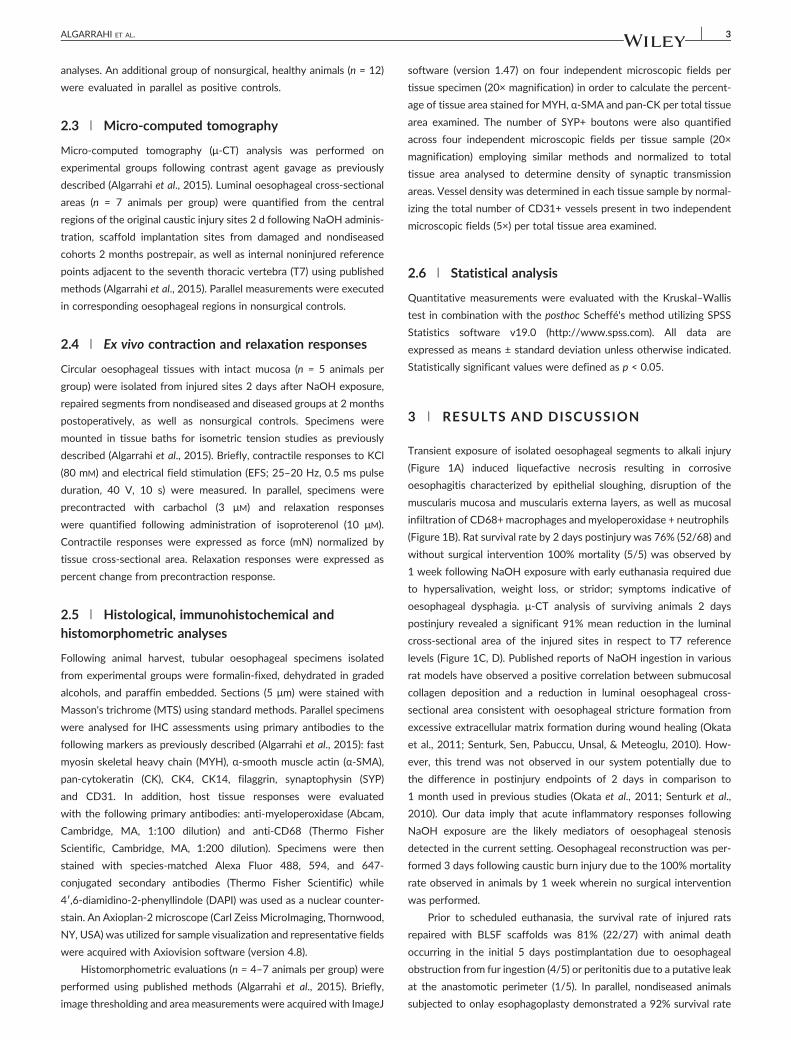
Transient exposure of isolated oesophageal segments to alkali injury
(Figure 1A) induced liquefactive necrosis resulting in corrosive
oesophagitis characterized by epithelial sloughing, disruption of the
muscularis mucosa and muscularis externa layers, as well as mucosal
infiltration of CD68+ macrophages and myeloperoxidase + neutrophils
(Figure 1B). Rat survival rate by 2 days postinjury was 76% (52/68) and
without surgical intervention 100% mortality (5/5) was observed by
1 week following NaOH exposure with early euthanasia required due
to hypersalivation, weight loss, or stridor; symptoms indicative of
oesophageal dysphagia. μ‐CT analysis of surviving animals 2 days
postinjury revealed a significant 91% mean reduction in the luminal
cross‐sectional area of the injured sites in respect to T7 reference
levels (Figure 1C, D). Published reports of NaOH ingestion in various
rat models have observed a positive correlation between submucosal
collagen deposition and a reduction in luminal oesophageal cross‐
sectional area consistent with oesophageal stricture formation from
excessive extracellular matrix formation during wound healing (Okata
et al., 2011; Senturk, Sen, Pabuccu, Unsal, & Meteoglu, 2010). How-
ever, this trend was not observed in our system potentially due to
the difference in postinjury endpoints of 2 days in comparison to
1 month used in previous studies (Okata et al., 2011; Senturk et al.,
2010). Our data imply that acute inflammatory responses following
NaOH exposure are the likely mediators of oesophageal stenosis
detected in the current setting. Oesophageal reconstruction was per-
formed 3 days following caustic burn injury due to the 100% mortality
rate observed in animals by 1 week wherein no surgical intervention
was performed.
Prior to scheduled euthanasia, the survival rate of injured rats
repaired with BLSF scaffolds was 81% (22/27) with animal death
occurring in the initial 5 days postimplantation due to oesophageal
obstruction from fur ingestion (4/5) or peritonitis due to a putative leak
at the anastomotic perimeter (1/5). In parallel, nondiseased animals
subjected to onlay esophagoplasty demonstrated a 92% survival rate

FIGURE 1 Characterization of alkali oesophageal injury and oesophageal stenosis. (a) Photomicrographs of isolated oesophageal segment prior toand 2 days following transient exposure to 40% NaOH. Scale bars = 7 mm. (b) representative cross‐sections of injured oesophageal segmentdescribed in (a) following MTS staining (top row) and IHC analyses (bottom row). Top row: Scale bars = 1.25 mm for gross and 400 μm for magnifiedviews. Bottom row: Respective marker expression is displayed in red (Alexa Fluor 594 labelling) and blue denotes DAPI nuclear counterstain. Scalebars = 200 μm. (c) representative three‐dimensional images acquired by μ‐CT analysis of injured oesophagus described in (a) following contrastagent (red) gavage. Injured site denoted in yellow with blue labels denote proximal/distal marking sutures of isolated segment. E = oesophagus;S = stomach. (d) quantification of luminal oesophageal cross‐sectional areas from the central region of the injury site (ST) described in (c) ornoninjured reference point adjacent toT7 (*) = p < 0.05 in comparison toT7 control region [Colour figure can be viewed at wileyonlinelibrary.com]
4 ALGARRAHI ET AL.
(11/12) with mortality from obstruction secondary to fur ingestion
noted in one replicate at 1 month following grafting. None of the
surviving rats in either group displayed clinical symptoms of dysphagia
over the course of the study period. In addition, both experimental
cohorts were capable of solid food consumption after a 3‐day liquid
diet and exhibited comparable degrees of weight gain by 2 months
postoperatively (Figure 2A). Global tissue evaluations at 2 months
postoperatively of both nondiseased and injured implant groups
revealed prominent host tissue ingrowth throughout the original graft
sites with no evidence of significant axial contraction between proxi-
mal and distal marking sutures (Figure 2B). Endpoint μ‐CT evaluations
of repaired injury sites revealed preservation of organ continuity with
no evidence of anatomical anomalies (Figure 2C). In comparison to
baseline levels prior to scaffold implantation, repaired diseased
conduits displayed a significant 4.5‐fold increase in luminal cross‐
sectional area, which was statistically similar to nonsurgical controls
(Figure 2D). These results highlight the ability of BLSF grafts to
promote de novo tissue formation and restore function at caustic
oesophageal injury sites.
Constructive tissue remodelling and host tissue responses were
temporally evaluated in damaged oesophagi following BLSF matrix
implantation by MTS analysis (Figure 3). At 1 week postoperatively, a
fibrovascular scar populated by mononuclear inflammatory cells as well
as myofibroblasts was evident at graft sites and lined by a stratified
squamous, keratinized epithelium. The de novo oesophageal wall
contained residual scaffold fragments with putative sites of macro-
phage phagocytosis located around the perimeters. Invasion of host
skeletal muscle fibres and smooth muscle bundles were localized at
the peripheral boundaries of the neotissues. At 1 months postimplan-
tation, an ECM‐rich lamina propria had developed and both the de
novo muscularis mucosa and muscularis externa had further integrated
into the graft region. In addition, organization of the muscularis externa
into circular and outer longitudinal skeletal muscle layers was apparent
at the edges of the neotissues; however, fibrosis still persisted toward
internal areas. The de novo muscularis externa at 2 months following
repair of injured sites demonstrated a qualitative increase in skeletal
muscle density within the central regions of the graft site in compari-
son to early timepoints. Areas of fibrosis had also diminished at this
stage of regeneration and no chronic inflammatory reactions were
noted. The structural architecture of neotissues generated in the set-
ting of caustic damage was qualitatively similar to the nondiseased
repair group at 2 months postoperatively. However, maturation of
the muscularis mucosa and muscularis externa compartments in both
these cohorts was notably underdeveloped in respect to nonsurgical
controls. Finally, the temporal stages of wound healing encountered
during repair of caustic injury sites with BLSF scaffolds were found
to mimic the regenerative responses previously observed in our
nondiseased model of oesophagoplasty (Algarrahi et al., 2015).

FIGURE 2 Evaluation of neotissue formation and oesophageal function in injured and repaired groups. (a) body weight evaluations of nondiseased(BLSF‐ND) and diseased (BLSF‐ST) animals over the course of the 2 months of scaffold implantation period. Means ± standard deviation per datapoint. (b) Neotissues present within the original graft sites at 2 months postoperatively. Proximal/distal and lateral marking sutures are respectivelydesignated by red and black arrows. (c) representative three‐dimensional μ‐CT images of oesophagi 2 days after NaOH exposure (ST) and in BLSF‐ND and BLSF‐ST groups at 2 months postoperatively detailed in (b) following contrast agent (red) gavage. Repaired sites in nondiseased anddiseased groups are coded in yellow while proximal/distal marking sutures are colored in blue. E = oesophagus. S = stomach. (d) quantification ofluminal oesophageal cross‐sectional areas in yellow regions described in (c) and in nonsurgical controls (NSC). (*) = p < 0.05 in comparison to allother groups [Colour figure can be viewed at wileyonlinelibrary.com]
FIGURE 3 MTS analyses of constructive remodelling at implant sites in experimental cohorts. (first row) Photomicrographs of gross oesophagealcross‐sections stained with MTS from nondiseased (BLSF‐ND) and diseased (BLSF‐ST) groups containing original graft site (bracketed) as well asnonsurgical controls (NSC). Scale bars = 1.25 mm. (second row) magnification of global regenerative areas (RA) bracketed in first row or the nativetissue in NSC. Scale bars = 400 μm. (third row) magnified boxed green area from second row profiling muscularis externa development. (fourth row)magnified boxed red area from second row detailing epithelial formation. Scale bars for third and fourth rows = 100 μm [Colour figure can beviewed at wileyonlinelibrary.com]
ALGARRAHI ET AL. 5
IHC (Figure 4A) and parallel histomorphometric (Figure 4B)
evaluations were executed on experimental groups to characterize
further the phases of regeneration during repair of damaged regions
and compare the degree of tissue maturation achieved between
diseased and nondiseased counterparts. Pan‐CK+ epithelia were first
observed spanning reconstructed injury sites at 1 week following

FIGURE 4 Immunohistochemical and histomorphometric assessments of neotissues and controls. (a) Photomicrographs of protein expressionpatterns in repaired, nondiseased (BLSF‐ND) and diseased (BLSFST) graft sites as well as nonsurgical controls (NSC) of epithelia markers, pan‐CK, CK14, CK4, FG; endothelial and innervation markers, CD31 and SYP in mucosa; and contractile muscle markers, α‐SMA and MYH in muscularismucosa and muscularis externa, respectively. Arrowheads indicate SYP+ boutons. For all panels, respective marker expression is displayed in red(Alexa Fluor 594 labelling), green (Alexa Fluor 488 labelling), or white (Alexa Fluor 647 labelling) and blue denotes DAPI nuclear counterstain. For allpanels, scale bars = 200 μm. (b) Histomorphometric analyses of marker expression in regenerated tissues as well as NSC. (*) = p < 0.05 incomparison to respective NSC. (#) = p < 0.05 in comparison to all other groups [Colour figure can be viewed at wileyonlinelibrary.com]
6 ALGARRAHI ET AL.
matrix grafting. By 2 months postoperatively, no significant differences
in the extent of pan‐CK+ epithelia were noted between implant groups
or nonsurgical controls. In addition, distinct epithelial subpopulations
were present in all experimental groups and consisted of CK14 + basal
cells, polygonal CK4+ suprabasal cells, and flattened FG+ superficial
cells. Regenerated vascular networks containing vessels lined with
CD31+ endothelial cells were found in both nondiseased and diseased
graft sites at all examined timepoints. The mean vessel density was
significantly higher in consolidated tissues from all groups in respect
to nonsurgical control levels indicating an ongoing stage of tissue
remodelling (Zawicki, Jain, Schmid‐Schoenbein, & Chien, 1981; Jain,
2003). Analysis of de novo innervation revealed synaptic transmission
areas marked by SYP+ boutons in all neotissues. There was no signifi-
cant difference between the densities of SYP+ boutons in implant

FIGURE 5 Ex vivo contractility and relaxation behaviours in controls and repaired oesophageal segments. Contractile responses to (a) electric fieldstimulation (2–25 Hz) and (b) KCl (80 mM) in circular oesophageal rings from injured sites 2 days after NaOH exposure (ST), repaired segments fromnondiseased (BLSF‐ND) and diseased (BLSF‐ST) groups at 2 months postoperatively, as well as nonsurgical controls (NSC). Inset in (b) reflectsmagnification of injured and experimental responses. (c) relaxation activities in response to isoproterenol (10 μM) in cohorts detailed in (a)precontracted with carbachol (3 μM). Means ± standard errors per data point. (*) = p < 0.05 in comparison to ST group [Colour figure can be viewedat wileyonlinelibrary.com]
ALGARRAHI ET AL. 7
groups at 2 months postoperatively compared to nonsurgical control
levels. Characterization of the regenerated muscularis externa in
neotissues demonstrated the extent of MYH+ skeletal muscle in
diseased and nondiseased cohorts had respectively achieved 67%
and 76% of control values by 2 months of scaffold implantation. In
contrast to other tissue components, animals subjected to caustic
injury displayed a reduced capacity to support regeneration of the
muscularis mucosa in respect to noninjured subjects. The density of
α‐SMA+ smooth muscle bundles in this compartment was significantly
lower in the diseased setting reflecting 33% of nonsurgical control
levels by 2 months postoperatively, while nondiseased counterparts
were capable of supporting 67%. Taken together, these data
demonstrate the ability of BLSF grafts to promote the formation of
innervated, vascularized oesophageal tissues at sites of caustic injury;
however, the propensity for muscularis mucosal regeneration is muted
in comparison to nondiseased microenvironments.
Peristaltic waves generated from radially symmetrical contraction
and relaxation of oesophageal circular muscle is an essential mecha-
nism for propagation of food bolus through the digestive tract.
Contractile behaviours in repaired conduits from each implant group
as well as nonsurgical controls and injured segments prior to scaffold
implantation were assessed in ex vivo organ bath studies following
stimulation with EFS (Figure 5A) and KCl (Figure 5B). In addition,
relaxation properties of experimental groups precontracted with car-
bachol were analysed following isoproterenol treatment (Figure 5C).
Alkali injury to oesophageal tissues significantly decreased both con-
tractile and relaxation responses to all stimuli tested in comparison to
nonsurgical controls. These results are consistent with our histological
findings demonstrating extensive caustic damage to oesophageal mus-
cular components following transient exposure to NaOH. Following
2 months of BLSF matrix grafting, both repaired nondiseased and
diseased conduits demonstrated the ability to contract in response to
EFS and KCl stimulation to similar extents. The magnitude of KCl and
EFS‐induced force generation in these groups was substantially higher
than in injured controls, but less than nonsurgical counterparts, sug-
gesting partial recovery of contractile machinery elicited by membrane
depolarization as well as neuronal input, respectively. Parallel analysis
of relaxation patterns in experimental groups demonstrated similar
trends as observed for contractile responses.
4 | CONCLUSIONS
The data presented in this study demonstrate the ability of BLSF grafts
to promote functional repair of caustic injury sites in a rat model of
onlay oesophagoplasty. Reconstruction of damaged tissues with BLSF
scaffolds restored luminal oesophageal calibre, enabled solid food
consumption, and promoted the formation of innervated, vascularized
epithelial and muscular wall components. In addition, injured oesopha-
geal conduits repaired with BLSF grafts displayed contractile and relax-
ation properties. In comparison to nondiseased settings, BLSF matrices
displayed similar regenerative capacities during healing of damaged
regions except for a reduced ability to reconstitute the muscularis
mucosa to nonsurgical control levels. In summary, these results provide
preclinical validation for the potential of BLSF grafts to serve as off‐
the‐shelf scaffolds for reconstruction of oesophageal tissues following
caustic injury.
ACKNOWLEDGEMENTS
This research was supported by the NIH/NIDDK 1R01DK107568‐
01A1 (MAUNEY), Tissue Engineering Resource Center, NIH/NIBIB
P41 EB002520 (KAPLAN), and BX001790 (MPS). This research was

8 ALGARRAHI ET AL.
also supported through the vision and generosity of the Rainmaker
Group in honor of Dr Alan Retik, MD. We also acknowledge Dr Bryan
Sack, MD and Mr Kyle Costa BSc for technical support.
CONFLICT OF INTEREST
The authors declare no conflicts of interest.
AUTHOR CONTRIBUTIONS
JRM and KA designed and conceptualized this study. Acquisition of
data was carried out by KA, DF, VC, XY, AS, SA, FMS and MPS.
Analysis and interpretation of data was done by JRM, KA, DF, VC,
AS and MPS. Manuscript was drafted by JRM, KA, VC and, MPS, while
VC, MPS, CRE and JRM performed critical revision of the manuscript
for important intellectual content. Statistical analysis was done by
KA, VC and MPS. Funding was obtained by JRM and CRE. The entire
study was supervised by JRM.
REFERENCES
Aikawa, M., Miyazawa, M., Okamoto, K., Okada, K., Akimoto, N., Sato, H., …Ikada, Y. (2013). A bioabsorbable polymer patch for the treatment ofesophageal defect in a porcine model. Journal of Gastroenterology,48(7), 822–829.
Akbal, C., Lee, S. D., Packer, S. C., Davis, M. M., Rink, R. C., & Kaefer, M.(2006). Bladder augmentation with acellular dermal biomatrix in adiseased animal model. Journal of Urology, 176(4 Pt 2), 1706–1711.
Algarrahi, K., Franck, D., Ghezzi, C. E., Cristofaro, V., Yang, X., Sullivan, M. P.,… Mauney, J. R. (2015). Acellular bi‐layer silk fibroin scaffolds supportfunctional tissue regeneration in a rat model of onlay esophagoplasty.Biomaterials, 53, 149–159.
Badylak, S. F., Hoppo, T., Nieponice, A., Gilbert, T. W., Davison, J. M., &Jobe, B. A. (2011). Esophageal preservation in five male patients afterendoscopic inner‐layer circumferential resection in the setting ofsuperficial cancer: A regenerative medicine approach with a biologicscaffold. Tissue Engineering Part A, 17(11–12), 1643–1650.
Badylak, S., Meurling, S., Chen, M., Spievack, A., & Simmons‐Byrd, A.(2000). Resorbable bioscaffold for esophageal repair in a dog model.Journal of Pediatric Surgery, 35(7), 1097–1103.
Dall'Oglio, L., Caldaro, T., Foschia, F., Federici di Abriola, G., Rea, F., Romeo,E., … De Angelis, P. (2016). Endoscopic management of esophagealstenosis in children: New and traditional treatments. World Journal ofGastrointestinal Endoscopy, 25(4), 212–219.
Doede, T., Bondartschuk, M., Joerck, C., Schulze, E., & Goernig, M. (2009).Unsuccessful alloplastic esophageal replacement with porcine smallintestinal submucosa. Artificial Organs, 33(4), 328–333.
Dua, K. S., Hogan, W. J., Aadam, A. A., & Gasparri, M. (2016). In‐vivooesophageal regeneration in a human being by use of a non‐biologicalscaffold and extracellular matrix. Lancet, 388(10037), 55–61.
Ezemba, N., Eze, J. C., Nwafor, I. A., Etukokwu, K. C., & Orakwe, O. I. (2014).Colon interposition graft for corrosive esophageal stricture: Midtermfunctional outcome. World Journal of Surgery, 38(9), 2352–2357.
Jain, R. K. (2003). Molecular regulation of vessel maturation. NatureMedicine, 9(6), 685–693.
Lopes, M. F., Cabrita, A., Ilharco, J., Pessa, P., Paiva‐Carvalho, J., Pires, A., &Patrício, J. (2006). Esophageal replacement in rat using porcineintestinal submucosa as a patch or a tube‐shaped graft. Diseases ofthe Esophagus: Official Journal of the International Society for Diseasesof the Esophagus, 19(4), 254–259.
Okata, Y., Hisamatsu, C., Hasegawa, T., Nishijima, E., & Okita, Y. (2011).Development of a model of benign esophageal stricture in rats: Theoptimal concentration of sodium hydroxide for stricture formation.Pediatric Surgery International, 27(1), 73–80.
Ramareddy, R. S., & Alladi, A. (2016). Review of esophageal injuries andstenosis: Lessons learn and current concepts of management. Journalof Indian Association of Pediatric Surgery, 21(3), 139–143.
Reinberg, O. (2016). Esophageal replacements in children. Annals of theNew Yrok Academy of Science, 1381(1), 104–112.
Senturk, E., Sen, S., Pabuccu, E., Unsal, C., & Meteoglu, I. (2010). Newexperimental corrosive esophagitis model in rats. Pediatric SurgeryInternational, 26(3), 257–261.
Seth, A., Chung, Y. G., Gil, E. S., Tu, D., Franck, D., Di Vizio, D., …Mauney, J.R. (2013). The performance of silk scaffolds in a rat model ofaugmentation cystoplasty. Biomaterials, 34(20), 4758–4765.
Uygun, I. (2015). Caustic oesophagitis in children: Prevalence, the corrosiveagents involved, and management from primary care through tosurgery. Current Opinion in Otolaryngology and Head and Neck Surgery,23(6), 423–432.
Villoldo, G. M., Loresi, M., Giudice, C., Damia, O., Moldes, J. M., DeBadiola,F., … Argibay, P. (2013). Histologic changes after urethroplasty usingsmall intestinal submucosa unseeded with cells in rabbits with injuredurethra. Urology, 81(6), 1–5.
Zawicki, D. F., Jain, R. K., Schmid‐Schoenbein, G. W., & Chien, S. (1981).Dynamics of neovascularization in normal tissue. MicrovascularResearch, 21(1), 27–47.
Zhao, H., Zhou, Y., Feng, J., Wang, W., & Mei, Y. (2016). Literature analysisof the treatment of benign esophageal disease with stent. Indian Journalof Surgery, 78(1), 6–13.
How to cite this article: Algarrahi K, Franck D, Savarino A, et
al. Bilayer silk fibroin grafts support functional oesophageal
repair in a rodent model of caustic injury. J Tissue Eng Regen
Med. 2017;1‐8. https://doi.org/10.1002/term.2434

本文献由“学霸图书馆-文献云下载”收集自网络,仅供学习交流使用。
学霸图书馆(www.xuebalib.com)是一个“整合众多图书馆数据库资源,
提供一站式文献检索和下载服务”的24 小时在线不限IP
图书馆。
图书馆致力于便利、促进学习与科研,提供最强文献下载服务。
图书馆导航:
图书馆首页 文献云下载 图书馆入口 外文数据库大全 疑难文献辅助工具