binding site differences revealed by crystal structures of
TRANSCRIPT

doi:10.1006/jmbi.2001.4749 available online at http://www.idealibrary.com on J. Mol. Biol. (2001) 309, 181±192
Binding Site Differences Revealed by CrystalStructures of Plasmodium falciparum and BovineAcyl-CoA Binding Protein
D. M. F. van Aalten1*, K. G. Milne1, J. Y. Zou2, G. J. Kleywegt2
T. Bergfors2, M. A. J. Ferguson1, J. Knudsen3 and T. A. Jones2
1Wellcome Trust BiocentreDepartment of BiochemistryUniversity of Dundee, DundeeDD1 5EH, Scotland2Department of Cell andMolecular Biology, UppsalaUniversity, Biomedical CentreBox 596, SE-75124, UppsalaSweden3Institute of BiochemistryOdense UniversityDK-5320, Odense, Denmark
E-mail address of the [email protected]
Abbreviations used: ACBP, acyl-cprotein; bACBP, bovine ACBP; CoAcorrelation coef®cient; GPI, glycosyNCS, non-crystallographic symmetrreading frame; PCR, polymerase chPfACBP, ACBP from Plasmodium fabody re®nement; RMSD, root meanSA, simulated annealing.
0022-2836/01/010181±12 $35.00/0
Acyl-CoA binding protein (ACBP) maintains a pool of fatty acyl-CoAmolecules in the cell and plays a role in fatty acid metabolism. The bio-chemical properties of Plasmodium falciparum ACBP are describedtogether with the 2.0 AÊ resolution crystal structures of a P. falciparumACBP-acyl-CoA complex and of bovine ACBP in two crystal forms.Overall, the bovine ACBP crystal structures are similar to the NMR struc-tures published previously; however, the bovine and parasite ACBPstructures are less similar. The parasite ACBP is shown to have a differ-ent ligand-binding pocket, leading to an acyl-CoA binding speci®citydifferent from that of bovine ACBP. Several non-conservative differencesin residues that interact with the ligand were identi®ed between themammalian and parasite ACBPs. These, together with measured binding-speci®city differences, suggest that there is a potential for the design ofmolecules that might selectively block the acyl-CoA binding site.
# 2001 Academic Press
Keywords: acyl-CoA; structure; Plasmodium falciparum; drug design;X-ray crystallography
*Corresponding authorIntroduction
Human malaria is caused by the protozoan para-site Plasmodium falciparum. Although drugs areavailable, there is an increasing demand for newpharmaceuticals due to the emergence of drug-resistant parasites.1 The increasing wealth ofsequence data from both the P. falciparum and thehuman genome projects, in combination withstructural data, facilitate the design of novel drugleads on a rational basis. Here, we describe thecrystal structures of bovine and P. falciparum acyl-CoA binding protein, characterize differencesbetween the ligand-binding sites and assess thepotential for exploitation of these differences in
ing author:
oenzyme A-binding, coenzyme A; CC,
lphosphatidylinositol;y; ORF, openain reaction;lciparum; RBR, rigid
square deviation;
the design of blocker molecules selective for theP. falciparum protein.
Acyl-coenzyme A binding proteins (ACBPs) area family of 86 to 103 residue proteins with con-served amino acid sequences.2 A total of 30sequences have been described from a wide rangeof species and genomic sequence data, such asfrom Caenorhabditis elegans, have suggested theexistence of other proteins that contain ACBP-likedomains.3 Long chain acyl-CoA esters serve asimportant intermediates in lipid synthesis andfatty acid degradation. Besides this house-keepingfunction, a large body of evidence has accumulatedindicating that long-chain acyl-CoA esters alsohave an important function in the regulation ofintermediary metabolism and gene regulation.2,4 InTrypanosoma brucei, ACBP has been shown to be animportant component in glycosylphosphatidylino-sitol (GPI) biosynthesis,5 donating myristoyl-CoAfor the fatty acid remodelling reactions.6 ACBPstypically bind saturated and unsaturated C14-C22
acyl-CoA esters with 1:1 stoichiometry and a highdegree of af®nity (Kd � 2-10 nM).2 Long-chainacyl-CoA esters are amphipathic molecules whichcan partition into phospholipid bilayers and inhibita large number of cellular functions and enzymes
# 2001 Academic Press
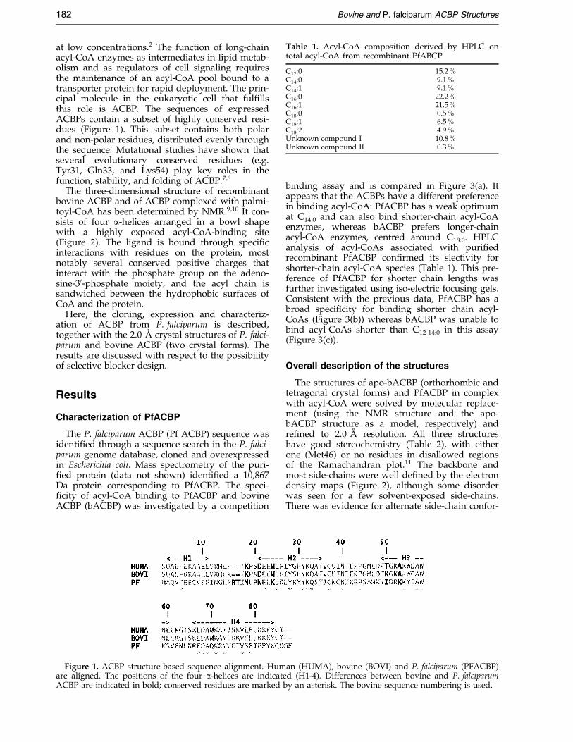
Table 1. Acyl-CoA composition derived by HPLC ontotal acyl-CoA from recombinant PfABCP
C12:0 15.2 %C14:0 9.1 %C14:1 9.1 %C16:0 22.2 %C16:1 21.5 %C18:0 0.5 %C18:1 6.5 %C18:2 4.9 %Unknown compound I 10.8 %Unknown compound II 0.3 %
182 Bovine and P. falciparum ACBP Structures
at low concentrations.2 The function of long-chainacyl-CoA enzymes as intermediates in lipid metab-olism and as regulators of cell signaling requiresthe maintenance of an acyl-CoA pool bound to atransporter protein for rapid deployment. The prin-cipal molecule in the eukaryotic cell that ful®llsthis role is ACBP. The sequences of expressedACBPs contain a subset of highly conserved resi-dues (Figure 1). This subset contains both polarand non-polar residues, distributed evenly throughthe sequence. Mutational studies have shown thatseveral evolutionary conserved residues (e.g.Tyr31, Gln33, and Lys54) play key roles in thefunction, stability, and folding of ACBP.7,8
The three-dimensional structure of recombinantbovine ACBP and of ACBP complexed with palmi-toyl-CoA has been determined by NMR.9,10 It con-sists of four a-helices arranged in a bowl shapewith a highly exposed acyl-CoA-binding site(Figure 2). The ligand is bound through speci®cinteractions with residues on the protein, mostnotably several conserved positive charges thatinteract with the phosphate group on the adeno-sine-30-phosphate moiety, and the acyl chain issandwiched between the hydrophobic surfaces ofCoA and the protein.
Here, the cloning, expression and characteriz-ation of ACBP from P. falciparum is described,together with the 2.0 AÊ crystal structures of P. falci-parum and bovine ACBP (two crystal forms). Theresults are discussed with respect to the possibilityof selective blocker design.
Results
Characterization of PfACBP
The P. falciparum ACBP (Pf ACBP) sequence wasidenti®ed through a sequence search in the P. falci-parum genome database, cloned and overexpressedin Escherichia coli. Mass spectrometry of the puri-®ed protein (data not shown) identi®ed a 10,867Da protein corresponding to PfACBP. The speci-®city of acyl-CoA binding to PfACBP and bovineACBP (bACBP) was investigated by a competition
Figure 1. ACBP structure-based sequence alignment. Humare aligned. The positions of the four a-helices are indicateACBP are indicated in bold; conserved residues are marked b
binding assay and is compared in Figure 3(a). Itappears that the ACBPs have a different preferencein binding acyl-CoA: PfACBP has a weak optimumat C14:0 and can also bind shorter-chain acyl-CoAenzymes, whereas bACBP prefers longer-chainacyl-CoA enzymes, centred around C18:0. HPLCanalysis of acyl-CoAs associated with puri®edrecombinant PfACBP con®rmed its slectivity forshorter-chain acyl-CoA species (Table 1). This pre-ference of PfACBP for shorter chain lengths wasfurther investigated using iso-electric focusing gels.Consistent with the previous data, PfACBP has abroad speci®city for binding shorter chain acyl-CoAs (Figure 3(b)) whereas bACBP was unable tobind acyl-CoAs shorter than C12-14:0 in this assay(Figure 3(c)).
Overall description of the structures
The structures of apo-bACBP (orthorhombic andtetragonal crystal forms) and PfACBP in complexwith acyl-CoA were solved by molecular replace-ment (using the NMR structure and the apo-bACBP structure as a model, respectively) andre®ned to 2.0 AÊ resolution. All three structureshave good stereochemistry (Table 2), with eitherone (Met46) or no residues in disallowed regionsof the Ramachandran plot.11 The backbone andmost side-chains were well de®ned by the electrondensity maps (Figure 2), although some disorderwas seen for a few solvent-exposed side-chains.There was evidence for alternate side-chain confor-
an (HUMA), bovine (BOVI) and P. falciparum (PFACBP)d (H1-4). Differences between bovine and P. falciparumy an asterisk. The bovine sequence numbering is used.

Figure 2. Overall structure and electron density maps. (a) Stereo image of bACBP in the tetragonal crystal formwith carbon atoms coloured green, nitrogen atoms light blue and oxygen atoms mangenta. For ACBP in the orthor-hombic crystal form, carbon atoms are shown in gold, nitrogen atoms in blue, and oxygen atoms in red. SigmaAweighted 2Fo ÿ Fc maps are shown in blue (orthorhombic bACBP) and orange (tetragonal bACBP). (b) PfACBP, withbackbone shown as blue ribbon. The acyl-CoA ligand is shown as a stick model. The Fo ÿ Fc maps just beforeinclusion of the adenosine-30-phosphate and the fatty acid are shown at 2.5 and 2.0 s, respectively (red).
Bovine and P. falciparum ACBP Structures 183
mations, some of which were modelled in thePfACBP structure. No density was present for thetwo C-terminal PfACBP residues, which weretherefore not modelled. The differences betweenPfACBP and bACBP (orthorhombic form) are sig-ni®cant with an RMSD value of 1.3 AÊ on all corre-sponding Ca atoms (see Table 3 and Figure 4(a)).
Although bACBP was co-crystallized with pal-mitoyl-CoA, no density was observed for theligand. PfACBP was not co-crystallized with anyadded acyl-CoA, yet strong density for the adeno-sine-30-phosphate and later the acyl chain wasobserved (Figure 2(b)). HPLC analysis con®rmedthe presence of a ligand (Table 1) and showed thatit was a mixture of acyl-CoA species. Density atthe end of the acyl chain was well de®ned, butfrom C2 onwards to the adenosine-30-phosphatethe density could not be interpreted unambigu-ously, and these atoms were not modelled. It ispossible that the protein accomodated a mixture of
acyl chain lengths by keeping CoA and the end ofthe acyl chain tethered, allowing the intermediateatoms to adopt different conformations. This isconsistent with the HPLC analysis and the disorderobserved in the crystal structure.
Comparison of the two bACBP crystal forms
The structures of bACBP in the two crystalforms are very similar. The three molecules in thetetragonal crystal form have RMSD values of0.5(�0.2) AÊ on Ca atoms to the molecule in theorthorhombic crystal form (Table 3). The maindifference is around the region Gly45 to Asp48. Inthe orthorhombic form, this region adopts a type Iturn structure, while a type II0 turn is present inthe tetragonal form. This causes the side-chain ofMet46 to point into opposite directions in the twostructures (Figure 2(a)). The electron density mapsare clear for this part of the structure in both forms
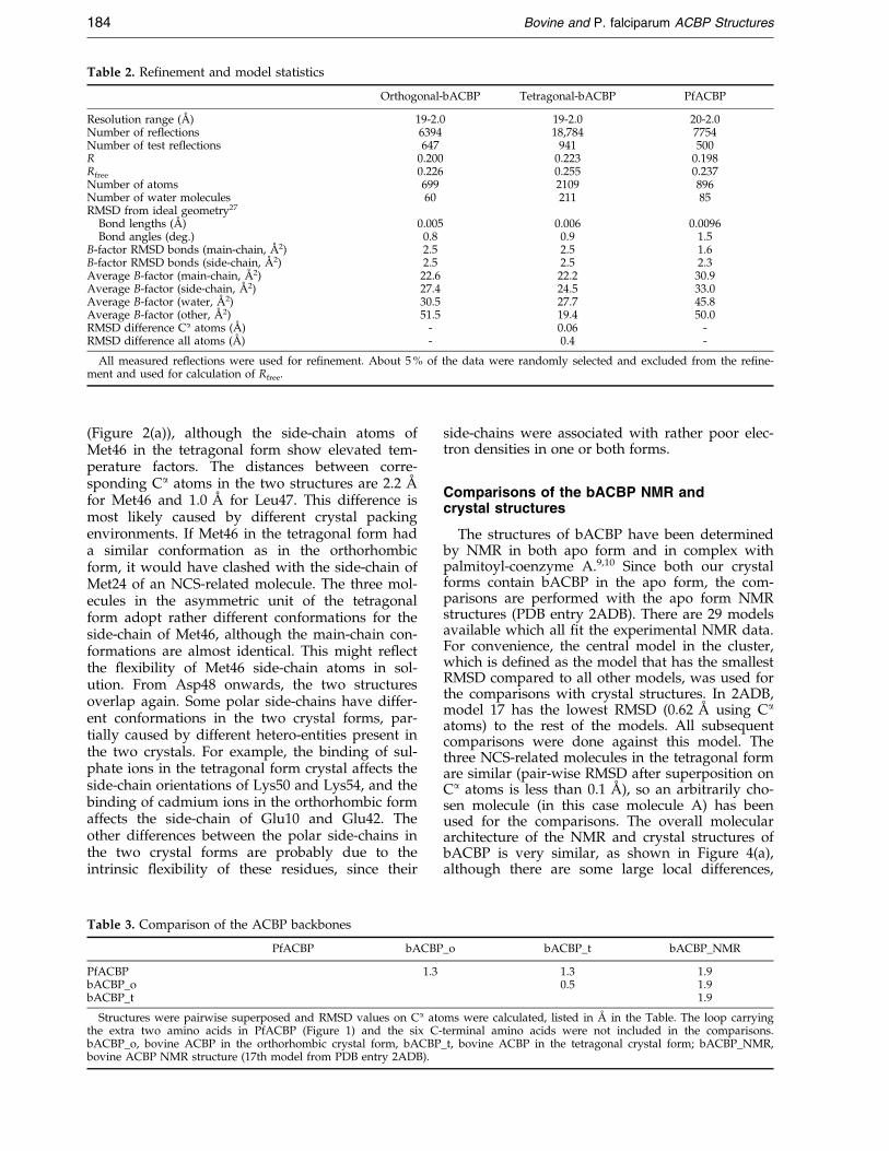
Table 2. Re®nement and model statistics
Orthogonal-bACBP Tetragonal-bACBP PfACBP
Resolution range (AÊ ) 19-2.0 19-2.0 20-2.0Number of reflections 6394 18,784 7754Number of test reflections 647 941 500R 0.200 0.223 0.198Rfree 0.226 0.255 0.237Number of atoms 699 2109 896Number of water molecules 60 211 85RMSD from ideal geometry27
Bond lengths (AÊ ) 0.005 0.006 0.0096Bond angles (deg.) 0.8 0.9 1.5
B-factor RMSD bonds (main-chain, AÊ 2) 2.5 2.5 1.6B-factor RMSD bonds (side-chain, AÊ 2) 2.5 2.5 2.3Average B-factor (main-chain, AÊ 2) 22.6 22.2 30.9Average B-factor (side-chain, AÊ 2) 27.4 24.5 33.0Average B-factor (water, AÊ 2) 30.5 27.7 45.8Average B-factor (other, AÊ 2) 51.5 19.4 50.0RMSD difference Ca atoms (AÊ ) - 0.06 -RMSD difference all atoms (AÊ ) - 0.4 -
All measured re¯ections were used for re®nement. About 5 % of the data were randomly selected and excluded from the re®ne-ment and used for calculation of Rfree.
184 Bovine and P. falciparum ACBP Structures
(Figure 2(a)), although the side-chain atoms ofMet46 in the tetragonal form show elevated tem-perature factors. The distances between corre-sponding Ca atoms in the two structures are 2.2 AÊ
for Met46 and 1.0 AÊ for Leu47. This difference ismost likely caused by different crystal packingenvironments. If Met46 in the tetragonal form hada similar conformation as in the orthorhombicform, it would have clashed with the side-chain ofMet24 of an NCS-related molecule. The three mol-ecules in the asymmetric unit of the tetragonalform adopt rather different conformations for theside-chain of Met46, although the main-chain con-formations are almost identical. This might re¯ectthe ¯exibility of Met46 side-chain atoms in sol-ution. From Asp48 onwards, the two structuresoverlap again. Some polar side-chains have differ-ent conformations in the two crystal forms, par-tially caused by different hetero-entities present inthe two crystals. For example, the binding of sul-phate ions in the tetragonal form crystal affects theside-chain orientations of Lys50 and Lys54, and thebinding of cadmium ions in the orthorhombic formaffects the side-chain of Glu10 and Glu42. Theother differences between the polar side-chains inthe two crystal forms are probably due to theintrinsic ¯exibility of these residues, since their
Table 3. Comparison of the ACBP backbones
PfACBP bACBP
PfACBP 1.3bACBP_obACBP_t
Structures were pairwise superposed and RMSD values on Ca atothe extra two amino acids in PfACBP (Figure 1) and the six C-bACBP_o, bovine ACBP in the orthorhombic crystal form, bACBPbovine ACBP NMR structure (17th model from PDB entry 2ADB).
side-chains were associated with rather poor elec-tron densities in one or both forms.
Comparisons of the bACBP NMR andcrystal structures
The structures of bACBP have been determinedby NMR in both apo form and in complex withpalmitoyl-coenzyme A.9,10 Since both our crystalforms contain bACBP in the apo form, the com-parisons are performed with the apo form NMRstructures (PDB entry 2ADB). There are 29 modelsavailable which all ®t the experimental NMR data.For convenience, the central model in the cluster,which is de®ned as the model that has the smallestRMSD compared to all other models, was used forthe comparisons with crystal structures. In 2ADB,model 17 has the lowest RMSD (0.62 AÊ using Ca
atoms) to the rest of the models. All subsequentcomparisons were done against this model. Thethree NCS-related molecules in the tetragonal formare similar (pair-wise RMSD after superposition onCa atoms is less than 0.1 AÊ ), so an arbitrarily cho-sen molecule (in this case molecule A) has beenused for the comparisons. The overall moleculararchitecture of the NMR and crystal structures ofbACBP is very similar, as shown in Figure 4(a),although there are some large local differences,
_o bACBP_t bACBP_NMR
1.3 1.90.5 1.9
1.9
ms were calculated, listed in AÊ in the Table. The loop carryingterminal amino acids were not included in the comparisons._t, bovine ACBP in the tetragonal crystal form; bACBP_NMR,
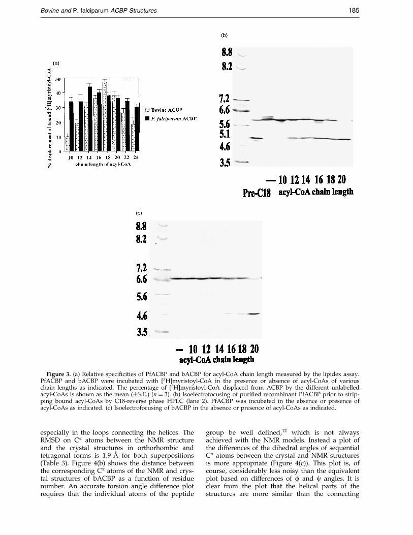
Figure 3. (a) Relative speci®cities of PfACBP and bACBP for acyl-CoA chain length measured by the lipidex assay.PfACBP and bACBP were incubated with [3H]myristoyl-CoA in the presence or absence of acyl-CoAs of variouschain lengths as indicated. The percentage of [3H]myristoyl-CoA displaced from ACBP by the different unlabelledacyl-CoAs is shown as the mean (�S.E.) (n � 3). (b) Isoelectrofocusing of puri®ed recombinant PfACBP prior to strip-ping bound acyl-CoAs by C18-reverse phase HPLC (lane 2). PfACBP was incubated in the absence or presence ofacyl-CoAs as indicated. (c) Isoelectrofocusing of bACBP in the absence or presence of acyl-CoAs as indicated.
Bovine and P. falciparum ACBP Structures 185
especially in the loops connecting the helices. TheRMSD on Ca atoms between the NMR structureand the crystal structures in orthorhombic andtetragonal forms is 1.9 AÊ for both superpositions(Table 3). Figure 4(b) shows the distance betweenthe corresponding Ca atoms of the NMR and crys-tal structures of bACBP as a function of residuenumber. An accurate torsion angle difference plotrequires that the individual atoms of the peptide
group be well de®ned,12 which is not alwaysachieved with the NMR models. Instead a plot ofthe differences of the dihedral angles of sequentialCa atoms between the crystal and NMR structuresis more appropriate (Figure 4(c)). This plot is, ofcourse, considerably less noisy than the equivalentplot based on differences of f and c angles. It isclear from the plot that the helical parts of thestructures are more similar than the connecting
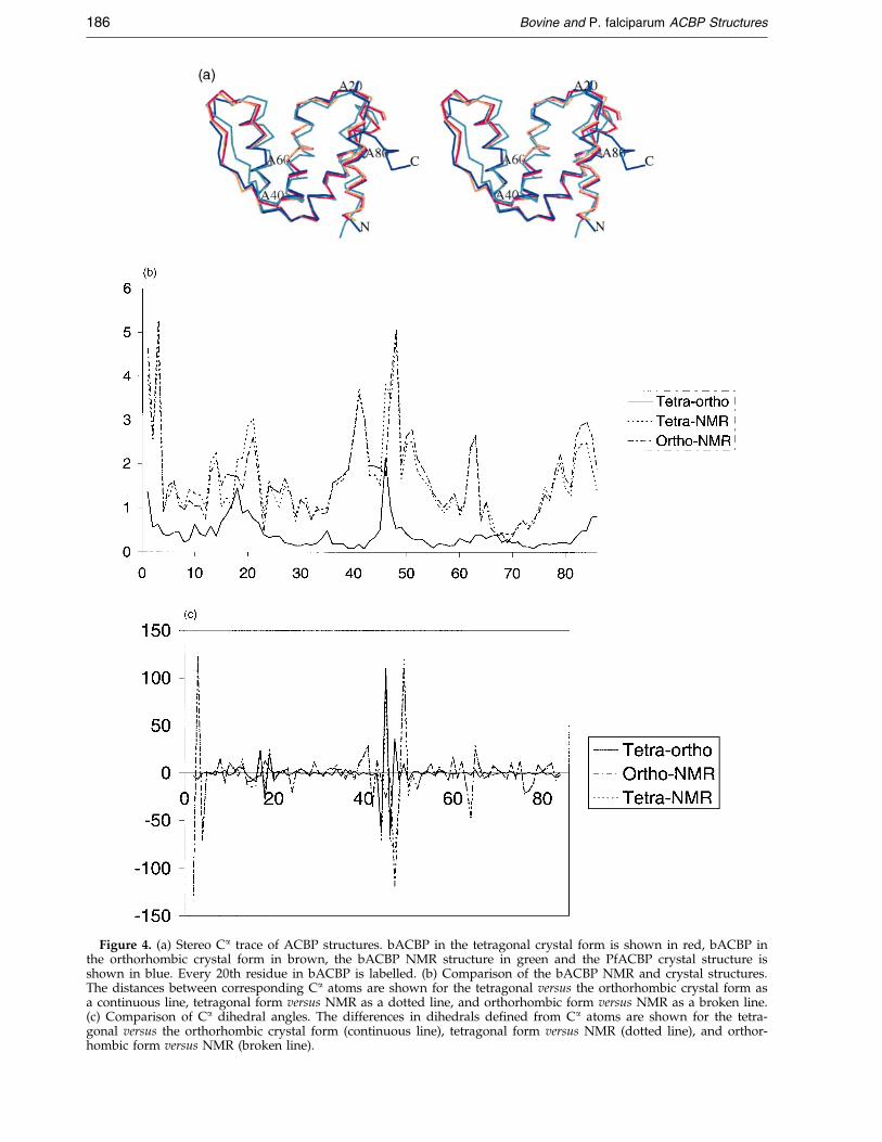
Figure 4. (a) Stereo Ca trace of ACBP structures. bACBP in the tetragonal crystal form is shown in red, bACBP inthe orthorhombic crystal form in brown, the bACBP NMR structure in green and the PfACBP crystal structure isshown in blue. Every 20th residue in bACBP is labelled. (b) Comparison of the bACBP NMR and crystal structures.The distances between corresponding Ca atoms are shown for the tetragonal versus the orthorhombic crystal form asa continuous line, tetragonal form versus NMR as a dotted line, and orthorhombic form versus NMR as a broken line.(c) Comparison of Ca dihedral angles. The differences in dihedrals de®ned from Ca atoms are shown for the tetra-gonal versus the orthorhombic crystal form (continuous line), tetragonal form versus NMR (dotted line), and orthor-hombic form versus NMR (broken line).
186 Bovine and P. falciparum ACBP Structures
Bovine and P. falciparum ACBP Structures 187
loops. The largest differences are found at theN-terminal region and the region around residues46-47. These two regions are not well de®ned inthe apo form NMR structures as there were only afew experimental restraints measured for theseregions.9
The search for exploitable differences
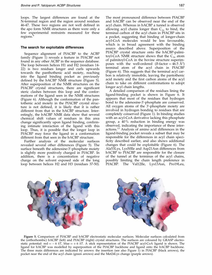
Sequence alignment of PfACBP to the ACBPfamily (Figure 1) reveals a feature in PfACBP notfound in any other ACBP in the sequence database.The loop between helices H1 and H2 (residues 14-22) is two residues longer and curls upwardstowards the panthothenic acid moiety, reachinginto the ligand binding pocket as previouslyde®ned by the bACBP NMR structure (Figure 5).After superposition of the NMR structures on thePfACBP crystal structures, there are signi®cantsteric clashes between this loop and the confor-mations of the ligand seen in the NMR structures(Figure 6). Although the conformation of the pan-tothenic acid moiety in the PfACBP crystal struc-ture is not de®ned, it is likely that it is ratherdifferent from that in the bACBP structure. Inter-estingly, the bACBP NMR data show that severalchemical shift values of residues in this areachange signi®cantly upon ligand binding, con®rm-ing intimate interaction of the ligand with thisloop. Thus, it is possible that the longer loop inPfACBP may force the ligand in a conformationdifferent from that seen in the bACBP structure.
Further analysis of the molecular surfacesrevealed several other differences (Figure 5). Thesurface beneath the adenosine-30-phosphate moietyis slightly more positively charged in PfACBP. Inaddition, there is a concentration of negativecharge on the solvent exposed side of the longloop between helices H2 and H3 (residues 37-50).
Figure 5. Comparison of PfACBP and bACBP electrostatithe (orthorhombic) bACBP (left) and PfACBP (right) crystalstatic potential: red � ÿ 6 kT, blue � � 6 kT. A stick represligand for bACBP was modelled by superposition of the PfThe three main differences are indicated by arrows: the inspocket near the end of the acyl chain (green arrows) and the
The most pronounced difference between PfACBPand bACBP can be observed near the end of theacyl chain. Whereas in bACBP a tunnel is observedallowing acyl chains longer than C16 to bind, theterminal carbon of the acyl chain in PfACBP sits ina pocket, suggesting that binding of longer-chainacyl-CoA molecules would be less favourable,which is in broad agreement with the bindingassays described above. Superposition of thePfACBP crystal structure onto the bACBP-palmi-toyl-CoA NMR structure shows that the C16 atomof palmitoyl-CoA in the bovine structure superim-poses with the well-ordered (B-factor � 46.5 AÊ 2)terminal atom of the acyl chain in PfACBP(Figure 6). This suggests that the terminal acyl car-bon is relatively immobile, leaving the pantothenicacid moiety and the ®rst carbon atoms of the acylchain to take on different conformations to adoptlonger acyl chain lengths.
A detailed comparison of the residues lining theligand-binding pocket is shown in Figure 6. Itappears that most of the residues that hydrogenbond to the adenosine-30-phosphate are conserved.All oxygen atoms of the 30-phosphate moiety areinvolved in hydrogen bonding to residues that arecompletely conserved (Figure 1). In binding studieswith an acyl-CoA derivative lacking this phosphategroup, a 40 % reduction in binding energy wasobserved, indicating the importance of these inter-actions.13 Analysis of amino acid differences in theligand-binding pocket reveals a subset that may beresponsible for the differences in acyl chain speci-®city described earlier, and also shows additionalchanges that could be exploitable (Figure 6). TheAla53Lys, Lys50Ile and Asp21Asn differences frombACBP to PfACBP are responsible for the closureof the tunnel at the terminus of the acyl chains,possibly limiting the chain length preference inPfACBP. The Val12Ile, Lys13Asn, Pro19Leu,
c molecular surfaces. Molecular surfaces calculated fromstructures. The surfaces are coloured by GRASP electro-entation of the PfACBP acyl-CoA ligand is shown. TheACBP backbone and ligand onto the bACBP backbone.ertion (see also Figure 1) in PfACBP (black arrows), theMet24Lys change (purple arrows).
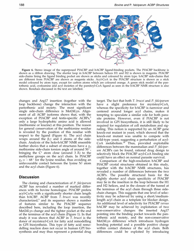
Figure 6. Stereo image of the superposed PfACBP and bACBP ligand-binding pockets. The PfACBP backbone isshown as a ribbon drawing. The shorter loop in bACBP between helices H1 and H2 is shown in magenta. PfACBPside-chains lining the ligand binding pocket are shown as sticks and coloured by atom type. bACBP side-chains thatare different from PfACBP are shown as magenta sticks. Acyl-CoA in the PfACBP structure is shown as a stickmodel coloured by atom type, except for carbon atoms which are coloured orange. A green stick model of the pan-tothenic acid, cysteamine and acyl moieties of the pamitoyl-CoA ligand as seen in the bACBP NMR structure is alsoshown. Residues discussed in the text are labelled.
188 Bovine and P. falciparum ACBP Structures
changes and Arg17 insertion (together with theloop backbone) change the interaction with thepantothenic acid moiety. The most signi®cantsingle side-chain difference is Met24Lys. Align-ment of all ACBP isoforms shows that, with theexception of PfACBP and testis-speci®c ACBPs,3
only a large hydrophobic amino acid is allowed(methionine or leucine) at this position. The reasonfor general conservation of the methionine residueis revealed by the position of this residue withrespect to the ligand (Figure 6). The acyl chainwraps around the b,g and d atoms of the side-chain. Analysis of the holo-bACBP NMR ensemblefurther shows that a subset of structures have a w3
methionine side-chain torsion angle of around 50 �,bringing the Ce atom close (around 3 AÊ ) to themethylene groups on the acyl chain. In PfACBPw3 � ÿ 68 � for the lysine residue, thus avoiding anunfavourable contact between the lysine Nz atomand the acyl chain (Figure 6).
Discussion
The cloning and characterization of P. falciparumACBP has revealed a number of marked differ-ences with its bovine homologue. PfACBP prefersacyl-CoAs with a signi®cantly shorter chain lengththan bACBP. ACBP from T. brucei was recentlycharacterized,5 and its sequence shows a numberof features similar to the PfACBP sequencedescribed here, including the loop insertion, theMet24Lys change and the tighter pocket at the endof the terminus of the acyl chain (Figure 1). In thatstudy it was shown that ACBP in T. brucei is thedonor of myristoyl-CoA for fatty acid remodellingin GPI biosynthesis. This type of fatty acid remo-delling reaction does not occur in human GPI bio-synthesis and may thus represent a potential drug
target. The fact that both T. brucei and P. falciparumhave a slight preference for myristoyl-CoA,whereas the speci®city for bACBP is narrower andcentered around longer acyl chains, makes ittempting to speculate a similar role for both para-site proteins. However, even if PfACBP is notinvolved in GPI biosynthesis, it is still likely to berequired for regulation of cell metabolism and sig-naling. This notion is supported by an ACBP geneknock-out mutant in yeast, which showed that theknock-out mutant was unable to compete withwild-type yeast, suggesting a perturbation of acyl-CoA metabolism.14 Thus, provided exploitabledifferences between the mammalian and P. falcipar-um ACBPs can be found, rational drug design toselectively block the PfACBP acyl-CoA binding sitecould have an effect on normal parasite survival.
Comparison of the high-resolution bACBP andPfACBP crystal structures obtained in this study,together with the bACBP NMR structures, hasrevealed a number of differences between the twoACBPs. The possible structural basis for theslightly shorter acyl chain speci®city of PfACBPmay lie in the insertion in the loop between the H1and H2 helices, and in the closure of the tunnel atthe terminus of the acyl chain through three side-chain changes. This suggests that one level of selec-tivity may be achieved by using a short/medium-length acyl chain as a template for blocker design.An additional level of selectivity for PfACBP versusbACBP may be achieved by exploitation of twonon-conservative changes: the inserted Arg17pointing into the binding pocket towards the pan-tothenic acid moiety, and the non-conservativeMet24Lys difference which introduces hydrogenbonding potential and a possible positive chargewithin contact distance of the acyl chain. Bothdifferences could be exploited by introducing

Bovine and P. falciparum ACBP Structures 189
hydrogen bonding acceptors and/or negativecharges on a template molecule for selective block-ing of the PfACBP binding site.
Materials and Methods
Cloning, expression, purification andcharacterization of P. falciparum ACBP
A genomic DNA clone of P. falciparum (accessionnumber AA549985) was kindly provided by J. Dame(Department of Pathobiology, University of Florida).Routine DNA manipulations were performed in E. colistrain DH5a. All chemicals were of the highest gradeavailable from BDH-Merck or Sigma and restrictionenzymes were from Promega. DNA sequencing ofdouble-stranded DNA was accomplished by the dideox-ynucleotide chain termination method by automatedcycle sequencing using the dye terminator method (ABIPRISM big dye terminator kit; Perkin Elmer).
A BLAST search of the P. falciparum genomicsequences in the TIGR database was performed usingthe gene encoding T. brucei ACBP which has beendescribed recently.5 An open reading frame (ORF) corre-sponding to the P. falciparum ACBP sequence was ident-i®ed (accession number AA549985). The gene wasobtained by polymerase chain reaction (PCR) ampli®ca-tion of the P. falciparum ORF from genomic DNA, usingforward primer 50-ggcatatggcacaagtatttg-30 and reverseprimer 50-ggctcgagttattccccatcttg-30. The forward primercontained an NdeI site and the reverse primer contains aXhoI site for cloning. Ampli®cation conditions were94 �C for 45 seconds, 50 �C for one minute and 72 �C forone minute for 30 cycles. The PCR product was blunt-end ligated into the SmaI site of pUC18 (Sureclone kit;Amersham Pharmacia Biotech) giving the plasmidpUC18-Pf.ACBP. The insert was excised by digestionwith NdeI and XhoI and ligated into the NdeI and XhoIcloning site of the pET16b expression vector (Novagen).The pET16b-Pf.ACBP plasmid was transformed intocompetent BL21 (DE3) E. coli cells. A single colony wasgrown up in Luria-Bertani broth containing 0.1 mg/mlampicillin. When the culture reached an A600 of 0.5 in avolume of one litre, 1 mM isopropyl-D-thiogalactopyra-noside was added to induce expression of recombinantP. falciparum ACBP. Cultures were grown for anadditional three hours at 37 �C and then harvested bycentrifugation (4000 g, ten minutes, 4 �C). Cells wereresuspended in 100 ml of lysing buffer (20 mM Bis-Trispropane, 20 mM Tris-HCl pH 7.5, 300 mM NaCl, 1 mMphenylmethanesulfonyl ¯uoride, 2 mg/ml leupeptin,0.2 mM N-tosyl-L-lysine chloromethylketone and0.1 mg/ml lysozyme) for 15 minutes at 30 �C and dis-rupted by sonication (6 � 30 second pulses interruptedwith cooling on ice). Cell debris was removed by cen-trifugation at 10,000 g for ten minutes prior to applyingthe supernatant to a Ni2�-resin column (1.6 cm � 12 cmChelating Sepharose Fast Flow; Amersham PharmaciaBiotechnology). The histidine-tagged recombinant pro-tein was eluted from the column with a 10 mM to 1 Mlinear gradient of imidazole in 20 mM Bis-Tris propane,20 mM Tris-HCl (pH 7.5), 300 mM NaCl at 3 ml/minute.Fractions were desalted and concentrated with a Centri-con plus-20 (Amicon) ®ltration unit and treated with50 mg factor Xa protease (New England Biolabs) permilligram recombinant protein for three days at 4 �C indigestion buffer (20 mM Tris-HCl (pH 8.0), 100 mMNaCl, 2 mM CaCl2). The recombinant protein was
further puri®ed on a gel permeation column(1 cm � 30 cm Superdex 75 HR; Amersham PharmaciaBiotechnology) in 50 mM Na-Hepes (pH 7.5), 300 mMKCl at 0.5 ml/minute. The sample was concentrated to20 mg/ml using a Centricon plus-20 and stored at 4 �C.
The acyl-CoA content of PfACBP puri®ed from E. coliwas characterized by high performance liquid chroma-tography (HPLC). A 100 ml sample of puri®ed, PfACBP(15 mg/ml) was applied on an ODS 10/100 column(4.6 mm � 250 mm) equilibrated with 30 % solvent B(80 % acetonitrile in 20 mM KH2PO4, pH 5.3) in solventA (20 % acetonitrile in 20 mM KH2PO4, pH 5.3), withoutany pretreatment. The CoA esters were eluted with thefollowing gradient of solvent B in solvent A: 30 % B in Afor 20 minutes, 30-40 % B in 30 minutes, 40-45 % B in tenminutes, 45-60 % B in 20 minutes, 60-85 % B in ®veminutes, and 85 % B for ®ve minutes. The ¯ow-rate was1.0 ml/minute.
An acyl-CoA competition binding assay was per-formed as described.15 Recombinant P. falciparum andbovine ACBP were stripped of bound acyl-CoAs usingreverse phase HPLC.16 Aliquots of 0.05 pmol ACBP in25 ml binding buffer (10 mM potassium phosphate,pH 7.4) were mixed with 0.1 pmol of unlabelled acyl-CoA and 0.1 pmol [3H]myristoyl-CoA in 75 ml of bindingbuffer. The samples were incubated at 37 �C for 30 min-utes, chilled on ice for ten minutes and mixed with0.2 ml of an ice-cold 50 % slurry of Lipidex 1000 (Canber-ra Packard) in binding buffer. After 100 minutes on ice,the samples were centrifuged at 12,000 g for ®ve minutesat 0 �C. The radioactivity in 100 ml of the resulting super-natant was determined by scintillation counting. Theassay was carried out in triplicate and controls withoutACBP were performed.
An acyl-CoA binding speci®city study was performedusing isoelectric focusing analysis as described.16 Ali-quots of 5 mg ACBP were incubated with 0.5 mg of acyl-CoA (C10-20) in 10 ml of 10 mM potassium phosphate(pH 7.4) at 37 �C for ten minutes and resolved on Novexisoelectric focusing gels with ampholytes for the pHrange 3-10.
Crystallization
Recombinant bovine ACBP (bACBP) was preparedand puri®ed as described.17 All crystallization trials werecarried out using the hanging drop vapour diffusionmethod. Tetragonal bACBP crystals were obtained usinga reservoir solution composed of 25 % polyethylene gly-col 5000 monomethyl ether (PEG 5000 MME), 40 mMNi(SO4)2, 40 mM Hepes buffer (pH 7.5). A drop contain-ing 4 ml of reservoir and 4 ml of 18 mg/ml protein sol-ution was equilibrated against 1 ml of the reservoirsolution at 4 �C. Bi-pyramidal crystals appeared withinone week reaching a full size of approximately0.2 mm � 0.3 mm � 0.3 mm.
Orthorhombic bACBP crystals were obtained by mix-ing a 4 ml solution of 15 % PEG 8000, 5 mM CdCl2,10 mM Hepes buffer (pH 7.5) with 4 ml of 10 mg/mlbACBP complexed with palmitoyl-CoA at a 1:1 molarconcentration ratio. The drops were equilibrated against25 ml of 0.2 M NaCl at 20 �C. No crystals were observedduring the ®rst two months after the experiments wereset up. One crystal with well-de®ned shape was even-tually found after the drops were allowed to equilibratefor ten months. This crystal was used both for spacegroup determination with precession photography andfor intensity data collection.
190 Bovine and P. falciparum ACBP Structures
PfACBP was crystallized using the hanging dropvapour diffusion method. Protein solution (2 ml) contain-ing 15 mg/ml protein, 50 mM Hepes (pH 7.5) and300 mM KCl was mixed with 2 ml of well solution(10 mM NiCl2, 100 mM Tris (pH 8.5), 20 % PEG 2000MME). Trays were stored at 20 �C, and crystals appearedafter two to three days, reaching a maximum size of0.05 mm � 0.05 mm � 0.3 mm.
Data collection
Precession photography of the orthorhombic bACBPcrystal form showed that it belonged to P21212 orP212121. Intensity data were then collected to a resolutionof 2.0 AÊ using a Hamlin/Xuong (UCSD-type) area detec-tor mounted on a Rigaku rotating anode generatorequipped with graphite monochromator. Softwaredescribed by Howard et al.18 was used for data collectionand processing. The space group was determined to beP212121 with unit cell dimensions a � 26.1 AÊ , b � 54.8 AÊ ,and c � 65.8 AÊ . Details of data processing are given inTable 4.
Systematic absences obtained for the bi-pyramidalbACBP crystal form by precession photography showedit to be in a tetragonal space group. Diffraction datasetswere collected to a resolution of 2.4 AÊ at room tempera-ture using a Hamlin/Xuong (UCSD-type) area detectorand an R-Axis IIc imaging plate detector mounted on aRigaku rotating anode X-ray generator. The images wereintegrated and scaled using the programs from the HKLsuite.19 The space group was determined to be either P41
or P43 with cell parameters a � b � 47.8 AÊ andc � 128.8 AÊ . The two data sets were merged to give acomplete data set to 2.4 AÊ resolution and used for mol-ecular replacement calculations. At a later stage, a dataset to a resolution of 2.0 AÊ was collected from a singlecrystal using a MAR image plate detector at the beam-line X11 at the EMBL Outstation, DESY, Hamburg, andprocessed using the HKL suite19 (Table 4).
Diffraction data for PfACBP were collected on beam-line BW7A, EMBL Outstation, DESY, Hamburg, using aMAR CCD detector. The crystal was cryoprotected byadding 50 % 2-methane-2,4-pentanediol to the motherliquor for 60 seconds, after which the crystal was frozenin a nitrogen stream. Initial oscillation images indicateda tetragonal lattice with a � b � 48.7 AÊ , c � 48.4 AÊ . A fulldataset was collected to 2.0 AÊ resolution and processedwith the HKL suite19 (Table 4). Analysis of the Matthew's
Table 4. Details of data collection, processing and merging
Ortho-bACBP
Temperature (K) 298Wavelength (AÊ ) 1.54Space group P212121
Cell dimensions (AÊ ) a � 26.1, b � 54.8, c � 65.8Resolution range (AÊ ) 8-2.0(2.15-2.0)No. observed refl. 24,078 (2567)No. unique refl. 6473 (1196)Redundancy 3.7 (2.1)hI/sIi 32.7 (9.3)Completeness (%) 94.9(91.2)Rmerge 0.040(0.076)NCS NoneVMatthews (AÊ 3/Da) 2.4
Values in parentheses are for the highest resolution shell. Ortho-bAbACBP crystal form.
coef®cient indicated that only a space group with foursymmetry operations was allowed, and the systematicabsences along the 4-fold axis indicated a P41 or P43
space group.
Structure determination and refinement
All structures were solved by molecular replacementusing AMoRe.20 Model building was performed usingO,21 and re®nement with CNS.22 Quality of intermediatemodels was monitored using PROCHECK23 and WHATIF.24
The solution of the bACBP crystal structure has beenbrie¯y described.25 For the bACBP orthorhombic crystalform, the search models were derived from the NMRmodels of bACBP complexed with palmitoyl-coenzymeA10 (PDB entry 1ACA) , which were kindly provided byF.M. Poulsen (Carlsberg Laboratory, Denmark) beforetheir general release from the PDB. A solution wasfound using an ensemble of the 14 most similar modelsfrom the family of 20 NMR structures10 superimposedon top of each other as a search model. A solution wasfound with AMoRe with a CC of 0.441 (R-factor 0.525,next best solution had CC 0.437 and R-factor 0.540)within the resolution range 15-3.0 AÊ . The crystal packingof the best solution looked good and the maps calculatedusing phases from the model looked convincing for mostof the protein. Despite rather high-resolution diffractiondata collected for the orthorhombic crystal form, there®nement, which included simulated annealing, provedto be dif®cult due to the lack of independent phase infor-mation, and Rfree stalled at 0.46 (R � 0.36). The partiallyre®ned model, however, was suf®ciently improved to beused to solve the bACBP tetragonal crystal form(described below). Using this re®ned model, the orthor-hombic crystal form could be re®ned further. By alternat-ing SA re®nement, manual rebuilding, and addition ofsolvent molecules, the model was re®ned with CNS22
using the amplitude-based maximum likelihood func-tion. All data between 20.0 and 2.0 AÊ resolution wereused in the re®nement and the bulk solvent correctionimplemented in CNS was applied, resulting in the ®nalmodel (Table 2) with an R-factor of 0.200 (Rfree � 0.226)(Figure 2(a)). A search for the palmitoyl-CoA ligand inthe crystal was unsuccessful, as no electron densitywhich could account for whole or part of the ligand wasfound. A possibility is that the ligand was hydrolysedduring the relatively long period of crystallization.
Tetra-bACBP PfACBP
120 1000.928 1.03P41 P43
a � b � 47.8, c � 128.8 a � b � 48.7, c � 48.440-2.0(2.07-2.0) 25-2.0(2.07-2.0)50,106 (5001) 28,439 (2526)18831 (1917) 7754 (736)
2.7 (2.6) 3.7 (3.4)13.8 (3.6) 16.2 (2.7)96.5(98.8) 99.4(95.3)
0.066(0.292) 0.058(0.516)3-fold None
2.5 2.6
CBP, bACBP orthorhombic crystal form; tetra-ACBP, tetragonal
Bovine and P. falciparum ACBP Structures 191
As described above, the partially re®ned structure forthe bACBP orthorhombic crystal form was used as asearch model for molecular replacement with the bACBPtetragonal form re¯ection data. After the rotation func-tion, a translation function search was carried out inboth P41 and P43 , with the results being consistently bet-ter for the former. The best solution after rotation andtranslation function had a CC of 0.291 and R-factor of0.517 (next best: CC 0.277, R-factor 0.526) for databetween 15.0 and 4.0 AÊ resolution. These were improvedafter RBR to CC of 0.402 and R-factor of 0.525 in the res-olution range 15.0-2.7 AÊ . Keeping this solution ®xed, asecond translation search was performed. A solution forthe second molecule was found and combining thesetwo solutions, the correlation coef®cient improved to0.498 (R-factor 0.490) after RBR. The search for a thirdmolecule from these rotation peaks was unsuccessful.However, analysis of the crystal packing on a graphicsdisplay, revealed a well-de®ned void for a third mol-ecule. Therefore another rotation search using databetween 15.0 and 3.0 AÊ resolution was carried out. The48th peak in the rotation function gave the best score inthe combined translation function with AMoRe. AfterRBR using data between 15.0-2.7 AÊ resolution of allthree molecules, the correlation coef®cient was 0.587 andthe R-factor was 0.451 (the next best solution had CCand R-factor of 0.522 and 0.487, respectively). The threemolecules were related by 3-fold NCS along the a-axis.Several cycles of rebuilding and simulated annealingre®nement with NCS produced a model with an Rfree of0.259 (R � 0.207) for data to 2.4 AÊ resolution. At thisstage a 2.0 AÊ dataset was collected at the EMBL-Outsta-tion, DESY, Hamburg (Table 4). Re®nement was contin-ued using CNS22 with this high-resolution data, whichinvolved mainly adjusting some side-chain confor-mations and inclusion of more solvent molecules. Alldata between 20.0 and 2.0 AÊ resolution were used in there®nement and the bulk solvent correction implementedin CNS was applied. The ®nal model has an R-factor of0.223 (Rfree � 0.255) (Table 2; Figure 2(a)).
Initial attempts to solve the PfACBP structure usingthe NMR ensemble of bACBP structures as searchmodels were unsuccessful. Using the tetragonal bACBPcrystal structure, a solution was found (R � 0.463,CC � 0.313 after RBR) in P43, with a poly-alanine back-bone model plus side-chains conserved between bACBPand PfACBP. An initial map revealed good density formost of the backbone and it was possible to identifymost of the new side-chains. Since good quality diffrac-tion data were collected (Table 4), this initial set ofphases was used as input for the autobuilding procedurewith warpNtrace.26 Standard protocols were used, witha relatively large number (1000) of cycles of rebuildingand re®nement. A total of 75 out of the 90 residues wereautomatically built, including their side-chains. After aninitial round of model building in O21 and re®nement inCNS22 the maps revealed interpretable density for themissing pieces of backbone and side-chains (R � 0.295,Rfree � 0.344). After the next macrocycle, which includedsimulated annealing, well-de®ned density in the bindingsite (Figure 2(b)) could be observed for the nucleotidemoiety of CoA, which was included (R � 0.267,Rfree � 0.312). The new maps revealed two strong peaksclose to His0 and His49, and density for a fatty acid(Figure 2(b)). Using the phases of the model and theanomalous differences from the re¯ection data set, amap was calculated that showed the same two peaks(>4s). The peaks were interpreted as coordinated Ni2 �,which were the only heavy atoms present in the crystalli-
zation solution. A string of 13 carbon atoms was mod-elled in the density for the acyl chain (R � 0.206,Rfree � 0.255). Further inclusion of solvent molecules, twosulphate ions and some alternate side-chain confor-mations resulted in the ®nal model (R � 0.198,Rfree � 0.237) (Table 4).
PDB accession codes
The coordinates and structure factors have beendeposited in the RCSB data bank with accession codes1HBK, 1HB6, and 1HB8.
Acknowledgements
We thank the EMBL-Hamburg outstation for use ofbeamlines BW7A and X11 at DESY, and Flemming Poul-sen for access to the coordinates of the bACBP NMRstructure prior to their public release. K.G.M. is a BeitMemorial Fellow, D.v.A. is supported by a WellcomeTrust Career Development Research Fellowship.
References
1. Brockman, A., Price, R. N., van Vught, M., Heppner,D. G., Walsh, D., Sookto, P., Wimomwattrawatee,T., Looareesuwam, S., White, N. J. & Nosten, F.(2000). Plasmodium falciparum antimalarial drugsusceptibility on the north-western border of Thai-land during ®ve years of etensive use of artesunate-me¯oquine. Trans. Roy. Soc. Trop. Med. Hygiene, 94,537-544.
2. Faergeman, N. J. & Knudsen, J. (1997). Role of long-chain fatty acyl-coA esters in the regulation ofmetabolism and in cell signalling. Biochem. J. 323, 1-12.
3. Kragelund, B. B., Knudsen, J. & Poulsen, F. M.(1999). Acyl-coenzyme a binding protein (ACBP).Biochim. Biophys. Acta, 1441, 150-161.
4. van Aalten, D. M. F., DiRusso, C. C., Knudsen, J. &Wierenga, R. K. (2000). Crystal structure of FadR, afatty acid-responsive transcription factor with anovel acyl coenzyme A-binding fold. EMBO J. 19,5167-5177.
5. Milne, K. G. & Ferguson, M. A. J. (2000). Cloning,expression and characterisation of the acyl-coAbinding protein in African trypanosomes. J. Biol.Chem. 275, 12503-12508.
6. Masterson, W. I., Raper, J., Doering, T. L., Hart,G. W. & Englund, P. T. (1990). Fatty acid remodel-ing: a novel reaction sequence in the biosynthesis oftrypanosome glycosyl phosphatidylinositol mem-brane anchors. Cell, 62, 73-80.
7. Kragelund, B. B., Poulsen, K., Andersen, K. V.,Baldursson, T., Kroel, J. B., Neergard, T. B., Jepsen,J., Roepstoff, P., Kristiansen, K., Poulsen, F. M. &Knudsen, J. (1999). Conserved residues and theirrole in the structure, function and stability of acyl-coenzyme A binding protein. Biochemistry, 38, 2386-2394.
8. Kragelund, B. B., Osmark, P., Neergaard, T. B.,Schioedt, J., Kristiansen, K., Knudsen, J. & Poulsen,F. M. (1999). The formation of a native-like structurecontaining eight conserved hydrophobic residues israte limiting in two-state protein folding of ACBP.Nature Struct. Biol. 6, 594-601.

192 Bovine and P. falciparum ACBP Structures
9. Andersen, K. V. & Poulsen, F. M. (1992). Three-dimensional structure in solution of acyl-coenzyme-A binding-protein from bovine liver. J. Mol. Biol.226, 1131-1141.
10. Kragelund, B. B., Andersen, K. V., Madsen, J. C.,Knudsen, J. & Poulsen, F. M. (1993). Three-dimen-sional structure of the complex between acyl-coen-zyme A binding protein and palmitoyl-coenzyme A.J. Mol. Biol. 230, 1260-1277.
11. Kleywegt, G. J. & Jones, T. A. (1996). Phi/psi-chology: Ramachandran revisited. Structure, 4, 1395-1400.
12. Korn, A. P. & Rose, D. R. (1994). Torsion angledifferences as a means of pinpointing local polypep-tide-chain trajectory changes for identical proteins indifferent conformational states. Protein Eng. 7, 961-967.
13. Faergeman, N. J., Sigurskjold, B. W., Kragelund,B. B., Andersen, K. V. & Knudsen, J. (1996). Thermo-dynamics of ligand binding to acyl-coenzyme Abinding protein studied by titration calorimetry.Biochemistry, 35, 14118-14126.
14. Schjeling, C. K., Hummel, R., Hansen, J. K.,Boersting, C., Mikkelsen, J. M., Kristiansen, K. &Knudsen, J. (1996). Disruption of the gene encodingthe acyl-coA-binding protein (acb1) perturbs acyl-coA metabolism in Saccharomyces cerevisiae. J. Biol.Chem. 271, 22514-22521.
15. Rosendal, J., Ertberg, P. & Knudsen, J. (1993).Characterisation of ligand binding to acyl-coA bind-ing protein. Biochem. J. 290, 321-326.
16. Knudsen, J., Faergeman, N. J., Skott, J., Hummel, R.,Borsting, C., Rose, T. M., Andersen, J. S., Hojrup, P.,Roepstorff, P. & Kristiansen, K. (1994). Yeast acyl-coA binding protein: acyl-coA binding af®nity andeffect on intracellular acyl-coA pool-size. Biochem. J.302, 479-485.
17. Mandrup, S., Hojrup, P., Kristiansen, K. & Knudsen,J. (1991). Gene synthesis, expression in E. coli, puri®-cation and characterization of the recombinant
bovine acyl-coA binding protein. Biochem. J. 276,817-823.
18. Howard, A. J., Nielsen, C. & Xuong, N. H. (1985).Software for a diffractometer with multiwire areadetector. Methods Enzymol. 114, 452-472.
19. Otwinowski, Z. & Minor, W. (1997). Processing ofX-ray diffraction data collected in oscillation mode.Methods Enzymol. 276, 307-326.
20. Navaza, J. (1994). AMoRe: an automated packagefor molecular replacement. Acta Crystallog. sect. A,50, 157-163.
21. Jones, T. A., Zou, J. Y., Cowan, S. W. & Kjeldgaard,M. (1991). Improved methods for building proteinmodels in electron density maps and the location oferrors in these models. Acta Crystallog. sect. A, 47,110-119.
22. Brunger, A. T., Adams, P. D., Clore, G. M., Gros, P.,Grosse-Kunstleve, R. W., Jiang, J.-S., Kuszewski, J.,Nilges, M., Pannu, N. S., Read, R. J., Rice, L. M.,Simonson, T. & Warren, G. L. (1998). Crystallo-graphy and NMR system: a new software systemfor macromolecular structure determination. ActaCrystallog. sect. D, 54, 905-921.
23. Laskowski, R. A., McArthur, M. W., Moss, D. S. &Thornton, J. M. (1993). PROCHECK: a program tocheck the stereochemical quality of protein struc-tures. J. Appl. Crystallog. 26, 283-291.
24. Vriend, G. (1990). WHAT IF: a molecular modelingand drug design program. J. Mol. Graph. 8, 52-56.
25. Kleywegt, G. J. (1996). Use of non-crystallographicsymmetry in protein structure re®nement. Acta Crys-tallog. sect. D, 52, 842-857.
26. Perrakis, A., Morris, R. & Lamzin, V. S. (1999).Automated protein model building combined withiterative structure re®nement. Nature Struct. Biol. 6,458-463.
27. Engh, R. A. & Huber, R. (1991). Accurate bond andangle parameters for X-ray protein-structure re®ne-ment. Acta Crystallog. sect. A, 47, 392-400.
Edited by R. Huber
(Received 2 November 2000; received in revised form 19 March 2001; accepted 19 March 2001)