bio 210 lab instructor: dr. rebecca clarke chapter 14: the brain
TRANSCRIPT
BIO 210 LabInstructor: Dr. Rebecca Clarke
Chapter 14: The Brain
Human BrainContains almost 98% of body’s neural
tissueAverage weight about 3 lb (1.4 kg)

6 Major Regions of the BrainCerebrumCerebellumDiencephalonMesencephalonPonsMedulla oblongata
The BrainEmbryological Development
Determines organization of brain structures
Neural tubeOrigin of brain
Enlarges into three primary brain vesicles
prosencephalon
mesencephalon
rhombencephalon

The BrainFive Secondary Brain Vesicles
Telencephalon
Diencephalon
Mesencephalon
Metencephalon
Myelencephalon
The BrainOrigins of Brain Structures
Diencephalon and mesencephalon persist
Telencephalon:
Becomes cerebrum
Metencephalon
Forms cerebellum and pons
Myelencephalon
Becomes medulla oblongata

The Brain
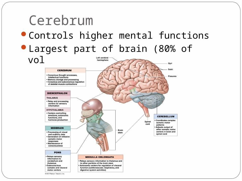
CerebrumControls higher mental functionsLargest part of brain (80% of volume)
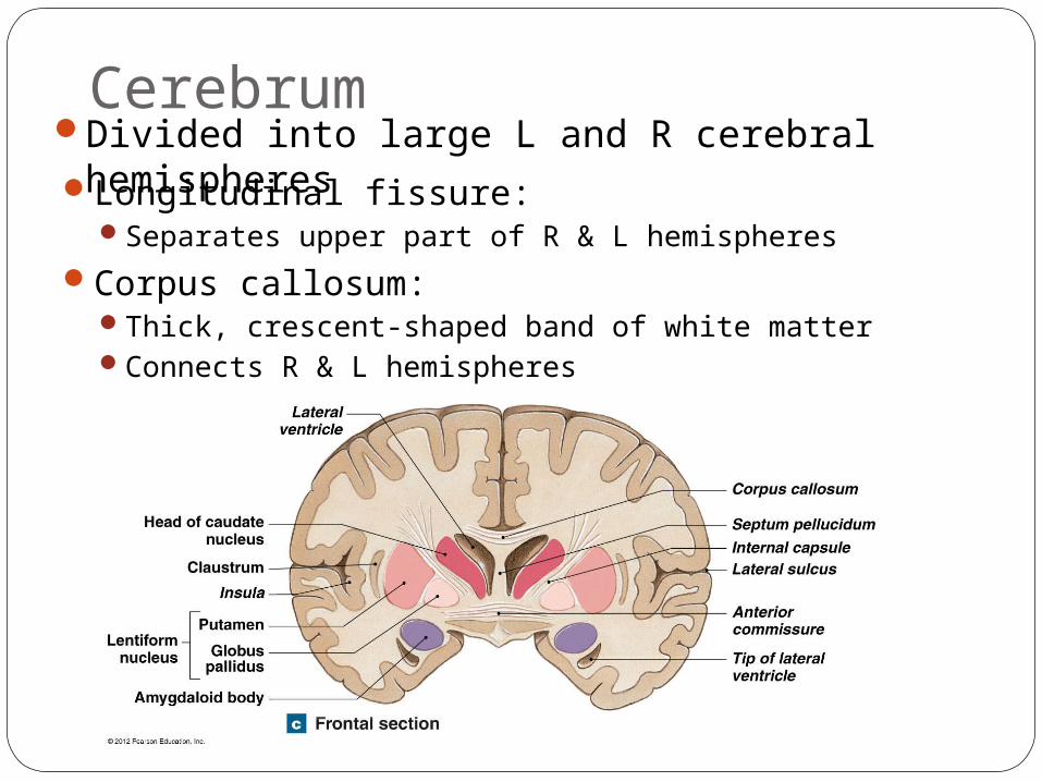
CerebrumDivided into large L and R cerebral
hemispheresLongitudinal fissure:Separates upper part of R & L hemispheres
Corpus callosum:Thick, crescent-shaped band of white matterConnects R & L hemispheres
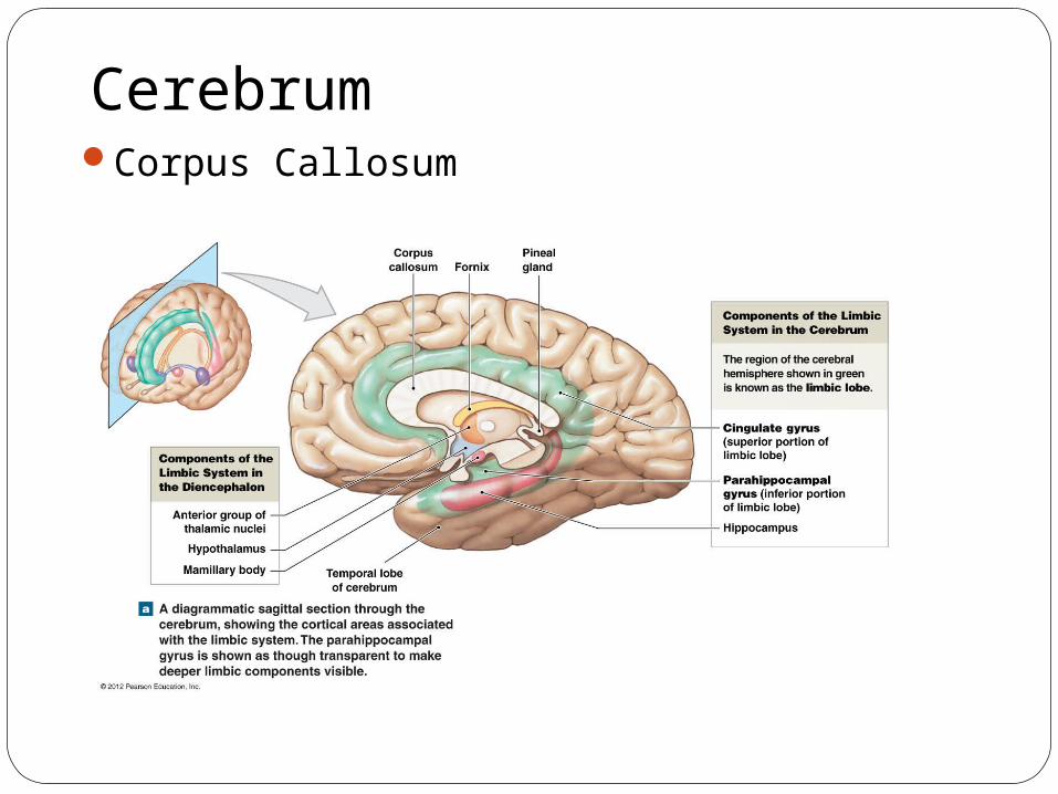
Figure 14–11a
CerebrumCorpus Callosum
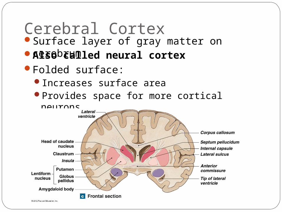
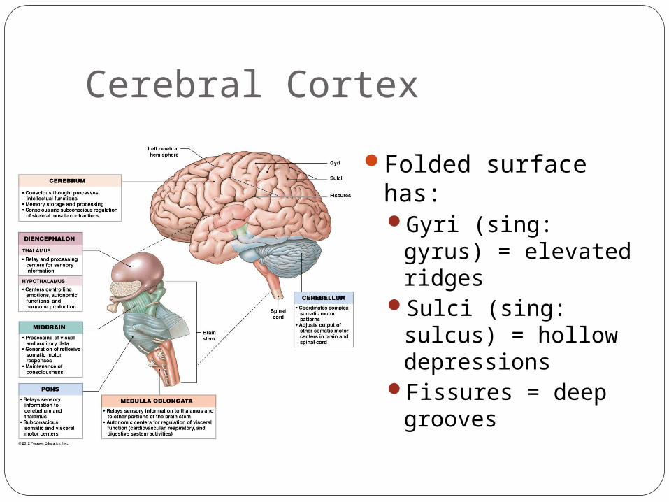
Cerebral CortexSurface layer of gray matter on cerebrumAlso called neural cortexFolded surface:
Increases surface areaProvides space for more cortical
neurons
Cerebral Cortex
Folded surface has:Gyri (sing: gyrus) =
elevated ridgesSulci (sing: sulcus)
= hollow depressions
Fissures = deep grooves
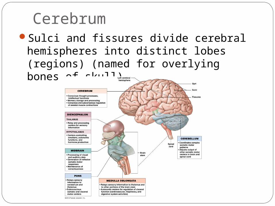
CerebrumSulci and fissures divide cerebral
hemispheres into distinct lobes (regions) (named for overlying bones of skull)
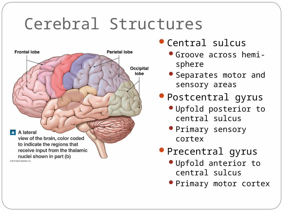
Cerebral StructuresCentral sulcus
Groove across hemi-sphere
Separates motor and sensory areas
Postcentral gyrusUpfold posterior to
central sulcusPrimary sensory cortex
Precentral gyrusUpfold anterior to
central sulcusPrimary motor cortex
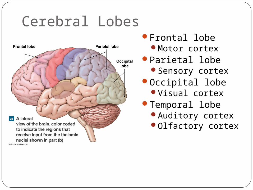
Cerebral LobesFrontal lobe
Motor cortexParietal lobe
Sensory cortex Occipital lobe
Visual cortex Temporal lobe
Auditory cortexOlfactory cortex
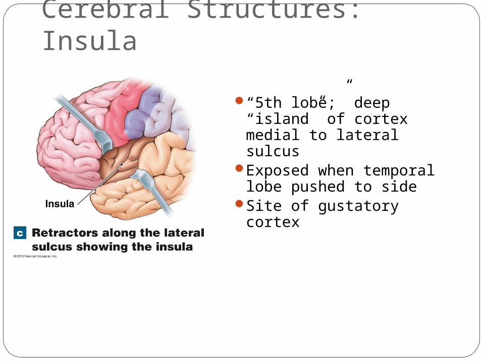
Cerebral Structures: Insula
“5th lobe;” deep “island” of cortex medial to lateral sulcus
Exposed when temporal lobe pushed to side
Site of gustatory cortex

Cerebral Gray MatterFound in:
Cerebral cortexCerebral nuclei
Cerebral White MatterMyelinated nerve fibersMakes up most of interior of cerebrum Located:
Deep to cerebral cortexAround cerebral nuclei
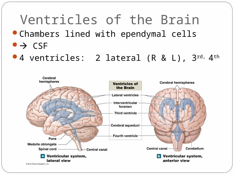
Ventricles of the BrainChambers lined with ependymal cells CSF4 ventricles: 2 lateral (R & L), 3rd, 4th
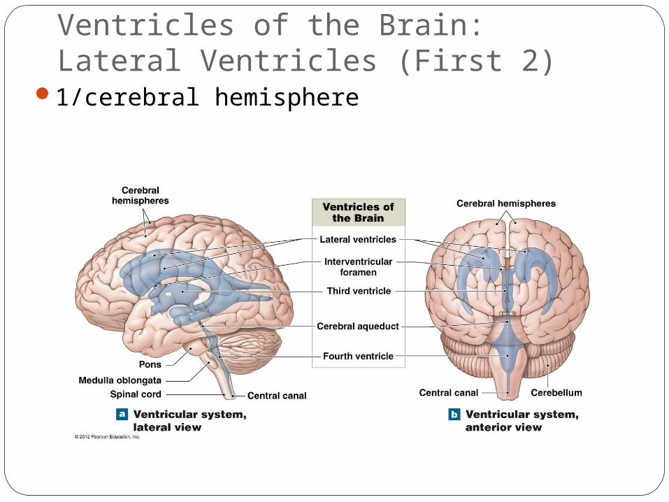
Ventricles of the Brain:Lateral Ventricles (First 2)
1/cerebral hemisphere
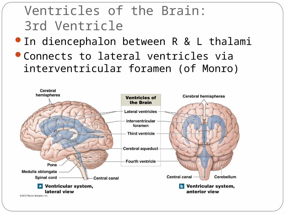
Ventricles of the Brain:3rd Ventricle
In diencephalon between R & L thalamiConnects to lateral ventricles via
interventricular foramen (of Monro)
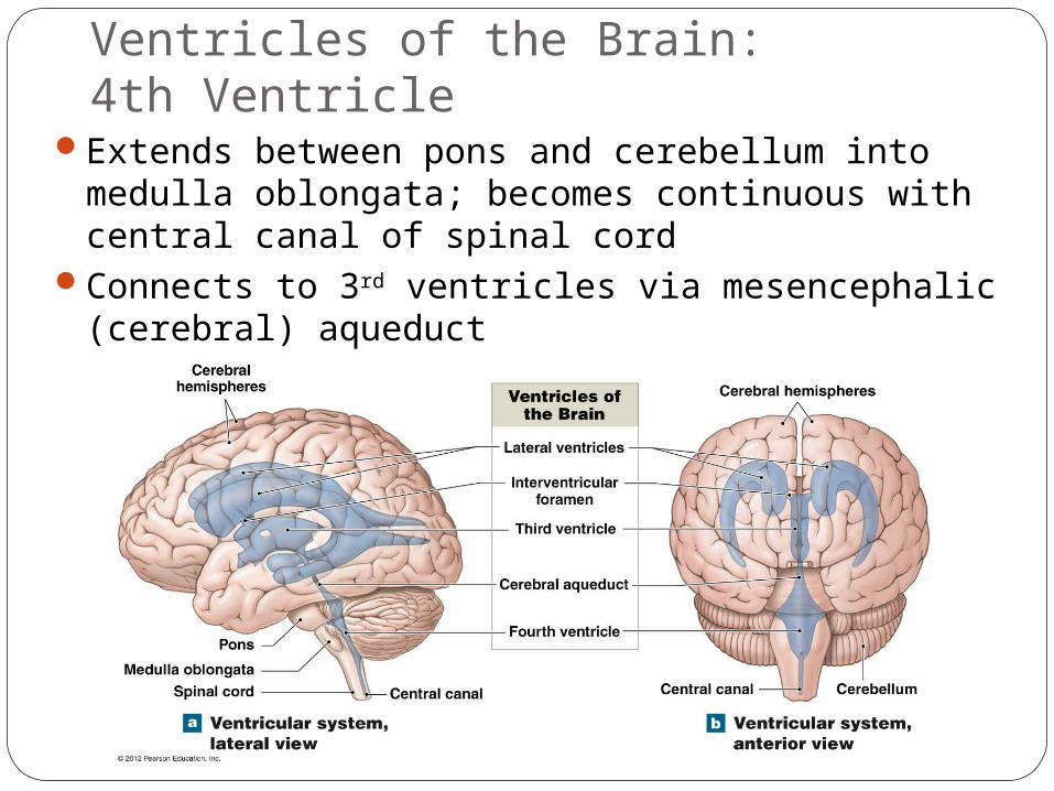
Ventricles of the Brain:4th Ventricle
Extends between pons and cerebellum into medulla oblongata; becomes continuous with central canal of spinal cord
Connects to 3rd ventricles via mesencephalic (cerebral) aqueduct
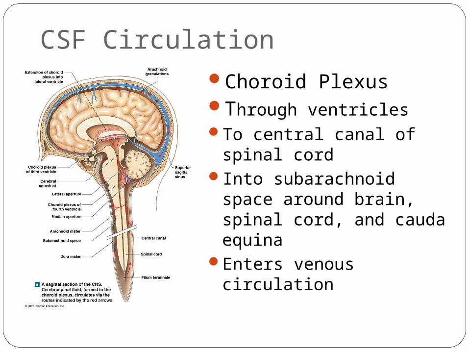
CSF CirculationChoroid PlexusThrough ventriclesTo central canal of spinal
cordInto subarachnoid space
around brain, spinal cord, and cauda equina
Enters venous circulation
Cranial MeningesHave 3 layers (like spinal meninges):
Dura materArachnoid materPia mater
Are continuous with spinal meningesProtect the brain from cranial trauma (act
as “air bags”)
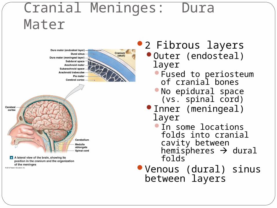
Cranial Meninges: Dura Mater2 Fibrous layers
Outer (endosteal) layerFused to periosteum of
cranial bonesNo epidural space (vs.
spinal cord)Inner (meningeal)
layerIn some locations folds
into cranial cavity between hemispheres dural folds
Venous (dural) sinus between layers
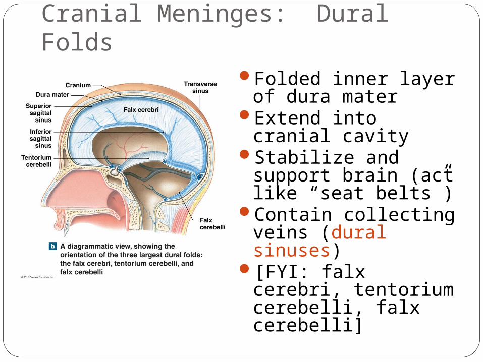
Cranial Meninges: Dural FoldsFolded inner layer of
dura materExtend into cranial
cavityStabilize and support
brain (act like “seat belts”)
Contain collecting veins (dural sinuses)
[FYI: falx cerebri, tentorium cerebelli, falx cerebelli]
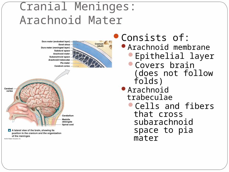
Cranial Meninges: Arachnoid Mater
Consists of:Arachnoid membrane
Epithelial layerCovers brain (does
not follow folds)Arachnoid trabeculae
Cells and fibers that cross subarachnoid space to pia mater

Cranial Meninges: Pia Mater
Attached to brain surface by astrocytes
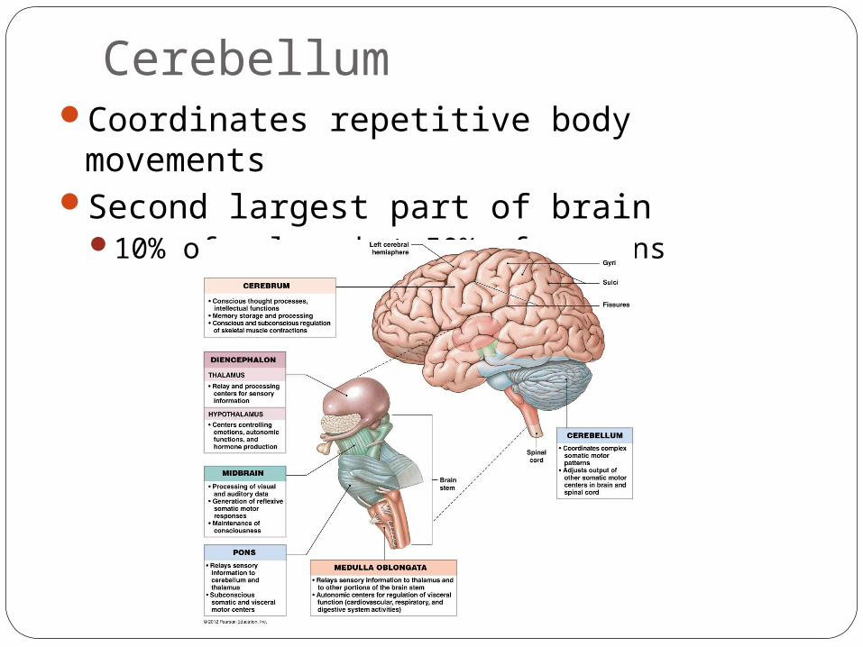
CerebellumCoordinates repetitive body movementsSecond largest part of brain
10% of volume but 50% of neurons
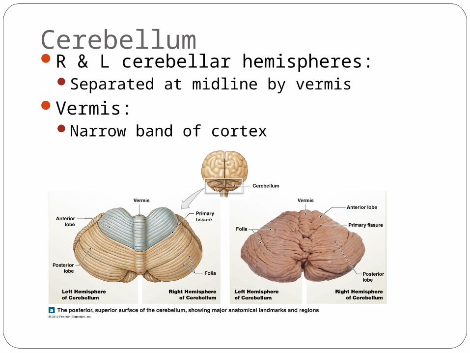
CerebellumR & L cerebellar hemispheres:
Separated at midline by vermisVermis:
Narrow band of cortex
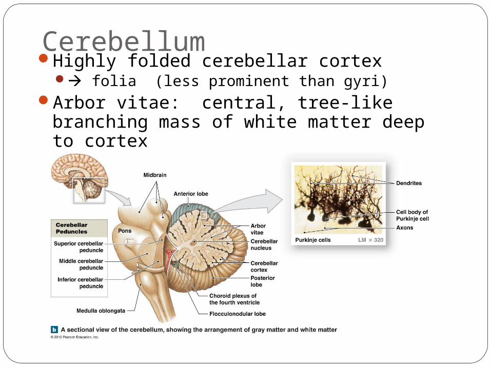
CerebellumHighly folded cerebellar cortex
folia (less prominent than gyri)Arbor vitae: central, tree-like branching
mass of white matter deep to cortex

DiencephalonLocated in central region of brain deep to
cerebrumLinks cerebrum with brain stem
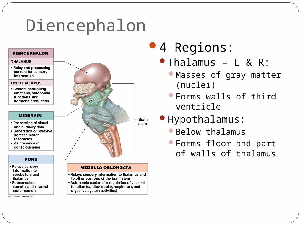
Diencephalon4 Regions:
Thalamus – L & R:Masses of gray matter
(nuclei)Forms walls of third
ventricleHypothalamus:
Below thalamusForms floor and part of
walls of thalamus
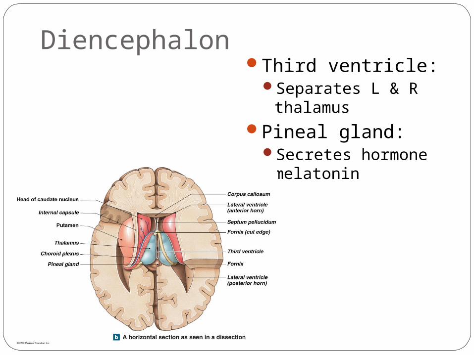
DiencephalonThird ventricle:
Separates L & R thalamus
Pineal gland:Secretes hormone
melatonin
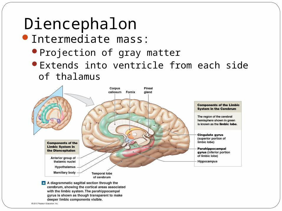
DiencephalonIntermediate mass:
Projection of gray matter Extends into ventricle from each side of
thalamus
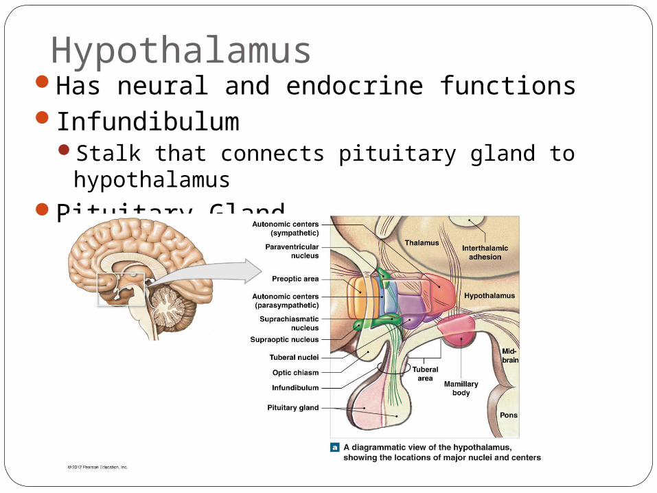
HypothalamusHas neural and endocrine functionsInfundibulum
Stalk that connects pituitary gland to hypothalamus
Pituitary Gland
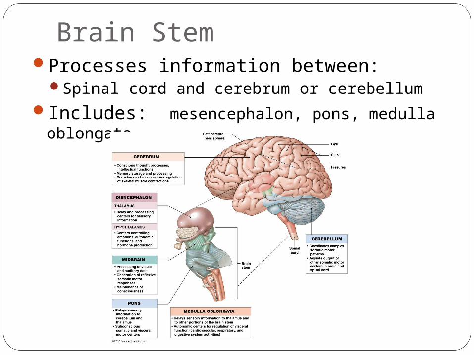
Brain StemProcesses information between:
Spinal cord and cerebrum or cerebellumIncludes: mesencephalon, pons, medulla
oblongata
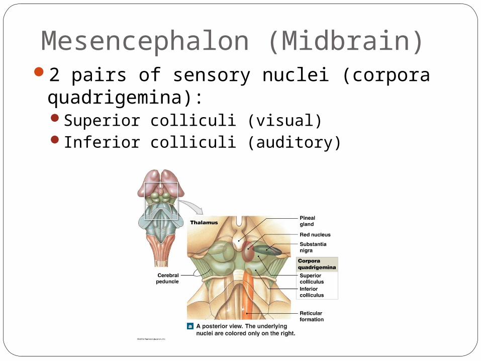
Mesencephalon (Midbrain)2 pairs of sensory nuclei (corpora
quadrigemina):Superior colliculi (visual)Inferior colliculi (auditory)
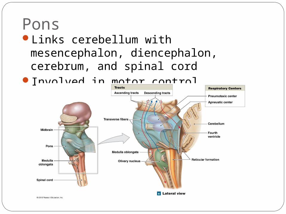
PonsLinks cerebellum with mesencephalon,
diencephalon, cerebrum, and spinal cordInvolved in motor control
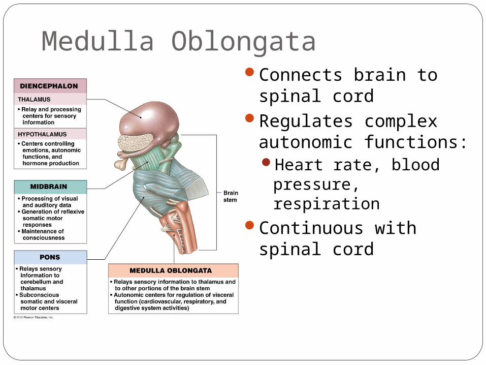
Medulla OblongataConnects brain to
spinal cordRegulates complex
autonomic functions: Heart rate, blood
pressure, respirationContinuous with spinal
cord