bio - zoology higher secondary - first year · 11/1/2019 · vertebrata and class amphibia. reasons...
TRANSCRIPT
Higher Secondary - First year
BIO - ZOOLOGY
PRACTICAL
www.Padasalai.Net
II
Bio-Zoology Practical - General Instruction
In order to get maximum benefit and good training it is necessary for the students to follow the following instructions.
1. The students must attend all practical classes. Each experiment in practicals has got important relevance to theory subjects.
2. Bring this practical manual to your practicals class.3. Bring the following objects to the practicals class – Pencils (HB), Pen,
Eraser, a scale and a small hand towel.4. Record the title, date and findings of the experiment in the observation
note book.5. Carefully listen to the instructions given by your Teacher.6. While observation slides or models draw the structure of the specimen
as you see it neatly in your observation note book. Use pencil for drawing.
7. While doing experiments neither consult your neighbours nor look into their readings or observations.
8. If the object under the microscope remains without proper focusing immediately bring it to the notice of the Teacher.
9. Do not touch or lift the models or equipments kept for your identification.
10. No need to draw diagrams from part III to VII in the record note. Relevant photograph can be collected, pasted and notes to be written.
www.Padasalai.Net
III
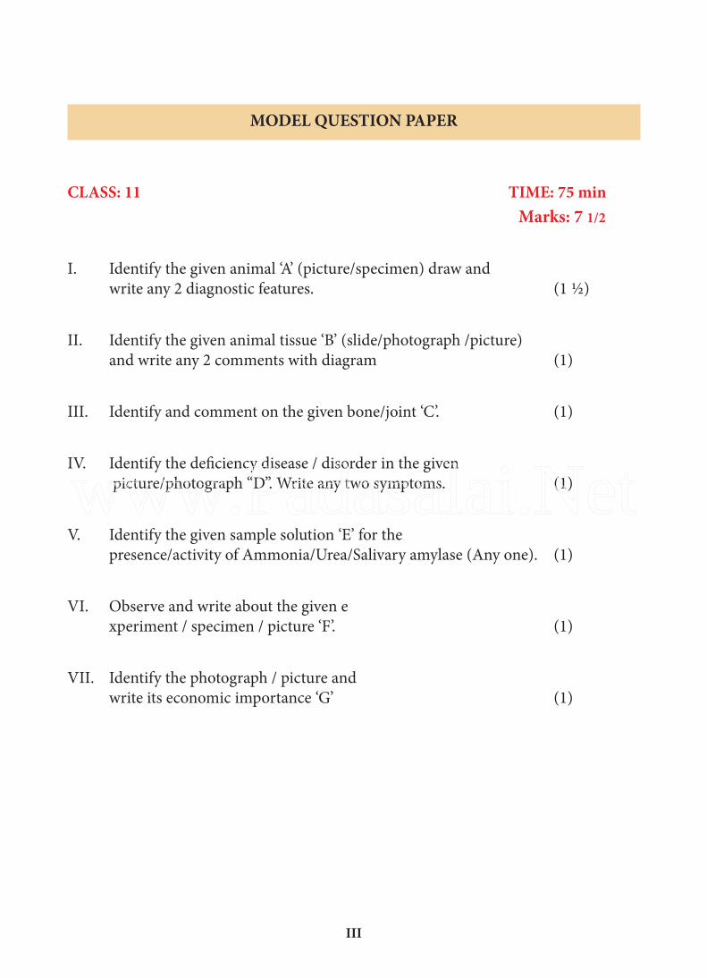
MODEL QUESTION PAPER
CLASS: 11 TIME: 75 min Marks: 7 1/2
I. Identify the given animal ‘A’ (picture/specimen) draw and write any 2 diagnostic features. (1 ½)
II. Identify the given animal tissue ‘B’ (slide/photograph /picture) and write any 2 comments with diagram (1)
III. Identify and comment on the given bone/joint ‘C’. (1)
IV. Identify the deficiency disease / disorder in the given picture/photograph “D”. Write any two symptoms. (1)
V. Identify the given sample solution ‘E’ for the presence/activity of Ammonia/Urea/Salivary amylase (Any one). (1)
VI. Observe and write about the given e xperiment / specimen / picture ‘F’. (1)
VII. Identify the photograph / picture and write its economic importance ‘G’ (1)
www.Padasalai.Net
IV
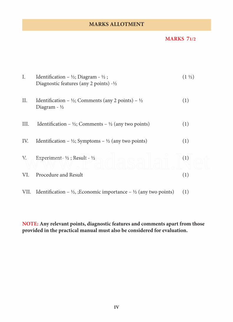
MARKS ALLOTMENT
MARKS 71/2
I. Identification – ½; Diagram - ½ ; (1 ½) Diagnostic features (any 2 points) -½
II. Identification – ½; Comments (any 2 points) – ½ (1) Diagram - ½
III. Identification – ½; Comments – ½ (any two points) (1)
IV. Identification – ½; Symptoms – ½ (any two points) (1)
V. Experiment- ½ ; Result - ½ (1)
VI. Procedure and Result (1)
VII. Identification – ½, ;Economic importance – ½ (any two points) (1)
NOTE: Any relevant points, diagnostic features and comments apart from those provided in the practical manual must also be considered for evaluation.
www.Padasalai.Net
V
QUESTION NO-I (A)
S.No List of Slides/Specimens Page No
1 Spongilla 1
2 Sea Anemone 1
3 Tapeworm 2
4 Ascaris 3
5 Earthworm 3
6 Cockroach 4
7 Pila 5
8 Starfish 5
9 Balanoglossus 6
10 Amphioxus 7
11 Ascidia 7
12 Shark 8
13 Seahorse 9
14 Frog 10
15 Calotes 10
16 Pigeon 11
17 Rat 12
QUESTION NO-II (B)
S.No List of Slides/Pictures/Photograph Page no
1 Squamous Epithelium 13
2 Columnar Epithelium 13
3 Cardiac Muscles 14
4 Smooth Muscles 14
5 Adipose Tissue 15
CONTENT
www.Padasalai.Net

VI
6 RBC 15
7 WBC 16
QUESTION NO-III (C)
S.No List of model/picture/photograph (Human) Page no
1 Humerus 17
2 Pectoral girdle 17
3 Pelvic girdle 18
4 Part of the Skull (Occipital, Frontal, Temporal, Parietal) 18
5 Rib cage (True ribs, Pseudo ribs, False ribs) 19
6 Ball and Socket joint 19
7 Pivot joint 20
QUESTION NO-IV (D)
S.No List of Slides/Pictures/Photograph Page no
1 Addison’s disease 21
2 Gigantism 21
3 Marasmus 22
4 Rickets 22
5 Exopthalmic Goitre 23
QUESTION NO-V (E)
S.No List of Experiments Page no
1 Test for Ammonia 24
2 Test for Urea 24
3 Test for Salivary Amylase 24
www.Padasalai.Net
VII
QUESTION NO-VI (F)
S.No List of Experiments Page no
1 Determine Your Eye Dominance 25
2 Determine Your Blind Spot 25
3 Identify the sex of cockroach (using hand lens) 26
4 Clitellum of earthworm (using hand lens) 26
QUESTION NO-VII (G)
S.No List of photograph/pictures Page no
1 Kangayam bull 27
2 Aquaponics 27
3 Honey bee 28
4 Bombyx mori 28
www.Padasalai.Net

VIII
www.Padasalai.Net
1
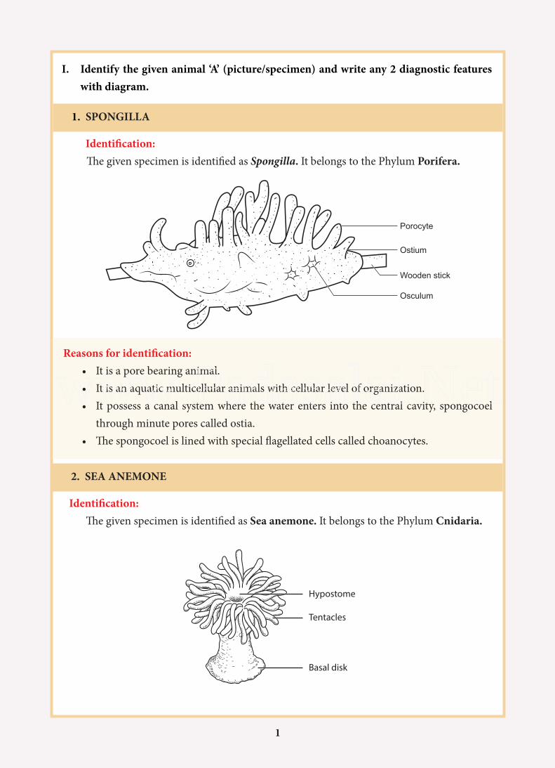
I. Identify the given animal ‘A’ (picture/specimen) and write any 2 diagnostic features with diagram.
1. SPONGILLA
Identification:The given specimen is identified as Spongilla. It belongs to the Phylum Porifera.
Reasons for identification: • It is a pore bearing animal.• It is an aquatic multicellular animals with cellular level of organization.• It possess a canal system where the water enters into the central cavity, spongocoel
through minute pores called ostia.• The spongocoel is lined with special flagellated cells called choanocytes.
2. SEA ANEMONE
Identification:The given specimen is identified as Sea anemone. It belongs to the Phylum Cnidaria.
Porocyte
Ostium
Osculum
Wooden stick
Hypostome
Tentacles
Basal disk
www.Padasalai.Net
2
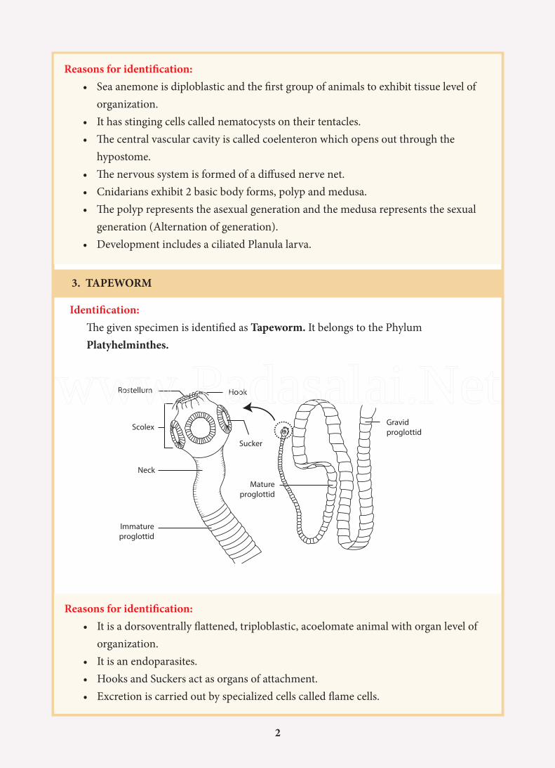
Reasons for identification:• Sea anemone is diploblastic and the first group of animals to exhibit tissue level of
organization.• It has stinging cells called nematocysts on their tentacles.• The central vascular cavity is called coelenteron which opens out through the
hypostome.• The nervous system is formed of a diffused nerve net.• Cnidarians exhibit 2 basic body forms, polyp and medusa.• The polyp represents the asexual generation and the medusa represents the sexual
generation (Alternation of generation).• Development includes a ciliated Planula larva.
3. TAPEWORM
Identification:The given specimen is identified as Tapeworm. It belongs to the Phylum Platyhelminthes.
Reasons for identification:• It is a dorsoventrally flattened, triploblastic, acoelomate animal with organ level of
organization.• It is an endoparasites.• Hooks and Suckers act as organs of attachment.• Excretion is carried out by specialized cells called flame cells.
Rostellum Hook
Scolex
Sucker
Neck
Immatureproglottid
Matureproglottid
Gravidproglottid
www.Padasalai.Net
3
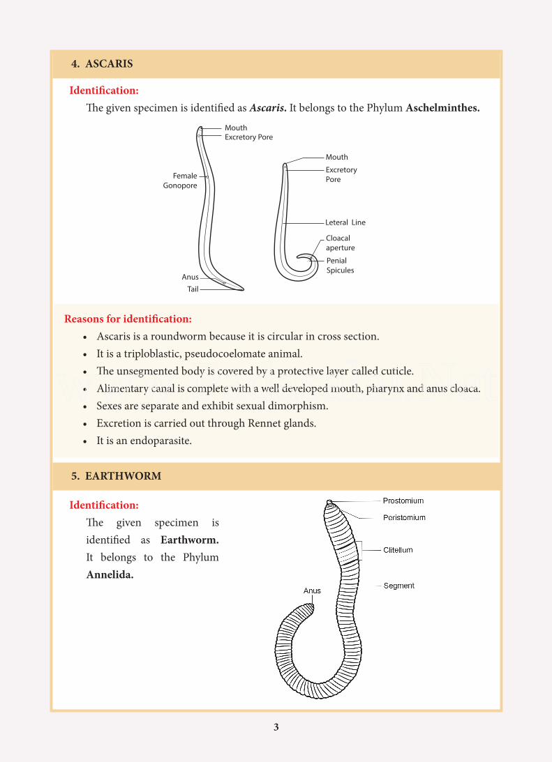
4. ASCARIS
Identification:The given specimen is identified as Ascaris. It belongs to the Phylum Aschelminthes.
Reasons for identification:• Ascaris is a roundworm because it is circular in cross section.• It is a triploblastic, pseudocoelomate animal.• The unsegmented body is covered by a protective layer called cuticle.• Alimentary canal is complete with a well developed mouth, pharynx and anus cloaca.• Sexes are separate and exhibit sexual dimorphism.• Excretion is carried out through Rennet glands.• It is an endoparasite.
5. EARTHWORM
Identification:The given specimen is identified as Earthworm. It belongs to the Phylum Annelida.
Mouth
Mouth
Excretory Pore
ExcretoryPore
Leteral Line
Cloacalaperture
PenialSpicules
Tail
Anus
FemaleGonopore
www.Padasalai.Net
4
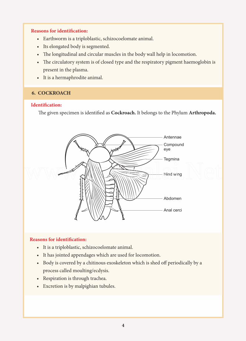
Reasons for identification: • Earthworm is a triploblastic, schizocoelomate animal.• Its elongated body is segmented.• The longitudinal and circular muscles in the body wall help in locomotion.• The circulatory system is of closed type and the respiratory pigment haemoglobin is
present in the plasma.• It is a hermaphrodite animal.
6. COCKROACH
Identification:The given specimen is identified as Cockroach. It belongs to the Phylum Arthropoda.
Reasons for identification:• It is a triploblastic, schizocoelomate animal.• It has jointed appendages which are used for locomotion.• Body is covered by a chitinous exoskeleton which is shed off periodically by a
process called moulting/ecdysis.• Respiration is through trachea.• Excretion is by malpighian tubules.
Antennae
Compoundeye
Tegmina
Hind wing
Abdomen
Anal cerci
www.Padasalai.Net
5
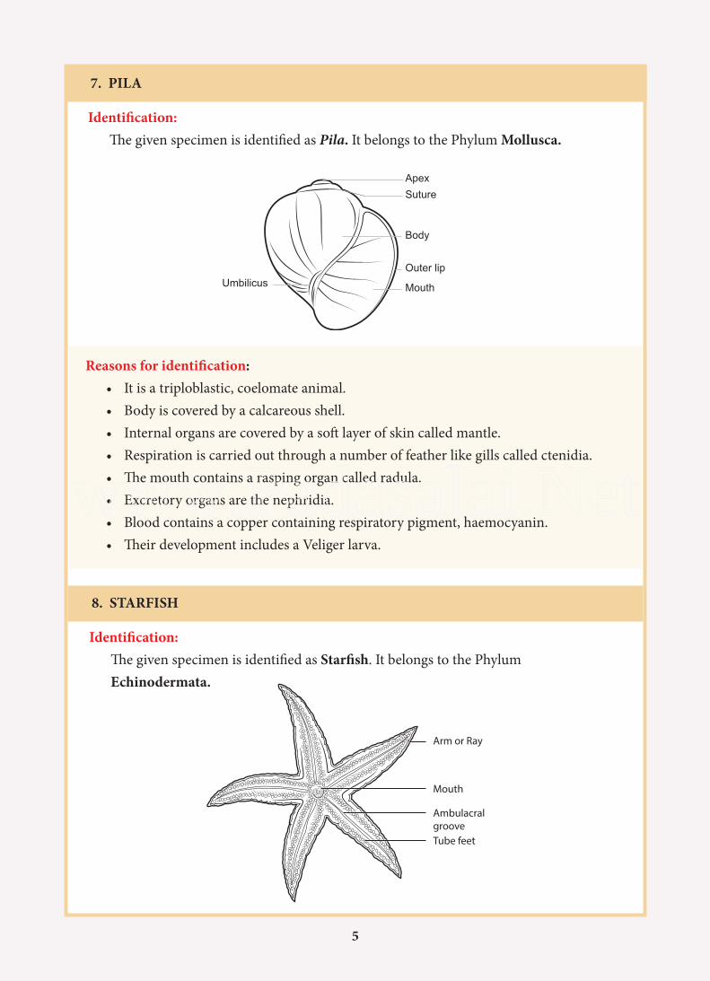
7. PILA
Identification:The given specimen is identified as Pila. It belongs to the Phylum Mollusca.
Reasons for identification:• It is a triploblastic, coelomate animal.• Body is covered by a calcareous shell.• Internal organs are covered by a soft layer of skin called mantle.• Respiration is carried out through a number of feather like gills called ctenidia.• The mouth contains a rasping organ called radula.• Excretory organs are the nephridia.• Blood contains a copper containing respiratory pigment, haemocyanin.• Their development includes a Veliger larva.
ApexSuture
Body
Outer lip
MouthUmbilicus
8. STARFISH
Identification:The given specimen is identified as Starfish. It belongs to the Phylum Echinodermata.
Arm or Ray
Mouth
AmbulacralgrooveTube feet
www.Padasalai.Net
6
9. BALANOGLOSSUS
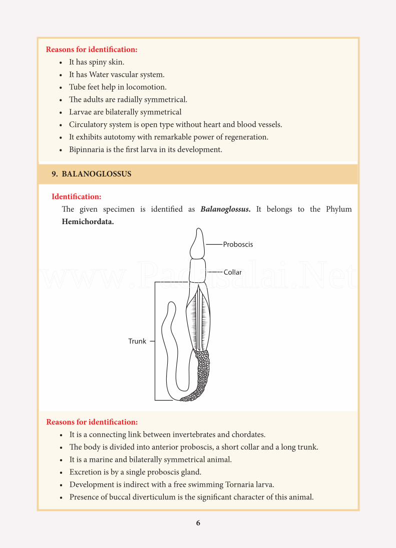
Reasons for identification:• It has spiny skin.• It has Water vascular system.• Tube feet help in locomotion.• The adults are radially symmetrical.• Larvae are bilaterally symmetrical• Circulatory system is open type without heart and blood vessels.• It exhibits autotomy with remarkable power of regeneration.• Bipinnaria is the first larva in its development.
Identification: The given specimen is identified as Balanoglossus. It belongs to the Phylum Hemichordata.
Reasons for identification:• It is a connecting link between invertebrates and chordates.• The body is divided into anterior proboscis, a short collar and a long trunk.• It is a marine and bilaterally symmetrical animal.• Excretion is by a single proboscis gland.• Development is indirect with a free swimming Tornaria larva.• Presence of buccal diverticulum is the significant character of this animal.
Proboscis
Collar
Trunk
www.Padasalai.Net
7
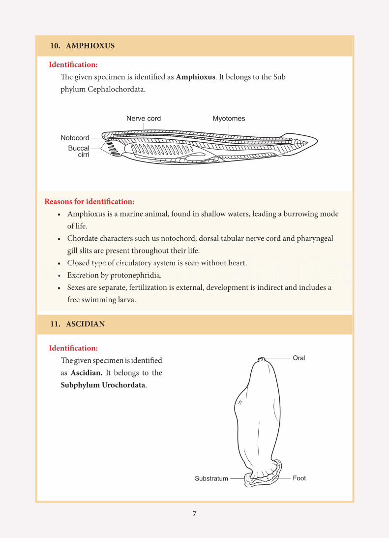
10. AMPHIOXUS
Identification:The given specimen is identified as Amphioxus. It belongs to the Sub phylum Cephalochordata.
Reasons for identification:• Amphioxus is a marine animal, found in shallow waters, leading a burrowing mode
of life.• Chordate characters such us notochord, dorsal tabular nerve cord and pharyngeal
gill slits are present throughout their life.• Closed type of circulatory system is seen without heart.• Excretion by protonephridia.• Sexes are separate, fertilization is external, development is indirect and includes a
free swimming larva.
Notocord
Nerve cord Myotomes
Buccalcirri
11. ASCIDIAN
Oral
FootSubstratum
Identification:The given specimen is identified as Ascidian. It belongs to the Subphylum Urochordata.
www.Padasalai.Net
8
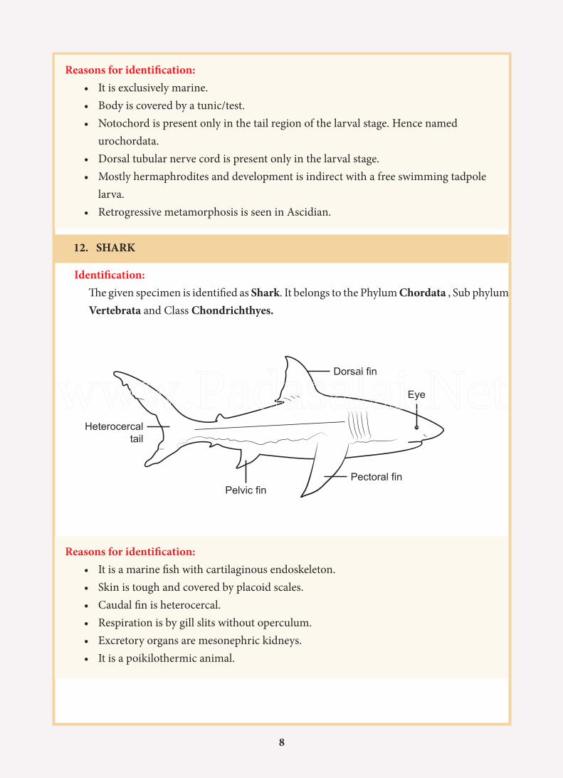
Reasons for identification:• It is exclusively marine.• Body is covered by a tunic/test.• Notochord is present only in the tail region of the larval stage. Hence named
urochordata.• Dorsal tubular nerve cord is present only in the larval stage.• Mostly hermaphrodites and development is indirect with a free swimming tadpole
larva.• Retrogressive metamorphosis is seen in Ascidian.
12. SHARK
Identification: The given specimen is identified as Shark. It belongs to the Phylum Chordata , Sub phylum Vertebrata and Class Chondrichthyes.
Dorsal fin
Pectoral finPelvic fin
Heterocercaltail
Eye
Reasons for identification:• It is a marine fish with cartilaginous endoskeleton.• Skin is tough and covered by placoid scales.• Caudal fin is heterocercal.• Respiration is by gill slits without operculum.• Excretory organs are mesonephric kidneys.• It is a poikilothermic animal.
www.Padasalai.Net
9
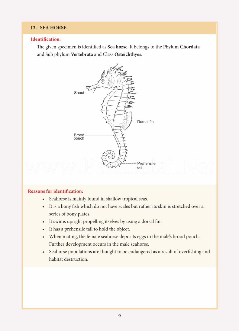
13. SEA HORSE
Identification: The given specimen is identified as Sea horse. It belongs to the Phylum Chordata and Sub phylum Vertebrata and Class Osteichthyes.
Reasons for identification:• Seahorse is mainly found in shallow tropical seas.• It is a bony fish which do not have scales but rather its skin is stretched over a
series of bony plates.• It swims upright propelling itselves by using a dorsal fin.• It has a prehensile tail to hold the object.• When mating, the female seahorse deposits eggs in the male’s brood pouch.
Further development occurs in the male seahorse.• Seahorse populations are thought to be endangered as a result of overfishing and
habitat destruction.
Snout
Broodpouch
Prehensiletail
Dorsal fin
www.Padasalai.Net
10
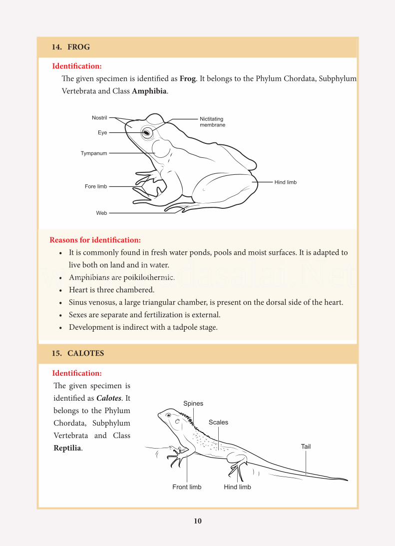
14. FROG
Identification: The given specimen is identified as Frog. It belongs to the Phylum Chordata, Subphylum
Vertebrata and Class Amphibia.
Reasons for identification:• It is commonly found in fresh water ponds, pools and moist surfaces. It is adapted to
live both on land and in water.• Amphibians are poikilothermic.• Heart is three chambered.• Sinus venosus, a large triangular chamber, is present on the dorsal side of the heart.• Sexes are separate and fertilization is external.• Development is indirect with a tadpole stage.
Nostril
Eye
Tympanum
Fore limbHind limb
Nictitatingmembrane
Web
15. CALOTES
Spines
Scales
Tail
Hind limbFront limb
Identification: The given specimen is
identified as Calotes. It belongs to the Phylum Chordata, Subphylum Vertebrata and Class Reptilia.
www.Padasalai.Net
11
Reasons for identification:• It is a terrestrial, poikilothermic animal.• Body is covered with dry horny scales.• Heart is 3 chambered.• Excretion is by metanephric kidneys and is uricotelic.• It is monoecious.• Fertilization is internal.• It is oviparous and lays cledoic eggs.
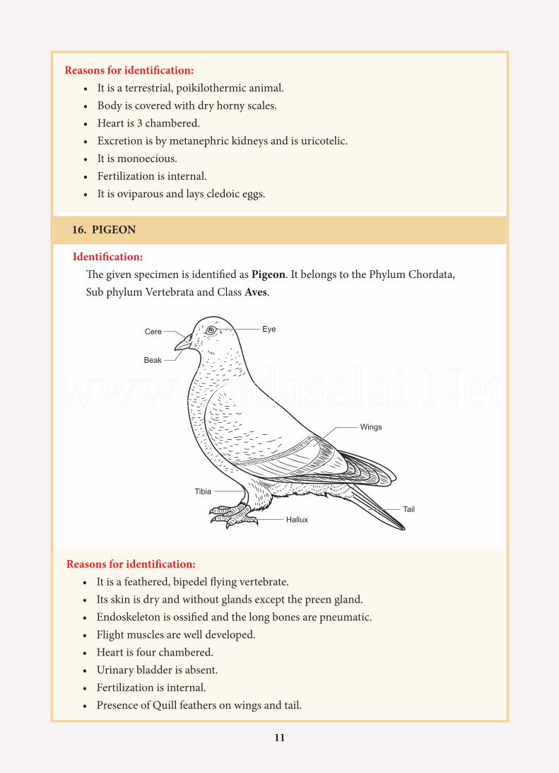
16. PIGEON
Identification: The given specimen is identified as Pigeon. It belongs to the Phylum Chordata, Sub phylum Vertebrata and Class Aves.
Reasons for identification:• It is a feathered, bipedel flying vertebrate.• Its skin is dry and without glands except the preen gland.• Endoskeleton is ossified and the long bones are pneumatic.• Flight muscles are well developed.• Heart is four chambered.• Urinary bladder is absent.• Fertilization is internal.• Presence of Quill feathers on wings and tail.
Eye
TailHallux
Tibia
Beak
Cere
Wings
www.Padasalai.Net
12
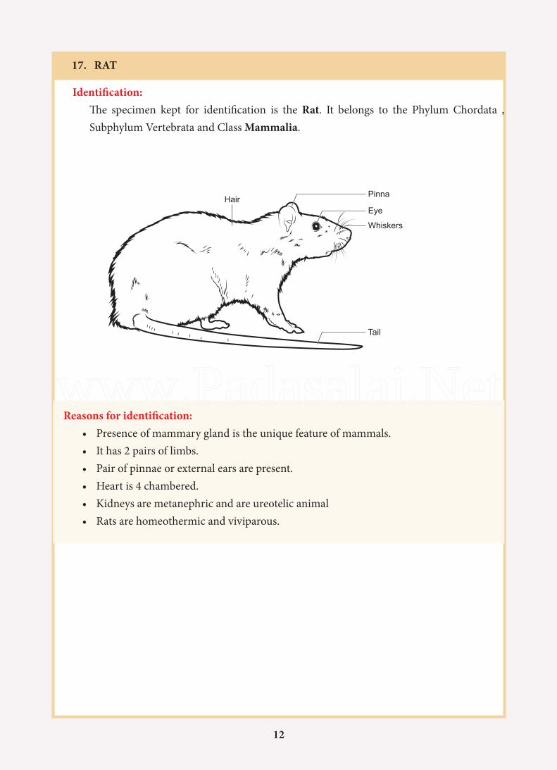
17. RAT
Identification: The specimen kept for identification is the Rat. It belongs to the Phylum Chordata , Subphylum Vertebrata and Class Mammalia.
Pinna
Eye
Tail
Whiskers
Hair
Reasons for identification:• Presence of mammary gland is the unique feature of mammals.• It has 2 pairs of limbs.• Pair of pinnae or external ears are present.• Heart is 4 chambered.• Kidneys are metanephric and are ureotelic animal• Rats are homeothermic and viviparous.
www.Padasalai.Net
13
Cell membrane
Nucleus
Basement
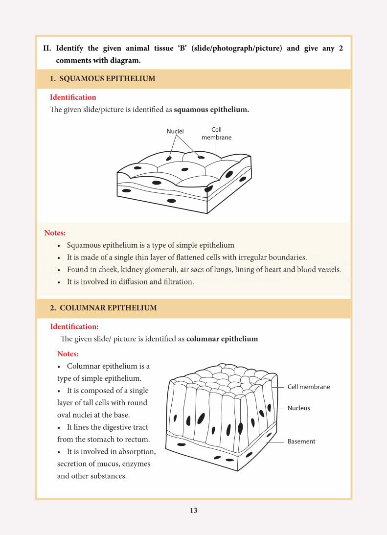
1. SQUAMOUS EPITHELIUM
Identification The given slide/picture is identified as squamous epithelium.
Notes:• Squamous epithelium is a type of simple epithelium• It is made of a single thin layer of flattened cells with irregular boundaries.• Found in cheek, kidney glomeruli, air sacs of lungs, lining of heart and blood vessels.• It is involved in diffusion and filtration.
II. Identify the given animal tissue ‘B’ (slide/photograph/picture) and give any 2 comments with diagram.
Nuclei Cellmembrane
2. COLUMNAR EPITHELIUM
Identification: The given slide/ picture is identified as columnar epithelium
Notes:• Columnar epithelium is a type of simple epithelium.• It is composed of a single layer of tall cells with round oval nuclei at the base.• It lines the digestive tract from the stomach to rectum.• It is involved in absorption, secretion of mucus, enzymes and other substances.
www.Padasalai.Net
14
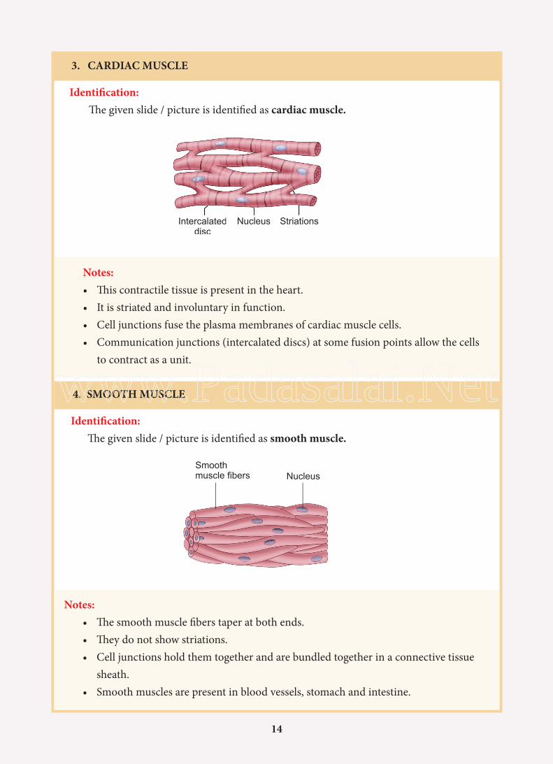
3. CARDIAC MUSCLE
Identification: The given slide / picture is identified as cardiac muscle.
Notes:• This contractile tissue is present in the heart.• It is striated and involuntary in function.• Cell junctions fuse the plasma membranes of cardiac muscle cells.• Communication junctions (intercalated discs) at some fusion points allow the cells
to contract as a unit.
4. SMOOTH MUSCLE
Identification:The given slide / picture is identified as smooth muscle.
Notes:• The smooth muscle fibers taper at both ends.• They do not show striations.• Cell junctions hold them together and are bundled together in a connective tissue
sheath.• Smooth muscles are present in blood vessels, stomach and intestine.
NucleusSmoothmuscle fibers
www.Padasalai.Net
15
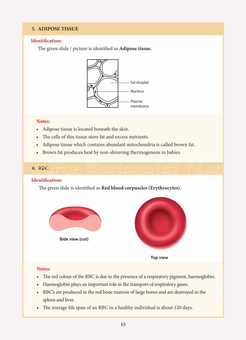
5. ADIPOSE TISSUE
Identification: The given slide / picture is identified as Adipose tissue.
Notes:• Adipose tissue is located beneath the skin.• The cells of this tissue store fat and excess nutrients.• Adipose tissue which contains abundant mitochondria is called brown fat.• Brown fat produces heat by non-shivering thermogenesis in babies.
6. RBC
Identification:The given slide is identified as Red blood corpuscles (Erythrocytes).
Fat droplet
Nucleus
Plasmamembrane
Notes:• The red colour of the RBC is due to the presence of a respiratory pigment, haemoglobin.• Haemoglobin plays an important role in the transport of respiratory gases.• RBC’s are produced in the red bone marrow of large bones and are destroyed in the
spleen and liver.• The average life span of an RBC in a healthy individual is about 120 days.
www.Padasalai.Net

16
7. WBC
Identification:The given slide is identified as white blood corpuscles (leucocytes).
Notes:• Leucocytes are colourless, amoeboid, nucleated cells devoid of haemoglobin and
other pigments.• Based on the presence (or) absence of granules, WBC’s are divided into two types,
granulocytes (Neutrophil, Basophil and Eosinophil) and agranulocytes (Lymphocyte and Monocyte).
• WBCs are involved in protecting the body against pathogens.• The life span of a white blood cell ranges from 13 to 20 days. These are destroyed in
the lymphatic system.
Eosinophils Basophils Neutrophils
Monocytes Lymphocytes
Macrophages Plasma cells
www.Padasalai.Net
17
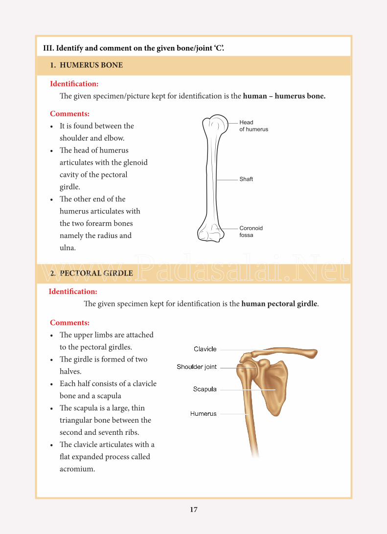
Headof humerus
Shaft
Coronoidfossa
2. PECTORAL GIRDLE
Identification: The given specimen kept for identification is the human pectoral girdle.
1. HUMERUS BONE
Identification: The given specimen/picture kept for identification is the human – humerus bone.
Comments:• The upper limbs are attached
to the pectoral girdles.• The girdle is formed of two
halves.• Each half consists of a clavicle
bone and a scapula• The scapula is a large, thin
triangular bone between the second and seventh ribs.
• The clavicle articulates with a flat expanded process called acromium.
III. Identify and comment on the given bone/joint ‘C’.
Comments:• It is found between the
shoulder and elbow.• The head of humerus
articulates with the glenoid cavity of the pectoral girdle.
• The other end of the humerus articulates with the two forearm bones namely the radius and ulna.
www.Padasalai.Net
18
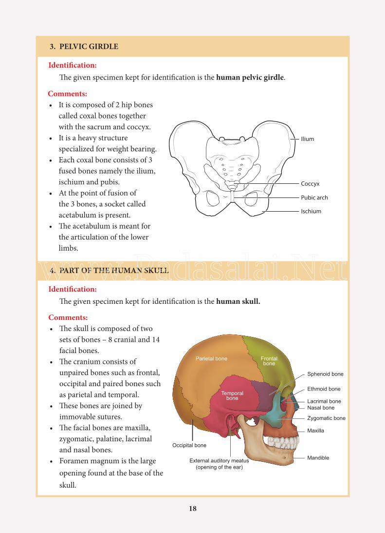
3. PELVIC GIRDLE
Identification: The given specimen kept for identification is the human pelvic girdle.
Comments:• It is composed of 2 hip bones
called coxal bones together with the sacrum and coccyx.
• It is a heavy structure specialized for weight bearing.
• Each coxal bone consists of 3 fused bones namely the ilium, ischium and pubis.
• At the point of fusion of the 3 bones, a socket called acetabulum is present.
• The acetabulum is meant for the articulation of the lower limbs.
4. PART OF THE HUMAN SKULL
Identification: The given specimen kept for identification is the human skull.
Comments:• The skull is composed of two
sets of bones – 8 cranial and 14 facial bones.
• The cranium consists of unpaired bones such as frontal, occipital and paired bones such as parietal and temporal.
• These bones are joined by immovable sutures.
• The facial bones are maxilla, zygomatic, palatine, lacrimal and nasal bones.
• Foramen magnum is the large opening found at the base of the skull.
Ilium
Coccyx
Pubic arch
Ischium
External auditory meatus(opening of the ear)
Parietal bone Frontalbone
Temporalbone
Sphenoid bone
Ethmoid bone
Lacrimal bone
Zygomatic bone
Maxilla
Mandible
Occipital bone
Nasal bone
www.Padasalai.Net

19
5. RIB CAGE
Identification: The given specimen kept for identification is the human ribcage.
Comments:• There are 12 pairs of ribs.• Each rib is connected dorsally to the vertebral column and ventrally to the sternum.• The first 7 pairs of ribs are called true ribs.• The 8th, 9th and 10th pairs of ribs do not articulate with the sternum but is joined with the 7th rib. They are called as false ribs.• The last 11th and 12th pairs of ribs are not connected with sternum. They are called as floating ribs.
Sternum
True ribs
False ribs
Floting ribs
6. BALL AND SOCKET JOINT
Identification: The specimen/model/picture kept for identification is the Ball and Socket joint.
Comments:• It is a type of synovial joint.• In this type, the ball shaped
rounded bone fits into the cup like depression of another bone.
• It allows multi directional movements and rotation.
• This type of joints are found between the upper arm and shoulder and between the upper leg and hip.
Ball and
Socket joint
www.Padasalai.Net
20
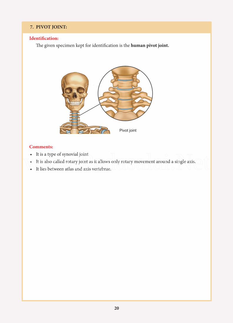
7. PIVOT JOINT:
Identification: The given specimen kept for identification is the human pivot joint.
Comments:• It is a type of synovial joint• It is also called rotary joint as it allows only rotary movement around a single axis.• It lies between atlas and axis vertebrae.
Pivot joint
www.Padasalai.Net
21
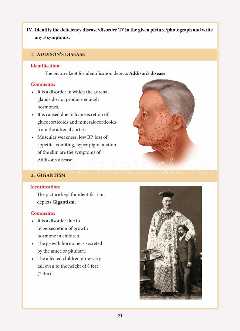
1. ADDISON’S DISEASE
Identification: The picture kept for identification depicts Addison’s disease.
Comments:• It is a disorder in which the adrenal
glands do not produce enough hormones.
• It is caused due to hyposecretion of glucocorticoids and mineralocorticoids from the adrenal cortex.
• Muscular weakness, low BP, loss of appetite, vomiting, hyper pigmentation of the skin are the symptoms of Addison’s disease.
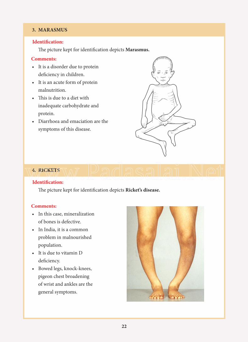
2. GIGANTISM
Identification: The picture kept for identification depicts Gigantism.
Comments:• It is a disorder due to
hypersecretion of growth hormone in children.
• The growth hormone is secreted by the anterior pituitary.
• The affected children grow very tall even to the height of 8 feet (2.4m).
IV. Identify the deficiency disease/disorder ‘D’ in the given picture/photograph and write any 3 symptoms.
www.Padasalai.Net
22
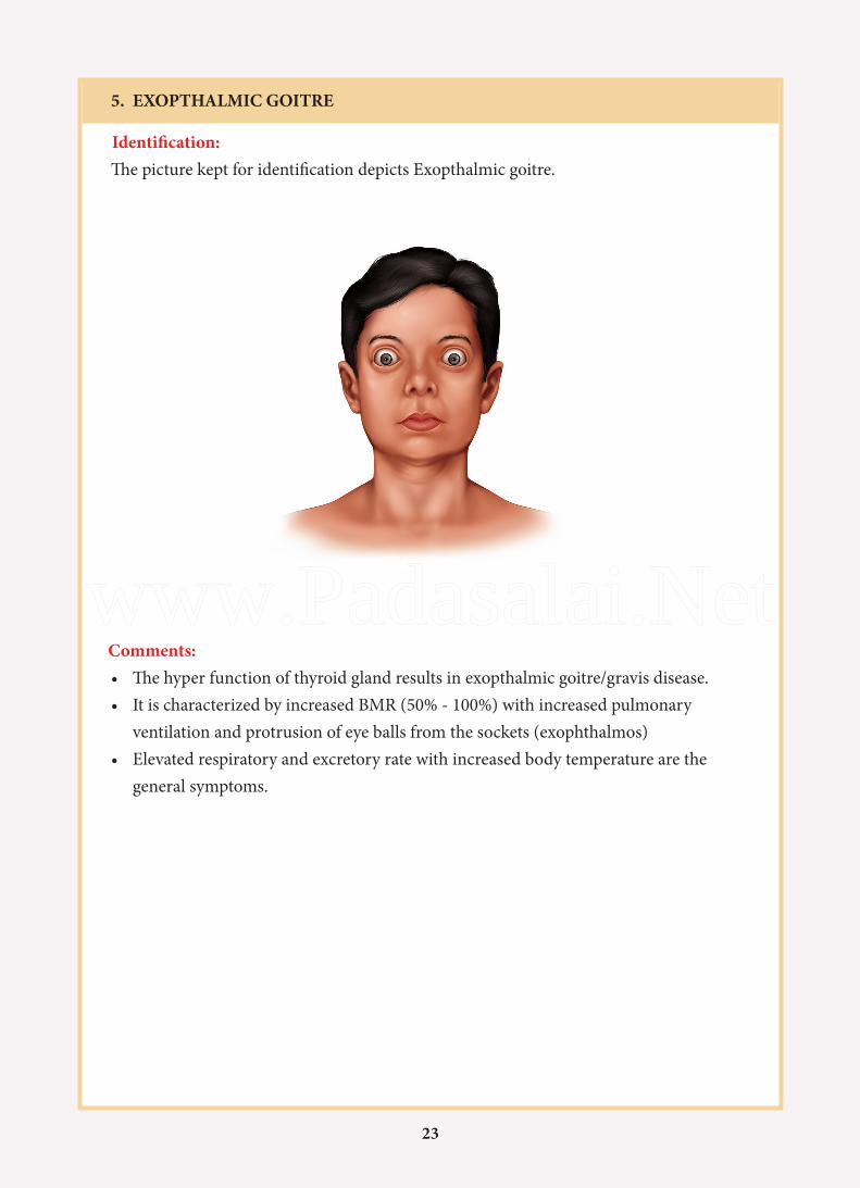
3. MARASMUS
Identification: The picture kept for identification depicts Marasmus.
4. RICKETS
Identification: The picture kept for identification depicts Ricket’s disease.
Comments:• It is a disorder due to protein
deficiency in children.• It is an acute form of protein
malnutrition.• This is due to a diet with
inadequate carbohydrate and protein.
• Diarrhoea and emaciation are the symptoms of this disease.
Comments:• In this case, mineralization
of bones is defective.• In India, it is a common
problem in malnourished population.
• It is due to vitamin D deficiency.
• Bowed legs, knock-knees, pigeon chest broadening of wrist and ankles are the general symptoms.
www.Padasalai.Net
23
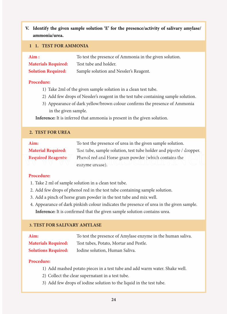
5. EXOPTHALMIC GOITRE
Identification: The picture kept for identification depicts Exopthalmic goitre.
Comments:• The hyper function of thyroid gland results in exopthalmic goitre/gravis disease.• It is characterized by increased BMR (50% - 100%) with increased pulmonary
ventilation and protrusion of eye balls from the sockets (exophthalmos)• Elevated respiratory and excretory rate with increased body temperature are the
general symptoms.
www.Padasalai.Net
24
1 1. TEST FOR AMMONIA
Aim : To test the presence of Ammonia in the given solution.Materials Required: Test tube and holder.Solution Required: Sample solution and Nessler’s Reagent.
Procedure:1) Take 2ml of the given sample solution in a clean test tube.2) Add few drops of Nessler’s reagent in the test tube containing sample solution.3) Appearance of dark yellow/brown colour confirms the presence of Ammonia
in the given sample.Inference: It is inferred that ammonia is present in the given solution.
2. TEST FOR UREA
Aim: To test the presence of urea in the given sample solution.Material Required: Test tube, sample solution, test tube holder and pipette / dropper.Required Reagents: Phenol red and Horse gram powder (which contains the
enzyme urease).
Procedure: 1. Take 2 ml of sample solution in a clean test tube. 2. Add few drops of phenol red in the test tube containing sample solution.3. Add a pinch of horse gram powder in the test tube and mix well.4. Appearance of dark pinkish colour indicates the presence of urea in the given sample.
Inference: It is confirmed that the given sample solution contains urea.
3. TEST FOR SALIVARY AMYLASE
Aim: To test the presence of Amylase enzyme in the human saliva.Materials Required: Test tubes, Potato, Mortar and Pestle.Solutions Required: Iodine solution, Human Saliva.
Procedure:1) Add mashed potato pieces in a test tube and add warm water. Shake well.2) Collect the clear supernatant in a test tube.3) Add few drops of iodine solution to the liquid in the test tube.
V. Identify the given sample solution ‘E’ for the presence/activity of salivary amylase/ammonia/urea.
24
www.Padasalai.Net
25
4) Note the bluish black (dark blue) colour in the test tube.5) Collect a few drops of saliva in a clean test tube.6) Transfer the saliva into the test tube containing the sample solution and
shake well.7) Leave the sample undisturbed for 5 minutes. Observe the colour change in the
sample solution.8) The solution gradually becomes colourless.9) This confirms the presence of amylase in the human saliva.
Inference: It is inferred that human saliva contains the enzyme amylase that digests the starch.
1. DETERMINE EYE DOMINANCE:
We’re all familiar with preferences for using a particular hand for jobs such as writing and throwing. Eye dominance is important for how we see and react to the world around us.
Procedure:
1. With both eyes open carefully focus on an object a few feet away.2. Close one eye, and then reopen it.3. Close the other eye, and then reopen it. Which eye seems more directly in line with the
object?a. If it is the right eye, you are right eye dominant.b. If it is the left eye, you are left eye dominant.c. If it is the middle of both eyes, you are central eye dominant.
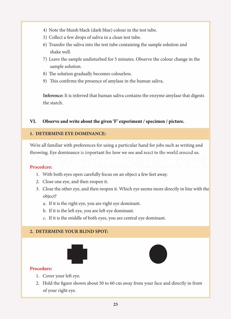
2. DETERMINE YOUR BLIND SPOT:
VI. Observe and write about the given ‘F’ experiment / specimen / picture.
Procedure:1. Cover your left eye.2. Hold the figure shown about 50 to 60 cm away from your face and directly in front
of your right eye.
25
www.Padasalai.Net
26
3. Stare at the cross in the shown figure. You can also see the circle.4. Continue to stare and slowly bring the figure nearer to your eye.5. Note the point at which the circle will seem to disappear. This is your blind
spot.6. Record the distance.7. Test your other eye in a similar manner, but focus on the circle and watch for
the cross to disappear.Result:
1) Blind spot of my right eye is ________cm2) Blind spot of my left eye is___________cm
3. Identify the sex of the cockroach by observing the given specimen/picture /model and write two reasons.
Identification :
Reasons:
4. Identify the part marked in the given specimen / picture of the earthworm and write its significance.
Identification (Part) :
Reasons:
26
www.Padasalai.Net
27
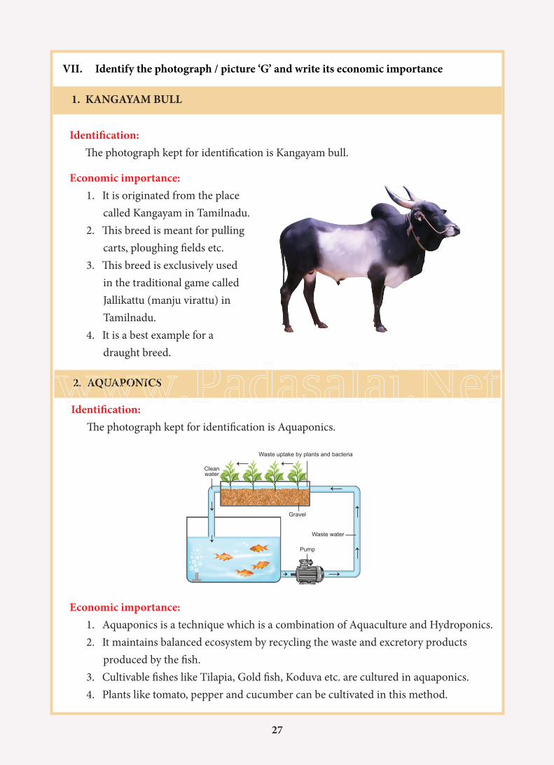
1. KANGAYAM BULL
VII. Identify the photograph / picture ‘G’ and write its economic importance
Identification: The photograph kept for identification is Kangayam bull.
Economic importance:1. It is originated from the place
called Kangayam in Tamilnadu.2. This breed is meant for pulling
carts, ploughing fields etc.3. This breed is exclusively used
in the traditional game called Jallikattu (manju virattu) in Tamilnadu.
4. It is a best example for a draught breed.
2. AQUAPONICS
Identification: The photograph kept for identification is Aquaponics.
Economic importance:1. Aquaponics is a technique which is a combination of Aquaculture and Hydroponics.2. It maintains balanced ecosystem by recycling the waste and excretory products
produced by the fish.3. Cultivable fishes like Tilapia, Gold fish, Koduva etc. are cultured in aquaponics.4. Plants like tomato, pepper and cucumber can be cultivated in this method.
Cleanwater
Gravel
Waste water
Pump
Waste uptake by plants and bacteria
27
www.Padasalai.Net

28
3. HONEY BEE
Identification: The photograph kept for identification is Honey bee.
Economic importance:1. The chief products of bee
keeping industry are honey and bee wax.
2. Honey is the healthier substitute for sugar.
3. It is used as an antiseptic, laxative and as a sedative.
4. Bee wax secreted by the abdomen of the worker bee is used for making candles, polishes for floors and furniture etc.
4. BOMBYX MORI
Identification: The photograph kept for identification is silkworm Bombyx mori
Economic importance:1. Silk fibre produced by this
silkworm is called mulberry silk.
2. It mainly feeds on mulberry leaves
3. It is used in manufacturing silk cloths, fishing fibres, tyres of racing cars, in medical dressings, parachutes etc.
4. It is exclusively cultivated in the states of Karnataka, Andra Pradesh and Tamilnadu.
Queen bee
Worker bee
Drone bee
28
www.Padasalai.Net
29
CHAIR PERSONS AND ADVISORY COMMITTEE:
Dr. SULTAN AHMED ISMAIL Scientist, Eco-science Research Foundation, Chennai.
Dr. P.K. KALEENA Associate Professor, Department of Zoology, Presidency College, Chennai.
SUBJECT EXPERT AND COORDINATOR:
Dr. S. SHAMEEM Deputy Director, SCERT, Chennai.
CONTENT WRITERS:
Mr. M. MAYILSAMYSenior Lecturer, DIET,Krishnagiri District.
Dr. S. GANESAPANDIANHead Master,Govt. Higher secondary School,Kadukkai valasai, Ramanadapuram District.
Zoology Practical - CLASS: XI
Mr. V. RAJENDRANP.G Assistant in Zoology,Govt. Higher Secondary School,Aragalur, Salem District.
Mrs. M. ANUSUA CATHERINA CHELLIAHPG Assistant in Zoology,Presidency GHSS, Egmore.
Mr. J.M. BRITTO FELICIOUSPG Assistant in Zoology,KLK GBHSS,Gummudipoondi,Thiruvallur District.
Mr. N. SENTHIL KUMARPG Assistant in Zoology,Govt Boys Higher Secondary School,Thalaivasal, Salem District.
Mrs. R.SURAMANJARIPG Assistant in Zoology,GRT.M.V Higher Secondary School,Ashok Nagar,Chennai – 83
COORDINATORSDr. V.T.SHANTHISeniour Lecturer,DIET,Tirur
Mrs. B.SELVILecturer, SCERT, Chennai.
LAYOUT AND ILLUSTRATIONS
Gopu RasuvelPrabhaVinod
29
www.Padasalai.Net

30
www.Padasalai.Net