bioactive glass coatings - materials science · bioactive glass coatings ... terials is usually...
TRANSCRIPT

Materials Science-Poland, Vol. 23, No. 1, 2005
Bioactive glass coatings
RAFAŁ SINDUT1, KATARZYNA CHOLEWA-KOWALSKA
1, JELENA NAJMAN2,
MARIA ŁĄCZKA1*, MARIA KARPOV
3, ANNA MARIA OSYCZKA3, PHOEBE LEBOY
3
1AGH-University of Science and Technology, Faculty of Materials Science and Ceramics, Department of Glass Technology and Amorphous Coating, 30-059 Cracow, Poland
2Cracow University of Technology, Faculty of Chemical Engineering and Technology, 31-155 Cracow, Poland
3University of Pennsylvania, Philadelphia, 19104 PA, USA
Four kinds of gel-derived materials of the CaO–P2O5–SiO2 (S2, II, I, A2) system were obtained in the form of thin coatings on microscope slides. The obtained materials differed from each other in the ratio of the basic components (CaO and SiO2). The coatings were characterised with regard to the state of the surface as well as to the phase composition of the materials. In order to determine any bioactive proper-ties of the gel-derived coatings in vitro, tests in simulated body fluid (SBF) were made and biochemical examinations using cultured human marrow stromal cells (hMSC) were conducted. It was found that surface crystallisation of hydroxyapatite (HAp) indicating the bioactivity of the material occurred in SBF only in the case of A2 coatings, which are characterised by the highest ratio of CaO:SiO2. Tests with hMSC showed that the A2 biomaterial promotes both the osteogenesis and remodelling of bone (osteo-clastogenesis).
Key words: biomaterials; bioactivity; sol-gel method
1. Introduction
Bioactive ceramic materials are based on the CaO–P2O5–SiO2 system [1, 2]. They comprise both glasses and glass-ceramic materials with hydroxyapatite and wollaston-ite as the main crystal phases. They are assumed to satisfy the following requirements:
• biocompatibility, • suitability for clinical applications, • ability to form a stable connection with the bone as well as to promote bone re-
generation.
_________ * Corresponding author, e-mail: [email protected].

R. SINDUT et al. 124
When in contact with body fluid, these materials form layers of hydroxyapatite (HAp) on their surfaces, through which the implant materials form permanent bonds with the bone in a living organism. The ability to form HAp on the surface of bioma-terials is usually indicated by results of simulated body fluid (SBF) tests, which allow in vitro character of changes on the biomaterial surface to be estimated after contact with SBF [3]; biomaterials are preliminarily defined by this basic bioactive property.
In recent years, great interest has arisen for a new generation of bioactive materi-als with increased bioactivity, interpreted mainly as the ability to stimulate faster re-generation of natural tissues [4, 5]. In order to obtain such materials, the sol-gel method is used, which enables biomaterials with a high degree of both chemical and biological surface activities to be produced [6]. This method allows the production of biomaterials in the form of powders and granules, dense and porous sinters, thin coat-ings, on bioinert substrates. So far, however, it has not been fully recognized which material parameters, such as the state of the surface, pore structure, chemical and phase composition, etc. affect the bioactive properties of these biomaterials. The lack of this knowledge does not permit fully controlled production processes to be carried out.
The aim of this study was to obtain bioactive gel-derived coatings with various characteristics (chemical properties, phase composition, surface roughness) and to evaluate their bioactive properties under in vitro conditions from the point of view of material parameters.
2. Materials and methods
Biomaterials from the system CaO–P2O5–SiO2 have been chosen for the investiga-tions. Their chemical compositions are given in Table 1.
Table 1. The chemical composition of the investigated materials
Symbol Chemical composition S2 II I A2
SiO2 80 72 64 40 CaO 16 24 30 54 P2O5 4 4 6 6 CaO/SiO2 0.2 0.33 0.47 1.35
Such a choice of chemical compositions allowed materials to be obtained with
various molar ratios of the components having considerable influence on the bioactive properties, i.e. CaO and SiO2.
To prepare the starting solutions, the following reagents were used: Si(OC2H5)4 (TEOS) (Merck), OP(OC2H5)3 (Merck), and Ca(NO3)2 4H2O (POCh). In addition to

Bioactive glass coatings
125
distilled water, ethanol was used as a solvent and hydrochloric acid (HCl) as a cata-lytic agent. The scheme of preparing the starting solutions is given in Fig. 1.
Fig. 1. A scheme for the preparation of coatings
The solutions were used to deposit coatings on bioinert substrates (microscopic glass slides). Thin coatings were deposited by dip coating, using a specially designed apparatus. The glasses with the deposited coatings were dried at ambient temperature and subsequently heated in an electric furnace at the temperature of 450 °C. The coat-ings obtained were either opaque or transparent. Coatings prepared in this way were subject to the following observations and investigations:
• evaluation of the quality of the coatings by visual and microscopic methods, • determination of the phase composition of the coating materials by X-ray diffrac-
tion analysis (XRD), using a Seiferd diffractometer and applying CuKα radiation; • determination of layers roughness according to ISO (DIS H287/1), using a pro-
filometer (Hammel Tester T500, Mommelwerke GmbH, Berlin), • testing bioactivity in vitro (the simulated body fluid (SBF) test) comprising: • investigation of the solubility of the coating materials in SBF within a period of
1–21 days; to this end, the concentrations of Ca ions in SBF were measured by the complexometric method (applying a solution of disodium versenate in the presence of calcess as an indicator),
• evaluation of the changes on the surfaces of the coatings after 1–21 days of con-tact with SBF by means of SEM observations (JEOL 5400, Tokyo, Japan), EDAX analysis (LINK ISIS 300), and XRD diffraction phase analysis (Seiferd diffractome-ter),
• in vitro tests of the A2 material with cultured human marrow stromal cells (hMSC); the aim of this test was to check if the obtained gel-derived material pro-motes both osteogenesis and bone remodelling (osteoclasogenesis). Three independ-

R. SINDUT et al. 126
ent samples of human marrow cells, isolated from the femurs of individuals undergo-ing total hip replacement, were cultured in a medium (α-MEM + 15% bovine serum FBS). When confluent, the adherent layer of hMSC was re-plated on either the A-2 gel-derived coating or on a tissue-cultured plastic. Cells were harvested for mRNA assays after 7 days with either ascorbate-2-P (Asc), Asc with bone morphogenetic protein BMP2 or dexamethasone (a synthetic glucocorticoid) Dex, or with both BMP2 and Dex. Alkaline phosphatase (ALP) and RANK-L were localised with immunocyto-chemistry. ALP was treated as a marker of osteogenesis and RANK-L as a marker of the promotion of bone remodelling (osteoclastogenesis) [7–9].
3. Results
3.1. Characteristics of the coatings
The coatings deposited on the microscope slides tightly covered the substrate and were characterized by very good adhesion. Coatings S2, I, and II were translucent, while coatings made from the A2 material were opaque (Fig. 2).
A2 S2
Fig. 2. Thin coatings of the A2 and S2 materials
on microscopic glass slides
It can be inferred from SEM observations (Figs. 3, 5) that the quality of the ob-tained coatings was good. At some places a few cracks, typically in the boundary area, were visible. In the case of the A2 coating, spherical micro areas could be observed, uniformly distributed on the entire surface, evidence of liquation and/or crystalliza-tion. The chemical compositions of the coatings were in agreement with the expected compositions of the gel-derived materials (results of EDAX analysis – Figs. 4, 6).

Bioactive glass coatings
127
Fig. 3. SEM image of the surface of a gel-derived A2 coating
Fig. 4. Results of EDAX analysis for the surface of a gel-derived A2 coating

R. SINDUT et al. 128
Fig. 5. SEM image of the surface of a gel-derived S2 coating
Fig. 6. Results of EDAX analysis for the surface of a gel-derived S2 coating

Bioactive glass coatings
129
Fig. 7. XRD patterns of gel-derived A2, II, I, and S2 coatings
X- ray diffraction analysis showed (Fig. 7) that the coatings S2, I, and II are com-pletely amorphous, while the coating made of the A2 material contains a crystal phase (phases) with calcium. On account of the low intensity of the reflexes, however, it is difficult to identify the character of these phases (phase).
Table 2. The surface properties of the investigated materials
Gel-derived layer Coefficient
S2 A2 I II
Ra 0.06 0.96 0.06 0.12 Rz 0.59 10.19 0.80 1.83 Rt 1.72 11.71 1.10 4.17
Three parameters defining the state of the surface coatings (roughness and topog-
raphy) have been measured (Table 2): • Ra – the arithmetic mean of the deviation of the filtered roughness profile from
the centre line within the measured length; • Rt – the vertical distance between the maximum and the highest points in the fil-
tered roughness profile within the reference length; • Rz – the height of ten points (upper level of the absolute values of the five highest
peaks and five lowest valleys within the measured length 1m). From the data given in Table 2, it appears that the A2 coating, in which crystalli-
zation occurred, was characterized by the largest roughness. In the case of the amor-phous coatings S2 and I, the roughness was considerably smaller, whereas the amor-
R. SINDUT et al. 130
phous coating II exhibited a roughness that could be defined as intermediate between that of A2 and the materials S2 and I.
3.2. Test in SBF
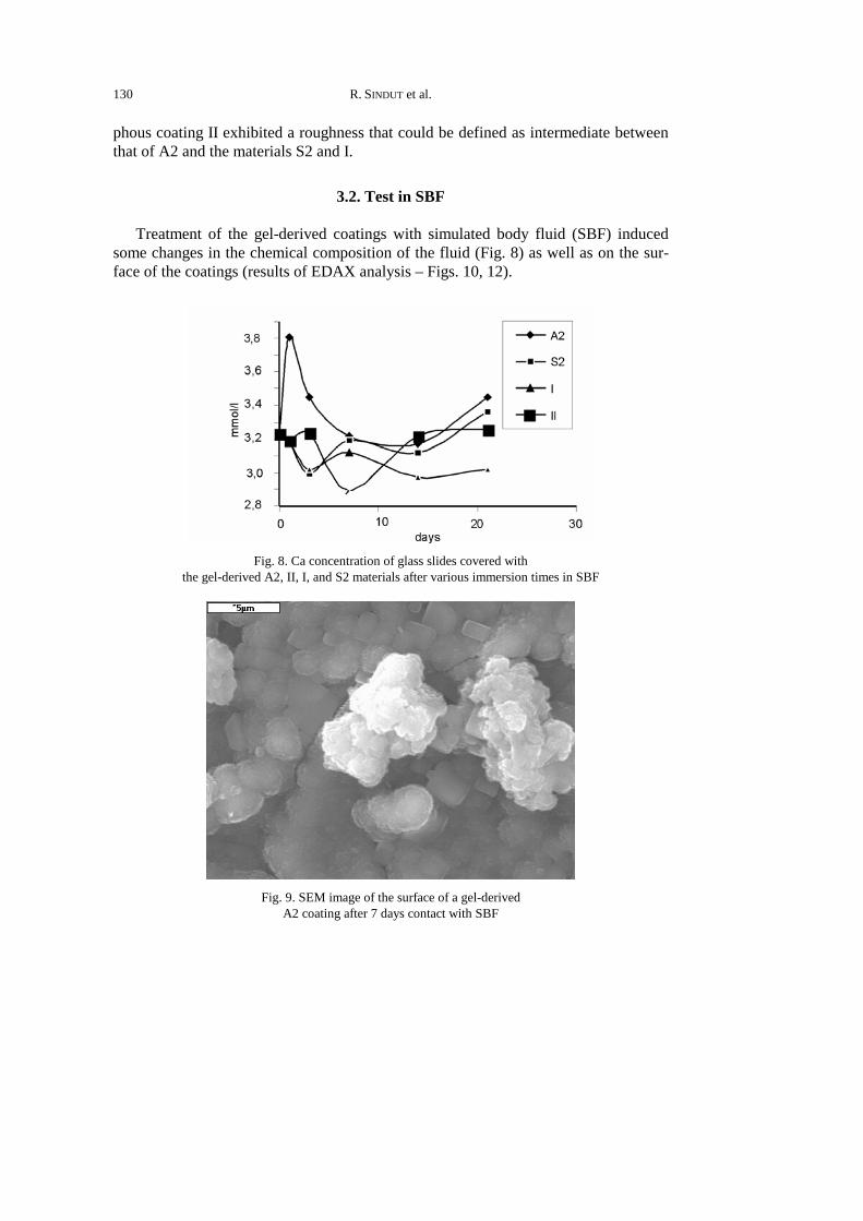
Treatment of the gel-derived coatings with simulated body fluid (SBF) induced some changes in the chemical composition of the fluid (Fig. 8) as well as on the sur-face of the coatings (results of EDAX analysis – Figs. 10, 12).
Fig. 8. Ca concentration of glass slides covered with
the gel-derived A2, II, I, and S2 materials after various immersion times in SBF
Fig. 9. SEM image of the surface of a gel-derived
A2 coating after 7 days contact with SBF

Bioactive glass coatings
131
Fig. 10. Results of EDAX analysis for the surface
of a gel-derived A2 coating after 7 days contact with SBF
Fig. 11. SEM image of the surface of a gel-derived
S2 coating after 7 days contact with SBF

R. SINDUT et al. 132
Fig. 12. Results of EDAX analysis for the surface
of a gel-derived S2 coating after 7 days contact with SBF
Changes in SBF composition were caused by the solubility of the coating materi-als. This solubility was evaluated only with respect to the calcium content in SBF. In the case of other components of the coatings, no measurable solubilities have been ob-served. From among the four examined gel-derived materials, only the A2 coating, for which the CaO:SiO2 ratio is the highest, exhibited a considerable loss of calcium already 1 day after contact with SBF. The consequence of this was an increase in the Ca concen-tration in the SBF. With further contact, the content of Ca in SBF showed a tendency to fall, which might be connected to the surface crystallization of hydroxyapatite. In the case of other coatings, the Ca concentration in SBF, independently of the duration of contact with the biomaterial, was close to the starting concentration.
Changes in chemical composition and SEM images (resulting from contact with SBF) were observed only in the case of the A2 material (Figs. 9, 10). From EDAX analysis (Fig. 10) it appears that already after 7 days after immersion, the concentra-tion of calcium and phosphorus in the surface layer of the material increased. This is an indication of the surface crystallization of calcium phosphates. With prolonged contact time with SBF (up to 21 days), the content of Ca and P on the surface increased, while the content of silicon decreased.

Bioactive glass coatings
133
Fig. 13. XRD patterns of gel-derived A2, II, I, and S2 coatings after 7 days contact with SBF
Changes in the chemical composition were accompanied by a distinct change in the morphology of the layer surface. The surface became covered with spherical forms (Fig. 9), which after 21 days completely covered the primary layer. XRD inves-tigations (Fig. 13) showed that the spherical forms appearing on the surface were composed of hydroxyapatite. The described phenomenon was observed for layers characterized by a lower ratio of CaO to SiO2 (Figs. 11–13). Since the results of the tests with SBF show that only the A2 material is able to produce hydroxyapatite on its surface, it was chosen for in vitro tests with human marrow cells.
3.3. In vitro test with human marrow stromal cells
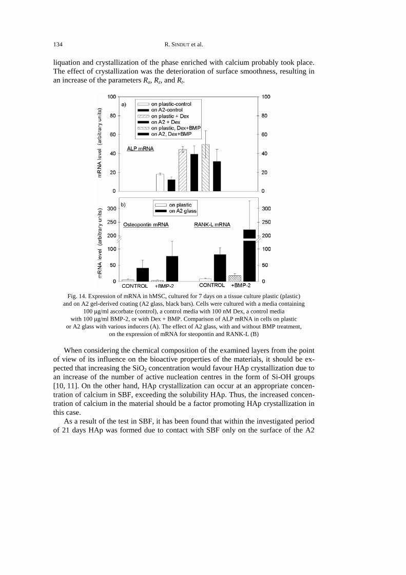
Cells cultured directly on the A2 surface showed a stimulation pattern for alkaline phosphates (ALP) similar to that of cells cultured on plastic, though the levels on A2 were slightly lower (Fig. 14a). The A2 glass, however, had a far more profound effect on the expression of osteopontin and RANK-L (Fig. 14b). MSC cultured on a A2 surface had osteopontin and RANK-L mRNA levels 5–10 times higher than those of cells on plastic, and addition of BPM caused a further increase in the mRNA levels.
4. Discussion
The sol-gel process enabled coatings with various chemical compositions to be ob-tained in a relatively simple way on glass plates. The phase composition of the layers depended on the molar ratio CaO:SiO2. At the highest value of this ratio (ca. 12), the
R. SINDUT et al. 134
liquation and crystallization of the phase enriched with calcium probably took place. The effect of crystallization was the deterioration of surface smoothness, resulting in an increase of the parameters Ra, Rz, and Rt.
Fig. 14. Expression of mRNA in hMSC, cultured for 7 days on a tissue culture plastic (plastic)
and on A2 gel-derived coating (A2 glass, black bars). Cells were cultured with a media containing 100 µg/ml ascorbate (control), a control media with 100 nM Dex, a control media
with 100 µg/ml BMP-2, or with Dex + BMP. Comparison of ALP mRNA in cells on plastic or A2 glass with various inducers (A). The effect of A2 glass, with and without BMP treatment,
on the expression of mRNA for steopontin and RANK-L (B)
When considering the chemical composition of the examined layers from the point of view of its influence on the bioactive properties of the materials, it should be ex-pected that increasing the SiO2 concentration would favour HAp crystallization due to an increase of the number of active nucleation centres in the form of Si-OH groups [10, 11]. On the other hand, HAp crystallization can occur at an appropriate concen-tration of calcium in SBF, exceeding the solubility HAp. Thus, the increased concen-tration of calcium in the material should be a factor promoting HAp crystallization in this case.
As a result of the test in SBF, it has been found that within the investigated period of 21 days HAp was formed due to contact with SBF only on the surface of the A2

Bioactive glass coatings
135
material, which is characterized by the highest CaO:SiO2 ratio. At the same time, only in A2 layers a considerable solubility of Ca in SBF was observed. Thus, it can be assumed that an appropriate level of calcium solubility, determined by the value of the CaO:SiO2 ratio in CaO–P2O5–SiO2 gel-derived coatings, is the factor that determines the ability of HAp to form on the surface. Accordingly, it can be expected that coat-ings with appropriately high CaO:SiO2 ratios can be bioactive. This statement, how-ever, needs to be verified in in vitro conditions.
At the same time, our in vitro study identifies the bioactive gel-derived A2 mate-rial to potentially support the growth and osteoblast differentiation of human bone marrow stromal cells. Surfaces coated with the A2 sol-gel glass were found to permit the adherence, proliferation, and differentiation of hMSC at levels comparable to those seen for tissue culture plastics. Furthermore, the A2 glass induces the expres-sion of both osteopontin, which promotes the migration of osteoblast precursors, and RANK-L, which induces the differentiation of osteoclast precursors. Our data indicate that those MSC that exhibit increased ALP levels are the same cells that show elevated RANK-L levels. There may be, however, an inverse relationship between the levels of ALP and RANK-L. The ability to promote both osteogenesis and osteoclas-togenesis may facilitate not only new bone formation, but also the subsequent remod-elling of the new bone. Since hMSC are believed to be stem cells that play a key role in providing new osteoblasts for bone repair and remodelling, A2 gel-derived glass is significantly promising as a coating for orthopaedic and dental implants.
5. Conclusions
The bioactive properties of CaO–P2O5–SiO2 gel-derived coatings, determined on the basis of HAp surface crystallization capability caused by contact with simulated body fluid (SBF), are determined for different CaO:SiO2 ratios. The increase of CaO content at the expense of SiO2 leads to a higher solubility of Ca in SBF and promotes the surface crystallization of HAp.
The material for which the surface crystallization of Hap occurs as a result of contact with SBF, promotes both osteogenesis and osteoclastogenesis, which may facilitate the formation and remodelling of the newly formed bone.
Acknowledgements
The research was financially supported by the Polish State Committee for Scientific Research, project No. PBZ-KBN-082-T08.
References
[1] HENCH L.L., Am. Ceram. Soc. Bull., 77 (1998), 67. [2] HENCH LL., J. Am. Ceram. Soc., 74 (1991), 1487. [3] KOKUBO T., KUSHITANI H., OHTSUKI C., SAKKA K., YAMAMURO T., J. Mater. Sci. Mater. Med., 3,
(1992), 78.

R. SINDUT et al. 136
[4] LI.R., CLARK A.E., HENCH LL. J. Appl. Biomat., 2 (1991), 231. [5] LACZKA M., CHOLEWA-KOWALSKA K., LACZKA-OSYCZKA A.M., TWORZYDLO M., TURYNA B., J. Bio-
med. Mat. Res., 52 (2000), 601. [6] DISLICH H., J. Noncryst. Sol., 73 (1985), 599. [7] SAKOU T., Bone, 22 (1998), 591. [8] LACZKA-OSYCZKA A.M., LACZKA M., KASUGAI S., OHYA K., J. Biomed. Mat. Res., 42 (1998), 433. [9] SANTOS E.M., RADIN S., SHENKER B.J., SHAPIRO I.M., DUCHEYNE P., J. Biomed. Mat. Res., 41
(1998), 87. [10] KOKUBO T., J. Noncryst. Sol., 120 (1990), 138. [11] DUCHEYNE P., QIU Q., Biomaterials, 20 (1999), 2287.
Received 13 August 2004 Revised 6 December 2004