biochem evals 3
TRANSCRIPT

8/7/2019 BIOCHEM EVALS 3
http://slidepdf.com/reader/full/biochem-evals-3 1/8
BIOCHEMISTRY REVIEW: EVALUATION 3 SET D 1
SUBJECT: BIOCHEMISTRY
TOPIC: EVALUATION #3 Set D
(Translation, Gene Regulation, Heme
Metabolism, Hemostasis)
LECTURER: PROF. SHEILA TORRES & DR. MARIA
ESPERANZA UY
DATE: MARCH 2011
This review will only answer questions 1-28 as Heme Metabolism and
Hemostasis are not part of the Comprehensive Exam. Please read
Chapters 37 & 38 in Harper‟s Illustrated Biochemistry (28th Ed) for
additional information although most bases are lifted from the book.
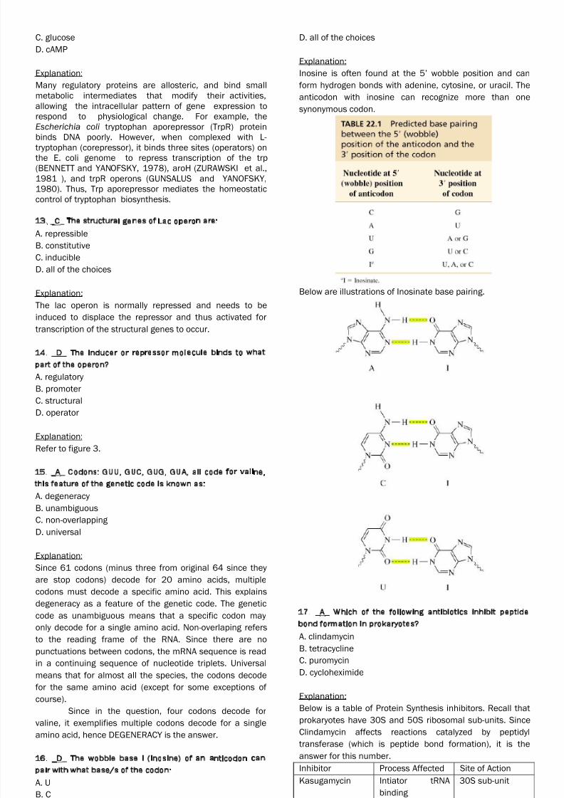
REVIEW PROPER
1. _B_ Which of the following is NOT a valid statement
about the genetic code?
A. Exceptions to the “standard” genetic code have been
found in some species and in mitochondria of others.B. There are 64 possible codons, all of which code for
amino acids
C. The genetic code is redundant with some amino acids
represented by as many as six different codons.
D. The genetic code is read without punctuation, such that
loss of one nucleotide can change the coding for all amino
acids downstream from the loss.
Explanation:
To start, we should define translation. Translation, simply
put, is the conversion of genetic information (in form of
mRNA) into their protein product that they decode for. Next,we define what a codon is. A codon is a triplet code
composed of three nucleotides. B is the answer because
through probability, we can get 64 possible codons. Since
there are only FOUR nucleotides, and there are only 3
nucleotides in a codon, then we raise 4 to 3. 43 = 64. Next,
what makes this statement WRONG? It‟s because of its
latter part. NOT ALL of the codons CODE FOR AMINO ACIDS.
3 codons called nonsense codons code for STOP CODONS
which terminate the translation process, namely, UAA, UAG,
UGA.
Above is a table of the different codons and the amino acids they
encode. The codons are written in the 5’→3’ direction. The third
base of each codon (in bold type) plays a lesser role in specifying
an amino acid than the first two. There are three stop codons and
one initiation codon (AUG; which also decodes for Methionine). All
the AAs except met and trp have more than one codon. In most
cases, codons that specify the same AA differ only at the third
base.
2. _A_ In sickle cell anemia A-T mutation occurred within
the β8-globin gene resulting to the change of val to glu
amino acid in β-globin. This mutation is known as:
A. transversion, missense
B. transition, nonsense
C. transition, missense
D. transversion, nonsense
Explanation:
Mutations are changes in the DNA sequence. These may
happen as single base changes called POINT MUTATIONS
and could be of two types: (1) TRANSITION, which is
pyrimidine → pyrimidine or purine → purine substitution,
and (2) TRANSVERSION, which is pyrimidine → any of the
two purines or purine → any of the two pyrimidines.
Again,
Transition Transversion
Purine → purine or
pyrimidine → pyrimidine
Purine → pyrimidine or
Pyrimidine → purine
The effects of point mutations may either be:
SILENT MUTATIONs – wherein there is no
detectable effect because the codon still decoded
for the same amino acid.
o Recall the “Wobble Effect” wherein
change in the wobble base (the third
nucleotide in the codon) had so significant
effect on the amino acid produced (i.e.
AGA and AGU both decode Arg).
MISSENSE MUTATIONs – when the point mutation
results in the production of a different amino acid.
o Acceptable missense – when the proteinproduct cannot be distinguished from the
normal one
o Partially acceptable missense – when
protein function of the peptide product is
rendered partially abnormal
o Unacceptable missense – when protein
product molecule is rendered incapable of
functioning normally.
NONSENSE MUTATIONs – when the point mutation
results in premature termination of the polypeptide

8/7/2019 BIOCHEM EVALS 3
http://slidepdf.com/reader/full/biochem-evals-3 2/8
BIOCHEMISTRY REVIEW: EVALUATION 3 SET D 2
chain, hence the codon coded for was a STOP
CODON.
Review:
Purines Pyrmidines
Adenine Thymidine
Guanine Cytosine
Since the mutation above was A-T (meaning its purine to
pyrimidine) it is a TRANSVERSION and since the protein
product decoded a different protein (valine to glutamate), it
is a MISSENSE MUTATION. This justifies that the answer is
letter A.
3. _A_ How many nucleotides are there in a couple of
codons?
A. 6
B. 4
C. 3
D. 2
Explanation:Remember, a codon is a triplet code, meaning it is
composed of three nucleotides. Having a couple, by this I
think Professor Torres was pertaining to a PAIR („cause
couple seems so romantic. HAHA), then we would have 2
codons, summing up to 6 nucleotides.
4. _B_ A phosphorylated eIF-2α that inactivates eIF-2B
inhibits protein synthesis by:
A. blocking the formation of the 80S complex
B. preventing the formation of the 43S complex
C. blocking the reduction of secondary structure
D. all of the above
Explanation:
Translation happens in three phases, initiation, elongation,
and termination. Initiation involves many processes
explained below: (Refer to Figure 1 for the pathway)
A. Ribosomal dissociation
eIF-3 and eIF-1A bind to the dissociated 40S sub-unit that
delays the reassociation with 60S.
B. Formation of the 43S Preinitiation Complex
GTP binds to eIF-2 and this binary complex binds to met
tRNA (that carries the start codon AUG which is
methionine). This ternary complex binds to the 40S sub-unit
to form the 43S preinitiation complex.
*eIF-2 is one of the two control points for initiation of the
protein synthesis and is composed of alpha, beta, and
gamma sub-units. eIF-2α is phosphorylated by different
protein kinases that are activated under conditions of
stress, starvation, viral infection, etc. Phosphorylated eIF-
2α binds tightly to and inactivates the GTP -GDP recycling
protein eIF-2B that prevents the formation of the 43S
initiation complex. Hence, the answer is B.
C. Formation of the 48S Initiation Complex
The 5‟ terminal of mRNA is capped with a methyl-guanosyl
triphosphate cap that facilitates binding of mRNA to the
43S initiation complex. Cap-binding complex: eIF-4F
composed of eIF-4E and eIF-4G - eIF-4A. eIF-4B reduces the
complex secondary structure of the 5‟ end of the mRNA
through ATPase and ATP-dependent helicase activites.
mRNA + 43S Initiation complex = 48S Initiation Complex.
*eIF-3 is a key protein since it binds with high affinity to 4G of 4F
and links this with 40S sub-unit.
Formation of 80S Initiation complex
eIF-5 hydrolyzes the GTP bound to eIF-2 on the 48S
initiation complex that leads to release of all the other
initiation factors bound to the complex and facilitates rapid
binding of the 60S sub-unit to form the 80S ribosome.
5. _D_ A structural analog of tyrosinyl - tRNA that can cause
premature termination of translation:
A. bromouracil
B. rifampicin
C. tetracycline
D. puromycin
Explanation:
Puromycin is incorporated via the A site on the ribosome
into the carboxyl terminal position of a peptide but causes
the premature release of the polypeptide. This inhibitive
effect happens to both prokaryotes and eukaryotes.
Please refer to page 367 of Harper’s for the comparison of the
structures of tyrosinyl – tRNA and puromycin. Below is the
mechanism by which puromycin inhibits proteins synthesis.
Bromouracil is brominated uracil which can act as a base
analog for thymine in DNA and can induce DNA mutations.
It is a structural analog but NOT of the molecule of interest,
hence ruled out.
Rifampicin is an antibiotic that inhibits DNA – dependent
RNA polymerase in bacterial cells and inhibits the
translation to RNA and transcription to proteins. It prevents
translation but not in the way specified in the questions,
hence ruled out.
Tetracycline is an antibiotic that bind to the 30S sub-unit in
microbial ribosomes and inhibits protein synthesis by
blocking the site of attachment of the charged aminoacyl
tRNA that prevents introduction of new amino acids to the
growing peptide bond. Yes, it has the same function as
stated above but is NOT a structural analog of tyrosinyl-
tRNA.
6. _B_ Peptidyl transferase is an intergral part of what
ribosomal sub-unit?

8/7/2019 BIOCHEM EVALS 3
http://slidepdf.com/reader/full/biochem-evals-3 3/8
BIOCHEMISTRY REVIEW: EVALUATION 3 SET D 3
A. 30S
B. 60S
C. 40 S
D. 80S
Explanation:
Peptidyl transeferase is located on the P site of the 60S
ribosomal sub-unit. It is involved in the petide bond
formation in the elongation step of translation.
7. _C_ Huntington‟s disease is a neurodegenerative
disorder which is characterized by 35-120 CAG repeats in
the gene‟s coding region. It arose from what kind of
mutation?
A. transition
B. transversion
C. expansion
D. deletion
Explanation:
Trinucleotide repeat disorders are a set of geneticdisorders caused by trinucleotide repeat expansion, a kind
of mutation where trinucleotide repeats in
certain genes exceeding the normal, stable, threshold,
which differs per gene. During protein synthesis, the
expanded CAG repeats are translated into a series of
uninterrupted glutamine residues forming what is known as
a polyglutamine tract ("polyQ"). Such polyglutamine tracts
may be subject to increased aggregation. (Wikipedia)
8. _B_ A manipulated mutation that results to the
elimination or loss of function of the gene product:
A. neural mutationB. gene knockout
C. missense
D. same sense
Explanation:
Gene knockout is a genetic technique (meaning done by
man or by machine and is not natural) in which one of an
organism‟s genes is made inoperative (“knocked out” of
the organism).
9. _B_ Which of the following is not involved in translation?
A. Initiation Factor
B. Enhancer
C. ATP
D. GTP
Explanation:
The important “ingredients” needed for translation
are: tRNA, rRNA, mRNA, ribosomes, eIFs (or initiation
factors in general), ATP, GTP, and amino acids. Enhancers
are involved in TRANSCRIPTION, not translation.
10. _D_ Which of the following can initiate frameshift
mutation?
A. tautomerism of bases
B. alkylating agents
C. base analogs
D. intercalating agents
Explanation:
Frameshift mutations occur when the reading frame of the
codon is shifted either due to an insertion or deletion of a
nucleotide. Deletion or insertion however of three (or
multiples of three) will not shift the reading frame since
triplets are also added/removed. Insertion or deletion of
one or two nucelotides will result into translation of a
peptide with garbled peptide chain or probably formation of
a stop codon that will prematurely end the translational
process.
Tautomerism happens when a keto or enol structure is
formed. Guanine and thymine can have alternate molecular
structures based on different locations of a particular
hydrogen atom. A keto structure occurs when the hydrogen
atom bonds to a nitrogen atom within the ring. An enol
structure occurs when the hydrogen atom bonds to an
nearby oxygen atom that sticks out from the ring. Both
guanine and thymine can switch easily from one tautomer
to another. The change in shape affects the three-
dimensional shape of the molecule. These may cause other
mutations in the genetic code but not frameshift mutations.
Alkylating agents do not cause frameshift mutations
because they only add alkyl groups (alkanes, alkenes,
alkynes) to the molecule. They do not change the reading
frame.
Base analogs are molecules that are structurally similar to
the nucleotide in the DNA chain but cannot cause a
frameshift mutation because they do not change the
reading frame. They may cause a mutation like a point
mutation due to confusion in reading the analog but not a
frameshift mutation.
Intercalating agents are ligands that are small enough,
polycyclic, aromatic, and planar that can fit in between
base pairs of the DNA strand. Since they resemble
insertions, they can cause shifting of the reading frame and
therefore, frameshift mutations.
11. _B_ Photolyases directly repair what kind of damage?
A. base locked in its enol-form
B. thymine dimers
C. apurinic site
D. apyrimidinic site
Explanation:Photolyases are DNA-linked enzymes that repair DNA
damage caused by exposure to UV rays. Specifically, bind
complementary DNA strands and break ceratin pyrimidine
dimers that form when thymine or cytosine bases
covalently link on the same strand and form bulges on the
chain.
12. _A_ Co-repressor of tryptophan:
A. tryptophan
B. lactose
8/7/2019 BIOCHEM EVALS 3
http://slidepdf.com/reader/full/biochem-evals-3 4/8
BIOCHEMISTRY REVIEW: EVALUATION 3 SET D 4
C. glucose
D. cAMP
Explanation:
Many regulatory proteins are allosteric, and bind small
metabolic intermediates that modify their activities,
allowing the intracellular pattern of gene expression to
respond to physiological change. For example, the
Escherichia coli tryptophan aporepressor (TrpR) protein
binds DNA poorly. However, when complexed with L-tryptophan (corepressor), it binds three sites (operators) on
the E. coli genome to repress transcription of the trp
(BENNETT and YANOFSKY, 1978), aroH (ZURAWSKI et al.,
1981 ), and trpR operons (GUNSALUS and YANOFSKY,
1980). Thus, Trp aporepressor mediates the homeostatic
control of tryptophan biosynthesis.
13. _C_ The structural genes of Lac operon are:
A. repressible
B. constitutive
C. inducible
D. all of the choices
Explanation:
The lac operon is normally repressed and needs to be
induced to displace the repressor and thus activated for
transcription of the structural genes to occur.
14. _D_ The inducer or repressor molecule binds to what
part of the operon?
A. regulatory
B. promoter
C. structural
D. operator
Explanation:
Refer to figure 3.
15. _A_ Codons: GUU, GUC, GUG, GUA, all code for valine,
this feature of the genetic code is known as:
A. degeneracy
B. unambiguous
C. non-overlapping
D. universal
Explanation:Since 61 codons (minus three from original 64 since they
are stop codons) decode for 20 amino acids, multiple
codons must decode a specific amino acid. This explains
degeneracy as a feature of the genetic code. The genetic
code as unambiguous means that a specific codon may
only decode for a single amino acid. Non-overlaping refers
to the reading frame of the RNA. Since there are no
punctuations between codons, the mRNA sequence is read
in a continuing sequence of nucleotide triplets. Universal
means that for almost all the species, the codons decode
for the same amino acid (except for some exceptions of
course).Since in the question, four codons decode for
valine, it exemplifies multiple codons decode for a single
amino acid, hence DEGENERACY is the answer.
16. _D_ The wobble base I (inosine) of an anticodon can
pair with what base/s of the codon:
A. U
B. C
C. A
D. all of the choices
Explanation:
Inosine is often found at the 5‟ wobble position and can
form hydrogen bonds with adenine, cytosine, or uracil. The
anticodon with inosine can recognize more than one
synonymous codon.
Below are illustrations of Inosinate base pairing.
17. _A_ Which of the following antibiotics inhibit peptide
bond formation in prokaryotes?
A. clindamycin
B. tetracycline
C. puromycin
D. cycloheximide
Explanation:Below is a table of Protein Synthesis inhibitors. Recall that
prokaryotes have 30S and 50S ribosomal sub-units. Since
Clindamycin affects reactions catalyzed by peptidyl
transferase (which is peptide bond formation), it is the
answer for this number.
Inhibitor Process Affected Site of Action
Kasugamycin Intiator tRNA
binding
30S sub-unit
Streptomycin Initiation, 30S sub-unit
8/7/2019 BIOCHEM EVALS 3
http://slidepdf.com/reader/full/biochem-evals-3 5/8
BIOCHEMISTRY REVIEW: EVALUATION 3 SET D 5
elongation
Tetracycline Aminoacyl tRNA
binding
A-site
Erythromycin Peptidyl
transferase
50S sub-unit
Lincomycin Peptidyl
transferase
50S sub-unit
Clindamycin Peptidyl
transferase
50S sub-unit
Chloramphenicol Peptidyl
transferase
50S sub-unit
18. _D_ A eukaryotic mRNA can form how many
polypeptide after one round of translation?
A. four
B. three
C. two
D. one
Explanation:Translation of mRNA starts at the 5' terminal where the
amino terminal is formed. The sequence is read from 5' to
3' and ends with the formation of the carboxy terminal.
Recall that transcription of DNA into RNA will form the 5'
end of the RNA first, hence in prokaryotes (which have no
compartmentalization since they have a nucleiod region
instead of a membrane-bound nucleus), translation can
readily proceed transcription and occur simultaneously. In
eukaryotes, since they have a membrane bound nucleus
where transcription occurs, the mRNA has to be brought
out into the cytoplasm first where translation will occur.
Hence, after once round of translation, only ONE
polypeptide chain can be formed from eukaryotic mRNA.
19. _D_ Which of the following nucleic acid base is not
present in codons?
A. adenine
B. cytosine
C. guanine
D. thymine
Explanation:
If you remember in RNA synthesis, when RNA is transcribed
from DNA, the complementary base for adenine is NOT
thymine, but uracil. Hence, in codons (which are found in
RNA sequences) there is NO THYMINE, just uracils.
20. _B_ In one form of Thalassemia, codon 17 of the β-
chain is changed from UGG to UGA. This kind of mutation
is:
A. same sense
B. nonsense
C. missense
D. gene knockout
Explanation:
As explained earlier, point mutations are mutations that
happen on single base nucleotides. In this case, there was
a point mutation on the third nucleotide in the codon. Also,
codons can also encode for non-amino acid products like
the STOP CODONS, which are UAA, UAG, and UGA. Since the
point mutation resulted in UGA, the translation process is
prematurely stopped and this kind of mutation is called a
NONSENSE mutation.
21. _C_ The enzyme that is involved during tRNA charging
and amino acid activation:
A. translocase
B. peptidase
C. aminoacyl synthetase
D. DNA glycosylase
Explanation:
Charging or recognition and attachment of amino acids is
catalyzed by AMINOACYL tRNA SYNTHETASES that catalyze
esterification of the AA to the tRNA. This step is energy
requiring (specifically ATP).
22. _D_ Which of the following events of translation is/are
GTP/ATP requiring?
A. charging of tRNA
B. entry of aminoacyl tRNa into the A site
C. formation of peptide bond
D. all of the above
Explanation:
As discussed in previous questions, all these processes
require energy either in the form of ATP or GTP.
23. _A_ Which of the following congenital defect/s could
not carry out excision repair?
A. Xeroderma pigmentosum
B. Fanconi‟s syndrome
C. Bloom‟s syndrome
D. all of the choices
Explanation:Xeroderma Pigementosum is an autosomal
recessive genetic disorder of DNA repair in which the ability
to repair damage caused by ultraviolet (UV) light is
deficient. The most common defect in xeroderma
pigmentosum is an autosomal recessive genetic defect in
which nucleotide excision repair (NER) enzymes are
mutated, leading to a reduction in or elimination of
Nucleotide Excision Repair. If left unchecked, damage
caused by UV light can cause mutations in
individual cell's DNA.
Fanconi‟s disease is a disease of the proximal
renal tubules of the kidney in which glucose, amino acids,
uric acid, phosphate and bicarbonate are excreted in the
urine rather than absorbed. It has no relation with base
excision repair.
Bloom‟s syndrome is a rare autosomal recessive
chromosomal disorder characterized by a high frequency of
breaks and rearrangements in an affected person‟s
chromosomes.
24. _B_ An enzyme that removes damaged base during
base excision repair:
A. Dam methylase
B. DNA glycosylase
C. AP endonucease
D. ABC exonuclease
Explanation:
Base Excision Repair is initiated by DNA glycosylases, which
recognize and remove specific damaged or inappropriate
bases, forming AP sites. These are then cleaved by an AP

8/7/2019 BIOCHEM EVALS 3
http://slidepdf.com/reader/full/biochem-evals-3 6/8
BIOCHEMISTRY REVIEW: EVALUATION 3 SET D 6
endonuclease. The resulting single-strand break can then
be processed by either short-patch (where a single
nucleotide is replaced) or long-patch Base Excision Repair
(where 2-10 new nucleotides are synthesized). (Wikipedia)
25. _A_ Which of the following gene/alleles of blood arose
from frameshift mutation in the glycoslytransferase gene?
A. O
B. B
C. A
D. all of the choices
Explanation:
The ABO blood group system is determined by what type of
glucosyltransferases are expressed in the body.
The ABO gene locus expressing the glucosyltransferases
has three main alleleic forms: A, B, and O. The A allele
encodes a glycosyltransferase that bonds α-N-
acetylgalactosamine to D-galactose end of H antigen,
producing the A antigen. The B allele encodes
a glycosyltransferase that joins α-D-galactose bonded to D-
galactose end of H antigen, creating the B antigen. In case
of O allele the exon 6 contains a deletion that results in a
loss of enzymatic activity. The O allele differs slightly from
the A allele by deletion of a single nucleotide -Guanine at
position 261. The deletion causes a frameshift and results
in translation of an almost entirely different protein that
lacks enzymatic activity. This results in H antigen remaining
unchanged in case of O groups.
The combination of glucosyltransferases by both alleles
present in each person determines whether there is a AB,
A, B or O blood type. (Wikipedia)
26. _B_ Spontaneous deamination of cytosine converts this
base into:
A. adenine
B. uracil
C. thymine
D. guanine
Explanation:
Spontaneous deamination of cytosine happens through
action of the enzyme cytosine deaminase that hydrolyzes
cytosine into uracil and liberates ammonia as a by-product.
Reaction is seen below: (Cytosine + H2O → Uracil + NH3)
27. _B_ The following are some components of 48s
intiation complex EXCEPT:
A. mRNA
B. 60S
C. 40S
D. charged tRNA
Explanation:
Recall as initiation of translation was discussed earlier: the
48S complex is formed from the capped mRNA and the
43S preinitiation complex (ternary complex [met tRNA] +
40S sub-unit with bound eIF-3 and eIF-1A). Hence, the 60S
sub-unit is not part of the 48S complex.
28. _C_ This eukaryotic initiation factor prevents the re-
association of the 60S to 40S ribosomal sub-units:
A. eIF1
B. eIF2
c. eIF3
d. eIF5
Explanation:
eIF1A and eIF3 bind to the 40S sub-unit and delays the
reassociation of 40S and 60S sub-units. eIF-3 plays a more
important role since it also has high affinity for the 4F sub-
unit. (Refer to Figure 2 for summary.)
-------------------------------END OF TRANSCRIPTION----------------------------
Supplementary figure on the structure of tRNA.
8/7/2019 BIOCHEM EVALS 3
http://slidepdf.com/reader/full/biochem-evals-3 7/8
BIOCHEMISTRY REVIEW: EVALUATION 3 SET D 7
FIGURE 1
FIGURE 2

8/7/2019 BIOCHEM EVALS 3
http://slidepdf.com/reader/full/biochem-evals-3 8/8
BIOCHEMISTRY REVIEW: EVALUATION 3 SET D 8
FIGURE 3