biochemical and biological characterization of - molecular cancer
TRANSCRIPT

Rappa et al. Molecular Cancer 2013, 12:62http://www.molecular-cancer.com/content/12/1/62
RESEARCH Open Access
Biochemical and biological characterization ofexosomes containing prominin-1/CD133Germana Rappa1, Javier Mercapide1, Fabio Anzanello1, Robert M Pope2 and Aurelio Lorico1*
Abstract
Exosomes can be viewed as complex “messages” packaged to survive trips to other cells in the localmicroenvironment and, through body fluids, to distant sites. A large body of evidence indicates a pro-metastaticrole for certain types of cancer exosomes. We previously reported that prominin-1 had a pro-metastatic role inmelanoma cells and that microvesicles released from metastatic melanoma cells expressed high levels ofprominin-1. With the goal to explore the mechanisms that govern proteo-lipidic-microRNA sorting in cancerexosomes and their potential contribution(s) to the metastatic phenotype, we here employed prominin-1-basedimmunomagnetic separation in combination with filtration and ultracentrifugation to purify prominin-1-expressingexosomes (prom1-exo) from melanoma and colon carcinoma cells. Prom1-exo contained 154 proteins, including allof the 14 proteins most frequently expressed in exosomes, and multiple pro-metastatic proteins, including CD44,MAPK4K, GTP-binding proteins, ADAM10 and Annexin A2. Their lipid composition resembled that of raftmicrodomains, with a great enrichment in lyso-phosphatidylcholine, lyso-phosphatidyl-ethanolamine andsphingomyelin. The abundance of tetraspanins and of tetraspanin-associated proteins, together with the high levelsof sphingomyelin, suggests that proteolipidic assemblies, probably tetraspanin webs, might be the essentialstructural determinant in the release process of prominin-1 of stem and cancer stem cells. Micro-RNA profilingrevealed 49 species of micro-RNA present at higher concentrations in prom1-exo than in parental cells, including 20with cancer-related function. Extensive accumulation of prom1-exo was observed 3 h after their addition to culturesof melanoma and bone marrow-derived stromal cells (MSC). Short-term co-culture of melanoma cells and MSCresulted in heterologous prominin-1 transfer. Exposure of MSC to prom1-exo increased their invasiveness. Our studysupports the concept that specific populations of cancer exosomes contain multiple determinants of the metastaticpotential of the cells from which they are derived.
Keywords: Exosomes, Melanoma, Prominin-1, Proteomics, Lipidomics, micro-RNA
IntroductionDevelopment of effective anti-cancer strategies based onprevention and targeting of metastatic disease is of highpriority, particularly for melanoma, a disease for whichthe development of metastasis is by far the major causeof patients’ death [1]. Tumor-derived exosomes, smallextracellular vesicles that perform diverse cellular func-tions including intercellular communication, antigenpresentation, and transfer of proteins, RNA and lipids,have been recently implicated in the metastatic process.Exosomes originate by a sequential process of inward
* Correspondence: [email protected] Research Center, Roseman University of Health Sciences, Las Vegas,NV 89135, USAFull list of author information is available at the end of the article
© 2013 Rappa et al.; licensee BioMed CentralCommons Attribution License (http://creativecreproduction in any medium, provided the or
budding of late endosomes, producing multivesicularbodies (MVBs), followed by release of internal microve-sicles into the microenvironment by fusion of the MVBswith the plasma membrane [2]. Cancer exosomes mayhave a role in the cross-talk between primary tumorsand bone marrow-derived stromal cells (MSC), repro-gramming MSC and other non-tumor cells to supportlocal cancer growth as well as to prime pre-metastaticniche(s) [3-7]. However, difficulties in obtaining homo-geneous exosomal preparations result in incompleteunderstanding of exosome formation, composition andfunctions [8]. We recently reported a novel identificationof the extracellular release of prominin-1-containingmembrane microvesicles from human FEMX-I meta-static melanoma cells, and suggested that prominin-1
Ltd. This is an Open Access article distributed under the terms of the Creativeommons.org/licenses/by/2.0), which permits unrestricted use, distribution, andiginal work is properly cited.

Rappa et al. Molecular Cancer 2013, 12:62 Page 2 of 17http://www.molecular-cancer.com/content/12/1/62
microvesicles influence the metastatic capacity of FEMX-Icells [9]. Our laboratory had previously shown thatprominin-1 knock-down resulted in decreased metastaticpotential of FEMX-I cells in immune-deficient mice [10].Prominin-1, a pentaspanning transmembrane protein ori-ginally identified as a surface marker of both neural [11]and hematopoietic [12] stem and progenitor cells, isexpressed in both established melanoma cell lines and clin-ical specimens derived from melanoma patients [13-16].Here, we have employed immune-selection for-
prominin-1 to isolate and characterize a homogenouspreparation of exosomes, presumably engineered fromFEMX-I melanoma cells to perform unique and pro-metastatic tasks in the local microenvironment.
Experimental proceduresCell CultureThe human FEMX-I cell line was originally derived from alymph node metastasis of a patient with malignantmelanoma [17]. Cells were routinely cultured in RPMI(Mediatech Inc., Manassas, VA, http://www.cellgro.com)supplemented with 10% fetal bovine serum (FBS) (AtlantaBiologicals, Lawrenceville, GA, http://www.atlantabio.com) at 37°C in a 5% CO2 humidified incubator and usedbetween passages 3 and 15. Human MSC were obtainedfrom Dr. Prockop, Texas A & M. They were isolated from1 to 4-ml bone marrow aspirates taken from the iliac crestof normal adult donors after informed consent and undera protocol approved by the Texas A & M Institutional Re-view Board, prepared as described by Larson et al. [18],and frozen at passage 1. For expansion, MSC were platedin a 75-cm2 culture dish, and incubated for 1 day, to re-cover viable adherent cells. Cultures contained approxi-mately 50% of rapidly self-renewing cells (RS) and 50% oflarger, more slowly dividing and more mature cells (MS).MSC were then replated at 50 cells per cm2 and incubatedfor 10 days before lentiviral transduction. With time inculture, the percentage of RS cells decreased progressivelyto less of 10% of the total cells. All cell lines were stored inaliquots in liquid nitrogen and kept in culture for less than3 months. Complete culture medium for MSC consistedof α-minimal essential medium (Gibco, Grand Island,NY), 17% fetal bovine serum (lot selected for rapid growthof MSC) (Atlanta Biologicals), 100 units/ml penicillin, 100μg/ml streptomycin, and 2 mM L-glutamine. Cells wereroutinely tested for mycoplasma contamination by theVenor GeM mycoplasma detection kit (Sigma-Aldrich, St.Louis, MO) and by DAPI staining and authenticated bymorphology check every two weeks.
Preparation of microvesicles and exosomesFor preparation of FEMX-I microvesicles (“classical” prep-aration) and exosomes (prom1-exo), cells were enzymati-cally detached and cultured for six days as spheroids in
serum-free medium, consisting of Dulbecco’s modifiedEagle’s medium in the presence of B27 supplement (bothfrom Gibco) in tissue culture plates, as previously de-scribed [9]. At time of harvest, the pH of the medium was6.7. “Classical” microvesicle preparations were performedby differential centrifugation at 4°C at 300 × g for 5 min,then at 500 × g for 5 min., at 1,200 × g for 20 min. and at10,000 × g for 30 min, followed by centrifugation at200,000 × g for 60 min at 4°C. Because these preparationsare likely to contain a mixture of both exosomes andother microvesicles, we have used the generic termmicrovesicles in this study to include the exosome pool.Prom1-exo were prepared by differential centrifugation at4°C at 300 × g for 5 min, then at 500 × g for 5 min., at1,200 × g for 20 min. and at 10,000 × g for 30 min, followedby filtration with a 0.22 μm low-protein binding Millex-GVfilters (Millipore); the 10,000 × g supernatant was then con-centrated by Amicon Ultracel-100K (Millipore) tubesaccording to the manufacturer’s instructions. The concen-trate was diluted 1:1 (v/v) with PBS; incubated with anti-IgG microbeads (Miltenyi Biotec, Auburn, CA) for 90 minat 4°C, and passed through LS-columns according to themanufacturer’s instructions. The flow-through was col-lected, incubated with anti-human-prominin-1 beads(Miltenyi) for 1 h, and passed through LS-columns. Afterwashings, the column was removed from the magnet andprom1-exo were flushed down with 10 ml of cold PBS.Prom1-exo were then centrifuged at 200,000 g for 60 minat 4°C and resuspended in PBS. Each exosomal preparationwas checked by nanoparticle tracking analysis for size dis-tribution and microparticle concentration and by Westernblotting for expression of prominin-1. Exosomes andmicrovesicles were stained with PKH67 (Sigma-Aldrich, St.Louis, MO), according to the manufacturer’s protocol.
Nanoparticle tracking analysis (NTA)We used the light-scattering characteristics of 488 nmlaser light on microvesicle preparations undergoingBrownian motion injected by continuous flow intothe sample chamber of an LM10 unit (Nanosight,Amesbury, UK). Three videos of 60–90 seconds wererecorded of each sample. Data analysis was performedwith NTA 2.3 software (Nanosight). The diffusion coeffi-cient and hydrodynamic radius were determined usingthe Stokes–Einstein equation, and results were displayedas a particle size distribution. Data are presented as theaverage and standard deviation of the three video re-cordings. Since NTA is most accurate between particleconcentrations in the range of 2 × 10⁸ to 2 × 10⁹/ml,when samples contained higher numbers of particles,they were diluted before analysis and the relative con-centration calculated according to the dilution factor.Control 100 and 200 nm beads were supplied byNanosight. NTA of a small sample of any given
Rappa et al. Molecular Cancer 2013, 12:62 Page 3 of 17http://www.molecular-cancer.com/content/12/1/62
preparation revealed that they were essentially monodis-perse, excluding the problem of aggregation, which maysignificantly impact on a biological system.
Prominin-1-EGFP fusion plasmid and transfectionWe employed the eukaryotic expression plasmid en-hanced GFP (pEGFP)–N1-prominin-1, containing theentire coding sequence of human prominin-1 fusedin-frame to the N-terminus of GFP [19], to transfectFEMX-I cells, as previously described [9].
Protein processing and LC-MS/MS
ElectrophoresisThree independent preparations of prom1-exo were ana-lyzed by LC-MS/MS. Samples, 5 μg each according toresults of Bradford assays, were individually mixed with20 μl LDS buffer, divided into four fractions and loadedon NuPage 4–12% Bis-Tris precast gels (Invitrogen,Carlsbad, CA). Two exterior lanes were loaded withSharp pre-stained protein ladder standards (Invitrogen)and the gel was electrophoresed according the manufac-turer’s recommendations. Two lanes, one containingSharp prestained standards and one containing onefourth of the total sample, were visualized using a silvernitrate protocol (QuickSilver, Pierce, Madison, WI), thenrealigned with the unstained gel section to create a tem-plate for excision. The three remaining, unstained laneswere segmented into 14 equal sections and subjected toin-gel tryptic digestion following the procedure ofShevchenko et al. [20]. Briefly, the protocol calls forreduction with 10 mM DTT and alkylation with 55 mMiodoacetamide (SIGMA-Aldrich, St. Louis, Mo). Eachsegment was prepared with successive wash and dehy-dration steps using 50 mM ammonium bicarbonate(AmBic) or 50% acetonitrile containing 50 mM AmBic,respectively. Finally, the shrunken gel segments wererehydrated with ice cold AmBic containing 12 ng/ml se-quencing grade trypsin (Promega, Madison, WI), andallowed to swell on ice for three hours. Digestions werethen carried out for 16 h at 57°C. The quality of thedigested supernatant was determined prior to lyophi-lization by spotting 1 μl aliquots mixed 1:1 with asaturated solution of alpha-cyano-4-hydroxycinnamicacid (CHCA) acid in 0.1% trifluroacetic acid (Pierce) and50% acetonitrile onto a stainless steel target plate withsubsequent MALDI/TOF analysis on a Autoflex IIITOF/TOF (Bruker, Billerica, MA). The remainder of thegel extract was diluted prior to loading on home-brewStageTips desalting microtip using as previously de-scribed[21]. Material eluted below 50% acetonitrilewas lyophilized and the concentrated peptides wererehydrated in 15 μL of 0.1% formic acid with 5% LC/
MS-grade acetonitrile and 4uL was used for each LCinjection.
LC-MS/MS analysisUsing a Dionex 3000 nanoRSLC series HPLC system(Thermo-Electron, Waltham, MA) recovered peptideswere loaded at 2 μl/min onto a 200 μm id by 2.5 cmprecolumn (New Objective, Woburn, MA) packed with5 μm YMC ODS-C18 beads (Waters, Milford, MA). Fol-lowing an on-line desalting step, trap flow was reroutedthrough a self-packed 75 um id × 9 cm analytical col-umn containing 3 μm Halo solid-core C-18 particleswith 300 Angstrom pore size. A distal spray opening 8to 10 microns in diameter restricted the hand-packedcolumn. A linear gradient from 95% buffer A [0.1% for-mic acid, 5% acetonitrile and 94.9% LCMS grade water]to 55% buffer B [90% ACN, 9.9% water and 0.1% FA]was delivered at 200 μl/min over 70 min using a secondnano-capacity pump. Following this, the composition ofbuffer B was ramped to 80% over 5 min, maintained for5 min and finally decreased to 5% over the final 10 min.LC effluent was directed to the electrospray source of
a linear ion-trap mass spectrometer (LTQ/XL, Thermo-Electron, USA). MS/MS spectra were acquired in a data-dependent acquisition mode that automatically selectedand fragmented the five most abundant peaks from eachMS spectrum. MS.MS scans were recorded in centroidmode targeting 8000 counts. The trap was filled for amaximum of 10 ms prior to isolation of the target pep-tide at an average value 1E04.
Database searchingTandem mass spectra were processed and charge statesascertained without deisotoping by Mascot Distiller ver-sion 2.4. All MS/MS samples were analyzed using batchprocessing with the Mascot Daemon interface (version2.4, Matrix Science) and MASCOT search engine (ver-sion 2.4 Matrix Science) [21]. All spectral files were alsosearched using Spectrum Mill Proteomics Workbench(Rev.Rev A.03.02.060, Agilent Technologies, Santa Clara,CA) and X! Tandem (The GPM, thegpm.org; versionCYCLONE (2010.12.01.1)). All three engines were set upto search SwissProt_2012_09.fasta (selected for Homosapiens, Nov. 24 2012, 20,235 entries) assuming the di-gestion enzyme trypsin and considering up to twomissed cleavages. X! Tandem searches were restricted tothe subset of proteins assigned with either Mascot orSpectrum Mill.Mascot, Spectrum Mill and X! Tandem were searched
with a fragment ion mass tolerance of 0.40 Da and a par-ent ion tolerance of 1.8 Da. Mascot’s Carbamidomethy-lation of cysteine was specified in Mascot and X!Tandem as a fixed modification. Oxidation of methio-nine, carbamidomethylation of lysine were specified in
Rappa et al. Molecular Cancer 2013, 12:62 Page 4 of 17http://www.molecular-cancer.com/content/12/1/62
X!Tandem and Mascot as variable modifications. Oxida-tion of methionine was the only variable modificationspecified in SpectrumMill.
Criteria for protein identificationScaffold (version Scaffold_4.0.0, Proteome Software Inc.,Portland, OR) was used to validate MS/MS based pep-tide and protein identifications. Peptide identificationswere accepted if they could be established at greaterthan 90.0% probability by the Peptide Prophet algorithm[22]. Protein identifications were accepted if they couldbe established at greater than 99.0% probability andcontained at least 4 identified peptides. The ProteinProphet algorithm as implemented in Scaffold_4.0.0,assigned protein probabilities [23]. Proteins that contai-ned similar peptides and could not be differentiatedbased on MS/MS analysis alone were grouped to satisfythe principles of parsimony. Specifically the fragmenta-tion patterns of distinct peptides from families ofhomologous proteins were inspected manually using theprotocol described by Tabb et al. [24]. Hence, validatingat least four unique peptides for each protein listed in-dividually minimized protein ambiguity. Peptide FalseDiscovery Rates (FDR) were also estimated using Target:Decoy search as described by Elias and Gygi [25,26],with FDR = 2 × (no. of PSM in the decoy)/(No. of allPSM), where PSM are the peptide spectral matches withbetter than 90% probability as described above. TheFDR calculated by this approach, 0.1%, likely benefitsfrom probabilistically merging multiple search algo-rithms [23].
ImmunoblottingFor immunoblotting, microvesicles and prom1-exo re-suspended in PBS were checked for consistency byNTA. Aliquots of microvesicles, exosomes and FEMX-Itotal cell lysates containing 1–10 μg of protein weremixed 1:1 with SDS sample buffer (NuSep, Bogart, GA)containing 2% 2-mercaptoethanol, boiled for 5 min, andloaded onto a 8% Tris/Glycine/SDS gel. Electrophoreticseparation of proteins was performed at a constant volt-age of 120 V for 2 h, and electrophoretic transfer of theproteins into Hybond ECL membrane (GE Healthcare,Waukesha, WI) was carried out at constant amperage(30 mA) for 15 h. The blots were blocked with 5% drymilk in Tris-buffered saline containing 0.05% Tween 20(pH 7.5), antibody, and incubated with W6B3C1 anti-prominin-1 (Miltenyi Biotec, Auburn, CA), or alix 3A9clone (Cell Signaling Technology, Danvers, MA) at1:1000 dilution in TBS-T for 5 h at room temperature.After washing with TBS-T, blots were incubated withIRDye 800CW secondary antibody (Li-Cor Biosciences,Lincoln, NE) in TBS-T (1:20,000) for 45 min at roomtemperature. Finally, blots were washed with TBS-T,
scanned by Odyssey infrared imaging system and analy-zed by Odyssey 2.1 application software (Li-Cor Biosci-ences). Gel band densitometric quantification was per-formed employing the ImageJ64 software (rsbweb.nih.gov/ij).
ESI-MS/MS lipid profilingAn automated electrospray ionization (ESI)-tandemmass spectrometry approach was used, and data acquisi-tion and analysis were carried out as described previ-ously [27,28] with modifications. The lipid extracts fromthe FEMX-I cell pellets were dissolved in 1 ml chloro-form. An aliquot of 50 μl of each extract in chloroformwas used for analysis. Precise amounts of internal stan-dards, obtained and quantified as previously described[29], were added in the following quantities (with somesmall variation in amounts in different batches of in-ternal standards): 0.6 nmol di12:0-PC, 0.6 nmol di24:1-PC, 0.6 nmol 13:0-lysoPC, 0.6 nmol 19:0-lysoPC, 0.3 nmoldi12:0-PE, 0.3 nmol di23:0-PE, 0.3 nmol 14:0-lysoPE,0.3 nmol 18:0-lysoPE, 0.3 nmol di14:0-PG, 0.3 nmoldi20:0(phytanoyl)-PG, 0.3 nmol di14:0-PA, 0.3 nmoldi20:0(phytanoyl)-PA, 0.2 nmol di14:0-PS, 0.2 nmol di20:0(phytanoyl)-PS, and 0.23 nmol 16:0–18:0-PI. The sampleand internal standard mixture was combined with sol-vents, such that the ratio of chloroform/methanol/300mM ammonium acetate in water was 300/665/35, and thefinal volume was 1.4 ml. The microvesicle samples wereprepared similarly, except that the entire sample was ana-lyzed, 1/3 of the above standard amounts were added, andthe final volume was 0.75 ml. The unfractionated lipidsamples with internal standards were introduced by con-tinuous infusion into the ESI source on a triple quadru-pole MS/MS (API 4000, Applied Biosystems, Foster City,CA). Samples were introduced using an autosampler (LCMini PAL, CTC Analytics AG, Zwingen, Switzerland) fit-ted with the required injection loop for the acquisitiontime and presented to the ESI needle at 30 μl/min. Se-quential precursor and neutral loss scans of the extractsproduce a series of spectra with each spectrum revealing aset of lipid species containing a common head group frag-ment. Lipid species were detected with the followingscans: PC and lysoPC, [M+H]+ ions in positive ion modewith Precursor of 184.1 (Pre 184.1); PE and lysoPE, [M +H]+ ions in positive ion mode with Neutral Loss of 141.0(NL 141.0); PG, [M+NH4]
+ in positive ion mode with NL189.0 for PG; PI, [M+NH4]
+ in positive ion mode withNL 277.0; PS, [M+H]+ in positive ion mode with NL185.0; PA, [M+NH4]
+ in positive ion mode with NL115.0. SM was determined from the same mass spectrumas PC (precursors of m/z 184 in positive mode) [27,30]and by comparison with PC internal standards using amolar response factor for SM (in comparison with PC) de-termined experimentally to be 0.39. The collision gas
Rappa et al. Molecular Cancer 2013, 12:62 Page 5 of 17http://www.molecular-cancer.com/content/12/1/62
pressure was set at 2 (arbitrary units). The collision ener-gies, with nitrogen in the collision cell, were +28 V for PE,+40 V for PC (and SM), +25 V for PA, PI and PS, and +20V for PG. Declustering potentials were +100 V for alllipids. Entrance potentials were +15 V for PE and +14 Vfor PC (and SM), PA, PG, PI, and PS. Exit potentials were+11 V for PE and +14 V for PC (and SM), PA, PG, PI, PS.The scan speed was 50 or 100 u per sec. The mass ana-lyzers were adjusted to a resolution of 0.7 u full width athalf height. For each spectrum, 9 to 150 continuum scanswere averaged in multiple channel analyzer (MCA) mode.The source temperature (heated nebulizer) was 100°C, theinterface heater was on, +5.5 kV or −4.5 kV were appliedto the electrospray capillary, the curtain gas was set at 20(arbitrary units), and the two ion source gases were set at45 (arbitrary units). The background of each spectrumwas subtracted, the data were smoothed, and peak areasintegrated using a custom script and Applied BiosystemsAnalyst software, and the data were isotopically decon-voluted. The first and typically every 11th set of mass spec-tra were acquired on the internal standard mixture only.Peaks corresponding to the target lipids in these spectrawere identified and molar amounts calculated in compari-son to the two internal standards on the same lipid class,except for PI, which was quantified in relation to a singleinternal standard. Ether-linked (alk(en)yl,acyl) lipids werequantified in comparison to the diacyl compounds withthe same head groups without correction for response fac-tors for these compounds as compared to their diacyl ana-logs. To correct for chemical or instrumental noise in thesamples, the molar amount of each lipid metabolitedetected in the “internal standards only” spectra wassubtracted from the molar amount of each metabolite cal-culated in each set of sample spectra. The data from each“internal standards only” set of spectra was used to cor-rect the data from the following 10 samples. Finally, thedata were corrected for the fraction of the sample ana-lyzed and normalized to the mg protein to produce datain the units nmol/mg.
miRNA profilingThe miRNA profiling array was carried out usingApplied Biological Materials miRNA profiling service(ABM C201). Total RNA from FEMX-I cells andexosomes was prepared byQiazol extraction followed bypoly-A tailing reactions and miRNA cDNA synthesis(ABM C204). 250 ng of cell’s total RNA and exosomes’RNA were used in cDNA synthesis. Both cells’ andexosomes’ cDNA synthesis were carried out simultan-eously and equal volume of cDNA synthesis reactionproduct was used in the subsequent profiling. The Ctvalues for each miRNA-specific cDNA were comparedbetween FEMX-I cells and exosomes. Real-time qPCRreactions and instrumental analysis was performed using
Roche LightCycler480. Lists of miRNAs were generatedby pair-wise comparison of our expression data sets(cells vs exosomes). Differentially expressed miRNAswere analyzed by the Ingenuity Pathway Analysis soft-ware (Ingenuity Systems, Redwood City, CA) to identifythe biological functions that were most significant to thedata sets.
ImmunofluorescenceCells were seeded on poly-L-lysine coated chamberslides and grown overnight. Following aspiration ofmedia, cells were fixed in 4% paraformaldehyde (PFA),washed with PBS, permeabilized in 0.5% Tween 20 andblocked with goat serum. After washing with PBS, cellswere incubated overnight at 4°C with primary antibodiesin 1% BSA-PBS, followed by washes and a 45-minutesincubation at room temperature with fluorochrome-labeled secondary antibody in 1% BSA in PBS. Fluo-rescent cells were analyzed by a CKX41 fluorescenceinverted microscope (Olympus, Center Valley, PA).
Invasion assayIn vitro invasion assays were performed in BioCoat inva-sion chambers holding matrigel-coated-8 μm-pore PETmembrane cell culture inserts, using non-coated insertsas control (both from BD Biosciences, San Jose, CA),according to the manufacturer's directions. The matrigellayers of the invasion chambers were rehydrated withserum-free medium. The lower chambers were filledwith medium containing 2% FBS, and equal aliquots ofcells, pre-incubated for 3 h with or without prominin-1-purified exosomes, were added in serum-free mediumto the inserts. Following 24 h incubation at 37°C, thecells on the upper side of the membrane were gently re-moved with wet sterilized cotton swabs. The cells on thelower surface of the membranes were fixed with 4%para-formaldehyde for 10 min, and then stained withDAPI. The number of cells was counted in 8–12 ran-domly selected 10X-microscopic fields per insert usingan Olympus CKX41 fluorescence microscope (OlympusAmerica Corp., Center Valley, PA), and matrigel inva-siveness expressed as the percentage of the number ofmatrigel-invading cells with respect to the control ofchemotactic migration.
ResultsExosomal preparationWe previously reported that human FEMX-I metastaticmelanoma cells released into the extracellular mediumprominin-1-expressing microvesicles [9]. To investigatetheir nature, we cultured FEMX-I cells as spheroidsunder serum-free conditions for six days and compareda “classical” microvesicle preparation, based on differen-tial centrifugation [9], with a prominin-1+ preparation,

Rappa et al. Molecular Cancer 2013, 12:62 Page 6 of 17http://www.molecular-cancer.com/content/12/1/62
illustrated in Figure 1A, based on the combination ofdifferential centrifugation, filtration and prominin-1-based immuno-magnetic selection (prom1-exo). Thefinal pH at time of harvest was 6.7, which resembledin vivo tumor growth conditions, where low pH condi-tion is a hallmark of tumor malignancy, particularly formalignant melanoma cells, which, differently from nor-mal cells, can survive in an acidic microenvironment[31]. Low pH conditions reportedly increase exosomerelease and uptake by cancer cells [32]. We used serum-free medium in the present study because serum supple-ments (such as fetal calf serum) often contain vesicles aswell as aggregates of serum proteins, which may interferewith the isolation and characterization of FEMX-Iexosomes. By NTA, we determined both size distributionand relative concentration of microvesicles and prom1-exo in the supernatants of FEMX-I cells. As shown byNTA of PKH67-stained microvesicles, the “classical”microvesicle preparation showed several peaks, rangingfrom 70 to 550 nm, while prom1-exo yielded a single peakof about 100 nm (Figure 1B); persistent binding of anti-prominin-1 50 nm-immunomagnetic beads resulted in anapparent over-estimation of the exosomal size and broad-ening of the size distribution peak. The concentration ofmicrovesicles and prom1-exo in FEMX-I supernatantwere 3 ± 0.4 ×10⁹/ml and 0.35 ± 0.2 × 10⁹/ml, respectively.
Figure 1 Isolation and characterization of prom1-exo from FEMX-I cecentrifugation and of prom1-exo by a combination of differential centrifugtracking analysis shows size distribution of a “classical” ultracentrifugation-bimmunomagnetic preparation (prom1-exo), both from serum-free culture mpreparations were stained with the membrane dye PKH67 and fluorescencintensities of 80 and 120 nm for microvesicles and 90 nm for exosomes, reprominin-1 microvesicles resulted in an over-estimation of their size distrib
We then employed the same methodology (Figure 1A) toinvestigate whether it was possible to isolate prom1-exofrom different prominin-1-expressing cancer cell lines.We found that prominin-1-immunomagnetic selectionresulted in isolation of cancer exosomes also from humanprominin-1-expressing Caco-2 colon carcinoma cells(Figure 1B). An approximately 10-fold difference in con-centration of microvesicles and prom1-exo was found alsoin the cell supernatants of Caco-2 cells (1.5 ± 0.35 × 10⁹/mland 0.18 ± 0.05 × 10⁹/ml, respectively). Similarly to whatwe observed in FEMX-I cells, Caco-2 cells microvesicleshad a broad size range, while prom1-exo had a single100 nm-peak.
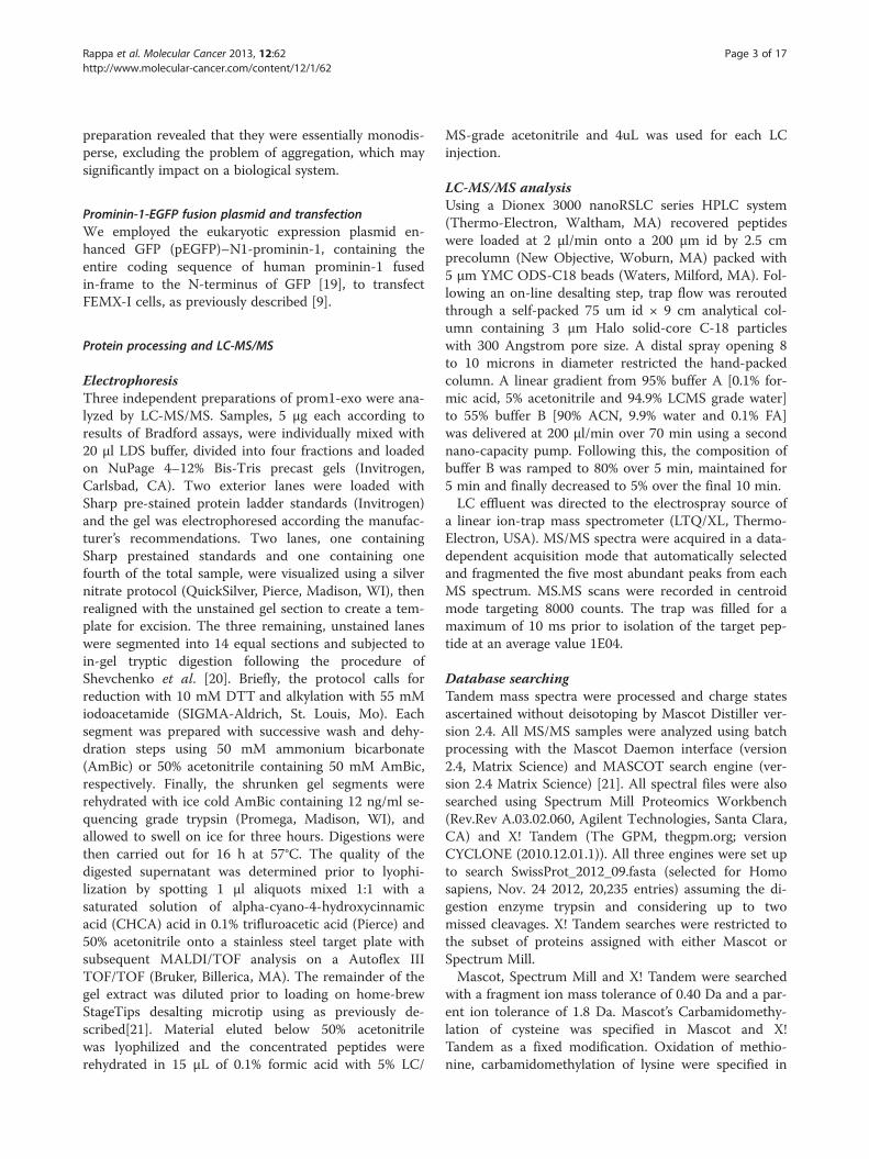
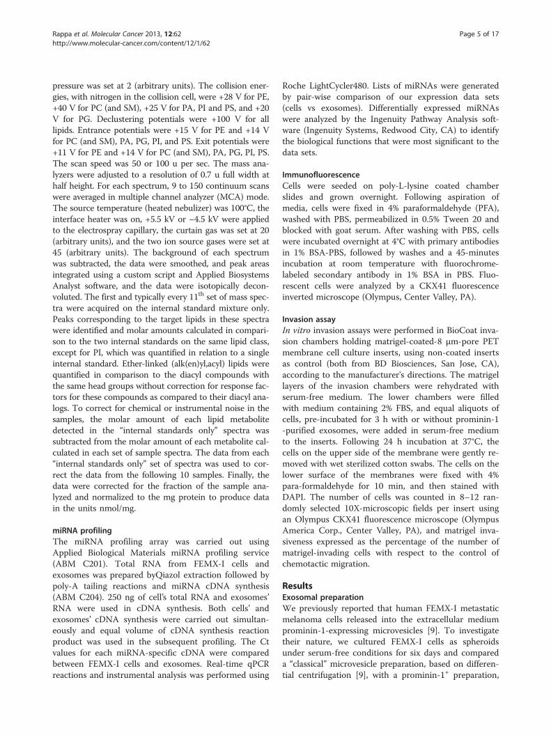
Proteome of prominin-1+ microvesiclesComparison of total cell lysates, microvesicles andprom1-exo from FEMX-I cells by Western blotting(Figure 2) revealed a great enrichment in prominin-1and in the exosomal protein alix in prom1-exo vs. theFEMX-I cells themselves (53- and 184-fold for promi-nin-1 and alix, respectively) and vs. microvesicles(78- and 168-fold for prominin-1 and alix, respectively).To investigate whether they had the biochemical charac-teristics of bona fide exosomes, we analyzed theproteolipidic composition of prom1-exo from FEMX-Icells. Three independent preparations were used to
lls. A. Scheme of isolation of “classical” microvesicles by differentialation, filtration and immuno-magnetic separation. B. Microvesicleased preparation of microvesicles and a prominin-1-basededium of the human FEMX-I metastatic melanoma cell line. Both
e analyzed by a 488 nm laser. Nanotracking analysis gives mean peakspectively. The persistent binding of magnetic beads (50 nm) to theution.
Figure 2 Enrichment of prominin-1 and alix in prom1-exo. Immunoblotting analysis of total cell lysates, microvesicles (MVs), and prom1-exofrom FEMX-I cells. 1 and 10 μg of total proteins were loaded per lane for total cell lysates and MVs and 1 μg for prom1-exo, and analyzed asdescribed under Experimental Procedures.
Rappa et al. Molecular Cancer 2013, 12:62 Page 7 of 17http://www.molecular-cancer.com/content/12/1/62
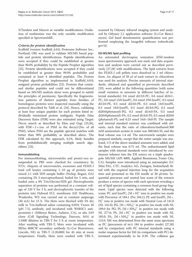
determine their protein composition via MS/MS massspectrometry (see search methods in Additional file 1:Table S1). A total of 282 proteins were confidentlyassigned across all three samples (Additional file 2:Table S2 and Additional file 3: Table S3). We furtherhighlighted an ensemble of proteins among which couldbe verified with two or more stringent peptide spectralmatches (PSM) in all three replicates or those observedwith three or more stringent PSM in any two replicates(Additional file 4: Table S4). This subset of 154 proteinsis highly enriched for physiological processes (Additionalfile 5: Figure S1), involving membrane bound vesicles[count 40, p-value 6.4E-21] and endocytosis [count 20,p-value 7.3E-11] complexes, and including all of the 14proteins most expressed in exosomes according to thecompilation of peer-reviewed data hosted on theExocarta site [33] (Table 1). Since the biogenesis ofexosomes takes place in late endosomes to end up inmultivesicular bodies (MVB), we first checked our list ofproteins for those known to be involved with thatparticular compartment (Table 2). Reassuringly, weidentified the bro1 domain-containing proteins alix and
Table 1 Prom1-exo composition includes all the 14 most-expr
Protein name Gene symbol Acce
Heat shock cognate 71 kDa protein HSPA8
CD9 Antigen CD9
Glyceraldehyde-3-phosphate dehydrogenase GAPDH
Actin, cytoplasmic 1 ACTB
CD63 Antigen CD63
CD81 Antigen CD81
Annexin A2 ANXA2
Alpha-enolase ENO1
Heat shock protein HSP 90-alpha HSP90AA1
Elongation factor 1-alpha 1 EEF1A1
Pyruvate kinase isozymes M1/M2 PKM
14-3-3 protein epsilon YWHAE
Syntenin-1 SDCBP
Programmed cell death 6-interacting protein PDCD6IP
brox, known to function in association with the ESCRT(Endosomal Sorting Complex Required for Transport)pathway to help mediate intraluminal vesicle formation atmultivesicular bodies and the abscission stage of cytokin-esis. Various ESCRT components, central to MVB biogen-esis, were identified in prom1-exo, including five ESCRT-Iproteins [34], three ESCRT-III proteins and many otherESCRT-associated proteins (Table 2). Other proteins, re-lated to their endosomal origin, were identified, includingmembrane transport and fusion proteins (GTPases,Annexin A2, A4, A5, A6 and A11); eight tetraspanins(TSPAN 4,6,9,14; CD63; CD81; CD82; CD9), and five Rabproteins (Additional file 4: Table S4). Interestingly,the immunosuppressive Immunoglobulin superfamilymember 8 (IgSF8), also named CD81 partner 3,known to interact with CD81, CD9 and CD82 as wellas with integrin alpha-3/beta-1 and integrin alpha-4/beta-1, was highly expressed. The absence of endo-plasmic reticulum proteins, such as calnexin andGrp78, and of Golgi proteins, such as GM130, indi-cated no contamination of vesicles of other compart-ments in prom1-exo preparations.
essed exosomal proteins (Exocarta)
ssion number Max N. of unique peptides Max % coverage
P11142 46 75
P21926 8 29
P04406 19 66
P60709 22 74
P08962 5 22
P60033 7 32
P07355 17 52
P06733 20 62
P07900 13 6
P68104 8 29
P14618 21 59
P62258 6 36
O00560 20 86
Q8WUM4 63 75

Table 2 Prom1-exo composition includes many ESCRTand ESCRT-associated proteins
Proteincategory
Genename
Accessionnumber
Max N. ofuniquepeptides
Max %coverage
ESCRT-I VPS-28 Q9UK41 9 57
VPS-37B Q9H9H4 9 55
FAM125A Q96EYS 6 37
FAM125B Q9H7P6 5 38
TSG101 Q99816 13 37
ESCRT-III CHMP2A O43633 3 16
CHMP4B Q9H444 6 35
CHMP5 Q9NZZ3 4 31
ESCRT-associatedproteins
Brox Q5VW32 10 40
PDCD6IP Q8WUM4 63 75
VPS-4A Q9UN37 9 22
MITD1 Q8VW92 6 36
IST1 P53990 11 33
HSPA1A P08107 17 46
HSPA8 P11142 46 75
Rappa et al. Molecular Cancer 2013, 12:62 Page 8 of 17http://www.molecular-cancer.com/content/12/1/62
Other cancer-related proteins and/or proteins impli-cated in cancer progression were identified, includingCD44 [35], Hsp70 [36], annexin A2 [37-40], as well ascomponents involved in Wnt (SFRP1 = secreted frizzled-related protein 1) and Ras signaling, including the GTP-binding proteins Rap1b and Rap2b, reportedly involvedin the activation of ERKs [41], the 14-3-3 protein, afamily of exosomal proteins that have a matrix
Figure 3 Co-localization of prominin-1 with CD29 in FEMX-I cells. Inserepresent areas of peri-nuclear co-localization of prominin-1 and CD29. Pro
metalloproteinase-1 stimulating effect for dermal fibro-blasts [42], and disintegrin and metalloproteinasedomain-containing protein 10 (ADAM 10) (Additionalfile 4: Table S4). A perinuclear pool of prominin-1, asso-ciated with integrin-beta 1 (CD29), expressed in FEMX-Iexosomes, was detected by fluorescence microscopy(Figure 3). Interestingly, a striking correspondence be-tween prominin-1 and CD29 in FEMX-I cells was ob-served, suggesting their co-localization in endosomalcompartments.
Prom1-exo have a typical lipid raft compositionA lipid composition analysis of prom1-exo and parentalFEMX-I cells was performed through ESI MS/MS(Figures 4 and 5). A typical lipid raft composition ofprom1-exo was observed, with 400% increase in sphingo-myelin, 240% increase in phosphatidylserine, 290% inphosphatidylglycerol, 2150% in lyso-phosphatidylethanol-amine) and 1190% in lyso-phosphatidylchoxline. A greatnumber of membrane lipids were significantly differentbetween prom1-exo and the membrane compartment ofparental FEMX-I cells (Table 3). To offset the elevatedsphingolipid levels, phosphatidylcholine levels were de-creased by 26%, resulting in similar choline-containinglipid levels between prom1-exo and the FEMX-I plasmamembrane. A 45% decrease in phosphatidylinositolcontent of prom1-exo also partially accounted for theobserved increase in raft-associated lipid species ofthe exosomes.
Specific “loading” of miRNAs in prom1-exoThe miRNA “cargo” of prom1-exo was significantlydifferent from the parental cell content. Of the 1,058
ts in the upper panels were enlarged in the lower panels. Arrowsminin-1, red. CD29, green; DAPI, blue. Bars, 25 μm.

Figure 4 Different membrane lipid distribution between parental FEMX-I cells and prom1-exo. An automated ESI-tandem massspectrometry approach was used. The lipid extracts from cells and microvesicles were dissolved in 1 ml chloroform. An aliquot of 50 μl of eachextract in chloroform was used for each analysis. To correct for chemical or instrumental noise in the samples, the molar amount of each lipidmetabolite detected in the “internal standards only” spectra was subtracted from the molar amount of each metabolite calculated in each set ofsample spectra. The data from each “internal standards only” set of spectra was used to correct the data from the following 10 samples. Finally,the data were corrected for the fraction of the sample analyzed and normalized to the mg protein to produce data in the units nmol/mg. Dataare presented as percent of total lipids analyzed. *, p < 0.05; **, p < 0.01 (unpaired Student’s t test). SM-DSM, sphingomyelin-dihydrosphingomyelin; PS, phosphatidylserine; PG, phosphatidylglycerol; e-PE, ether-linked phosphatidylethanolamine; e-PC, ether-linkedphosphatidylcholine; PI, phosphatidylinositol; PA, phosphatidic acid.
Rappa et al. Molecular Cancer 2013, 12:62 Page 9 of 17http://www.molecular-cancer.com/content/12/1/62
miRNA species investigated, only 49 were over-expressed in prom1-exo (Table 3), including miRNAsknown to mediate immune tolerance, and 13 cancer/metastasis-associated miRNAs. In particular, miR-216b,a well-known tumor and metastasis suppressor mi-RNA, that targets Ras [43,44], was highly expressed inprom1-exo and undetectable in FEMX-I cells, indicat-ing a detoxification role for prom1-exo; let-7i, associ-ated with metastatic progression [45-47], was found tobe expressed at levels 53-fold higher in prom1-exothan in FEMX-I cells. Also, miR-10a, reportedly in-volved in the metastatic process and immune-escaping[48,49] was 3.2-fold higher in prom1-exo than in par-ental cells.
Transfer of prom1-exo to adjacent FEMX-I and MSCExposure of FEMX-I cells to PKH-67-labeled prom1-exofor 3 h resulted in massive green perinuclear fluores-cence (Figure 6A), co-localized with the red fluorescenceof the intracellular pool of prominin-1 upon incubationof the cells with phycoerythrin-conjugated monoclonals(Figures 6A and 3A-B). Interestingly, also exposure ofhuman MSC to PKH-67-labeled prom1-exo for 3 hresulted in intra-cellular localization of fluorescentprom1-exo and of prom1-exo-associated prominin-1(Figure 6A); for MSC, the punctate pattern differed fromthe perinuclear accumulation for FEMX-I cells,
presumably for the lack of an endogenous pool ofprominin-1 in MSC. Since a shorter (1 h)-exposure ofFEMX-I to PKH-67-labeled prom1-exo resulted in ex-clusive, although minor, perinuclear accumulation ofgreen fluorescence (data not shown), the complete ab-sence of puncta in FEMX-I cells may be due to the rapidkinetics of intracellular exosome trafficking or turnover.To confirm the intracellular delivery of prominin-1 byprom1-exo, MSC were incubated with prom1-exo pre-pared from FEMX-I cells transiently transfected with aprominin-1-GFP fusion plasmid. After 3 h, extensivefluorescence from prominin-1-GFP was detected inthe intracellular compartment of MSC (Figure 6B),confirming that prominin-1 was effectively delivered toMSC. To investigate whether direct transfer of prom1-exo occurred from FEMX-I to MSC in mixed cultures,we co-cultured the cells at 5:1 ratio (FEMX-I:MSC) for24 h, and analyzed the expression of prominin-1 by im-munofluorescence. Figure 6 clearly shows transfer ofprominin-1 from FEMX-I to the intracellular compart-ment of MSC. The apparent contrast between themassive uptake of exosomes in Figure 6 and the rela-tively low transfer of exosomes from FEMX-I to MSCin Figure 7 may be explained by the technical diffe-rences of the two experiments (sudden addition ofexosomes in Figure 6 and gradual release of exosomesin Figure 7).
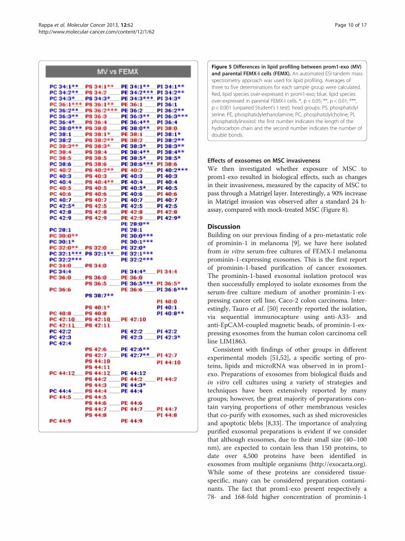
Figure 5 Differences in lipid profiling between prom1-exo (MV)and parental FEMX-I cells (FEMX). An automated ESI-tandem massspectrometry approach was used for lipid profiling. Averages ofthree to five determinations for each sample group were calculated.Red, lipid species over-expressed in prom1-exo; blue, lipid speciesover-expressed in parental FEMX-I cells. *, p < 0.05; **, p < 0.01; ***,p < 0.001 (unpaired Student’s t test); head groups: PS, phosphatidylserine; PE, phosphatidylethanolamine; PC, phosphatidylcholine; PI,phosphatidylinositol; the first number indicates the length of thehydrocarbon chain and the second number indicates the number ofdouble bonds.
Rappa et al. Molecular Cancer 2013, 12:62 Page 10 of 17http://www.molecular-cancer.com/content/12/1/62
Effects of exosomes on MSC invasivenessWe then investigated whether exposure of MSC toprom1-exo resulted in biological effects, such as changesin their invasiveness, measured by the capacity of MSC topass through a Matrigel layer. Interestingly, a 90% increasein Matrigel invasion was observed after a standard 24 h-assay, compared with mock-treated MSC (Figure 8).
DiscussionBuilding on our previous finding of a pro-metastatic roleof prominin-1 in melanoma [9], we have here isolatedfrom in vitro serum-free cultures of FEMX-I melanomaprominin-1-expressing exosomes. This is the first reportof prominin-1-based purification of cancer exosomes.The prominin-1-based exosomal isolation protocol wasthen successfully employed to isolate exosomes from theserum-free culture medium of another prominin-1-ex-pressing cancer cell line, Caco-2 colon carcinoma. Inter-estingly, Tauro et al. [50] recently reported the isolation,via sequential immunocapture using anti-A33- andanti-EpCAM-coupled magnetic beads, of prominin-1-ex-pressing exosomes from the human colon carcinoma cellline LIM1863.Consistent with findings of other groups in different
experimental models [51,52], a specific sorting of pro-teins, lipids and microRNA was observed in in prom1-exo. Preparations of exosomes from biological fluids andin vitro cell cultures using a variety of strategies andtechniques have been extensively reported by manygroups; however, the great majority of preparations con-tain varying proportions of other membranous vesiclesthat co-purify with exosomes, such as shed microvesiclesand apoptotic blebs [8,33]. The importance of analyzingpurified exosomal preparations is evident if we considerthat although exosomes, due to their small size (40–100nm), are expected to contain less than 150 proteins, todate over 4,500 proteins have been identified inexosomes from multiple organisms (http://exocarta.org).While some of these proteins are considered tissue-specific, many can be considered preparation contami-nants. The fact that prom1-exo present respectively a78- and 168-fold higher concentration of prominin-1
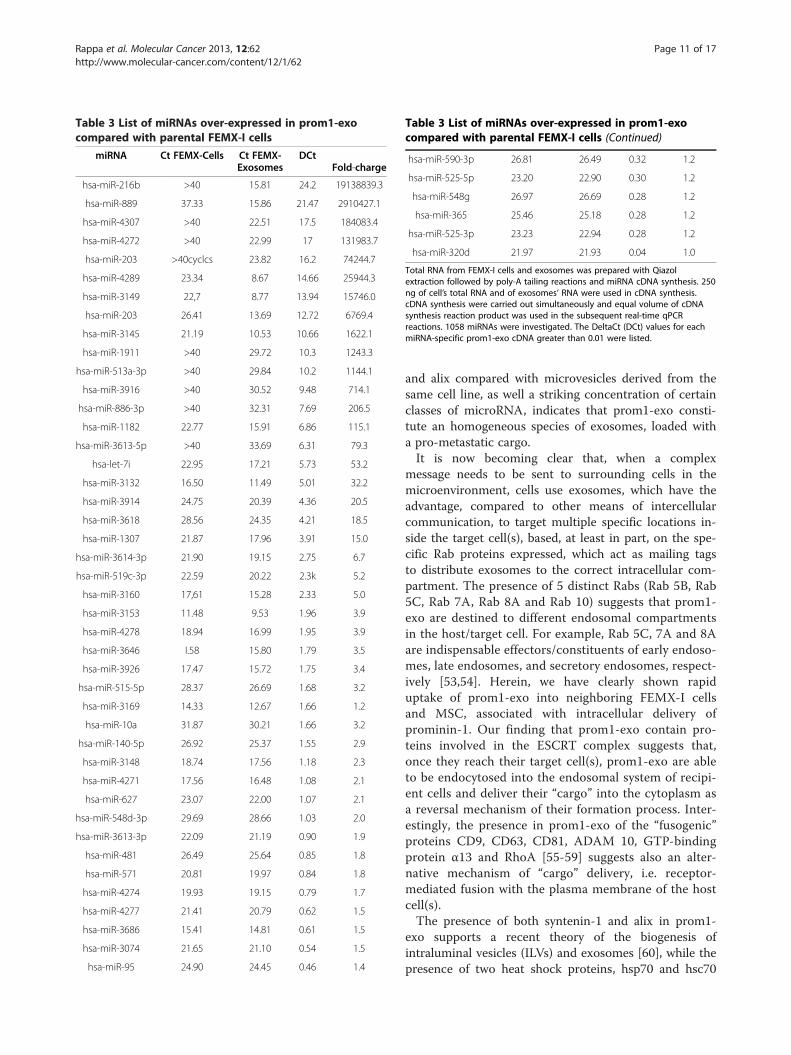
Table 3 List of miRNAs over-expressed in prom1-exocompared with parental FEMX-I cells
miRNA Ct FEMX-Cells Ct FEMX-Exosomes
DCtFold-charge
hsa-miR-216b >40 15.81 24.2 19138839.3
hsa-miR-889 37.33 15.86 21.47 2910427.1
hsa-miR-4307 >40 22.51 17.5 184083.4
hsa-miR-4272 >40 22.99 17 131983.7
hsa-miR-203 >40cyclcs 23.82 16.2 74244.7
hsa-miR-4289 23.34 8.67 14.66 25944.3
hsa-miR-3149 22,7 8.77 13.94 15746.0
hsa-miR-203 26.41 13.69 12.72 6769.4
hsa-miR-3145 21.19 10.53 10.66 1622.1
hsa-miR-1911 >40 29.72 10.3 1243.3
hsa-miR-513a-3p >40 29.84 10.2 1144.1
hsa-miR-3916 >40 30.52 9.48 714.1
hsa-miR-886-3p >40 32.31 7.69 206.5
hsa-miR-1182 22.77 15.91 6.86 115.1
hsa-miR-3613-5p >40 33.69 6.31 79.3
hsa-let-7i 22.95 17.21 5.73 53.2
hsa-miR-3132 16.50 11.49 5.01 32.2
hsa-miR-3914 24.75 20.39 4.36 20.5
hsa-miR-3618 28.56 24.35 4.21 18.5
hsa-miR-1307 21.87 17.96 3.91 15.0
hsa-miR-3614-3p 21.90 19.15 2.75 6.7
hsa-miR-519c-3p 22.59 20.22 2.3k 5.2
hsa-miR-3160 17,61 15.28 2.33 5.0
hsa-miR-3153 11.48 9.53 1.96 3.9
hsa-miR-4278 18.94 16.99 1.95 3.9
hsa-miR-3646 I.58 15.80 1.79 3.5
hsa-miR-3926 17.47 15.72 1.75 3.4
hsa-miR-515-5p 28.37 26.69 1.68 3.2
hsa-miR-3169 14.33 12.67 1.66 1.2
hsa-miR-10a 31.87 30.21 1.66 3.2
hsa-miR-140-5p 26.92 25.37 1.55 2.9
hsa-miR-3148 18.74 17.56 1.18 2.3
hsa-miR-4271 17.56 16.48 1.08 2.1
hsa-miR-627 23.07 22.00 1.07 2.1
hsa-miR-548d-3p 29.69 28.66 1.03 2.0
hsa-miR-3613-3p 22.09 21.19 0.90 1.9
hsa-miR-481 26.49 25.64 0.85 1.8
hsa-miR-571 20.81 19.97 0.84 1.8
hsa-miR-4274 19.93 19.15 0.79 1.7
hsa-miR-4277 21.41 20.79 0.62 1.5
hsa-miR-3686 15.41 14.81 0.61 1.5
hsa-miR-3074 21.65 21.10 0.54 1.5
hsa-miR-95 24.90 24.45 0.46 1.4
Table 3 List of miRNAs over-expressed in prom1-exocompared with parental FEMX-I cells (Continued)
hsa-miR-590-3p 26.81 26.49 0.32 1.2
hsa-miR-525-5p 23.20 22.90 0.30 1.2
hsa-miR-548g 26.97 26.69 0.28 1.2
hsa-miR-365 25.46 25.18 0.28 1.2
hsa-miR-525-3p 23.23 22.94 0.28 1.2
hsa-miR-320d 21.97 21.93 0.04 1.0
Total RNA from FEMX-I cells and exosomes was prepared with Qiazolextraction followed by poly-A tailing reactions and miRNA cDNA synthesis. 250ng of cell’s total RNA and of exosomes’ RNA were used in cDNA synthesis.cDNA synthesis were carried out simultaneously and equal volume of cDNAsynthesis reaction product was used in the subsequent real-time qPCRreactions. 1058 miRNAs were investigated. The DeltaCt (DCt) values for eachmiRNA-specific prom1-exo cDNA greater than 0.01 were listed.
Rappa et al. Molecular Cancer 2013, 12:62 Page 11 of 17http://www.molecular-cancer.com/content/12/1/62
and alix compared with microvesicles derived from thesame cell line, as well a striking concentration of certainclasses of microRNA, indicates that prom1-exo consti-tute an homogeneous species of exosomes, loaded witha pro-metastatic cargo.It is now becoming clear that, when a complex
message needs to be sent to surrounding cells in themicroenvironment, cells use exosomes, which have theadvantage, compared to other means of intercellularcommunication, to target multiple specific locations in-side the target cell(s), based, at least in part, on the spe-cific Rab proteins expressed, which act as mailing tagsto distribute exosomes to the correct intracellular com-partment. The presence of 5 distinct Rabs (Rab 5B, Rab5C, Rab 7A, Rab 8A and Rab 10) suggests that prom1-exo are destined to different endosomal compartmentsin the host/target cell. For example, Rab 5C, 7A and 8Aare indispensable effectors/constituents of early endoso-mes, late endosomes, and secretory endosomes, respect-ively [53,54]. Herein, we have clearly shown rapiduptake of prom1-exo into neighboring FEMX-I cellsand MSC, associated with intracellular delivery ofprominin-1. Our finding that prom1-exo contain pro-teins involved in the ESCRT complex suggests that,once they reach their target cell(s), prom1-exo are ableto be endocytosed into the endosomal system of recipi-ent cells and deliver their “cargo” into the cytoplasm asa reversal mechanism of their formation process. Inter-estingly, the presence in prom1-exo of the “fusogenic”proteins CD9, CD63, CD81, ADAM 10, GTP-bindingprotein α13 and RhoA [55-59] suggests also an alter-native mechanism of “cargo” delivery, i.e. receptor-mediated fusion with the plasma membrane of the hostcell(s).The presence of both syntenin-1 and alix in prom1-
exo supports a recent theory of the biogenesis ofintraluminal vesicles (ILVs) and exosomes [60], while thepresence of two heat shock proteins, hsp70 and hsc70
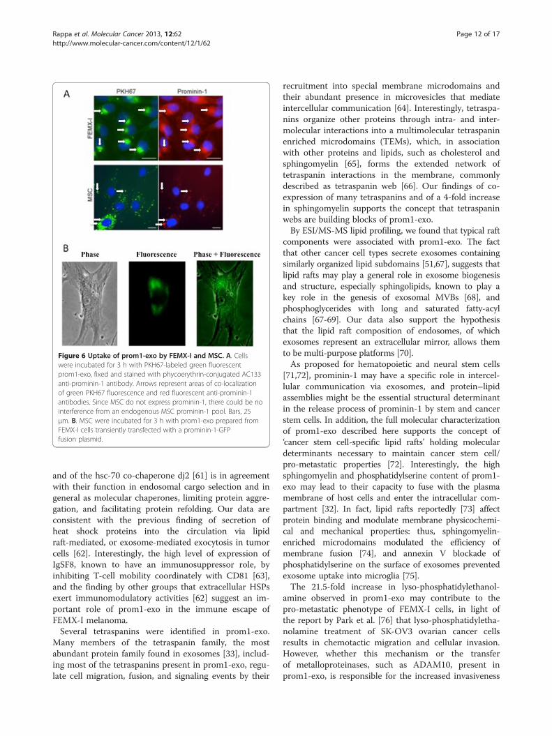
Figure 6 Uptake of prom1-exo by FEMX-I and MSC. A. Cellswere incubated for 3 h with PKH67-labeled green fluorescentprom1-exo, fixed and stained with phycoerythrin-conjugated AC133anti-prominin-1 antibody. Arrows represent areas of co-localizationof green PKH67 fluorescence and red fluorescent anti-prominin-1antibodies. Since MSC do not express prominin-1, there could be nointerference from an endogenous MSC prominin-1 pool. Bars, 25μm. B. MSC were incubated for 3 h with prom1-exo prepared fromFEMX-I cells transiently transfected with a prominin-1-GFPfusion plasmid.
Rappa et al. Molecular Cancer 2013, 12:62 Page 12 of 17http://www.molecular-cancer.com/content/12/1/62
and of the hsc-70 co-chaperone dj2 [61] is in agreementwith their function in endosomal cargo selection and ingeneral as molecular chaperones, limiting protein aggre-gation, and facilitating protein refolding. Our data areconsistent with the previous finding of secretion ofheat shock proteins into the circulation via lipidraft-mediated, or exosome-mediated exocytosis in tumorcells [62]. Interestingly, the high level of expression ofIgSF8, known to have an immunosuppressor role, byinhibiting T-cell mobility coordinately with CD81 [63],and the finding by other groups that extracellular HSPsexert immunomodulatory activities [62] suggest an im-portant role of prom1-exo in the immune escape ofFEMX-I melanoma.Several tetraspanins were identified in prom1-exo.
Many members of the tetraspanin family, the mostabundant protein family found in exosomes [33], includ-ing most of the tetraspanins present in prom1-exo, regu-late cell migration, fusion, and signaling events by their
recruitment into special membrane microdomains andtheir abundant presence in microvesicles that mediateintercellular communication [64]. Interestingly, tetraspa-nins organize other proteins through intra- and inter-molecular interactions into a multimolecular tetraspaninenriched microdomains (TEMs), which, in associationwith other proteins and lipids, such as cholesterol andsphingomyelin [65], forms the extended network oftetraspanin interactions in the membrane, commonlydescribed as tetraspanin web [66]. Our findings of co-expression of many tetraspanins and of a 4-fold increasein sphingomyelin supports the concept that tetraspaninwebs are building blocks of prom1-exo.By ESI/MS-MS lipid profiling, we found that typical raft
components were associated with prom1-exo. The factthat other cancer cell types secrete exosomes containingsimilarly organized lipid subdomains [51,67], suggests thatlipid rafts may play a general role in exosome biogenesisand structure, especially sphingolipids, known to play akey role in the genesis of exosomal MVBs [68], andphosphoglycerides with long and saturated fatty-acylchains [67-69]. Our data also support the hypothesisthat the lipid raft composition of endosomes, of whichexosomes represent an extracellular mirror, allows themto be multi-purpose platforms [70].As proposed for hematopoietic and neural stem cells
[71,72], prominin-1 may have a specific role in intercel-lular communication via exosomes, and protein–lipidassemblies might be the essential structural determinantin the release process of prominin-1 by stem and cancerstem cells. In addition, the full molecular characterizationof prom1-exo described here supports the concept of‘cancer stem cell-specific lipid rafts’ holding moleculardeterminants necessary to maintain cancer stem cell/pro-metastatic properties [72]. Interestingly, the highsphingomyelin and phosphatidylserine content of prom1-exo may lead to their capacity to fuse with the plasmamembrane of host cells and enter the intracellular com-partment [32]. In fact, lipid rafts reportedly [73] affectprotein binding and modulate membrane physicochemi-cal and mechanical properties: thus, sphingomyelin-enriched microdomains modulated the efficiency ofmembrane fusion [74], and annexin V blockade ofphosphatidylserine on the surface of exosomes preventedexosome uptake into microglia [75].The 21.5-fold increase in lyso-phosphatidylethanol-
amine observed in prom1-exo may contribute to thepro-metastatic phenotype of FEMX-I cells, in light ofthe report by Park et al. [76] that lyso-phosphatidyletha-nolamine treatment of SK-OV3 ovarian cancer cellsresults in chemotactic migration and cellular invasion.However, whether this mechanism or the transferof metalloproteinases, such as ADAM10, present inprom1-exo, is responsible for the increased invasiveness

Figure 7 Co-culture of MSC and FEMX-I cells shows uptake of prominin-1 by MSC. MSC and FEMX-I cells were cultured for 24 h at 1:5 ratio.After fixation and permeabilization, expression of prominin-1 was analyzed by immunofluorescence employing phycoerythrin-conjugated AC133anti-prominin-1 antibody. Since MSC do not express prominin-1, there could be no interference from an endogenous MSC prominin-1 pool.Insets in the upper panels were enlarged in the lower panels. Arrows indicate some areas of prominin-1 positivity inside a MSC. Red, prominin-1;blue, DAPI. Bars, 25 μm.
Rappa et al. Molecular Cancer 2013, 12:62 Page 13 of 17http://www.molecular-cancer.com/content/12/1/62
of MSC upon exposure to prom1-exo can not be con-cluded from the present study.Consistent with other previous studies [77-79], a con-
siderable difference in the miRNA profile of cancerexosomes and the originating cancer cells was observedin the present study. Specifically, 49 miRNA were foundto be over-expressed in prom1-exo, including miRNAsknown to mediate immune tolerance, and 13 cancer/me-tastasis-associated miRNAs. This is in apparent contrastwith the claim from several authors [80,81] that themiRNA content of circulating exosomes is similar tothat of the originating cancer cells. The cancer-associated loss of miRNA expression often leads to aproliferative advantage and aggressive behavior throughlargely unknown mechanisms. The finding of very highlevels of miR-216b in prom1-exo, coupled with un-detectable levels in parental FEMX-I cells, is intriguingin light of reports that miR-216b suppresses tumorgrowth and invasion by targeting KRAS in nasopharyn-geal carcinoma [43] and inhibits cell proliferation andcolony formation through Ras inhibition in a pancreaticcancer model [44]. Similarly, a 53-fold lower level oflet-7i was observed in FEMX-I cells compared withprom1-exo. Since underexpression of let-7i was found tocharacterize metastatic progression of oral carcinoma[45] and to have a crucial role in colorectal cancer
metastasis [46], it is conceivable that exosomal removalof both miR-216b and let-7i from the intracellular com-partment plays a significant role in the malignant pheno-type of FEMX-I melanoma. While removal of somespecies of microRNAs may have a detoxification role,exosomal delivery of other species of microRNA, such asmiR-10a, to other cells in the microenvironment mayplay an important role in FEMX-I melanoma immuno-escape. In fact, miR-10a, present in prom1-exo at levels3.2-fold higher than in parental cells, was recently shownto attenuate the phenotypic conversion of inducible T(reg) cells into follicular helper T cells and limit differen-tiation into the T(H)17 subset of helper T cells [48].Also, miR-10a reportedly stimulates cell invasion,suggesting a potential mechanism for the pro-invasiveeffect of prom1-exo on MSC [49]. Therefore, prom1-exomay accomplish for FEMX-I melanoma cells a doublerole of cell detoxification via excretion and of modula-tion of the function of other cell types, in particularMSC, in the microenvironment. Our data, suggesting apro-malignant role of prom1-exo, are consistent with arecent report by Peinado et al. [7] that exosomes fromhighly metastatic melanomas increased the metastaticbehavior of primary tumors by permanently ‘educating’bone marrow progenitors through the receptor tyrosinekinase MET. To metastasize, tumor cells need to send

Figure 8 Enhanced invasiveness of MSC through matrigelinduced by prominin-1-purified exosomes. In vitro invasionassays were performed in BioCoat invasion chambers holdingmatrigel-coated-8 μm-pore PET membrane cell culture inserts, usingnon-coated inserts as control. The lower chambers were filled withmedium containing 2% FBS, and equal aliquots of MSC, pre-incubated with or without prominin-1-purified exosomes, wereadded in serum-free medium to the inserts. Following 24 hincubation at 37°C, as recommended by the manufacturer, the cellson the upper side of the membrane were gently removed with wetcotton swabs. The cells on the lower surface of the membraneswere fixed with 4% para-formaldehyde for 10 min, and then stainedwith DAPI. Matrigel invasiveness is expressed as the percentage ofthe number of matrigel-invading cells respect to the control ofchemotactic migration. Columns, mean values of three separateexperiments; bars, SD; *p < 0.05, unpaired Student’s t test.
Rappa et al. Molecular Cancer 2013, 12:62 Page 14 of 17http://www.molecular-cancer.com/content/12/1/62
complex messages intended to subvert the normal func-tion of their immediate neighbors, fertilize vascu-logenesis and find or recruit a susceptible berth.However, messages of all sorts are being identified inmany functional exosomal studies, and if we samplethem stochastically it will be difficult to see the wholepicture. Prom1-exo, homogenous cancer organelles ex-pressing a cancer stem cell marker, are more likely tohave a concordant message(s), and this makes them es-pecially interesting to gain insight into the mechanismsby which exosomes contribute to the malignant pheno-type. In addition, our characterization of prom1-exofrom FEMX-I cells may be employed as a model for in-vestigating the rules that govern the formation of mem-brane microdomains: in contrast to rafts, exosomes areremarkably stable structures that can be purified withoutthe intervention of destructive techniques such as deter-gents or ultrasounds. Our model, therefore, in addition
to allowing progress in the understanding of the role(s)of cancer-derived exosomes in the metastatic process,can also shed light on the natural process of selectiveproteolipidic sorting in biological membranes and traf-ficking in living cells. Further studies are warranted todetermine what part of their cargo and which molecularmechanisms exosomes, and in particular prom1-exo,utilize to modify the phenotype of the different cells inthe local tumor microenvironment and exert specificroles in the metastatic phenotype.
Additional files
Additional file 1: Table S1. Search conditions for proteomic LC-MS/MSdata sets. MS/S data was acquired during 70 min gradients run on hand-packed capillary columns as described in Materials and Methods. Theeffluent was interfaced to an ESI source and peptides were recordedwith data-dependent scanning using a top 5 method on an LTQ/XL iontrap (Thermo). Table 1 describes data processing, search conditionscommon to both the MASCOT and Spectrum Mill ProteomicsWorkbench applied in this work as well as particular features of theSwissProt database used.
Additional file 2: Table S2. Protein assignments. Use of the Peptideand Protein Prophet algorithms to condense independent searches ofthe same data sets provided 282 proteins (including keratins) across atotal of three biological isolations of prom1-exo with an FDR of 0.1%, aminimum of three peptides and a protein sensitivity of 99%. Additionalfile 2 lists proteins matching these criteria.
Additional file 3: Table S3. All Peptides attributed to proteins fromAdditional file 2. All peptides identified in three replicate isolations ofprom1-exo. Scaffold 4.0.0 was used to rescore the results of MASCOTand SpectrumMill searches. Scaffold generates an adaptive discriminatescoring using Peptide and Protein Prophet algorithms. Complete resultsare listed in Additional file 3: Table S3.
Additional file 4: Table S4. Selection of the most observable proteinsassociated with prom1-exo. Tables 1 and 2 of the manuscript illustrateprotein enrichment for physiological processes involving endosome andESCRT complexes. These proteins are highlighted among an ensembleverified with two or more stringent peptide spectral matches (PSM) in allthree replicates or those observed with three or more stringent PSM inany two replicates. Additional file 4: Table S4 is a complete list of these154 proteins.
Additional file 5: Figure S1. Enrichment for physiological processes ofthe most observable proteins associated with prom1-exo.
AbbreviationProm1-exo: Prominin-1-expressing exosomes; MSC: Bone marrow-derivedstromal cells; NTA: Nanoparticle tracking analysis; ESI: Electrospray ionization;TEM: Tetraspanin enriched microdomain; MVB: Multivesicular body;VPS: Vacuolar protein sorting; ESCRT: Endosomal sorting complex requiredfor transport.
Competing interestsThe authors declare that they have no competing interests.
Authors’ contributionsGR and AL designed and executed most of the experiments and wrote themanuscript. JM analyzed proteins by Western blotting and performed theinvasion assays. FA prepared microvesicles and exosomes. RMP analyzedproteins by mass spectrometry, wrote the proteomics part of the manuscriptand helped writing the manuscript. All authors read and approved the finalmanuscript.

Rappa et al. Molecular Cancer 2013, 12:62 Page 15 of 17http://www.molecular-cancer.com/content/12/1/62
AcknowledgementsWe thank Harry Rosenberg, Renee Coffman and Ronald R. Fiscus for theirsupport and encouragement; Gerd Schmitz and Tatiana Konovalova foradvice on the interpretation and statistical analysis of lipidome data; RobertPiper and Giuseppe Pizzorno for reviewing the manuscript; Thuc (Tim) Le forhelpful advice; Darwin Prockop for human MSC through grant # P40RR017447from NCRR of the US NIH; and Duncan Griffiths of Nanosight for assistancewith the exosome counting and analysis. Lipid analysis was performed at theKansas Lipidomics Research Center, supported by NSF grants MCB 0455318,0920663, DBI 0521587, and EPS-0236913 with matching support from theState of Kansas through Kansas Technology Enterprise Corporation andKansas State University, and by K-INBRE (NIH Grant P20 RR16475). Massspectrometry analysis was performed in the Roy J. Carver CharitableTrust-supported CCOM Proteomics Facility at the University of Iowa.
Author details1Cancer Research Center, Roseman University of Health Sciences, Las Vegas,NV 89135, USA. 2Department of Medical Administration, University of IowaCarver College of Medicine, Iowa City, Iowa, USA.
Received: 2 April 2013 Accepted: 5 June 2013Published: 14 June 2013
References1. Jemal A, Siegel R, Xu J, Ward E: Cancer statistics. CA Cancer J Clin 2010,
60:277–300.2. Thery C, Zitvogel L, Amigorena S: Exosomes: composition, biogenesis and
function. Nat Rev Immunol 2002, 2:569–579.3. Peinado H, Lavotshkin S, Lyden D: The secreted factors responsible for
pre-metastatic niche formation: old sayings and new thoughts.Semin Cancer Biol 2011, 21:139–146.
4. Hood JL, San RS, Wickline SA: Exosomes released by melanoma cellsprepare sentinel lymph nodes for tumor metastasis. Cancer Res 2011,71:3792–3801.
5. Jung T, Castellana D, Klingbeil P, Cuesta Hernandez I, Vitacolonna M, OrlickyDJ, Roffler SR, Brodt P, Zoller M: CD44v6 dependence of premetastaticniche preparation by exosomes. Neoplasia 2009, 11:1093–1105.
6. Valadi H, Ekstrom K, Bossios A, Sjostrand M, Lee JJ, Lotvall JO: Exosome-mediated transfer of mRNAs and microRNAs is a novel mechanism ofgenetic exchange between cells. Nat Cell Biol 2007, 9:654–659.
7. Peinado H, Aleckovic M, Lavotshkin S, Matei I, Costa-Silva B, Moreno-Bueno G,Hergueta-Redondo M, Williams C, Garcia-Santos G, Ghajar C, Nitadori-HoshinoA, Hoffman C, Badal K, Garcia BA, Callahan MK, Yuan J, Martins VR, Skog J,Kaplan RN, Brady MS, Wolchok JD, Chapman PB, Kang Y, Bromberg J, Lyden D:Melanoma exosomes educate bone marrow progenitor cells toward a pro-metastatic phenotype through MET. Nat Med 2012, 18:883–891.
8. Tauro BJ, Greening DW, Mathias RA, Ji H, Mathivanan S, Scott AM, SimpsonRJ: Comparison of ultracentrifugation, density gradient separation, andimmunoaffinity capture methods for isolating human colon cancer cellline LIM1863-derived exosomes. Methods 2012, 56:293–304.
9. Rappa G, Mercapide J, Anzanello F, Le M, T T, Johlfs MG, Fiscus RR,Wilsch-Brauninger M, Corbeil D, Lorico A: Wnt interaction and extracellularrelease of prominin-1/CD133 in human malignant melanoma cells. ExpCell Res 2013, 319:810–819.
10. Rappa G, Fodstad O, Lorico A: The stem cell-associated antigen CD133(Prominin-1) is a molecular therapeutic target for metastatic melanoma.Stem Cells 2008, 26:3008–3017.
11. Weigmann A, Corbeil D, Hellwig A, Huttner WB: Prominin, a novelmicrovilli-specific polytopic membrane protein of the apical surface ofepithelial cells, is targeted to plasmalemmal protrusions of non-epithelialcells. Proc Natl Acad Sci USA 1997, 94:12425–12430.
12. Yin AH, Miraglia S, Zanjani ED, Almeida-Porada G, Ogawa M, Leary AG,Olweus J, Kearney J, Buck DW: AC133, a novel marker for humanhematopoietic stem and progenitor cells. Blood 1997, 90:5002–5012.
13. Klein WM, Wu BP, Zhao S, Wu H, Klein-Szanto AJ, Tahan SR: Increasedexpression of stem cell markers in malignant melanoma. Mod Pathol2007, 20:102–107.
14. Frank NY, Margaryan A, Huang Y, Schatton T, Waaga-Gasser AM, Gasser M,Sayegh MH, Sadee W, Frank MH: ABCB5-mediated doxorubicin transportand chemoresistance in human malignant melanoma. Cancer Res 2005,65:4320–4333.
15. Monzani E, Facchetti F, Galmozzi E, Corsini E, Benetti A, Cavazzin C, Gritti A,Piccinini A, Porro D, Santinami M, Invernici G, Parati E, Alessandri G, La PortaCA: Melanoma contains CD133 and ABCG2 positive cells with enhancedtumourigenic potential. Eur J Cancer 2007, 43:935–946.
16. Lorico A, Mercapide J, Rappa G: Prominin-1 (CD133) and MetastaticMelanoma: Current Knowledge and Therapeutic Perspectives. Adv ExpMed Biol 2013, 777:197–211.
17. Fodstad O, Kjonniksen I, Aamdal S, Nesland JM, Boyd MR, Pihl A:Extrapulmonary, tissue-specific metastasis formation in nude miceinjected with FEMX-I human melanoma cells. Cancer Res 1988,48:4382–4388.
18. Larson BL, Ylostalo J, Prockop DJ: Human multipotent stromal cellsundergo sharp transition from division to development in culture.Stem Cells 2008, 26:193–201.
19. Giebel B, Corbeil D, Beckmann J, Hohn J, Freund D, Giesen K, Fischer J,Kogler G, Wernet P: Segregation of lipid raft markers including CD133 inpolarized human hematopoietic stem and progenitor cells. Blood 2004,104:2332–2338.
20. Shevchenko A, Tomas H, Havlis J, Olsen JV, Mann M: In-gel digestion formass spectrometric characterization of proteins and proteomes.Nat Protoc 2006, 1:2856–2860.
21. Rappsilber J, Mann M, Ishihama Y: Protocol for micro-purification,enrichment, pre-fractionation and storage of peptides for proteomicsusing StageTips. Nat Protoc 2007, 2:1896–1906.
22. Keller A, Nesvizhskii AI, Kolker E, Aebersold R: Empirical statistical model toestimate the accuracy of peptide identifications made by MS/MS anddatabase search. Anal Chem 2002, 74:5383–5392.
23. Nesvizhskii AI, Keller A, Kolker E, Aebersold R: A statistical model foridentifying proteins by tandem mass spectrometry. Anal Chem 2003,75:4646–4658.
24. Tabb DL, Friedman DB, Ham AJ: Verification of automated peptideidentifications from proteomic tandem mass spectra. Nat Protoc 2006,1:2213–2222.
25. Elias JE, Gygi SP: Target-decoy search strategy for mass spectrometry-based proteomics. Methods Mol Biol 2010, 604:55–71.
26. Elias JE, Gygi SP: Target-decoy search strategy for increased confidence inlarge-scale protein identifications by mass spectrometry. Nat Methods2007, 4:207–214.
27. Brugger B, Erben G, Sandhoff R, Wieland FT, Lehmann WD: Quantitativeanalysis of biological membrane lipids at the low picomole level bynano-electrospray ionization tandem mass spectrometry. Proc Natl AcadSci USA 1997, 94:2339–2344.
28. Devaiah SP, Roth MR, Baughman E, Li M, Tamura P, Jeannotte R, Welti R,Wang X: Quantitative profiling of polar glycerolipid species from organsof wild-type Arabidopsis and a phospholipase Dalpha1 knockoutmutant. Phytochemistry 2006, 67:1907–1924.
29. Welti R, Li W, Li M, Sang Y, Biesiada H, Zhou HE, Rajashekar CB, Williams TD,Wang X: Profiling membrane lipids in plant stress responses. Role ofphospholipase D alpha in freezing-induced lipid changes in Arabidopsis.J Biol Chem 2002, 277:31994–32002.
30. Liebisch G, Lieser B, Rathenberg J, Drobnik W, Schmitz G: High-throughput quantification of phosphatidylcholine and sphingomyelinby electrospray ionization tandem mass spectrometry coupled withisotope correction algorithm. Biochim Biophys Acta 2004,1686:108–117.
31. Lugini L, Matarrese P, Tinari A, Lozupone F, Federici C, Iessi E, Gentile M,Luciani F, Parmiani G, Rivoltini L, Malorni W, Fais S: Cannibalism of livelymphocytes by human metastatic but not primary melanoma cells.Cancer Res 2006, 66:3629–3638.
32. Parolini I, Federici C, Raggi C, Lugini L, Palleschi S, De Milito A, Coscia C, IessiE, Logozzi M, Molinari A, Colone M, Tatti M, Sargiacomo M, Fais S:Microenvironmental pH is a key factor for exosome traffic in tumor cells.J Biol Chem 2009, 284:34211–34222.
33. Mathivanan S, Fahner CJ, Reid GE, Simpson RJ: ExoCarta 2012: database ofexosomal proteins, RNA and lipids. Nucleic Acids Res 2012, 40:D1241–1244.
34. Gill DJ, Teo H, Sun J, Perisic O, Veprintsev DB, Emr SD, Williams RL:Structural insight into the ESCRT-I/-II link and its role in MVB trafficking.EMBO J 2007, 26:600–612.
35. Misra S, Heldin P, Hascall VC, Karamanos NK, Skandalis SS, Markwald RR,Ghatak S: Hyaluronan-CD44 interactions as potential targets for cancertherapy. FEBS J 2011, 278:1429–1443.
Rappa et al. Molecular Cancer 2013, 12:62 Page 16 of 17http://www.molecular-cancer.com/content/12/1/62
36. Boroughs LK, Antonyak MA, Johnson JL, Cerione RA: A unique role for heatshock protein 70 and its binding partner tissue transglutaminase incancer cell migration. J Biol Chem 2011, 286:37094–37107.
37. Inokuchi J, Narula N, Yee DS, Skarecky DW, Lau A, Ornstein DK, Tyson DR:Annexin A2 positively contributes to the malignant phenotype andsecretion of IL-6 in DU145 prostate cancer cells. Int J Cancer 2009,124:68–74.
38. Lokman NA, Ween MP, Oehler MK, Ricciardelli C: The role of annexin A2 intumorigenesis and cancer progression. Cancer Microenviron, 4:199–208.
39. Zhai H, Acharya S, Gravanis I, Mehmood S, Seidman RJ, Shroyer KR, HajjarKA, Tsirka SE: Annexin A2 promotes glioma cell invasion and tumorprogression. J Neurosci, 31:14346–14360.
40. Wang CY, Chen CL, Tseng YL, Fang YT, Lin YS, Su WC, Chen CC, Chang KC,Wang YC, Lin CF: Annexin A2 silencing induces G2 arrest of non-smallcell lung cancer cells through p53-dependent and -independentmechanisms. J Biol Chem 2012, 287:32512–32524.
41. Leevers SJ, Marshall CJ: Activation of extracellular signal-regulated kinase,ERK2, by p21ras oncoprotein. EMBO J 1992, 11:569–574.
42. Medina A, Ghahary A: Transdifferentiated circulating monocytes releaseexosomes containing 14-3-3 proteins with matrix metalloproteinase-1stimulating effect for dermal fibroblasts. Wound Repair Regen 2010,18:245–253.
43. Deng M, Tang H, Zhou Y, Zhou M, Xiong W, Zheng Y, Ye Q, Zeng X, Liao Q,Guo X, Li X, Ma J, Li G: miR-216b suppresses tumor growth and invasionby targeting KRAS in nasopharyngeal carcinoma. J Cell Sci 2011,124:2997–3005.
44. Ali S, Banerjee S, Logna F, Bao B, Philip PA, Korc M, Sarkar FH: Inactivationof Ink4a/Arf leads to deregulated expression of miRNAs in K-Rastransgenic mouse model of pancreatic cancer. J Cell Physiol 2012,227:3373–3380.
45. Scapoli L, Palmieri A, Lo Muzio L, Pezzetti F, Rubini C, Girardi A, Farinella F,Mazzotta M, Carinci F: MicroRNA expression profiling of oral carcinomaidentifies new markers of tumor progression. Int J ImmunopatholPharmacol 2010, 23:1229–1234.
46. Zhang P, Ma Y, Wang F, Yang J, Liu Z, Peng J, Qin H: Comprehensive geneand microRNA expression profiling reveals the crucial role of hsa-let-7iand its target genes in colorectal cancer metastasis. Mol Biol Rep 2012,39:1471–1478.
47. Iorio MV, Ferracin M, Liu CG, Veronese A, Spizzo R, Sabbioni S, Magri E,Pedriali M, Fabbri M, Campiglio M, Menard S, Palazzo JP, Rosenberg A,Musiani P, Volinia S, Nenci I, Calin GA, Querzoli P, Negrini M, Croce CM:MicroRNA gene expression deregulation in human breast cancer.Cancer Res 2005, 65:7065–7070.
48. Takahashi H, Kanno T, Nakayamada S, Hirahara K, Sciume G, Muljo SA,Kuchen S, Casellas R, Wei L, Kanno Y, O'Shea JJ: TGF-beta and retinoic acidinduce the microRNA miR-10a, which targets Bcl-6 and constrains theplasticity of helper T cells. Nat Immunol 2012, 13:587–595.
49. Yan Y, Luo YC, Wan HY, Wang J, Zhang PP, Liu M, Li X, Li S, Tang H: miR-10a is involved in metastatic process by regulating EphA4-mediatedepithelial-mesenchymal transition and adhesion in hepatoma cells.Hepatology 2013, 57(2):667–670.
50. Tauro BJ, Greening DW, Mathias RA, Mathivanan S, Ji H, Simpson RJ: TwoDistinct Populations of Exosomes Are Released from LIM1863 ColonCarcinoma Cell-derived Organoids. Mol Cell Proteomics, 12:587–598.
51. de Gassart A, Geminard C, Fevrier B, Raposo G, Vidal M: Lipid raft-associated protein sorting in exosomes. Blood 2003, 102:4336–4344.
52. Pant S, Hilton H, Burczynski ME: The multifaceted exosome: biogenesis,role in normal and aberrant cellular function, and frontiers forpharmacological and biomarker opportunities. Biochem Pharmacol 2012,83:1484–1494.
53. Stenmark H: Rab GTPases as coordinators of vesicle traffic. Nat Rev MolCell Biol 2009, 10:513–525.
54. Stenmark H, Olkkonen VM: The Rab GTPase family. Genome Biol 2001,2. REVIEWS3007.
55. Chen EH, Olson EN: Unveiling the mechanisms of cell-cell fusion.Science 2005, 308:369–373.
56. Tachibana I, Hemler ME: Role of transmembrane 4 superfamily (TM4SF)proteins CD9 and CD81 in muscle cell fusion and myotube maintenance.J Cell Biol 1999, 146:893–904.
57. Parthasarathy V, Martin F, Higginbottom A, Murray H, Moseley GW, Read RC,Mal G, Hulme R, Monk PN, Partridge LJ: Distinct roles for tetraspanins CD9,
CD63 and CD81 in the formation of multinucleated giant cells.Immunology 2009, 127:237–248.
58. Wolfsberg TG, Primakoff P, Myles DG, White JM: ADAM, a novel family ofmembrane proteins containing A Disintegrin And Metalloproteasedomain: multipotential functions in cell-cell and cell-matrix interactions.J Cell Biol 1995, 131:275–278.
59. Carloni V, Mazzocca A, Mello T, Galli A, Capaccioli S: Cell fusion promoteschemoresistance in metastatic colon carcinoma. Oncogene 2013,32(21):2649–2660.
60. Baietti MF, Zhang Z, Mortier E, Melchior A, Degeest G, Geeraerts A, IvarssonY, Depoortere F, Coomans C, Vermeiren E, Zimmermann P, David G:Syndecan-syntenin-ALIX regulates the biogenesis of exosomes. Nat CellBiol 2012, 14:677–685.
61. Terada K, Mori M: Human DnaJ homologs dj2 and dj3, and bag-1 arepositive cochaperones of hsc70. J Biol Chem 2000, 275:24728–24734.
62. Kim HP, Morse D, Choi AM: Heat-shock proteins: new keys to thedevelopment of cytoprotective therapies. Expert Opin Ther Targets 2006,10:759–769.
63. Clark KL, Zeng Z, Langford AL, Bowen SM, Todd SC: PGRL is a majorCD81-associated protein on lymphocytes and distinguishes a new familyof cell surface proteins. J Immunol 2001, 167:5115–5121.
64. Zoller M: Tetraspanins: push and pull in suppressing and promotingmetastasis. Nat Rev Cancer 2009, 9:40–55.
65. Claas C, Stipp CS, Hemler ME: Evaluation of prototype transmembrane 4superfamily protein complexes and their relation to lipid rafts. J BiolChem 2001, 276:7974–7984.
66. Rubinstein E: The complexity of tetraspanins. Biochem Soc Trans 2011,39:501–505.
67. Subra C, Laulagnier K, Perret B, Record M: Exosome lipidomics unravels lipidsorting at the level of multivesicular bodies. Biochimie 2007, 89:205–212.
68. Trajkovic K, Hsu C, Chiantia S, Rajendran L, Wenzel D, Wieland F, Schwille P,Brugger B, Simons M: Ceramide triggers budding of exosome vesiclesinto multivesicular endosomes. Science 2008, 319:1244–1247.
69. Wubbolts R, Leckie RS, Veenhuizen PT, Schwarzmann G, Mobius W,Hoernschemeyer J, Slot JW, Geuze HJ, Stoorvogel W: Proteomic andbiochemical analyses of human B cell-derived exosomes. Potentialimplications for their function and multivesicular body formation. J BiolChem 2003, 278:10963–10972.
70. Gould GW, Lippincott-Schwartz J: New roles for endosomes: fromvesicular carriers to multi-purpose platforms. Nat Rev Mol Cell Biol 2009,10:287–292.
71. Marzesco AM, Janich P, Wilsch-Brauninger M, Dubreuil V, Langenfeld K,Corbeil D, Huttner WB: Release of extracellular membrane particlescarrying the stem cell marker prominin-1 (CD133) from neuralprogenitors and other epithelial cells. J Cell Sci 2005, 118:2849–2858.
72. Bauer N, Wilsch-Brauninger M, Karbanova J, Fonseca AV, Strauss D, FreundD, Thiele C, Huttner WB, Bornhauser M, Corbeil D: Haematopoietic stemcell differentiation promotes the release of prominin-1/CD133-containing membrane vesicles–a role of the endocytic-exocytic pathway.EMBO Mol Med 2011, 3:398–409.
73. Teissier E, Pecheur EI: Lipids as modulators of membrane fusion mediatedby viral fusion proteins. Eur Biophys J 2007, 36:887–899.
74. Rogasevskaia T, Coorssen JR: Sphingomyelin-enriched microdomainsdefine the efficiency of native Ca(2+)-triggered membrane fusion. J CellSci 2006, 119:2688–2694.
75. Yuyama K, Sun H, Mitsutake S, Igarashi Y: Sphingolipid-modulatedexosome secretion promotes clearance of amyloid-beta by microglia.J Biol Chem 2012, 287:10977–10989.
76. Park KS, Lee HY, Lee SY, Kim MK, Kim SD, Kim JM, Yun J, Im DS, Bae YS:Lysophosphatidylethanolamine stimulates chemotactic migration andcellular invasion in SK-OV3 human ovarian cancer cells: involvement ofpertussis toxin-sensitive G-protein coupled receptor. FEBS Lett 2007,581:4411–4416.
77. Mittelbrunn M, Gutierrez-Vazquez C, Villarroya-Beltri C, Gonzalez S, Sanchez-Cabo F, Gonzalez MA, Bernad A, Sanchez-Madrid F: Unidirectional transferof microRNA-loaded exosomes from T cells to antigen-presenting cells.Nat Commun 2011, 2:282.
78. Skog J, Wurdinger T, van Rijn S, Meijer DH, Gainche L, Sena-Esteves M, CurryWT Jr, Carter BS, Krichevsky AM, Breakefield XO: Glioblastoma microvesiclestransport RNA and proteins that promote tumour growth and providediagnostic biomarkers. Nat Cell Biol 2008, 10:1470–1476.
Rappa et al. Molecular Cancer 2013, 12:62 Page 17 of 17http://www.molecular-cancer.com/content/12/1/62
79. Zomer A, Vendrig T, Hopmans ES, van Eijndhoven M, Middeldorp JM, PegtelDM: Exosomes: Fit to deliver small RNA. Commun Integr Biol 2010,3(5):447–450.
80. Rabinowits G, Gercel-Taylor C, Day JM, Taylor DD, Kloecker GH: ExosomalmicroRNA: a diagnostic marker for lung cancer. Clin Lung Cancer 2009,10:42–46.
81. Taylor DD, Gercel-Taylor C: MicroRNA signatures of tumor-derivedexosomes as diagnostic biomarkers of ovarian cancer. Gynecol Oncol2008, 110:13–21.
doi:10.1186/1476-4598-12-62Cite this article as: Rappa et al.: Biochemical and biologicalcharacterization of exosomes containing prominin-1/CD133. MolecularCancer 2013 12:62.
Submit your next manuscript to BioMed Centraland take full advantage of:
• Convenient online submission
• Thorough peer review
• No space constraints or color figure charges
• Immediate publication on acceptance
• Inclusion in PubMed, CAS, Scopus and Google Scholar
• Research which is freely available for redistribution
Submit your manuscript at www.biomedcentral.com/submit