biochemical and morphological effects of cigarette smoke...
TRANSCRIPT

[CANCER RESEARCH 47, 2045-2049, April 15, 1987]
Biochemical and Morphological Effects of Cigarette Smoke Condensate and Its
Fractions on Normal Human Bronchial Epithelial Cells in VitroJ. C. Willey, R. C. Grafstrom,1 C. E. Moser, Jr., C. Ozanne, K. Sundqvist,1and C. C. Harris2
Laboratory of Human Carcinogenesis, National Cancer Institute, NIH, Bethesda, Maryland 20892 [J. C. W., C. E. M., C. O., C. C. H.J; and Department of Toxicology,Karolinska Institute, S-10401 Stockholm, Sweden [R. C. G., K. SJ
ABSTRACT
We investigated the effect of cigarette smoke condensate (CSC), twobasic fractions (Bu and HI,,)of CSC, the ethanol-extracted weakly acidicfraction (\V.\,.). and the methanol-extracted neutral fraction (%,„„,,,)onthe clonal growth rate, plasminogen activator (PA) activity, cross-linkedenvelope (CLE) formation, and ornithine decarboxylase activity, epidermal growth factor (EGF) binding, thiol levels, and DNA single strandbreaks in cultured human bronchial cells. Neither CSC nor any of thefractions were mitogenic over the range 0.01-100 Mg/ml.AH were growthinhibitory at higher concentrations. The 40% growth inhibitory concentrations for CSC, B,., BIb,WA«,and N^t were 10,10, 10, 3, and l »ig/ml, respectively. Effects on CLE formation, morphology, PA, and ornithine decarboxylase activities, EGF binding, and thiol levels were evaluated using 40% growth inhibitory concentrations. We found that CSCand all fractions caused an increased formation of CLEs, from a baselineof 0.5% in the untreated cells to a maximum increase of 25% induced byN,,,,,>i,.A squamous morphological change was observed within l h afterexposure to N^h, WA«,and CSC. The B,. and B,b fractions had littleeffect. Only N^,h increased PA significantly, from 2.5 ±0.4 to 5.1 ±0.3units/mg cellular protein. CSC and the WA«and N«*(N™,.,,> WA«>CSC) fractions caused a decrease in EGF binding, in each case reachinga maximum effect after a 10-12-h incubation. This effect on EGF bindingwas further characterized in the case of N„„,,i,.In untreated normal humanbronchial epithelial cells, by Scan-hard analysis the Adwas 2.0 nM andthere were 1.2 x 10s receptors/cell. In cells incubated in medium containing Na^t, (3 Mg/ml) the kt was 3.2 nM and there were 1.1 x 10*
receptors/cell. Thus, inhibition of EGF binding by \,,,.,,, was due primarily to a decrease in the affinity. At the 40% growth inhibitory concentrations neither CSC nor any of the fractions significantly affected intracel-lular thiol levels. While a 3-h incubation in medium containing CSCcaused significant DNA single strand breaks only at a concentration of100 fiu/nil. N„,„,,,caused a marked effect at 5 Mi/ml. Neither CSC norany of the fractions had an effect on ornithine decarboxylase activity.Due to the effects of the N„„,,i,fraction on growth, morphology, EGFbinding, PA activity, and formation of single strand breaks we considerit to be the most likely portion of CSC to contain compounds with actionssimilar to those of the phorbol ester, indole alkaloid, and polyacetatetumor promoters.
INTRODUCTION
Epidemiológica! studies have established that cigarette smoking markedly increases the risk of developing bronchial carcinoma (1) and that this increased risk is at least in part due totumor promotion (2). By using the mouse skin two-stage car-cinogenesis model as well as inhalation studies in a variety ofanimals, it has been determined that most of the tumor initia-
Received 7/22/86; revised 6/10/86, 1/13/87; accepted 1/16/87.The costs of publication of this article were defrayed in part by the payment
of page charges. This article must therefore be hereby marked advertisement inaccordance with 18 U.S.C. Section 1734 solely to indicate this fact.
1Supported in part by the Swedish Cancer Society, the Swedish NationalBoard for Laboratory Animals, the Workers Environment Health Fund, and theSwedish Tobacco Company.
1To whom requests for reprints should be addressed, at Building 37, Room
2C09, National Cancer Institute, NIH, Bethesda, Maryland 20892.3The abbreviations used are: CSC, cigarette smoke condensate; Bu and H„„
basic fractions of CSC; WA,, ethanol-extracted, weakly acidic fraction; N™„h,methanol-extracted neutral fraction; PA, plasminogen activator; ODC, ornithinedecarboxylase; EGF, epidermal growth factor; CLE, cross-linked envelope; SSB,single strand break; Mild, normal human bronchial epithelial; LHC, Laboratoryof Human Carcinogenesis; HBS, HEPES [4-(2-hydroxyethyl)-I-piperazine-eth-anesulfonic acid] buffered saline; SDS, sodium dodecyl sulfate; K .,,,.concentrationthat causes 40% inhibition; TPA, 12-0-tetradecanoylphorbol-13-acetate.
tors are in the Bu fraction of CSC3 (Fig. 1) and include several4- and 5-ring aromatic hydrocarbons (3). CSC-derived tumorpromoters for mouse skin reside primarily in the stronglypolaric neutral subfraction (Nmeoh)(4), the weakly acidic fraction(WA«)and the phenolic fraction (5) (Fig. 1).
Because it is not known what compounds may serve as tumorpromoters for NHBE or what their effects might be, we recentlyinvestigated the effects of representative compounds from threedifferent chemical classes of tumor promoters that are active inthe mouse skin Carcinogenesis model (6, 7). We reported thatTPA, teleocidin B, aplysiatoxin, and debromoaplysiatoxin eachinduce terminal squamous differentiation in the entire population of NHBE cells in culture. In order to determine if theremight be compounds in CSC that would have similar effects onNHBE cells, we studied the effects of CSC, Nmeohand WA„onthe clonal growth rates, morphology, PA and ODC activities,and CLE formation. We also observed the effects of the fractions BU and Blbwhich are relatively less active in mouse skintumor promotion studies. One of the early effects of TPA andother tumor promoters in many epithelial cell culture systemsis inhibition of EGF binding due to a decrease in the affinity ofthe EGF receptor for its ligand (8, 9). Therefore, we observedthe effect of CSC and CSC fractions on EGF binding in NHBEcells. Measurement of intracellular thiol levels provides a sensitive indication of the effects on cells of electrophiles such asreactive aldehydes and peroxides. From previous investigationsit is known that CSC decreases thiol levels in isolated suspensions of rat liver or lung cells (10). In addition, the clonalgrowth inhibitory effects of CSC on cultured human bronchialfibroblasts was partially prevented when cells were coincubatedwith N-acetylcysteine, a compound with nucleophile propertiessimilar to glutathione. Therefore, we measured the effects ofCSC and CSC fractions on thiol levels in NHBE cells. Finally,because CSC has been reported to be mutagenic (11) andclastogenic (12) in other systems, we measured CSC effects onDNA SSB formation.
MATERIALS AND METHODS
Chemicals and Reagents. We purchased human fibronectin, receptorgrade EGF and 125I-radiolabeled EGF from Collaborative Research,Waltham, MA; 6- and 24-well plastic culture dishes from Costar,Cambridge, MA; Lux 60-mm plastic culture dishes from Miles Laboratories, Inc., Naperville, IL; LHC medium from Biofluids, Rockville,MD; trypsin from Worthington Diagnostics Inc., Freehold, NJ; Vitro-gen from the Collagen Corp., Palo Alto, CA; plasminogen from Sigma,St. Louis, MO; and urokinase from Cal Biochem, La Jolla, CA. CSCand fractions were obtained from the Tobacco Health Institute, University of Kentucky, Lexington, KN.
Cell Culture Methods. Human bronchial tissue was obtained fromdonors at the time of autopsy or surgery. The tissue was dissected freeof peripheral lung tissue, cut with a scalpel into 2 x 3-cm fragmentsand placed into culture on a rocking platform for 4-6 days to enhancereversal of ischemia (13). The bronchi were then cut into smaller (0.5-cm2) pieces and incubated in LHC-8 medium (14), a serum-free medium
containing, in addition to other growth factors, bovine pituitary extractand EGF. The medium was replaced every 3-4 days. After 8-11 days
2045
on June 19, 2018. © 1987 American Association for Cancer Research. cancerres.aacrjournals.org Downloaded from

EFFECTS OF CIGARETTE SMOKE CONDENSATE ON BRONCHIAL CELLS
C, relative carcinogenic activityP, relative tumor-promoting activity
Fig. 1. Fractionation scheme of cigarette smoke participates and relativecarcinogenic and promoter activities of major fractions on mouse skin (4). '
of incubation, outgrowths of epithelial cells radiated outward from thetissue more than 1.5 cm. At this time, the expiants were transferred toa new culture dish to reestablish primary cultures.
The primary outgrowth cultures were dissociated into single cellsusing trypsin. The cells were washed twice with HBS, then incubatedat room temperature in a trypsin solution [1% polyvinyl pyrrol idinc.0.02% ethyleneglycol bis(/3-aminoethyl ether)-A',./V,./V",A''-tetracetic
acid, and 0.02% crystalline trypsin prepared in HBS]. Dissociated singlecells were suspended in 5 ml of medium containing 10% fetal bovineserum, pelleted (5 min, 1500 x g), resuspended in serum-free LHC-8
and used to inoculate experimental culture dishes. Primary epithelialcell cultures were used for all experiments. For each experiment, cellspooled from one donor or one group of donors were used to test theeffects of CSC and each of its fractions on a particular parameter, suchas the clonal growth rate. The group of donors used varied fromexperiment to experiment, but never within an experiment.
Clonal Growth Assays. Effects on cell division were measured usinga clonal growth rate assay (15). Sixty-mm culture dishes were inoculatedwith a clonal density of cells (200 cells/cm2). After 7-8 days of incu
bation in media containing the test compounds, the cells were fixedwith 10% formalin and stained with 0.25% aqueous crystal violet. Themean number of cells per clone in 18 randomly selected colonies (nineper replicate dish) was determined for each condition tested. To derivethe growth rate (population doublings per day) the log; of the averagenumber of cells per clone was divided by the number of days ofincubation. A computerized image analyzer (Artec 800) was programmed to count the number of cells per colony. Students' t test was
used to evaluate the significance of difference between experimentalgroups.
Plasminogen Activator Assay. Coated 16mm wells on 24-well plateswere inoculated with NHBE cells at a preconfluent density (10s cells/well; 5 x 10" cells/cm2) in 1 ml of LHC-8 medium. After a 24-hincubation at 37°C,PA activity was determined using a modification
of a previously described method (16). After 5 h of incubation with 250/¿Iof LHC-8 medium containing the test compounds, 25 /il of plasmin-ogen (final concentration 0.1-0.3 units/ml) was added and incubationwas continued for one more hour (a total of 6 h of incubation with thetest compound). Media were removed and centrifugea in an Eppendorf3200 microfuge for 30 s to pellet the cells, and 90 /il of medium wasincubated with 10 /il of benzyloxycarbonyl-glycyl-L-prolyl-L-argi-
4 D. Hoffman, personal communication.
nyl[MC]anilide (final concentration, 0.5 HIM; 1.0 mCi/mmole) for l hat 37°C.The reaction mixtures were then extracted three times each
with 2 ml of Econofluor II (New England Nuclear, Boston, MA) andradioactivity assayed. [l4C]anilide that was proteolytically cleaved from
the substrate was hydrophobic and was removed with the EconofluorII. The intact substrate was hydrophilic and remained behind. Plasminogen activity varied from lot to lot and was quantified using urokinaseas a standard.
Cross-Linked Envelope Assay. Coated 35-mm wells on 6-well plateswere inoculated with 2 x IO5cells (2 x IO4cells/cm2) in 2 ml of LHC-8 medium. Twenty-four h after inoculation, we assayed for the presenceof CLEs after a 6-h incubation with the test agent using a modificationof a method (17) previously described (18).
OIK Assay. ODC activity was measured by a modification of apreviously described method (19). For each assay, 16-mm wells on 24-well plates were inoculated with 10s cells (5 x IO4cells/cm2) in 1 ml ofdefined LHC medium (LHC-8 lacking EGF and bovine pituitary extract). Twenty-four h later, media were removed and replaced with 250ni of fresh media containing the test compounds. After a 6-h incubation,media were removed, and cells were quickly frozen on dry ice. Afterfreezing and thawing three times to lyse the cells, ODC activity wasquantified by measuring the release of 14CO2from labeled ornithineduring a 1-h incubation. The reaction mixture, with a final volume of100 /il, contained 40 mM Na phosphate, pH 7.2,1 HIMEDTA, 2.5 HIMdithiothreitol, 0.1 mM pyridoxal phosphate, 0.1 mM NaOH, 1.2 HIML-ornithine HC1, and 0.2 /¿Ciof [l4C]ornithine, with a final specific activityof 1.5-1.7 mCi/mol. Protein was assayed using a modified Lowrymethod (19).
Measurement of SSB Formation by DNA Alkaline Elution. Theprocedure used was developed and reviewed by Kohn et ai. (20). Thecells were filtered onto a 2-/im pore size polycarbonate filter (Nucleo-pore, Pleasanton, CA), lysed with 2% SDS/0.1 M glycine/0.2 M Nal-EDTA (pH 10.0); 2 ml of the same solution containing 0.5 mg/ml ofproteinase K was then pumped through the filter at 0.04 ml/min. Thissolution was followed by 0.02 M EDTA (acid form)/0.1% SDS plustetrapropylammonium hydroxide added in the amount required to givea pH of 12.2. Eluted fractions were collected and assayed for radioactivity as previously described (20). The combination of the polycarbonate filters, proteinase K digestion, and SDS in the eluting solutionminimized the cross-linking effect seen by agents that induce DNA-protein cross-links (20). In order to provide for an internal standard,3H-labeled LI210 cells that had received 300 rad at 0°Cwere included
in each assay.EGF Binding Assays. Coated wells (16 mm) on 24-well plates were
inoculated with IO5 cells in 1 ml of defined LHC medium (LHC-8
medium lacking EGF and bovine pituitary extract). After 24 h ofincubation to allow the cells to equilibrate, cells were washed with HBSand incubated for varying amounts of time at 37'C with the test agent.Media were then removed and replaced with media containing [I25I]-EGF in addition to the test agent, and cells were incubated at 37"C forl h or at 4°Cfor 2 h. Cells were then washed three times with HBS
and solubilized with 1% SDS, 1% Triton X-100, and 0.1 N NaOH.Radioactivity was measured in an 1KB 1275 gamma counter. Nonspecific EGF binding was determined by addition of 1 /tg/ml nonradioac-tive EGF.
Total Thiol Content. Cells were plated at 2 x 10s cells/60-mm dish48-72 h before assay of total thiol content. Cells were exposed to theindicated CSC fractions for 3 h in serum-free LHC medium. Afterwashing twice with phosphate buffered saline, cells were lysed andcellular protein was precipitated by addition of 0.7 ml 6.5% trichloro-acetic acid to the dish. The cellular precipitate was removed from thedish by scraping with a rubber policeman. The resulting suspensioncontaining protein and released cellular thiols was centrifugea at 1500x g for 8 min and 0.5 ml of supernatant analyzed for total contentaccording to Savi lie (21).
RESULTS
Clonal Growth Assays. Neither CSC nor any of the fractionsincreased the clonal growth rate of NHBE cells at concentra-
2046
on June 19, 2018. © 1987 American Association for Cancer Research. cancerres.aacrjournals.org Downloaded from

EFFECTS OF CIGARETTE SMOKE CONDENSATE ON BRONCHIAL CELLS
tions ranging from 0.01 to 100 Mg/ml under the conditions used(Fig. 2). CSC and each of the fractions were growth inhibitorywith the relative potency being Nme0h> WA,. > Bla = B!b =CSC. The IC40 values were 1,3, 10, 10, and 10 Mg/ml, respectively.
Morphology. CSC (10 Mg/ml) caused the cells to becomeelongated and to migrate on top of one another. With time theplanar surface area increased. Bh,and BH,(10 Mg/ml) also causedthe cells to become elongated, but their effect was less pronounced than that of CSC. W A«(3 Mg/ml) and Nmeoh(1 Mg/ml)each caused the cells to assume a squamoid appearance withincreased planar cell surface area and a centrally oriented,darkly staining nucleus, reminiscent of the morphologicalchange induced by TPA, teleocidin B, or aplysiatoxin (6, 7).The morphological changes induced by CSC, WA„or Nmcohbecome maximal only after 10-12 h.
CLE Formation. The IC4o concentration of CSC and each ofthe CSC fractions induced CLE formation to varying degrees(Table 1). Nmeohinduced 25% of the cells to form CLEs; WA«,20%; Bla, Blb, and CSC, 12%, 12%, and 15%, respectively.Again, induction of CLE formation became maximal after 10-12 h.
PA Activity. The Nme0hfraction induced PA activity by 270%above control (Table 1). CSC and the other fractions did notaffect PA activity significantly.
EGF Binding. For initial studies of the effect of CSC on EGFbinding, cells were preincubated in medium containing the testagent for varying periods of time at 37°Cfollowed by a 1-hincubation at 37°Cin medium containing the test agent plus[I25I]EGF (l HM). Under these conditions, specific binding of[I25I]EGF has reached a plateau and nonspecific binding repre
sents less than 5% of total binding (data not shown). While
<9
20 -
jjg/ml
Fig. 2. Effects of cigarette smoke condensate and its fractions on the clonalgrowth rate. Control growth rate was 0.95 ±0.04 PD/D. Results are the means±standard deviations from two identical experiments. O, CSC; D, BI.; A, Bu,:•.WA.; A, N_..
Table 1 Effects of CSC and fractions on NHBE cells
DMSOO.1%7
CSCBuBIbWA.IC«"
CLE*10
fin/ml 15 ±110 Mg/ml 12 ±210 Mg/ml 12 ±23 Mg/ml 20 ±31 Mg/ml 25 ±2PAC2.5
±0.42.6 ±0.81.8 ±0.72.2 ±1.22.9 ±0.65.1 ±0.4ODC*.2
±0.5.1 ±0.2.3 ±0.2.4 ±0.3.4 ±0.2.3 ±0.2EGF
binding'100
601101003010
" Concentration that produced a 40% reduction in clonal growth rate." Results given as a percentage of total cell population, ±SD.' PA activity, results given as nmol ["Cjanilide released/mg protein/h, ±SD.d ODC activity, results given as nmol "CO2 released/mg protein/h, ±SD.' Specific |IMI]EGF binding, percentage of control value (600-2000 cpm/
100,000 cells).7 DMSO, dimethyl sulfoxide.
maximum inhibition of specific [I25I]EGF binding by TPA is
observed after 1 h with an IC4o of 0.3 HM, in a separate seriesof experiments maximum inhibition with CSC was observedonly after a 10-h incubation and the IC4o was about 8 Mg/ml.When the CSC fractions were compared, both the WA«and theNm,.ohfractions inhibited specific [I25I]EGF binding with Nmeoh
being the most potent (Table 1). In an effort to determine themechanism of this inhibition, we measured [I25I]EGF bindingafter a 10-h preincubation at 37°Cof cells in medium containingNmeoh(3 Mg/ml) followed by a 2-h incubation at 4"C in mediumcontaining Nme0hand varying concentrations of [I25I]EGF (Fig.3). Specific binding of [I25I]EGF approaches a plateau after a2-h incubation at 4°C(data not shown). By Scatchard analysis,in the absence of Nme0hthe KAwas 2.0 and there were 1.2 x 10s
EGF receptors/cell. After incubation in the presence of Nmc0h,the KA was increased to 3.2 nM and there were 1.1 x 10s
receptors/cell.Thiol Levels. Incubation of cells for 3 h with the IC4oconcen
tration of CSC or CSC fractions did not significantly affectintracellular thiol levels (Fig. 4), however, when the cells wereincubated with a concentration of Nme0h10-fold greater thanthe IC40there was a statistically significant 27% decrease in thelevel of thiols.
DNA SSB Formation. Although addition of 100 Mg/ml CSCto the medium was necessary for observation of a significant
40
32
24
16
Kd Receptors/Cell
10BOUND (fmole/105 cells)
20
Fig. 3. Scatchard analysis of specific [12!I]EGF binding data, and effects of a10-h preincubation in medium containing N„„,,,,(3 ¿ig/ml).Correlation coefficients were 0.97 (r test > 99.9%) and 0.88 (r test > 99%) for Nm^,-exposed andcontrol cells, respectively. The data shown is from one experiment and is representative of two others. Points, average of two values with a range in each case of
100
60
20
10 30
leg/ml]
GO
Fig. 4. Effects of CSC and its fractions on the total thiol content of humanbronchial epithelial cells. G, WA.; A, B,.; •,CSC; O, BIb;•N»«*.
2047
on June 19, 2018. © 1987 American Association for Cancer Research. cancerres.aacrjournals.org Downloaded from
EFFECTS OF CIGARETTE SMOKE CONDENSATE ON BRONCHIAL CELLS
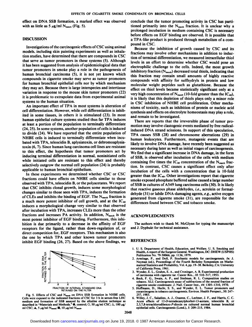
effect on DNA SSB formation, a marked effect was observedwith as little as 5 ng/ml Nmeoh(Fig. 5).
DISCUSSION
Investigations of the carcinogenic effects of CSC using animalmodels, including skin painting experiments as well as inhalation studies, have determined that there are compounds in CSCthat serve as tumor promoters in these systems (5). Althoughit has been suggested from analysis of epidemiológica! data thattumor promoters in smoke play a role in the pathogenesis ofhuman bronchial carcinoma (5), it is not yet known whichcompounds in cigarette smoke may serve as tumor promotersfor human bronchial epithelial cells nor by which mechanismthey may act. Because there is large interspecies and inten issuevariation in response to the mouse skin tumor promoters (22)it is problematic to extrapolate data from experimental animalsystems to the human situation.
An important effect of TPA in most systems is alteration ofcell differentiation. However, while cell differentiation is inhibited in some tissues, in others it is stimulated (23). In mosthuman epithelial culture systems studied thus far TPA inducesat least a portion of the population to terminally differentiate(24,25). In some systems, another population of cells is inducedto divide (24). We have reported that the entire population ofNHBE cells is induced to terminally differentiate when incubated with TPA, teleocidin B, aplysiatoxin, or debromoaplysia-toxin (6, 7). Since human lung carcinoma cell lines are resistantto this effect, the hypothesis that tumor promoters act byinducing terminal differentiation in normal, noninitiated cellswhile initiated cells are resistant to this effect and therebyselectively outgrow the normal cell population (24) is probablyapplicable to human bronchial epithelium.
In these experiments we determined whether CSC or CSCfractions could have effects on NHBE cells similar to thoseobserved with TPA, teleocidin B, or the polyacetates. We reportthat CSC inhibits clonal growth, induces some morphologicalchanges similar to those seen with TPA, induces the formationof CLEs and inhibits the binding of EGF. The Nmeohfraction isa much more potent inhibitor of cell growth, and at the !('.,,,
induces a morphological change very similar to that observedafter incubation with TPA, increases CLEs more than the otherfractions and increases PA activity. In addition, Nmc0his themost potent inhibitor of EGF binding. Furthermore, this inhibition is due primarily to a decrease in the affinity of EGFreceptors for the ligand, rather than down-regulation of, ordirect competition for, EGF receptors. This mechanism is alsothe one by which TPA and other known tumor promotersinhibit EGF binding (26, 27). Based on the above findings, we
=E .8
zzQ O ,6
FRACTION OF INTERNAL STANDARD DNARETAINED ON THE FILTER
Fig. 5. Effects of CSC and N,™*,on DNA SSB formation in NHBE cells.Cells were exposed to the indicated fractions of CSC for 3 h in serum-free LHC imedium and formation of SSB assayed by the alkaline elution technique asdescribed in "Materials and Methods." •,control; O, 50 ng/ml CSC; A, 100 >ig/
ml CSC; A, 5 Mg/ml N-*; •10 „g/mlN_».
2048
conclude that the tumor promoting activity in CSC has partitioned primarily into the Nmeohfraction. It is unclear why aprolonged incubation in medium containing CSC is necessarybefore effects on EGF binding are observed. It is possible thata TPA-like product is produced through metabolism of a compound in CSC.
Because the inhibition of growth caused by CSC and itsfractions may involve other mechanisms in addition to induction of terminal differentiation, we measured intracellular thiollevels in an effort to determine whether CSC would pose anelectrophilic challenge to the cells. Indeed, the most growthinhibitory fraction (Nmcoh)decreased total thiols, indicating thatthis fraction may contain small amounts of highly reactivecompounds with affinity for sulfhydryls in protein and lowmolecular weight peptides such as glutathione. Because theeffect on thiol levels became statistically significant only at avery high concentration of Nmc0h(10-fold greater than the 1C«)),it is likely that this depletion of thiols plays only a minor rolein CSC inhibition of NHBE cell proliferation. Other mechanisms of toxicity, such as inhibition of protein or nucleic acidsynthesis and effects on electrolyte homeostasis may play a role,and remain to be investigated.
There are reports that the irreversible phase of tumor promotion may involve clastogenic events mediated by free radical-induced DNA strand scissions. In support of this speculation,TPA causes SSB (28) and chromosome aberrations (29) inhuman leukocytes. Furthermore, mutagenic events that arelikely to involve DNA damage, have recently been suggested asnecessary during later as well as initial stages of carcinogenesis.We find that a significant increase in DNA damage, in the formof SSB, is observed after incubation of the cells with mediumcontaining five times the IC4o concentration of the Nmeohfraction. In contrast, CSC causes a significant effect only afterincubation of the cells with a concentration that is 10-foldgreater than the K '4„.Other investigations report that cigarette
smoke exposed phosphate buffered saline caused a large numberof SSB in cultures of AS49 lung carcinoma cells (30). It is likelythat reactive gaseous phase aldehydes, i.e.. acrolein or formaldehyde (31) or labile DNA-damaging agents, e.g., active oxygengenerated from cigarette smoke (31), are responsible for thedifferences found between CSC and tobacco smoke.
ACKNOWLEDGMENTS
The authors wish to thank M. McGlynn for typing the manuscriptand J. Dypbukt for technical assistance.
REFERENCES
1. is Department of Health, Education, and Welfare. U. S. Smoking andHealth, A report of the Surgeon General. Washington, DC: DHEW (USPHS)Publication No. 79-50066, pp. 1136, 1979.
2. Armitage, P., and Doll, P. Stochastic models for carcinogenesis. In: J.Neyman (ed.). Proceedings of the Fourth Berkeley Symposium on Mathematical Statistics and Probability, Vol. 4, pp. 19-38. Berkeley, CA: Universityof California Press, 1961.
3. Wynder, E. I .. Grahm, E. A., and Croninger, A. B. Experimental productionof carcinoma with cigarette tar. Cancer Res., IS: 510-517, 1953.
4. Bock, F. G., Swain, A. P., and Stedman, R. L. Composition studies ontobacco. XLI. Carcinogenesis assay of subfractions of the neutral fraction ofcigarette smoke condensate. J. Nati. Cancer Inst., 44:1305-1310, 1970.
5. Hoffmann, D., Hecht, S. S., and Wynder, E. L. Tumor promoters andcocarcinogens in tobacco carcinogenesis. Environ. Health Perspect., 50:247-257, 1983.
6. Willey, J. C, Saladino, A. J., Ozanne, C, Lechner, J. F., and Harris, C. C.Acute effects of 12-O-tetradecanylphorbol-13-acetate, teleocidin B, or2,3,7,8-tetrachlorodibenzo-/>-dioxin on cultured normal human bronchialepithelial cells. Carcinogenesis (Lond.), 5: 209-215, 1984.
on June 19, 2018. © 1987 American Association for Cancer Research. cancerres.aacrjournals.org Downloaded from
EFFECTS OF CIGARETTE SMOKE CONDENSATE ON BRONCHIAL CELLS
7. Willey, J. C, Moser, C. E., and Harris, C. C. The effects of aplysiotoxin anddebromoaplysiotoxin on normal human bronchial epithelial cells. J. CellBiol. Toxicol., I: 145-154, 1985.
8. Blumberg, P. M. in vitro studies on the mode of action of the phorbol esters,potent tumor promoters. Part 2, CRC Crit. Rev. Toxicol., 8: 199-234,1981.
9. Horowitz, A. D., Fujiki, H., Weinstein, I. B., Jeffrey, A., Okin, E., Moore,R. E., and Sugimura, T. Comparative effects of aplysiatoxin, debromaplsyia-toxin, and teleocidin on receptor binding and phospholipid metabolism.Cancer Res., 43:1529-1535, 1983.
10. Moldeus. P., Berggren, M., and Grafstrom, R. W-Acetylcysteine protectionagainst the loxicity of cigarette smoke and cigarette smoke condensâtesinvarious tissues and cells in vitro. EUT.J. Respir. Dis., in press, 1987.
11. Di-Marini. D. M. Genotoxicity of tobacco smoke and tobacco smoke condensate. Mutât.Res., 114: 59-98, 1983.
12. Leuchtenberger, C., Leuchtenberger, R., and Schneider, A. Effects of marijuana and tobacco smoke on human lung physiology. Nature (Lond.), 241:137-139, 1973.
13. Harris, C. C., Autrup, H., Stoner, G. D., and Trump, B. F. Carcinogenesisstudies in human respiratory epithelium. In: C. C. Harris, B. F. Trump, andG. D. Stoner (eds.). Methods in Cell Biology, Vol. 2. New York: AcademicPress, 1978.
14. Lechner, J. F., Stoner, G. D., Haugen, A., Autrup, H., Willey, J. C., Trump,B. F., and Harris, C. C. In vitro human bronchial epithelial model systemsfor carcinogenesis studies. In: M. Webber, and L. Sekely (eds), In VitroModels for Cancer Research. New York: CRC Press, Inc., in press, 1987.
15. Lechner, J. F., and Kaighn, M. E. Application of the principles of enzymekinetics to clonal growth assays: an approach for delineating interactionsamong growth promoting agents. J. Cell Physiol., 100: 519-530, 1979.
16. Kohn. D. B., Weber, M. J., Carl, P. L., Katzennellenbogen, J. A., andChakravarty, P. K. A peptidyl derivative of [3H]aniline as a sensitive stableprotease substrate. Anal. Biochem., 97: 269-276, 1979.
17. Sun, II.. and Green, H. Differentiation of the epidermal keratinocyte incell culture. Formation of the comified envelope. Cell, 9: 511-512, 1976.
18. Willey, J. C., Moser, C. E., Jr., Lechner, J. F., and Harris, C. C. Differentialeffects of 12-O-tetradecanoylphorbol-13-acetateon cultured normal and neo-plastic human bronchial epithelial cells. Cancer Res., 44: 5124-5126, 1984.
19. Lichti, U., and Gotlesman, M. Genetic evidence that a phorbol ester tumorpromoter stimulates ornithine decarboxylase activity by a pathway that is
independent of cyclic AMP-dependent protein kinases in CHO cells. J. CellPhysiol., 113:433-439, 1983.
20. Kohn, K. W., Ewig, R. A. G., Erickson, L. C., and Zwelling, L. A. Measurement of strand breaks and crosslinks by alkaline elution. In: P. C. Hanawaltand E. C. Friedberg (eds.), DNA Repair, A Laboratory Manual of ResearchProcedures, pp. 379-401. New York: Marcel Dekker, Inc., 1981.
21. Saville, B. A scheme for the colorimetrie determination of microgramamounts of thiols. Analyst, 83:670-672, 1958.
22. Harris, C. C. Concluding remarks: role of carcinogens, cocarcinogens andhost factors in cancer risk. In: C. C. Harris and H. Autrup (eds.), HumanCarcinogenesis, pp. 941-970. New York: Academic Press, 1984.
23. Yamasaki, H. Modulation of cell differentiation by tumor promoters. In- T.
J. Slaga (ed.), Mechanisms of Tumor Promotion, Vol. 3, Tumor Promotionand Co-carcinogenesis in Vitro. New York: CRC Press, Inc., in press, 1987.
24. Yuspa, S. J., Ben, T., Hennings, H., and Lichti, U. Divergent responses inepidermal basal cells exposed to the tumor promoter 12-O-tetradecanoyl-phorbol-13-acetate. Cancer Res., 42: 2344-2349, 1982.
25. Parkinson, E. K., and Emmerson, A. The effects of tumor promoters on themultiplication and morphology of cultured human epidermal keratinocytes.Carcinogenesis (Lond.), 3: 525-531, 1982.
26. Lee, L. S., and Weinstein, I. B. Mechanism of tumor promoter inhibition ofcellular binding of epidermal growth factor. Proc. Nati. Acad. Sci. USA, 76:5168-5172, 1979.
27. Shoyab, M., De Larco, J. E., and Todaro, G. J. Biologically active phorbolesters specifically alter affinity of epidermal growth factor membrane receptors. Nature (Lond.), 279: 387-391, 1979.
28. Birnboim, H. C. DNA strand breakage in human leukocytes exposed to atumor promoter, phorbol myristate acetate. Science (Wash. DC), 215:1247-1249, 1982.
29. l'inerii. I., and Cerutti, P. Tumor promoter phorbol-12-myristate acetateinduces chromosomal damage via indirect action. Nature (Lond.), 293:144-146, 1981.
30. Nakayama, T., Kaneko, M., Kodama, M., and Nagata, C. Cigarette smokeinduces DNA single-strand breaks in human cells. Nature (Lond.), 314:462-464, 1985.
31. Grafstrom, R. C, Willey, J. C, Sundqvist, K., and Harris, C. C. Toxicity oftobacco-related aldehydes in cultured human bronchial epithelial cells. AirForce Tech. Rep., 84: 255-265, 1985.
2049
on June 19, 2018. © 1987 American Association for Cancer Research. cancerres.aacrjournals.org Downloaded from

1987;47:2045-2049. Cancer Res J. C. Willey, R. C. Grafstrom, C. E. Moser, Jr., et al.
in VitroEpithelial Cells Condensate and Its Fractions on Normal Human Bronchial Biochemical and Morphological Effects of Cigarette Smoke
Updated version
http://cancerres.aacrjournals.org/content/47/8/2045
Access the most recent version of this article at:
E-mail alerts related to this article or journal.Sign up to receive free email-alerts
Subscriptions
Reprints and
To order reprints of this article or to subscribe to the journal, contact the AACR Publications
Permissions
Rightslink site. Click on "Request Permissions" which will take you to the Copyright Clearance Center's (CCC)
.http://cancerres.aacrjournals.org/content/47/8/2045To request permission to re-use all or part of this article, use this link
on June 19, 2018. © 1987 American Association for Cancer Research. cancerres.aacrjournals.org Downloaded from