biochemical studying of anabaena … journal of veterinary sciences 2013, 39:91-104 issn 110-2047...
TRANSCRIPT
Alexandria Journal of Veterinary Sciences 2013, 39:91-104 ISSN 110-2047 www.alexjvs.com
Biochemical Studying of Anabaena (Cyanobacteria) On Nile Tilapia
Sabreen E. Fadl1, M. Barakat
1, M. Elgohary
2,
1Biochemistry department, Animal Health Research Institute – kafrelshikh, Egypt.
2Fish diseases department, Animal Health Research Institute – kafrelshikh, Egypt.
Key words: ABSTRACT: Anabaena, Nile Tilapia, body weight, survival rate, immunity, A. hydrophila, hematological study, serum biochemistry.
An 6- An 6- weeks feeding trial was conducted to investigate the effect of Anabaena algae (cyanobacteria) supplemented to ration of Nile Tilapia on growth performance, survival rate, immunity, hematological studied, serum biochemistry and its protective effect against the challenge with A. hydrophila I/P injection . Sixty Nile tilapia were allotted into three equal groups: control fed on non- supplement ration, group fed on ration contain Anabaena 10 % and group fed on ration contain Anabaena 15 %, (control, A1 and A2) and were fed ration equal 3% of the total fish body weight. The results indicated that cyanobacteria supplementation improved (P≤0.05) final body weight, weight gain, feed intake and feed conversion ratio when compared with the control. Cyanobacteria supplementation improved hematological indices of blood in Nile tilapia fish. Moreover, it was observed that higher supplementation level (A2) of cyanobacteria increased blood serum protein and globulin concentrations but had no effect on albumin concentration. However, cyanobacteria deteriorate some blood serum parameters which indicate its damage effect on hepatic and renal cells. Experimental infection with A. hydrophila deteriorate most of the tested parameters and reduces kidneys function. Cyanobacteria inclusion in Nile tilapia diet had an protective effect against A. hyrophila infection.
Sabreen E. Fadl, researcher of Biochemistry, Animal Health Research Institute, kafrelshikh. Egypt.
E-mail: [email protected]
1. INTRODUCTION:
Documentation is required to evaluate the use of biocontrol system as an alternative method for inhibition the fish pathogenic bacteria in infected fish farms (Anne-Marie et al., 2003). Fishes are susceptible to a wide variety of bacterial pathogens (Schmidt et al., 2000). Many of these bacteria capable of causing disease are considered by some to be saprophytic in nature (Toranzo et al., 2005). These bacteria only become pathogens when fishes are physiologically unbalanced,
nutritionally deficient, or there are other stressors, i.e., poor water quality, overstocking, which allow opportunistic bacterial infections to proceed (Anderson, 1995). Bacterial fish diseases caused by Aeromonas species lead every year to considerable economic losses in aquaculture. Aeromonas hydrophila and other aeromonads are among the most common bacteria in freshwater habitats throughout the world (Kompanets et al; 1992). They frequently cause problems in both feral and cultured fish (Cipriano, 2001)
Fadl et. al. /Alexandria Journal of Veterinary Sciences 2013, 39: 91-104
29
it is responsible for heavy economic losses caused by both high mortality and deterioration of product quality (Karunasagar et al, 2003). The course of the disease usually runs in an acute manner. Clinical conditions associated with systemic infection result in mortality within 24–48 hours. In more chronic types of clinical conditions, eroded fins occur as well as skin lesions and sluggish swimming (Lio-Po et al., 1983). Cyanobacteria development became recently the most discussed topic all over the world (Carmichael, 1989). Besides negative effects such as deterioration of physicochemical parameters of aquatic environment accompanied by high pH due to photosynthetic activities, oxygen depletion and bad odor from decaying cyanobacterial biomass, cyanobacteria are able to produce a wide range of bioactive compounds (Carmichael, 2001). Cyanobacteria and fish coevolved in same habitats and thus the question arises whether cyanotoxins containing cyanobacteria given via the natural exposure route as a component of fish diet might affect fish physiology e.g. growth and cause toxin accumulation in fish. Some cyanobacterial species could be a prolific resource for substances with antibacterial activity. There are numerous reports concerning the inhibiting activities from Cyanobacteria against human pathogens (Ibraheem and Abdel-Raouf, 2007), fungi (Sunil and Puranik, 2007), mites (Abdel-Aziz and Abdel-Raouf, 2002), algae (Volk and Furkert, 2006), but there is no data about effects against fish pathogenic bacteria. However, under natural conditions cyanobacteria are also ingested by fish and thus it is of special interest to investigate the impact of cyanobacteria containing fish diets on performance of fish because they are at least in part a source of natural nutrition. To our knowledge, only few papers dealing with cyanobacteria containing diets on fish performance exist but because of ecological importance to reset fish meal it is important to investigate
the potential impacts of cyanobacteria biomass on fish performance and blood biochemical changes. Therefore, the main objective of the present study was to investigate the antibacterial activity of cyanobacterial species (Anabaena), and its effect on growth performance, immune response, haematological profile, blood biochemistry in Nile Tilapia.
2. MATERIALS and METHODS: This study was conducted in the Biochemistry Department, Animal Health Research Institute–kafrelshikh, Egypt to investigate the effect of dietary anabaena (cyanoabacteria) inclusion on growth performance, immune response and some blood serum biochemical changes of Nile tilapia fish. 2.1. Fish used and Husbandry: An 60 fish (mono sex males) Nile tilapia (O. niloticus) with an average body weight of about 20-25 gram, were obtained from the earthen nursing pond of a private local farm. Fish were randomly distributed through a total of three aquaria (20 fish for each aquaria). The fish were adapted to the experimental diets and feeding level for two weeks before the begun of the experimental period. The fish were fed on the experimental diets during 6 weeks of the experimental period. Fish were fed during the acclimation period on an artificial basal ration at a rate of 3% of the body weight 1 meal daily. Water was partially changed once every day, using dechlorinated fresh water. Aeration was provided using air blowers. This management was done according to Abdelhamid et al., (2002). 2.2. Experimental diet and design: An experimental diet was formulated contain 29% crude protein, 5% ether extract and 2235 Kcal ME/Kg to meet the requirement of Nile tilapia according to NRC (1994). Ingredient and proximate composition of the experimental diets are presented in Table 1. Fish were allotted into 3 equal groups. Where control group fed (C) on the experimental diet without

Fadl et. al. /Alexandria Journal of Veterinary Sciences 2013, 39: 91-104
29
Anabaena supplementation. While Anabaena, was added by the concentration of 10 % and 15 % for A1 and A2 respectively of the basal ration as
presented in table, 2. The ration was prepared every two weeks according to Shimeino, et al (1993).
Table (1): ingredients and chemical composition of the basal diet.
Physical composition Chemical composition
Ingredients % Items %
Corn grains 11.00 DM 89.4
Fish meal 5.00 ME* 2235 Kcal/Kg
Corn gluten meal 60% 6.00 CP 29.08
wheat bran 8.00 EE 5.73
Soybean meal, 44% 35.00 CF 6.3
Wheat Middling 10.00 Ash 13.36
Rice Polishing 12.50 NFE** 33.93
fish oil 2.14 P/E ratio*** 123.8
DDGS 9.00
Monocalcium phosphate 0.15
Limestone 0.70
Sodium chloride 0.10
DL-Methionine 0.18
Lysine 0.03
Premix 0.20
*Metabolizable energy: Protein (4.49 Kcal/g), Ether extract (8.5 kcal/g), Carbohydrate (3.48 Kcal/g) as reported from Shiau and Hang (1990). **NFE (Nitrogen free extract) calculated by difference. ***P/E ratio calculated as following (Protein to energy ratio in mg protein/Kcal ME). Table (2): Experimental outline.
Groups Diet type Anabaena* supplementation Control Basal ration --- A1 """"""" 10 % A2 """""" 15 %
*Anabaena (algae from Cyanobacteria Research Laboratory and The Field Drainage Dept., Sakha Agricultural Research Station - Kafr Elsheikh).
2.3. Experimental procedure: Fish were fed daily from artificial basal diet at a feeding rate of 3% of fresh biomass in each aquarium (six days per week) for 6 weeks. Fish were fed once daily at (12 pm) and adjusted at approximately 15-days intervals in response to weight gain. Fish were weighed at the beginning (initial weight)W0) and biweekly for a continuous 6 weeks. Total weight gain (TG) was calculated by the following equation, TG (g) = Wt1-Wt0 Where wt1 is the final body weight (g) and wt0 is the initial body weight (g) according to Castell and Tiews (1980). Survival rate (SR %) was calculated
by the following equation according to Castell and Tiews (1980). S.R= (No. of fish at end / No. of fish at the start) ×100. Hematogram and serum Biochemistry: At the end of the experiment, blood samples were taken from the caudal vein of fish by sterile syringe using EDTA solution used as an anticoagulant before (4 days) and after (7 days) challenge test. The blood samples were used for determining of erythrocyte and leukocyte count (Dacie and Lewis 1984), hemoglobin content (Vankampen, 1961) and packed cell volume (PCV) was calculated according to the formula mentioned by Britton (1963).
Serum was obtained before and after challenge test by centrifugation of blood at
3000 r/pm for 15 min and non-haemolyzed serum was stored in deep freezer for further
Fadl et. al. /Alexandria Journal of Veterinary Sciences 2013, 39: 91-104
29
biochemical analyses. Total protein content was determined calorimetrically according to Henry, (1964). Serum albumin was estimated by a colorimetric method according to Dumas and Biggs, (1972) using commercial kit. Globulin content was calculated by mathematical subtraction of albumin value from that of the total protein. Albumin/Globulin (A/G) ratio was calculated from data of albumin and globulin concentration. Activities of Aspartate Amninotansferase (AST), Alanine Aminotransferase (ALT), urea and Creatinin were determined calorimetrically according to Reitman and Frankel, (1957), serum triglyceride was determined according to Fossati, and Prencipe, (1982) while cholesterol according to Allain et al., (1974). Challenge Test: At the end of experiment, a total number of 31 fish (9 fish for control, 13 for A1 and 9 for A2) were challenged with pathogenic Aeromonas hydrophila at 0.2 ml dose of 24 hr. Saline from virulent bacterial broth of Aeromonas hydrophila 1 X 10 8 cells / ml was injected (0.2 ml / fish) interperitoneal (IP) according to Schaperclaus et al., (1992). Fishes were kept under observation for 7 days to record the clinical since and mortality rate according to Amos (1985). Mortality rate % was calculated = (No. of death in specific period/Total population during that period) x 100. 2.4. Statistical analysis: The obtained numerical data were statistically analyzed using S.P.S.S., (1997) for one-way analysis of variance. When F- test was significant, least significant difference was calculated according to Duncan (1955). 3. RESULTS and DISCUSSION: 3.1. Growth Performance: Effect of dietary Anabaena supplementation on Nile tilapia growth performance are presented in table 3. Statistical analysis of the obtained data revealed that no significant difference between fish body weight at the start of the experiment, while at the end of the experimental period it was observed that anabaena supplementation at 10 or 15 %
(groups 2 and 3) respectively increased (P≤0.05) final body weight and total gain throughout the whole experimental period of Nile tilapia fish by about (30.4% and125.77%) and (113.04% and 458.26%) respectively when compared with the control. Moreover, anabaena inclusion at 10 or 15% in Nile tilapia diet increased feed intake by about 38.3% and 14.2% respectively, however, improved feed conversion ratio by about 38.6% and 79.7% respectively when compared with the control. These results may possibly due to the improved feed intake and nutrient digestibility. Moreover, algae contains several nutrients especially vitamins and minerals that may help in fish growth promotion. The obtained data are supported by Zhao et al. (2006) they indicated that dietary intake of cyanobacteria could increase the growth of tilapia while there are no impacts on feed conversion efficiency. Moreover, feeding rate was higher for the diets containing highest cyanobacteria. The information regarding the relationship between feeding rate and growth rate is important in ensuring minimal feed loss and economic production cost (Hashim et al., 1999). Also, Abdel-Tawwab et al. (2008) who reported that fish fed diets containing Spirulina (5.0 - 10.0 g/kg) had significantly better growth and feed utilization as compared to fish fed the control diet. The results of various research studies show that, algae as dietary additives contribute to an increase in growth and feed utilization of cultured fish due to efficacious assimilation of dietary protein, improvement in physiological activity, stress response, starvation tolerance, and disease resistance (Hasan and Chakrabarti, 2009). The obtained data indicated that cyanobacteria inclusion in Nile tilapia diet at 10 % (A1 group) highly improved fish survival rate when compared with control or higher inclusion level of cyanobacteria (A2). these data disagree with those obtained by Zhao et al. (2006) they indicated that there were no significant differences in mortality between the fish fed different levels of cyanoacteria
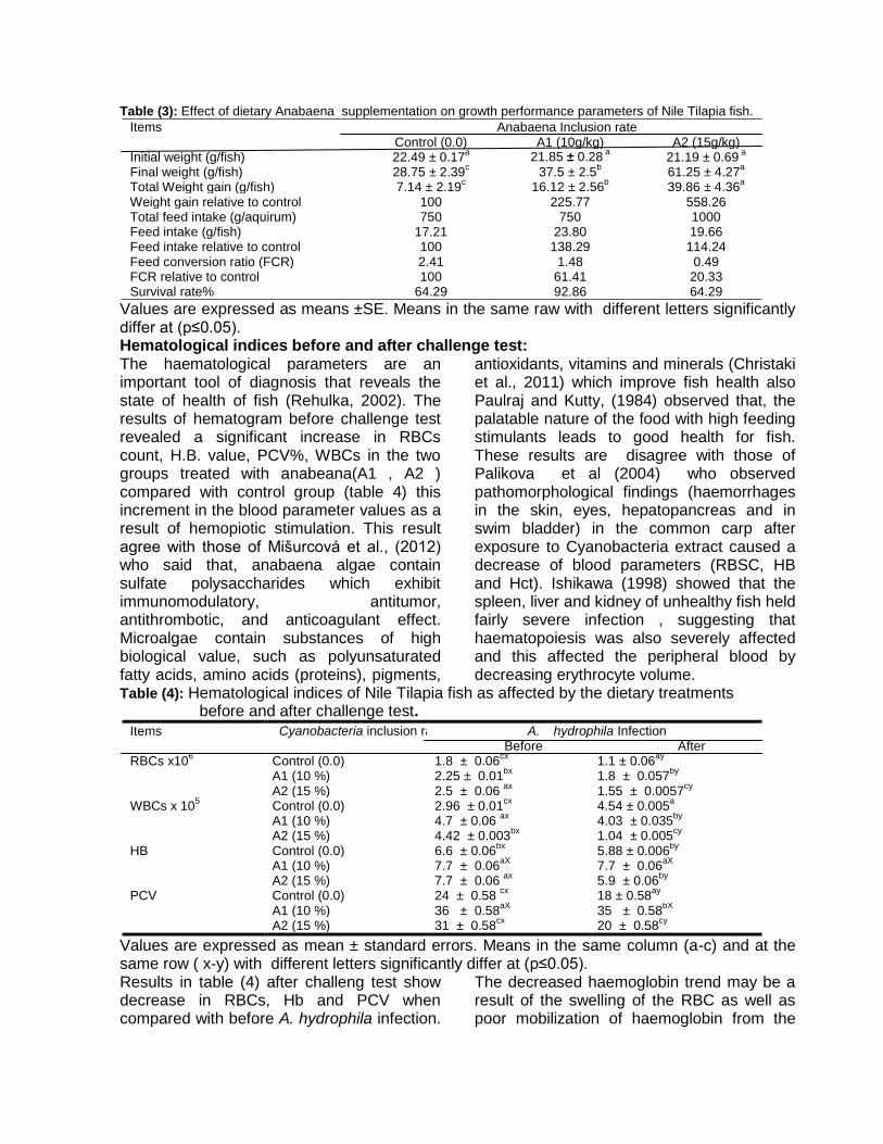
Table (3): Effect of dietary Anabaena supplementation on growth performance parameters of Nile Tilapia fish.
Items Anabaena Inclusion rate Control (0.0) A1 (10g/kg) A2 (15g/kg)
Initial weight (g/fish) 22.49 ± 0.17a 21.85 ± 0.28
a 21.19 ± 0.69
a
Final weight (g/fish) 28.75 ± 2.39c 37.5 ± 2.5
b 61.25 ± 4.27
a
Total Weight gain (g/fish) 7.14 ± 2.19c 16.12 ± 2.56
b 39.86 ± 4.36
a
Weight gain relative to control 100 225.77 558.26 Total feed intake (g/aquirum) 750 750 1000 Feed intake (g/fish) 17.21 23.80 19.66 Feed intake relative to control 100 138.29 114.24 Feed conversion ratio (FCR) 2.41 1.48 0.49 FCR relative to control 100 61.41 20.33 Survival rate% 64.29 92.86 64.29
Values are expressed as means ±SE. Means in the same raw with different letters significantly differ at (p≤0.05). Hematological indices before and after challenge test: The haematological parameters are an important tool of diagnosis that reveals the state of health of fish (Rehulka, 2002). The results of hematogram before challenge test revealed a significant increase in RBCs count, H.B. value, PCV%, WBCs in the two groups treated with anabeana(A1 , A2 ) compared with control group (table 4) this increment in the blood parameter values as a result of hemopiotic stimulation. This result agree with those of Mišurcová et al., (2012) who said that, anabaena algae contain sulfate polysaccharides which exhibit immunomodulatory, antitumor, antithrombotic, and anticoagulant effect. Microalgae contain substances of high biological value, such as polyunsaturated fatty acids, amino acids (proteins), pigments,
antioxidants, vitamins and minerals (Christaki et al., 2011) which improve fish health also Paulraj and Kutty, (1984) observed that, the palatable nature of the food with high feeding stimulants leads to good health for fish. These results are disagree with those of Palikova et al (2004) who observed pathomorphological findings (haemorrhages in the skin, eyes, hepatopancreas and in swim bladder) in the common carp after exposure to Cyanobacteria extract caused a decrease of blood parameters (RBSC, HB and Hct). Ishikawa (1998) showed that the spleen, liver and kidney of unhealthy fish held fairly severe infection , suggesting that haematopoiesis was also severely affected and this affected the peripheral blood by decreasing erythrocyte volume.
Table (4): Hematological indices of Nile Tilapia fish as affected by the dietary treatments before and after challenge test.
Items Cyanobacteria inclusion rate A. hydrophila Infection Before After
RBCs x106 Control (0.0) 1.8 ± 0.06
cx 1.1 ± 0.06
ay
A1 (10 %) 2.25 ± 0.01bx
1.8 ± 0.057by
A2 (15 %) 2.5 ± 0.06
ax 1.55 ± 0.0057
cy
WBCs x 105 Control (0.0) 2.96 ± 0.01
cx 4.54 ± 0.005
a
A1 (10 %) 4.7 ± 0.06 ax
4.03 ± 0.035by
A2 (15 %) 4.42 ± 0.003
bx 1.04 ± 0.005
cy
HB Control (0.0) 6.6 ± 0.06bx
5.88 ± 0.006by
A1 (10 %) 7.7 ± 0.06
aX 7.7 ± 0.06
aX
A2 (15 %) 7.7 ± 0.06 ax
5.9 ± 0.06by
PCV Control (0.0) 24 ± 0.58
cx 18 ± 0.58
ay
A1 (10 %) 36 ± 0.58aX
35 ± 0.58bX
A2 (15 %) 31 ± 0.58
cx 20 ± 0.58
cy
Values are expressed as mean ± standard errors. Means in the same column (a-c) and at the same row ( x-y) with different letters significantly differ at (p≤0.05). Results in table (4) after challeng test show decrease in RBCs, Hb and PCV when compared with before A. hydrophila infection.
The decreased haemoglobin trend may be a result of the swelling of the RBC as well as poor mobilization of haemoglobin from the

Fadl et. al. /Alexandria Journal of Veterinary Sciences 2013, 39: 91-104
29
spleen to other hemopoeitic organs (Scott and Rogers, 1981). The data support the present findings that the significant decrease in RBC and haemoglobin content is possibly due to hypo chromic microcytic anemia caused by A. hydrophila (Kumar and Ramulu, 2013). The destruction of erythrocytes and decline in haemoglobin content can be due to impaired osmoregulation and increase in the tissue damage by the pathogens and other stress factors. A significant reduction in haematocrit may be due to severe bacterial infection affecting the haemopoiesis mechanisms(Mona et al., 2011). However the WBC count increased especially in control infected group but decreased in A2 Changes in WBC count indicate the response to infections in fishes. The elevated count of WBC denotes the stressed state and might have resulted due to the direct stimulation of immunological mechanism to combat stressors. This result agree with Fernandez, and Mazon, (2003) who said that, WBC significant increased up to the 10th day after infection and then declined. Meanwhile the effect of A. hydrophila on anabaena groups was mild except in WBCs in A2, where decreased significantly compared with control infected group. This may be due to defense mechanism of WBCs.
The data support the present findings that the mortality rate in A2 is zero. Neveen and Ibraheem, (2008) said that, anabaena has inhibitory substance inhibit four species of aeromonas. 3.2. Blood serum units: Effect of dietary cyanobacteria inclusion (Anabaena) in Nile tilapia diet on some blood serum parameters are presented in table,( 5). Statistical analysis of the obtained data indicated that higher inclusion rate of Anabaena (Cyanobacteria) increased (P≤0.05) total blood serum protein concentration by about 9.6% when compared with control, while lower cyanobacteria (A1) decreased (P≥0.05) blood serum protein concentration by about 9.6%. However, cyanobacteria had no significant effect on blood serum albumin concentration when compared with control. Reduction of total protein levels in the low inclusion of Anabaena treated group (Hypoproteinemia) compared to control group, as was found by Kopp et al. (2010) who treated carp with Microcystis. Also Marzouk et al. (2013) who stated that plasma total protein analysis showed significantly (P<0.05) decreased levels (Hypoproteinemia) in the treated group than that in the control.
Table (5): Some blood serum parameters of Nile Tilapia fish as affected by the dietary treatments before and after challenge test.
Items Cyanobacteria
inclusion rate
A. hydrophila Infection
Before After
Total protein(g/dl) Control (0.0) 4.06 ± 0.01bx
4.1 ± 0.06 ax
A1 (10 %) 3.67 ± 0.02cx
3.48 ± 0.005bx
A2 (15 %) 4.45 ± 0.005ax
2.9 ± 0.058cy
Albumin (g/dl) Control (0.0) 1.86 ± 0.05ax
1.76 ± 0.005ay
A1 (10 %) 1.86 ± 0.02ax
1.66 ± 0.006by
A2 (15 %) 1.86 ± 0.001ax
1.17 ± 0.01cy
Globulin (g/dl) Control (0.0) 2.2 ± 0.03bx
2.34 ± 0.051ay
A1 (10 %) 1.81 ± 0.001cx
1.81 ± 0.001bx
A2 (15 %) 2.59 ± 0.01ax
1.73 ± 0.055 by
Values are expressed as mean ± standard errors. Means in the same column (A-c) and at the same row ( x-y) with different letters significantly differ at (p≤0.05).
The decreased levels of total protein in the present study could be due to the stressed fish which may increase cortisol secretion and consequently suppresses the
immunoglobulin function (Reddy & Leatherland 1998). Furthermore, under stress conditions, the protein consumed by fishes is not stored in the body tissue

Fadl et. al. /Alexandria Journal of Veterinary Sciences 2013, 39: 91-104
29
(Baskaran & Palanichamy, 1990) and hence, the stressed fish meet their extra energy requirements from body proteins, which are mobilized to produce glucose, that is made available for fishes by the process of gluconeogenesis. So, this depletion of the protein levels may have been due to its utilization for metabolic purposes. Cyanobacteria supplementation at higher level (A2) improved (P≤0.05) blood serum globulin concentration while lower inclusion rate (A1) decreased (P≤0.05) serum globulin concentration when compared with the control. Moreover, the measurement of albumin, globulin, and total protein in serum or plasma is of considerable diagnostic value in fish, as it affects the general nutritional status as well as the integrity of the vascular system and liver function. Experimental infection of Nile Tilapia with A. hydrophila decreased blood serum total protein and albumin concentrations by about (0.98% and 5.4%), (9.2% and 10.8%) and (34.8% and 37.1%) respectively when compared with the same group before infection. On the other hand it was observed that cyanobacteria supplementation at higher concentration (A2) increased blood serum globulin concentration after infection which indicate the immunostimulant effect of cyanobacteria at infection period. 3.3. Liver and Kidney Functions parameters: Blood serum urea analysis indicated significantly lower level (table 6) of Nile tilapia fish groups fed on diet supplemented by cyanobacteria (groups A1 and A2) by about 8.7% and 13.4% respectively when compared with the control. Moreover, statistical analysis of the obtained data indicated that significant increase of blood serum creatinine concentration of cyanobacteria treated groups when compared with the control. The obtained data are supported by Marzouk et al. (2013) they
reported that creatinine analyses indicated elevated levels in the treated group than that in the control. In contrary with the results of Kopp et al. (2010) who stated that the Creatinine levels of the common carp Cyprinus carpio exposed to toxic Microcystis were decreased, while Carbis et al. (1996) found no changes in Creatinine levels in that exposed carp. The difference may be related to different fish species. The increase in creatinine level in the present experiment may indicate kidney damage or malfunction. In comparison to previous studies we can conclude that the Nile tilapia, which forages on algae filtered from the water using tiny combs in the gills, is less susceptible to cyanobacterial toxins than non-phytoplanktonophagous fish such as the common carp. There are two possible hypotheses concerning the difference in susceptibility of the Nile tilapia and common carp. First, cyprinid fish have longer intestines with greater absorption capacity and are thus able to accummulate higher toxin concentrations. Second, fish such as the Nile tilapia digesting cyanobacteria come into greater contact with toxins and thus have better and more effective detoxification mechanisms. Cyanobacteria supplementation at low level (A1 group) significantly reduced AST blood serum concentration by about 14.8% while had no effect on blood serum ALT when compared with the control. On the other hand higher inclusion rate of Cyanobacteria (group A2) significantly elevated blood serum AST and ALT concentrations by about 33.3% and 112.5% respectively when compared with the control. The obtained data indicated that cyanobacteria (Anabaena) supplementation in Nile tilapia fish diet may damage hepatic cells and reduce liver functions at higher inclusion rate.
Fadl et. al. /Alexandria Journal of Veterinary Sciences 2013, 39: 91-104
29
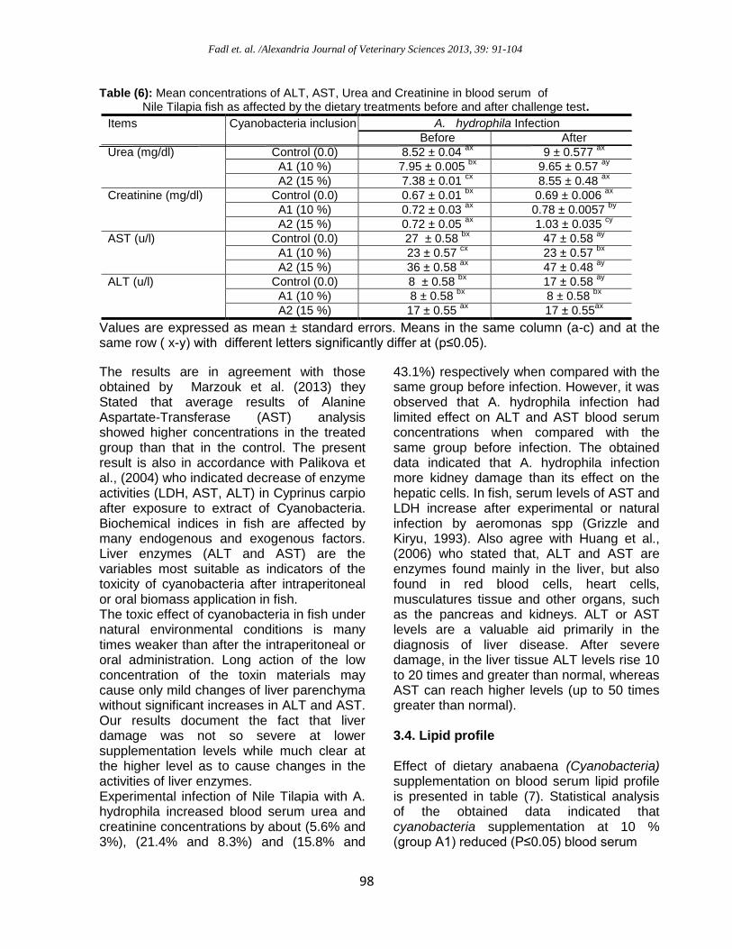
Table (6): Mean concentrations of ALT, AST, Urea and Creatinine in blood serum of Nile Tilapia fish as affected by the dietary treatments before and after challenge test.
Items Cyanobacteria inclusion rate A. hydrophila Infection
Before After
Urea (mg/dl)
Control (0.0) 8.52 ± 0.04 ax
9 ± 0.577 ax
A1 (10 %) 7.95 ± 0.005 bx
9.65 ± 0.57 ay
A2 (15 %) 7.38 ± 0.01 cx
8.55 ± 0.48 ax
Creatinine (mg/dl) Control (0.0) 0.67 ± 0.01 bx
0.69 ± 0.006 ax
A1 (10 %) 0.72 ± 0.03 ax
0.78 ± 0.0057 by
A2 (15 %) 0.72 ± 0.05 ax
1.03 ± 0.035 cy
AST (u/l)
Control (0.0) 27 ± 0.58 bx
47 ± 0.58 ay
A1 (10 %) 23 ± 0.57 cx
23 ± 0.57 bx
A2 (15 %) 36 ± 0.58 ax
47 ± 0.48 ay
ALT (u/l) Control (0.0) 8 ± 0.58 bx
17 ± 0.58 ay
A1 (10 %) 8 ± 0.58 bx
8 ± 0.58 bx
A2 (15 %) 17 ± 0.55 ax
17 ± 0.55ax
Values are expressed as mean ± standard errors. Means in the same column (a-c) and at the same row ( x-y) with different letters significantly differ at (p≤0.05).
The results are in agreement with those obtained by Marzouk et al. (2013) they Stated that average results of Alanine Aspartate-Transferase (AST) analysis showed higher concentrations in the treated group than that in the control. The present result is also in accordance with Palikova et al., (2004) who indicated decrease of enzyme activities (LDH, AST, ALT) in Cyprinus carpio after exposure to extract of Cyanobacteria. Biochemical indices in fish are affected by many endogenous and exogenous factors. Liver enzymes (ALT and AST) are the variables most suitable as indicators of the toxicity of cyanobacteria after intraperitoneal or oral biomass application in fish. The toxic effect of cyanobacteria in fish under natural environmental conditions is many times weaker than after the intraperitoneal or oral administration. Long action of the low concentration of the toxin materials may cause only mild changes of liver parenchyma without significant increases in ALT and AST. Our results document the fact that liver damage was not so severe at lower supplementation levels while much clear at the higher level as to cause changes in the activities of liver enzymes. Experimental infection of Nile Tilapia with A. hydrophila increased blood serum urea and creatinine concentrations by about (5.6% and 3%), (21.4% and 8.3%) and (15.8% and
43.1%) respectively when compared with the same group before infection. However, it was observed that A. hydrophila infection had limited effect on ALT and AST blood serum concentrations when compared with the same group before infection. The obtained data indicated that A. hydrophila infection more kidney damage than its effect on the hepatic cells. In fish, serum levels of AST and LDH increase after experimental or natural infection by aeromonas spp (Grizzle and Kiryu, 1993). Also agree with Huang et al., (2006) who stated that, ALT and AST are enzymes found mainly in the liver, but also found in red blood cells, heart cells, musculatures tissue and other organs, such as the pancreas and kidneys. ALT or AST levels are a valuable aid primarily in the diagnosis of liver disease. After severe damage, in the liver tissue ALT levels rise 10 to 20 times and greater than normal, whereas AST can reach higher levels (up to 50 times greater than normal). 3.4. Lipid profile Effect of dietary anabaena (Cyanobacteria) supplementation on blood serum lipid profile is presented in table (7). Statistical analysis of the obtained data indicated that cyanobacteria supplementation at 10 % (group A1) reduced (P≤0.05) blood serum
Fadl et. al. /Alexandria Journal of Veterinary Sciences 2013, 39: 91-104
22

Fadl et. al. /Alexandria Journal of Veterinary Sciences 2013, 39: 91-104
011
Table (7): Mean concentrations of cholesterol and triglycerides in blood serum of Nile Tilapia fish as affected by the dietary treatments before and after challenge test.
Items Cyanobacteria inclusion rate
A. hydrophila Infection
Before After
Cholesterol (mg/dl) Control (0.0) 156.2 ± 0.57bx
161.2 ± 0.57 ay
A1 (10 %) 131.2 ± 0.57 cx
141.9 ± 0.56 by
A2 (15 %) 168.7 ± 0.58ax
116.1 ± 0.48 cy
Triglycerides (mg/dl) Control (0.0) 313.0 ± 0.58 bx
263.6 ± 0.56 ay
A1 (10 %) 139.1 ± 0.5 cx
254.5 ± 0.55 by
A2 (15 %) 356.5 ± 0.57ax
209.0 ± 0.65 cy
Values are expressed as mean ± standard errors. Means in the same column (a-c) and at the same row ( x-y) with different letters significantly differ at (p≤0.05).
cholesterol and triglycerides by about 16.0% and 55.6% respectively while, higher supplementation level (group A2) increased (P≤0.05) both parameters by about 8% and 13.9% respectively when compared with the control. Experimental infection of Nile tilapia fish with A. hydrophila increased blood serum cholesterol concentration in Nile tilapia fed on the basal diet without treatment (control) or supplemented with 10 % anabaena (group A1) by about 3.2% and 8.2% respectively when compared with the same group before infection. On contrast, higher level of anabaena supplementation (group A2) decreased blood serum cholesterol supplementation when compared with the same group before infection. However, infection reduced blood serum triglycerides concentration except with lower supplementation of cyanobacteria. The decrease of cholesterol levels may indicate slight damage of the hepatopancreas, similar to the significant increase of lactate levels. The negative effect of different pollutants at sublethal
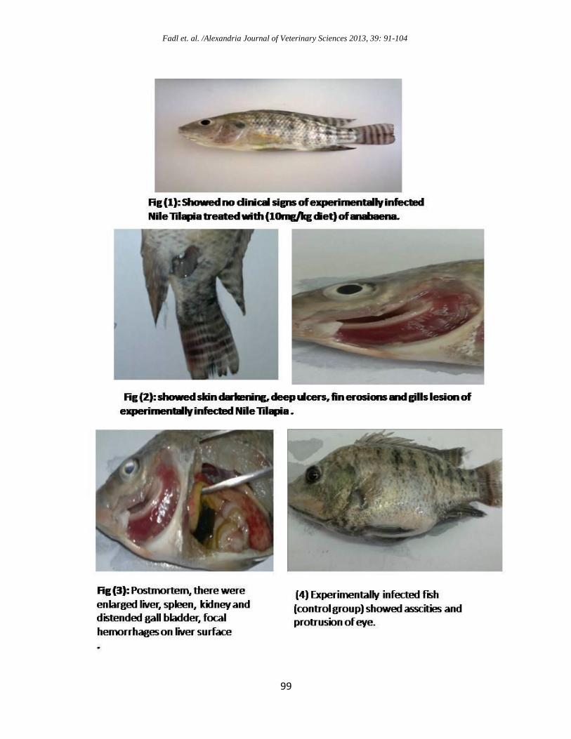
concentrations and stress of fish may be indicated by a decrease in cholesterol values (Svobodova et al. 2006). That explanation may clarify the reduction of cholesterol concentration in most experimental groups after infection. Challenge Test: Experimental infection of Nile Tilapia with aeromonas hydrophila mortality after I/P injection were lasted for 4 days then had stopped (table 8). Clinical findings showed in figure (1) in the first day after injection without any clinical signs and without any postmortem signs this result agree with Mai et al., (2008). In second day after infection clinical signs were in the form of skin darkening, ulcers on the skin varied from shallow to deep necrotizing ulcers, fin erosions, focal hemorrhages at base of fins and gills lesion (Fig. 2). postmortum, there were enlarged liver, spleen, kidney and gall bladder, focal hemorrhages on liver surface (Fig. 3). In thread day the abdomen may become distended as a result of an edema show (Fig . 4 and 5).

Fadl et. al. /Alexandria Journal of Veterinary Sciences 2013, 39: 91-104
010
Table (8): Mortality rate of Nile Tilapia fed with diet containing Anabeana challenged with Aeromonas hydrophila.
Items Anabaena (Cyanobacteria) inclusion rate 0.0 Control 10 % 15 %
Total No. 9 13 9 Dead No. 4 1 0 Survival% 55.56 92.31 100 Mortality% 44.44 11.11 0
Nile Tilapia fed on A1 (10 %) recorded lower mortality rates (11.11%) and zero mortality in A2 (15 %) compared to control (44.44%). Similar results were obtained by Neveen and Ibraheem, (2008) who observed that O. niloticus fed on anabaena had obtained high resistance against bacterial challenge. In addition, the feed additives improved fish immunity and decrease mortality of fish through improvements of the differential leucocytic count in fish as well as enhanced serum bactericidal activity. Honaucins, (2012) said that, Cyanobacteria may possess the ability to produce substances that could one day serve as anti-inflammatory agents and combat bacterial infections in humans. 4. CONCLUSION It could be concluded that anabaena (cyanobacteria) can be included in the Nile tilapia diet up to 15 % as growth promoters. Moreover, cyanobacteria could be deteriorate some blood biochemical parameters which indicate slight damage of the kidney and liver functions. However, cyanobacteria improve immune response of Nile tilapia fish and resistance against Aeromonas hydrophila infection. Further study needed to proof the results and determine the best inclusion rate of cyanobacteria in fish diets.
5. REFERENCES Abdel-Aziz, N.A.M., Abdel-Raouf, N. 2002.
Algal metabolites as alternative acaricide against Tetranychus urtica koch (Acari : Tetranychidae). International Conference for Development and the Environment In the Arab world, March, 26-28, Assuit University Center for Enviromental Studies, Assiut Univ., Assiut, Egypt. pp. 43- 51.
Abdelhamid, A. M., Khalil, F. F. M., El-Barbary, M. I., Zaki, V. H., Husien, H. S. 2002. Feeding Nile tilapia Biogen to detoxify aflatoxin diets. In Proceeding of the 1st Annual Scientific Conference of Animal and Fish Production. Mansoura. 208-230.
Abdel-Tawwab, M. Ahmad. M.H. Abdel-Hadi, Y.M., Seden, M.E. 2008. Use of SPIRULINA (Arthrospir Platensis) as a growth and immunity promoter for Nile tilapia (Oreochromis Niloticus (L.)) fry challenged with pathogenic AEROMONAS HYDROPHILA. 8th International Symposium on Tilapia in Aquaculture. 85:1015 – 1032.
Allain, C. C., Poon, L. S., Chan, C. S. G.1974. Enzymatic determination of total cholesterol in serum. Clin. Chem. 20, 470-275
Amos, H. K. 1985. Procedures for the detection and identification of certain fish pathogens. Am. Fish Soc. Oregan, 3rd ed.
Anderson DP (1995). Novel techniques for fish disease diagnosis. In Diseases in Asian Aquaculture II. Eds Shariff M, Arther JR, Subasinghe RP. Fish Health Section. Asian Fish. Soc. Manilla, pp. 27-39.
Anne-Marie, B., Asbjorn, H., Kristian, S.T., Fredrik, H.J. 2003. Removal of fish pathogenic bacteria in biological sand filters. Water Res. 37:2618-2626.
Baskaran, P., Palanichamy, S. 1990. Impact of agricultural (ammonium chloride) fertilizer on physiology and biochemistry of freshwater teleost fish, Oreochromis mossambicus. J. Ecobiol., 2: 97-106.
Britton, C. J. 1963. "Disorders of the Blood",9th ed. I. A. Churchill, Ld. London. United Kingdom.
Carbis, C. R. Mitchell, G. F., Anderson, J. W., McCauley I. 1996. The effects of microcystins on the serum biochemistry of
Fadl et. al. /Alexandria Journal of Veterinary Sciences 2013, 39: 91-104
019
carp, Cyprinus carpio L., when the toxins are administered by gavage, immersion and intraperitoneai routes. J. Fish. Dis. 19: 151-159.
Carmichael, W. W. 1989. Freshwater cyanobacteria (blue-green algae) toxins. In: Owenby CL, Odell GV, eds, Natural Toxins: Characterization, Pharmacology and Therapeutics, Pergamon Press, Oxford: pp 3–16.
Carmichael, W. W. 2001. Health effects of toxin producing cyanobacteria: ”The Cyanohabs”. Hum. Ecol. Risk Assesov.7: 1393–1407.
Castell, J. D., Tiews, K. 1980. Report of the EIFAC. IUNS and ICES working group on the standraization of methodology in fish nutration research. Hamburg, Fedral Republic of Germany, EIFAC Technology, 36: 24-30
Christaki, E., Florou-Paneri, P.; Bonos, E. 2011. "Microalgae: A novel ingredient in nutrition". Int. J. Food Sci. Nut. 62 (8): 794–799.
Cipriano, C. Rocco. C. 2001. Aeromonas hydrophila and Motile Aeromonas Septicemias of fish. Fish dis. leaflet 68: 25-30.
Decie, S., Lewis, S. 1984. Practical hematology 7th ed., Churchill Livingstone, London.
Dumas, B. T., Biggs, H. G. 1972. Standard Methods of Clinical Chemistry. Ed. Academic Press, New York.
Duncan, D. B.1955. Multiple Ranges and Multiple F – test. Biometerics, 11:1-42.
Fernandez, M. N., Mazon, A.F. 2003. Environmental pollution and fish gill morphology, In. Fish Adaptations (Val AL Kapoor, B.G. Eds.) Sci Pub Enfield, USA. 203- 231.
Fossati, P., Prencipe, L., 1982. Serum triglycerides determined colorimetrically with an enzyme that produces hydrogen peroxide. Clin. Chem. 28: 2077-2082.
Grizzle, J.M., Kiryu., Y 1993. Histopathology of gill, liver and pancrease, and serum enzyme levels of channel catfish infected with aeromonas hydrophila complex. J. Aqu. Animal Health 5:36-50.
Hasan, M. R., Chakrabarti, R. 2009. Use of algae and aquatic macrophytes as feed in small-scale aquaculture. A review. Food And Agriculture Organization of The United Nations Rome, 2009.
Hashim, R., Chong, A., Ali, A.1999.The effect of feeding rate on water quality and growth of red Tilapia fry culture in portable canvas tanks .J. Biosci.10:39-46.
Henry, R. J.1964. Colorimetric determination of total protein. In: Clinical Chemistry. Harper and Row Publ., New York, pp 181.
Honaucins, A. C., 2012. Potent Inhibitors of Inflammation and Bacterial Quorum Sensing: Synthetic Derivatives and Structure-Activity Relationships. Chemistry & Biology, 25:14-20.
Huang XJ, Choi YK, Im HS, Yarimaga O, Yoon E and Kim HS, 2006. aspartate ami-notransferase (AST/GOT) and alanin ami-notransferase (ALT/GPT) detection tech-niques. Sesors 6, 756-782.
Ibraheem, I.B.M., Abdel-Raou, N. 2007. Allelopathic activity of some local cyanobacterial extra-metabolites against some pathogenic bacteria. Egypt. J. Phycol. 8: 120-129.
Ishikawa, C.M. 1998. Oreochromis niloticus (Tilapia do Nilo) inoculados experimentalmente com Mycobacterium marimum ATCC 927. M. Sc. Thesis . University of Sao Paulo. Sao Paula, Brazil.
Karunasagar, I., Karunasagar, I., Otta, S. K. 2003. Disease problems affecting fish in tropical environments. J. Applied Aquacul. 13: 231-249.
Kompanets, E. V., Isaeva, N. M., Balakhnin, I. A. 1992. Bacteria of genus Aeromonas and their role in aquaculture .Microbial .Zh; 54 (4): 89-99.
Kopp, R.; Palikova, M.; Navratil, S.; Kubicek, Z.; Zikova, A. and Mares, J. (2010). Modulation of Biochemical and Haematological Indices of Silver Carp (Hypophthalmichthys molitrix Val.) Exposed to Toxic Cyanobacterial Water Bloom. Acta Vet. Brno., 79: 135–146.
Kumar, M.P. Ramulu, K.S. 2013. Hematological changes in pangasius hypophthalmus infected with Aeromonas

Fadl et. al. /Alexandria Journal of Veterinary Sciences 2013, 39: 91-104
019
hydrophila. Int. J. Food Agr. Vet. Sci. Vol. 3 (1) January-April, pp. 70-75.
Lio-Po, G. D., Pascual, J. P., Santos, J. G. 1983. Philippines. Fish Quarantine and fish diseases in Southeast Asia, report of a workshop held in Jakarta, Indonesia, 7–10 Dec. 1982. Ottawa Ont. IDRC (79pp.), 35–46.
Mai, D.I, Mostafa, M.M, Arab, R.M.H. Rezk, M.A. 2008. Prevalence of aeromonas hydrophila infection in wild and culture tilapia nilotica (O. Niloticus) in Egypt. 8th international symposium on tilapia in aquaculture.
Marzouk1, M.S.; Mostafa, M. ; Ibrahim, N.A.; Pick, F.R., Mahmoud, S. Sharaf, M.S. 2013. Effect of freshwater toxic and non toxic cyanobacteria, (Microcystis aeruginosa) strains on some biochemical parameters of Oreochromis niloticus. Egypt. J. Aquat. Biol. & Fish 17: 55-68.
Mišurcová, L., Škrovánková, S. A., Samek, D. A., Ambrožová, J., Machů, L. 2012. Health Benefits of Algal Polysaccharides in Human Nutrition. Advances in Food and Nut. Res. 66: 75–145
Mona, M. Ismael, Soliman, W. S., Amnah, Rayes, A.,H. 2011. Trials for Neem leaf extract treatment against MAS in Nile tilapia, Oreochromis niloticus Academia Arena, 2011:3-54-60
Neveen, A.R., Ibraheem, I.B.M. 2008. Antibiotic activity of two Anabaena species against four fish pathogenic Aeromonas species. African Journal of Biotechnology Vol. 7 (15), pp. 2644-2648, 4.
NRC (National Research Council), 1994. Nutrient requirement of fish. National Academy Press Washington D.C.
Palikova, M., Navratil, S., Krejcf, R., Sterba, F., Tichy, F., Kubala, L. 2004. Outcomes of repeated exposure of carp (Cyprinus carpio L) to Cyanobacteria extract. Acta. Vet. Brno., 73: 259- 265.
Paulraj, S. and Kutty, M.N. 1984. Food conversion efficiency and nitrogen balance on Cirrhinus mrigala fingerlings fed on the pelleted feeds compounded with wild legumes. J. Indian Institute of Sci. 65: p59-64.
Reddy, P. K., Leatherland, J. F. 1998. Stress Physiology, In: Fish disease and disorders, Vol.2 Non-infections Disorders. (Eds. Leatherland, J.F. & Woo, P.T.K.), Cabi Pub., 279-301.
Rehulka, J. 2002. Aeromonas causes severe skin lesions in rainbow trout (Oncorhynchus mykiss): clinical pathology, haematology and biochemistry. Acta Vet. Brno 71(3) 351-360.
Reitman, S., Frankel, S. 1957. A colorimetric method for the determination of serum glutamic oxaloaceitic and glutamic pyruvic transaminases. Am. J. Clin. Pathol. 28: 56-63.
S. P. S. S. Statistical package for the social sciences 1997. Revisions 6, spss Inc, Chicago, USA.
Schaperclaus, W., Kulow, H., Schreckenbach, K. 1992. Fish Disease. A.A. Balkema, Rotterdam, the Netherlands.
Schmidt, A.S., Bruun, M. S., Dalsgaard, I., Pedersen, K., Larsen, J. L. 2000. Occurrence of Antimicrobial Resistance in Fish-Pathogenic and Environmental Bacteria Associated with Four Danish Rainbow Trout Farms. Appl. Environ. Microbiol. 66 (11): 4908-4915.
Scott, A. L. Rogers, W. A. 1981. Hematological effects of prolonged sublethal hypoxia on channel catfish Ictalurus punctatus (Rafinesque). J. Fish Biol. 18: 591-601.
Shiau, S.Y. Haung, S.I. 1990. Optimal dietary protein level for hybrid tilapia (Oreochromis niloticus x O. aureus) reared in seawater. Aquacul., 81: 119-127.
Shimeino, S.T., Masumoto, Hujita. T. 1993. Alternative protein sources for fish meal rations of young yellowtail. Nippon Suisan Gakkaishi, 59.pp137-143. Vet. Med., Cairo Univ.
Sunil, T. P., Puranik, P. R. 2007. Screening of terrestrial and freshwater halotolerant cyanobacteria for antifungal activities. World J. Microbiol. Biotechnol., pp. 959-972.
Svobodova, Z, Vykusova B, Modra H, Jarkovsky J, Smutna M. (2006): Haematological and biochemical profile of

Fadl et. al. /Alexandria Journal of Veterinary Sciences 2013, 39: 91-104
019
harvest-size carp during harvest and post-harvest storage. Aquac Res 37: 959-965.
Toranzo, A.E, Beatriz, M. Jesús, L.R. 2005. A review of the main bacterial fish diseases in mariculture systems. Aquacult. 246: 4, 37-61.
Vankampen, E. J. 1961. Determination of haemoglobin. Clin. Chem. Acta, 5: 719-720.
Volk, R., Furkert, F. 2006. Antialgal, antibacterial and antifungal activity of two
metabolites produced and excreted by Cyanobacteria during growth. Microbiol. Res. 161: 180-186.
Zhao, M., Xie, S.Q., Zhu, X.M., Yang, Y.X., Gan, L.Q., Song, L.R., 2006. Effect of dietary cyanobacteria on growth and accumulation of microcystins in Nile tilapia (Oreochromis niloticus). Aquaculture 261, 960–966.