biochimica et biophysica acta - core.ac.uk · institut de biotecnologia i de biomedicina,...
TRANSCRIPT
Biochimica et Biophysica Acta 1843 (2014) 866–874
Contents lists available at ScienceDirect
Biochimica et Biophysica Acta
j ourna l homepage: www.e lsev ie r .com/ locate /bbamcr
Selection against toxic aggregation-prone protein sequences in bacteria
Susanna Navarro, Anna Villar-Piqué, Salvador Ventura ⁎Institut de Biotecnologia i de Biomedicina, Universitat Autònoma de Barcelona, E-08193 Bellaterra, SpainDepartament de Bioquímica i Biologia Molecular, Facultat de Biociències, Universitat Autònoma de Barcelona, E-08193 Bellaterra, Spain
Abbreviations: IPTG, isopropyl β-D-1-thiogalactopyrantetrazolium chloride; TMAO, trimethylamine n-oxide; Gwt, wild type; PDB, protein data bank; IP, propidium iodid⁎ Corresponding author at: Institut de Biotecnologia
Autònoma de Barcelona, E-08193 Bellaterra Spain. Tel.: +E-mail address: [email protected] (S. Ventura).
0167-4889/$ – see front matter © 2014 Elsevier B.V. All rhttp://dx.doi.org/10.1016/j.bbamcr.2014.01.020
a b s t r a c t
a r t i c l e i n f oArticle history:Received 14 December 2013Received in revised form 16 January 2014Accepted 21 January 2014Available online 26 January 2014
Keywords:Protein aggregationProtein foldingProtein evolutionAmyloid peptideBacteria
Despite genetic variation has the potential to arise newprotein functions, spontaneousmutations usually destabilizethe native fold.Misfolded proteins tend to form cytotoxic intracellular aggregates, decreasing cellfitness and leadingto degenerative disorders in humans. Therefore, it is thought that selection against proteinmisfolding and aggrega-tion constrains the evolution of protein sequences. However, obtaining experimental data to validate this hypothesishas been traditionally difficult. Herewe exploit bacteria as amodel organism to address this question. Using variantsof the Alzheimer's related Aβ42 peptide designed to exhibit different in vivo aggregation propensitieswe showherethat, in cell competition experiments, themost aggregation-prone variants are always purged out from the growingpopulation. Flow cytometry analysis of cellular metabolism and viability demonstrates that this purifying effect re-sponds to a clear correlation between physiological burden and intrinsic aggregation propensity. Interestingly, thefitness cost of aggregation appears to be associatedwith aggregation rates rather thanwith overall protein solubility.Accordingly, we show that, by reducing in vivo aggregation rates, the model osmolyte proline is able to buffer themetabolic impact of protein aggregation. Overall, our data provide experimental support for the role of toxic proteinaggregation on the cell fitness landscape and the evolution of natural protein sequences.
© 2014 Elsevier B.V. All rights reserved.
1. Introduction
Most genetic mutations decrease the probability of protein-coding se-quences to pack into compact and stable conformations [1]. Proteinmisfolding usually results in the formation of aggregates, which harmscells and reduces fitness [2]. In this way, protein aggregation is linked tothe onset of an increasing number of human degenerative disorders, in-cluding Alzheimer's or Parkinson's diseases [3,4]. Protein aggregation isa generic structural property of polypeptide chains that directly competeswith native protein folding [5,6]. Therefore, it might constrain the evolu-tion of proteins, in such away that protein sequenceswould be shaped bynatural selection to encode polypeptides that would not aggregate at thecellular concentrations and locations in which they should function [7,8],the so-called “Life on the Edge”hypothesis [9]. This purifying effect of pro-tein misfolding might explain why in all organisms, from bacteria tohumans, the specific rate of synonymous and non-synonymous substitu-tions is slower in highly expressed proteins [10,11], since their mutationwill generate relatively more misfolded species.
Despite the deleterious effect of protein aggregation on cell physiol-ogy is well documented [12,13], the magnitude of this effect has notbeen sufficiently characterized experimentally. Quantification of the in-fluence of genetic mutations on aggregation and fitness is essential to
oside; CTC, 5-cyano-2,3-ditolylFP, green fluorescent protein;ei de Biomedicina, Universitat34 93 586 8956.
ights reserved.
validate the purifying role of aggregation during the evolution of proteinsequences and might also provide molecular mechanistic insights onthe basis of human conformational disorders [14]. However, quantifyinghow sequential traits and the associated aggregation propensity impactcellular metabolism and the cell fitness landscape is a challenging taskbecause, in vivo, the intrinsic properties of proteins are modulated byan array of different factors [15–17]. Bacterial systems provide simplemodels to tackle this question under near-physiological conditions [18].In bacteria, protein aggregation has been shown to impair cell division,causing loss of cell reproductive ability or aging [19,20]. Importantly, theaggregates formed by amyloidogenic proteins in bacteria share manyconformational propertieswith those causing disease [21–23]. In partic-ular, we have shown previously that a fusion of the Alzheimer's relatedAβ42 peptide with GFP forms amyloid-like insoluble deposits whenexpressed in bacteria and impairs cell division [20,24]. Here we usedthismodel system to address the relationship between protein aggrega-tion propensity and protein evolution by analyzing the precise impact ofthree designed Aβ42 variants differing in their in vivo aggregation prop-erties on cell metabolism and viability using flow cytometry and mea-suring how these toxic effects correlate with the fitness cost theyimpose to the cell population.
2. Materials and methods
2.1. Bacterial strains and cell growth
Strains Escherichia coli BL21 DE3 transformed with pET-28a vec-tor (Novagen, INC., Madison, WI) encoding for Aβ42wt-GFP (Green
867S. Navarro et al. / Biochimica et Biophysica Acta 1843 (2014) 866–874
fluorescence protein), F19D and F19D/L34P variants were grown aero-bically in liquid Luria–Bertani (LB) medium containing appropriate an-tibiotics in a rotary shaker at 37 ºC and 250 rpm. Overnight cultureswere diluted 100-fold in LB and allowed to grow to an optical densityat 600 nm (OD600) of 0.5. At the indicated OD, protein expression wasinduced with 1 mM isopropyl β-D-1-thiogalactopyranoside (IPTG) and0.5 M of osmolytes proline and trimethylamine n-oxide (TMAO) wasadded, when necessary. Cultures were let to grow for 24 h.
2.2. Prediction of aggregation propensities
Theoretical aggregation-prone regions were predicted with TANGOusing the default settings [25].
2.3. Fluorescence measurements
After indicated incubation times, bacteria cells were harvested bycentrifugation andwashedwith phosphate-buffered saline (PBS buffer)at pH 7.0. GFP fluorescence in intact cells expressing the Aβ42wt-GFP,F19D and F19D/L34P variantswasmeasured on a Jasco FP-8200 fluores-cence spectrophotometer (Jasco Corporation, Japan) in the 500–600 nmrange using an excitation wavelength of 470 nm.
2.4. Optical fluorescence microscopy
10 μL of cultured cells was centrifuged andwashed in PBS buffer, anddeposited on top of glass slides. Images were obtained under UV lightusing a filter for GFP excitation (450–500 nm) and an emission filter(515–560 nm) in a Leica fluorescence microscope (Leica Microsystems,Germany).
2.5. Electrophoresis and immunoblots
E. coli cells expressing Aβ42wt-GFP, F19D and F19D/L34P variantswere lysed chemically. The cell lysate was centrifuged for 20 min at12,000 g. The supernatant was collected separately and the pellet wasdiluted in the same volume as supernantant. Samples of 10 μL of lysatewere separated on a 10% SDS-polyacrylamide gel electrophoresiscontaining 0.1% SDS. Proteins were transferred to a polyvinylidenedifluoride membrane (Immobilon, Millipore) with a Mini Trans-BlotCell (Bio-Rad). Membranes were blocked in 0.1 M Tris–HCl, pH 7.5,0.5% Tween 20, 1% Triton X-100, 5% bovine serum albumin o/n andprobed for GFP expression using an anti-GFP antibody (Roche) diluted1:1000 o/n in blocking solution. The secondary peroxidase-conjugatedantibody was goat anti-mouse IgG (BioRad) diluted 1:3000 in blockingsolution. Detection was carried out using an enhanced chemilumines-cence (ECL) SuperSignal kit (Pierce).
2.6. Bacterial growth and preparation for flow cytometry analysis
Overnight cultures of E. coli cells expressing Aβ42wt-GFP, F19D andF19D/L34P mutants were diluted 100-fold in LB and allowed to grow toan optical density at 600 nm (OD600) of 0.5. Then cultures were inducedwith 1mM IPTG and samples were taken from exponential and station-ary growth phases after 4, 8 and 24 h, respectively. Protein expressionlevels were comparable among Aβ42-GFP variants in these conditions.For analysis of osmolyte effects, proline and TMAO were added 30 minbefore protein induction. Each sample was vortexed and diluted to anOD of 0.2. Then diluted 10-fold in 0.22-μm filtered PBS. Subsequently,the cell suspension was centrifuged at 3500 g for 10 min, and the cellswere resuspended in 1 mL of PBS. All experiments were performedusing three biological replicates. Non-induced cultures of the three var-iants were incubated and analyzed in the samemanner to serve as neg-ative controls.
2.7. IP staining
To analyze cell cultures viability, samples were stained withpropidium iodide (PI) dye (BD Biosciences) to a final concentration of50 μM in PBS. To prepare killed bacteria as a positive control, 1 mL ofsample was heated at 90 ºC for 10min prior to PI staining. The analyzeddata correspond to the bacteria population gated for SSC vs FITC-A sothat cell debris and non-intact cells were excluded from the analysis.
2.8. CTC staining
To evaluate the respiratory activity of E. coli cultures expressingAβ42wt-GFP, F19D and F19D/L34P mutants, cells were stained with 5-cyano-2,3-ditolyl tetrazolium chloride (CTC) (Invitrogen). 1mL of dilut-ed cells in PBS was treated with 100 μL of the 50 mM CTC workingsolution, vortexed gently to mix and incubated for 30 min at 37 ºC,protected from light.
2.9. Flow cytometry
Flow cytometrywas performed using a BD FACSCanto flow cytometer(BD Biosciences) equipped with 488 nm and 635 nm lasers. Cells werefirst gated (P1) by forward scatter (FSC) and side scatter (SCC) both seton logarithmic amplification. A threshold was set on FSC to reduce elec-tronic background noise. Cells in P1 were then re-gated (P2) for greenfluorescence emission (GFP) to separate bacterial cells from cellular de-bris. GFP fluorescence emission was measured on a 530/30 nm bandpass filter and CTC fluorescence emission on a 670LP filter. All signalswere amplified. We acquired a total of 20,000 events logarithmically(five decades). Data were acquired with the FACSDiva Software (BDBiosciences). Data analysis was performed with the FlowJo software.
2.10. Competitive growth selection
Aβ42wt-GFP, F19D and F19D/L34P constructs were grown aerobi-cally in LB medium containing 50 μg/mL kanamicin at 37 ºC and250 rpm. The overnight cultureswere diluted 100-fold in a 50mL falcontube containing LB medium, 50 μg/mL kanamicin and 1 mM IPTG, andallowed to grow at 37 ºC. At 8, 24 and 48 h harvest time, the DNA waspurified using the Plamid Miniprep kit (Thermo Scientific) followingthemanufacturer instructions and sent for sequencing. Chromatogramsof the samples were analyzed with the software Sequence Scanner(Applied Biosciences) to assign the dominant construct.
3. Results
3.1. Design of Aβ42 peptide variants displaying different aggregationpropensity
We have shown previously that the bacterial IBs formed by theAlzheimer's disease-relatedAβ42 peptide display an amyloid-like struc-ture when this peptide is expressed alone [22] or as a fusion to differentfluorescent proteins [24]. We have constructed a library of 20 differentAβ42-GFP mutants differing only in the residue in position 19 of theAβ42 peptide, which is a Phe residue in the wild type sequence [26].All these fusions accumulate essentially in the insoluble cellular fraction.However, the presence of active GFP in such aggregates differs signifi-cantly, displaying a high correlation with the aggregation propensityof the Aβ42 mutants, due to the existence of an in vivo kinetic competi-tion between the folding of the GFP domain and the aggregation of thefusion protein, directed by the Aβ moiety [27]. The Asp mutant (F19D)exhibited the slowest aggregation rate [28]. Here we wanted to furtherincrease this dynamic range and generate an Aβ42-GFP variant that notonlywould aggregate slower than the F19Dmutant but would also shiftits solubility at equilibrium from beingmainly insoluble to mostly solu-ble, while keeping changes to a minimum. An analysis of the Aβ42
868 S. Navarro et al. / Biochimica et Biophysica Acta 1843 (2014) 866–874
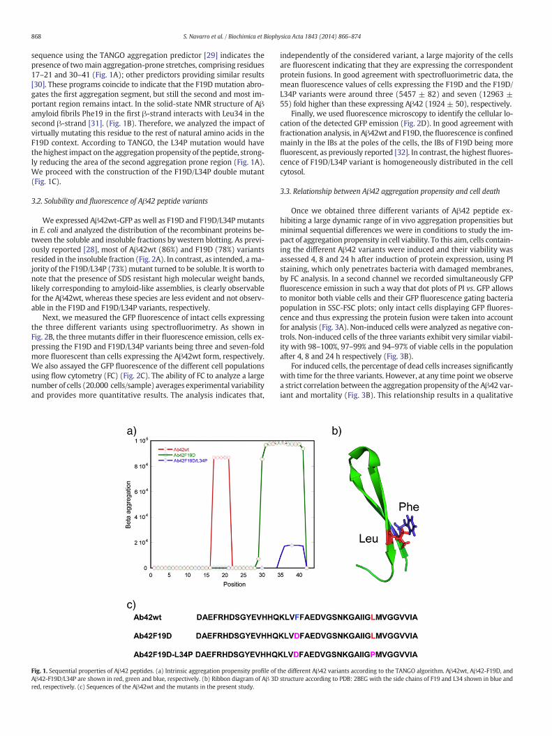
sequence using the TANGO aggregation predictor [29] indicates thepresence of twomain aggregation-prone stretches, comprising residues17–21 and 30–41 (Fig. 1A); other predictors providing similar results[30]. These programs coincide to indicate that the F19Dmutation abro-gates the first aggregation segment, but still the second and most im-portant region remains intact. In the solid-state NMR structure of Aβamyloid fibrils Phe19 in the first β-strand interacts with Leu34 in thesecond β-strand [31]. (Fig. 1B). Therefore, we analyzed the impact ofvirtually mutating this residue to the rest of natural amino acids in theF19D context. According to TANGO, the L34P mutation would havethe highest impact on the aggregation propensity of the peptide, strong-ly reducing the area of the second aggregation prone region (Fig. 1A).We proceed with the construction of the F19D/L34P double mutant(Fig. 1C).
3.2. Solubility and fluorescence of Aβ42 peptide variants
We expressed Aβ42wt-GFP as well as F19D and F19D/L34Pmutantsin E. coli and analyzed the distribution of the recombinant proteins be-tween the soluble and insoluble fractions by western blotting. As previ-ously reported [28], most of Aβ42wt (86%) and F19D (78%) variantsresided in the insoluble fraction (Fig. 2A). In contrast, as intended, ama-jority of the F19D/L34P (73%)mutant turned to be soluble. It is worth tonote that the presence of SDS resistant high molecular weight bands,likely corresponding to amyloid-like assemblies, is clearly observablefor the Aβ42wt, whereas these species are less evident and not observ-able in the F19D and F19D/L34P variants, respectively.
Next, we measured the GFP fluorescence of intact cells expressingthe three different variants using spectrofluorimetry. As shown inFig. 2B, the three mutants differ in their fluorescence emission, cells ex-pressing the F19D and F19D/L34P variants being three and seven-foldmore fluorescent than cells expressing the Aβ42wt form, respectively.We also assayed the GFP fluorescence of the different cell populationsusing flow cytometry (FC) (Fig. 2C). The ability of FC to analyze a largenumber of cells (20.000 cells/sample) averages experimental variabilityand provides more quantitative results. The analysis indicates that,
Fig. 1. Sequential properties of Aβ42 peptides. (a) Intrinsic aggregation propensity profile of tAβ42-F19D/L34P are shown in red, green and blue, respectively. (b) Ribbon diagram of Aβ 3Dred, respectively. (c) Sequences of the Aβ42wt and the mutants in the present study.
independently of the considered variant, a large majority of the cellsare fluorescent indicating that they are expressing the correspondentprotein fusions. In good agreement with spectrofluorimetric data, themean fluorescence values of cells expressing the F19D and the F19D/L34P variants were around three (5457 ± 82) and seven (12963 ±55) fold higher than these expressing Aβ42 (1924 ± 50), respectively.
Finally, we used fluorescence microscopy to identify the cellular lo-cation of the detected GFP emission (Fig. 2D). In good agreement withfractionation analysis, in Aβ42wt and F19D, the fluorescence is confinedmainly in the IBs at the poles of the cells, the IBs of F19D being morefluorescent, as previously reported [32]. In contrast, the highest fluores-cence of F19D/L34P variant is homogeneously distributed in the cellcytosol.
3.3. Relationship between Aβ42 aggregation propensity and cell death
Once we obtained three different variants of Aβ42 peptide ex-hibiting a large dynamic range of in vivo aggregation propensities butminimal sequential differences we were in conditions to study the im-pact of aggregation propensity in cell viability. To this aim, cells contain-ing the different Aβ42 variants were induced and their viability wasassessed 4, 8 and 24 h after induction of protein expression, using PIstaining, which only penetrates bacteria with damaged membranes,by FC analysis. In a second channel we recorded simultaneously GFPfluorescence emission in such a way that dot plots of PI vs. GFP allowsto monitor both viable cells and their GFP fluorescence gating bacteriapopulation in SSC-FSC plots; only intact cells displaying GFP fluores-cence and thus expressing the protein fusion were taken into accountfor analysis (Fig. 3A). Non-induced cells were analyzed as negative con-trols. Non-induced cells of the three variants exhibit very similar viabil-ity with 98–100%, 97–99% and 94–97% of viable cells in the populationafter 4, 8 and 24 h respectively (Fig. 3B).
For induced cells, the percentage of dead cells increases significantlywith time for the three variants. However, at any time point we observea strict correlation between the aggregation propensity of the Aβ42 var-iant and mortality (Fig. 3B). This relationship results in a qualitative
he different Aβ42 variants according to the TANGO algorithm. Aβ42wt, Aβ42-F19D, andstructure according to PDB: 2BEG with the side chains of F19 and L34 shown in blue and

Fig. 2. Intracellular aggregation properties of Aβ42 variants. (a) Immunoblot of SDS-PAGE of the total lysates (T) and the soluble (S) and the insoluble (I) fractions of Aβ42wt andAβ42F19D, Aβ42F19D/L34P expressing cells, probed with an antibody against GFP protein; * indicates the presence of SDS resistant oligomers, (b) GFP relative fluorescence signal ofAβ42wt and F19D, F19D/L34Pmutants in intact E. coli cells as measured by fluorescence spectroscopy, (c) histogram of Aβ42wt-GFP variants showing the average values of fluorescenceintensity at a wavelength of 530 nmas detected by flow cytometry for 20,000 total events, (d) visualization of GFP fluorescence in bacteria expressing Aβ42wt (left), Aβ42F19D (middle),Aβ42F19D/L34P (right) by optical fluorescence microscopy. Scale bar corresponds to 10 μm. All experiments correspond to 4 h after induction of protein expression.
869S. Navarro et al. / Biochimica et Biophysica Acta 1843 (2014) 866–874
correspondence between the mean fluorescence of the cells in the cul-ture and their viability; the higher the fluorescence of the variant, thelower the proportion of dead cells (Fig. 3A). In this way, at the endpointof the experiment, 58% of the cells expressing Aβ42wt are dead, whichcontrast with 94% of non-induced cells bearing the Aβ42wt plasmidbeing viable at this time point. The percentage of dead cells decreasesto 27% and 5% for F19D and F19D/L34P variants, respectively, stronglysuggesting that aggregation properties account for the observed differ-ences in cell viability.
3.4. Relationship between Aβ42 aggregation propensity and cellularmetabolic activity
We wondered whether the toxic effects exerted by the differentAβ42 variants could be associated to changes in the metabolic activityof the respective cells. To this aim the metabolic state of bacterial cellswas evaluated using the 5-cyano-2,3-ditolyl tetrazolium chloride(CTC) probe. CTC is reduced in respiring cells to a red insoluble fluores-cent formazan precipitate. As the membrane of viable cells is imperme-able to the chargedmolecules, positively charged formazan accumulatesintracellulary and this product is quantifiable by FC in individual cells.As above, in a second channel we recorded GFP fluorescence emissionin such a way that dot plots of CTC vs. GFP allows to monitor both met-abolically active cells and their GFP fluorescence; only intact cellsdisplaying GFP fluorescence and thus expressing the protein fusionwere taken into account for analysis in the case of induced cells(Fig. 4A). The respiratory activity of induced and non-induced cellswas assessed at times corresponding to 4, 8 and 24 h after induction ofprotein expression.
A large majority of non-induced cells remain metabolically active forthe three variants at 4 h and 8 h, with 95–99% and 97–99% of the cellsdisplaying respiratory activity (Fig. 4B). After 24 h 19% and 18% of non-induced cells are metabolically inactive for the more aggregation-proneAβ42wt and F19D variants, whereas this value is 7% for F19D/L34P,which suggests that this difference might be associated to certain leakyexpression at this time point.
For induced cells, the percentage of metabolically active cells de-creases with time for the three variants, but at any point time the pro-portion of Aβ42wt expressing active cells is significantly lower thanthat of cells expressing the mutant forms (Fig. 4B). At the endpoint ofthe experiment it is evident a correlation between the aggregation pro-pensity of the variant and themetabolic state of the cells, being 12%, 38%and 54% of the cells expressing Aβ42wt, F19D and F19D/L34P variantsmetabolically active, respectively. It is observed again a direct relation-ship between the mean fluorescence of the cells in the culture and thenumber of active cells (Fig. 4A). The toxic effect of protein aggregationis more evident in the cell respiratory activity than in overall cell viabil-ity, thus even if around 42% of the cells expressing Aβ42wt are aliveafter 24 h, 70% of them seem to bemetabolically compromised. The per-centage of metabolically inactive cells among those alive after 24 h isalso high for the F19D and F19D/L34P mutants, but lower than thosein Aβ42wt samples, with 48% and 43% of living cells displaying low re-spiratory activity, respectively.
We plotted the viability andmetabolic activity for the different Aβ42variants against time (Supplementary Fig. 1). Despite we only analyzedthree time points, it is evident that from the very beginning of the ex-periment the ratio of cell death and loss of respiratory activity is fasterfor the more aggregation-prone Aβ42wt variant. Despite the toxicityof the double mutant is always lower than that of F19D, their effects

Fig. 3. Impact of Aβ42 variants on cell viability. (a) Viability dot plot diagrams of IP staining of E. coli expressing Aβ42wt and Aβ42F19D, Aβ42F19D/L34P mutants evaluated by flow cy-tometry, after 24 h. Non-induced Aβ42wt cells are shown as a negative control. Red fluorescence corresponds to the PerCP-Cy5-A channel showing IP stained cells and Green fluorescencecorresponds to FITC-A channel showing theGFP fluorescence. The lower quadrants correspond to viable cells, and the upper quadrants to dead permeated cells. In the upper quadrants thepercentage of dead cells versus total cells is indicated, (b) percentage of dead cells at 4, 8 and 24 h culture, for non-induced (hatched bars) and induced (solid bars) bacteria, expressingAβ42wt (red) and Aβ42F19D (green), Aβ42F19D/L34P (blue) mutants as derived from flow cytometry analysis of 20,000 events.
870 S. Navarro et al. / Biochimica et Biophysica Acta 1843 (2014) 866–874
and rates are initially fairly similar (until 8 h). However, while the ratesof cell death and loss of respiratory activity for the Aβ42wt and F19D/L34P variants appear to be rather constant along the experiment, thetoxicity of F19D seems to be exacerbated at longer incubation times, ap-parently attaining rates comparable to that of the Aβ42wt form.
3.5. Aβ42 aggregation propensity determines cell fitness
In order to assess whether the aggregating properties of the differentAβ42 variantsmay affect cell fitness andhelp to explain the evolutionaryselection against aggregation-prone sequences in proteins, we grewmixed bacterial cultures, so that cells expressing one Aβ42 variant com-pete in the samemediumwith cells expressing another variant. After 8 h,24 h and 48 h of competition, the DNA was purified and sequenced toestablish the variants content of the mixed culture, allowing to monitorcell fitness in a simple manner; an approach previously used to addressthe role of aggregation gatekeepers in bacterial fitness [33]. Theresults from these assays are shown in Table 1 (each competition wasindependently repeated 5 times). Independently of the pair-wise com-bination, the Sanger sequencing only turned up a single sequence ineach individual replica, indicating the presence of a dominant variantin the competition experiment. For each pair-wise competition experi-ment we considered that one variant competed the other only if all rep-licates rendered the same sequence. After 8 h the F19D/L34P variantalready competes the Aβ42wt sequence, but it is not able to displacethe F19D mutant. In a similar manner, the presence of the Aβ42wtsequence is still detectable in Aβ42wtF19D competition experiments atthis time point. However, we observed that at longer incubation times(24 and 48 h) and independently of the competing pair, the less
aggregating Aβ42 variant always dominates the competition, indicatingthus that, in this model system, cell fitness is linked to aggregationpropensity.
3.6. Chemical chaperones modulate the intracellular aggregation rate ofAβ42 peptide
Osmolytes are present in the intracellular milieu of cells in a widerange of organisms to counterbalance stress conditions that might desta-bilize proteins, acting thus as chemical chaperones by preventing theirmisfolding and subsequent aggregation [34,35]. We analyzed if Glycerol,Betaine, Trehalose, TMAO and Proline had any effect on the aggregationof the Aβ42wt-GFP fusion by monitoring the fluorescence of induced in-tact cells grown in their absence and presence by spectrofluorimetry.Among the assayed osmolytes only TMAO and Pro were able to increasesignificantly GFP fluorescence (Fig. 5A). We fractionated the cells and an-alyzed whether this increase in fluorescence is linked to a higher level ofsoluble protein by western blotting. As it can be seen in Fig. 5B, the pres-ence of osmolytes does not have any noticeable effect on the distributionof the protein between the soluble and insoluble fractions, remaining in allcases essentially as insoluble aggregates, thepresence of SDS resistant spe-cies being detectable in all cases.We confirmed byfluorescencemicrosco-py that all the GFP fluorescence signal was located at the IBs in the cellpoles (Fig. 5C). However, the IBs formed in the presence of osmolyteswere significantly more fluorescent that those of cells grown in their ab-sence. This implies that the effect of the osmolytes emulates that of theF19D mutation, decreasing the aggregation rate of the Aβ moiety andthus allowing the GFP domain to fold significantly before aggregation oc-curs [20].
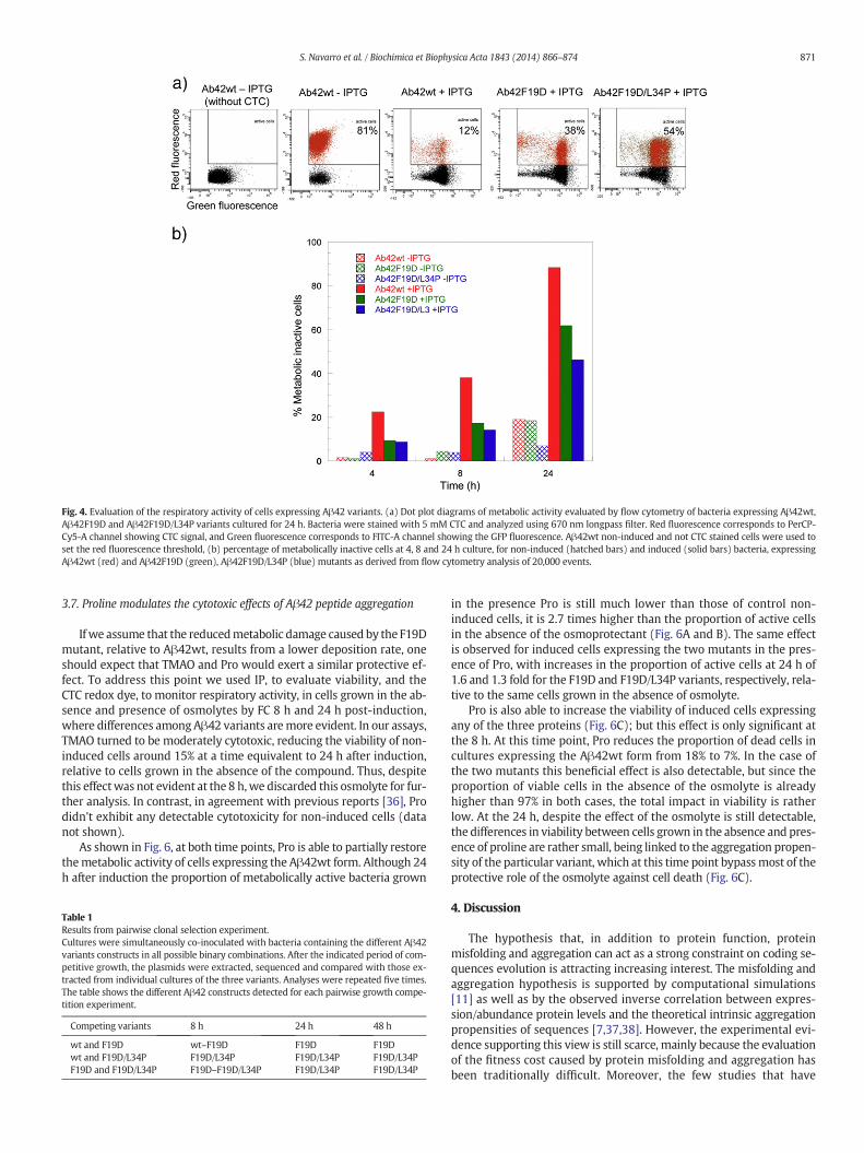
Fig. 4. Evaluation of the respiratory activity of cells expressing Aβ42 variants. (a) Dot plot diagrams of metabolic activity evaluated by flow cytometry of bacteria expressing Aβ42wt,Aβ42F19D and Aβ42F19D/L34P variants cultured for 24 h. Bacteria were stained with 5 mM CTC and analyzed using 670 nm longpass filter. Red fluorescence corresponds to PerCP-Cy5-A channel showing CTC signal, and Green fluorescence corresponds to FITC-A channel showing the GFP fluorescence. Aβ42wt non-induced and not CTC stained cells were used toset the red fluorescence threshold, (b) percentage of metabolically inactive cells at 4, 8 and 24 h culture, for non-induced (hatched bars) and induced (solid bars) bacteria, expressingAβ42wt (red) and Aβ42F19D (green), Aβ42F19D/L34P (blue) mutants as derived from flow cytometry analysis of 20,000 events.
871S. Navarro et al. / Biochimica et Biophysica Acta 1843 (2014) 866–874
3.7. Proline modulates the cytotoxic effects of Aβ42 peptide aggregation
If we assume that the reducedmetabolic damage caused by the F19Dmutant, relative to Aβ42wt, results from a lower deposition rate, oneshould expect that TMAO and Pro would exert a similar protective ef-fect. To address this point we used IP, to evaluate viability, and theCTC redox dye, to monitor respiratory activity, in cells grown in the ab-sence and presence of osmolytes by FC 8 h and 24 h post-induction,where differences amongAβ42 variants aremore evident. In our assays,TMAO turned to be moderately cytotoxic, reducing the viability of non-induced cells around 15% at a time equivalent to 24 h after induction,relative to cells grown in the absence of the compound. Thus, despitethis effectwas not evident at the 8 h,we discarded this osmolyte for fur-ther analysis. In contrast, in agreement with previous reports [36], Prodidn't exhibit any detectable cytotoxicity for non-induced cells (datanot shown).
As shown in Fig. 6, at both time points, Pro is able to partially restorethemetabolic activity of cells expressing the Aβ42wt form. Although 24h after induction the proportion of metabolically active bacteria grown
Table 1Results from pairwise clonal selection experiment.Cultures were simultaneously co-inoculated with bacteria containing the different Aβ42variants constructs in all possible binary combinations. After the indicated period of com-petitive growth, the plasmids were extracted, sequenced and compared with those ex-tracted from individual cultures of the three variants. Analyses were repeated five times.The table shows the different Aβ42 constructs detected for each pairwise growth compe-tition experiment.
Competing variants 8 h 24 h 48 h
wt and F19D wt–F19D F19D F19Dwt and F19D/L34P F19D/L34P F19D/L34P F19D/L34PF19D and F19D/L34P F19D–F19D/L34P F19D/L34P F19D/L34P
in the presence Pro is still much lower than those of control non-induced cells, it is 2.7 times higher than the proportion of active cellsin the absence of the osmoprotectant (Fig. 6A and B). The same effectis observed for induced cells expressing the two mutants in the pres-ence of Pro, with increases in the proportion of active cells at 24 h of1.6 and 1.3 fold for the F19D and F19D/L34P variants, respectively, rela-tive to the same cells grown in the absence of osmolyte.
Pro is also able to increase the viability of induced cells expressingany of the three proteins (Fig. 6C); but this effect is only significant atthe 8 h. At this time point, Pro reduces the proportion of dead cells incultures expressing the Aβ42wt form from 18% to 7%. In the case ofthe two mutants this beneficial effect is also detectable, but since theproportion of viable cells in the absence of the osmolyte is alreadyhigher than 97% in both cases, the total impact in viability is ratherlow. At the 24 h, despite the effect of the osmolyte is still detectable,the differences in viability between cells grown in the absence and pres-ence of proline are rather small, being linked to the aggregation propen-sity of the particular variant, which at this time point bypassmost of theprotective role of the osmolyte against cell death (Fig. 6C).
4. Discussion
The hypothesis that, in addition to protein function, proteinmisfolding and aggregation can act as a strong constraint on coding se-quences evolution is attracting increasing interest. The misfolding andaggregation hypothesis is supported by computational simulations[11] as well as by the observed inverse correlation between expres-sion/abundance protein levels and the theoretical intrinsic aggregationpropensities of sequences [7,37,38]. However, the experimental evi-dence supporting this view is still scarce, mainly because the evaluationof the fitness cost caused by protein misfolding and aggregation hasbeen traditionally difficult. Moreover, the few studies that have
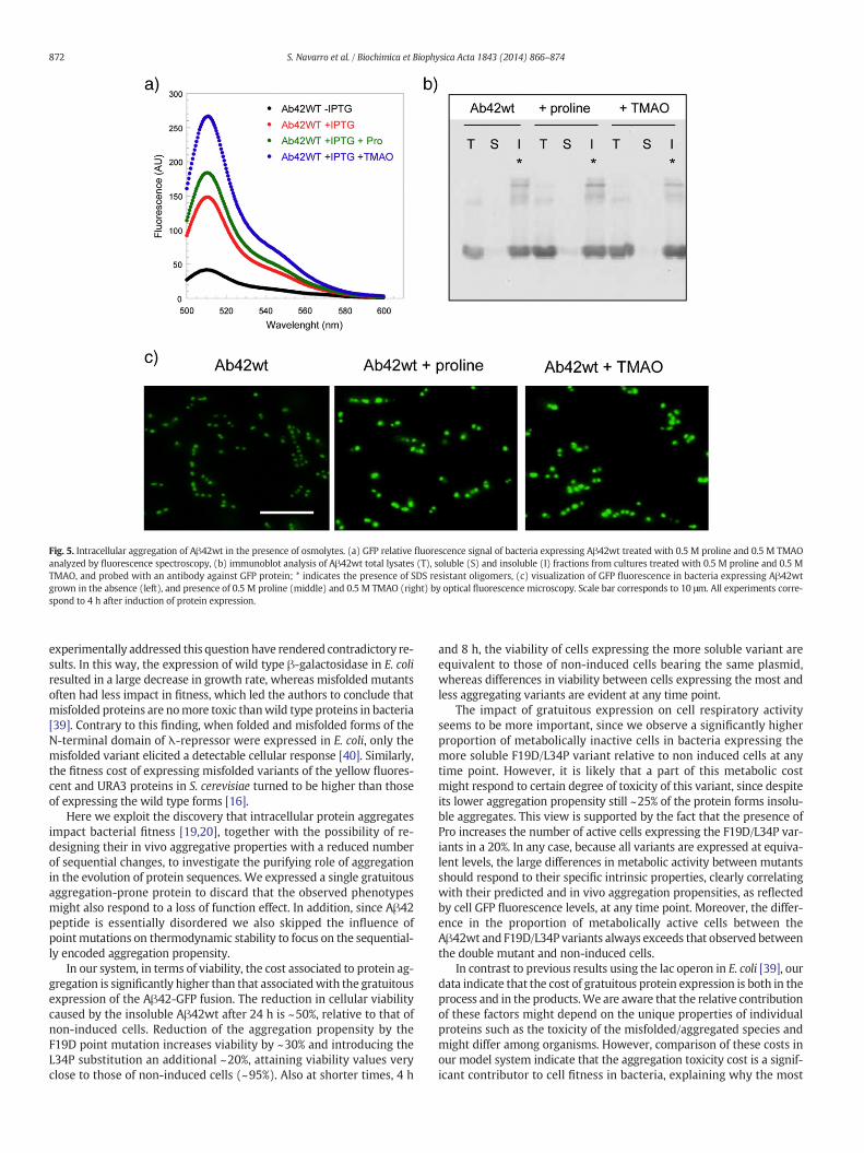
Fig. 5. Intracellular aggregation of Aβ42wt in the presence of osmolytes. (a) GFP relative fluorescence signal of bacteria expressing Aβ42wt treated with 0.5 M proline and 0.5 M TMAOanalyzed by fluorescence spectroscopy, (b) immunoblot analysis of Aβ42wt total lysates (T), soluble (S) and insoluble (I) fractions from cultures treated with 0.5 M proline and 0.5 MTMAO, and probed with an antibody against GFP protein; * indicates the presence of SDS resistant oligomers, (c) visualization of GFP fluorescence in bacteria expressing Aβ42wtgrown in the absence (left), and presence of 0.5 M proline (middle) and 0.5 M TMAO (right) by optical fluorescence microscopy. Scale bar corresponds to 10 μm. All experiments corre-spond to 4 h after induction of protein expression.
872 S. Navarro et al. / Biochimica et Biophysica Acta 1843 (2014) 866–874
experimentally addressed this question have rendered contradictory re-sults. In this way, the expression of wild type β-galactosidase in E. coliresulted in a large decrease in growth rate, whereas misfolded mutantsoften had less impact in fitness, which led the authors to conclude thatmisfolded proteins are nomore toxic thanwild type proteins in bacteria[39]. Contrary to this finding, when folded and misfolded forms of theN-terminal domain of λ-repressor were expressed in E. coli, only themisfolded variant elicited a detectable cellular response [40]. Similarly,the fitness cost of expressing misfolded variants of the yellow fluores-cent and URA3 proteins in S. cerevisiae turned to be higher than thoseof expressing the wild type forms [16].
Here we exploit the discovery that intracellular protein aggregatesimpact bacterial fitness [19,20], together with the possibility of re-designing their in vivo aggregative properties with a reduced numberof sequential changes, to investigate the purifying role of aggregationin the evolution of protein sequences. We expressed a single gratuitousaggregation-prone protein to discard that the observed phenotypesmight also respond to a loss of function effect. In addition, since Aβ42peptide is essentially disordered we also skipped the influence ofpointmutations on thermodynamic stability to focus on the sequential-ly encoded aggregation propensity.
In our system, in terms of viability, the cost associated to protein ag-gregation is significantly higher than that associatedwith the gratuitousexpression of the Aβ42-GFP fusion. The reduction in cellular viabilitycaused by the insoluble Aβ42wt after 24 h is ~50%, relative to that ofnon-induced cells. Reduction of the aggregation propensity by theF19D point mutation increases viability by ~30% and introducing theL34P substitution an additional ~20%, attaining viability values veryclose to those of non-induced cells (~95%). Also at shorter times, 4 h
and 8 h, the viability of cells expressing the more soluble variant areequivalent to those of non-induced cells bearing the same plasmid,whereas differences in viability between cells expressing the most andless aggregating variants are evident at any time point.
The impact of gratuitous expression on cell respiratory activityseems to be more important, since we observe a significantly higherproportion of metabolically inactive cells in bacteria expressing themore soluble F19D/L34P variant relative to non induced cells at anytime point. However, it is likely that a part of this metabolic costmight respond to certain degree of toxicity of this variant, since despiteits lower aggregation propensity still ~25% of the protein forms insolu-ble aggregates. This view is supported by the fact that the presence ofPro increases the number of active cells expressing the F19D/L34P var-iants in a 20%. In any case, because all variants are expressed at equiva-lent levels, the large differences in metabolic activity between mutantsshould respond to their specific intrinsic properties, clearly correlatingwith their predicted and in vivo aggregation propensities, as reflectedby cell GFP fluorescence levels, at any time point. Moreover, the differ-ence in the proportion of metabolically active cells between theAβ42wt and F19D/L34P variants always exceeds that observed betweenthe double mutant and non-induced cells.
In contrast to previous results using the lac operon in E. coli [39], ourdata indicate that the cost of gratuitous protein expression is both in theprocess and in the products.We are aware that the relative contributionof these factors might depend on the unique properties of individualproteins such as the toxicity of the misfolded/aggregated species andmight differ among organisms. However, comparison of these costs inour model system indicate that the aggregation toxicity cost is a signif-icant contributor to cell fitness in bacteria, explaining why the most
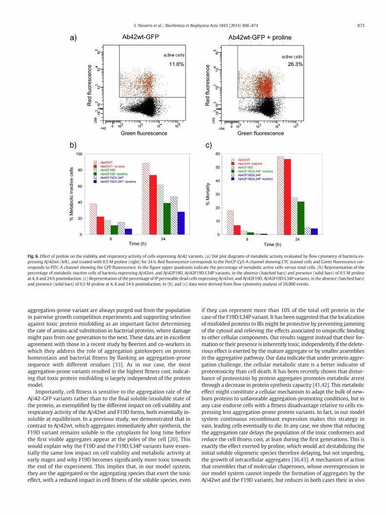
Fig. 6. Effect of proline on the viability and respiratory activity of cells expressing Aβ42 variants. (a) Dot plot diagrams of metabolic activity evaluated by flow cytometry of bacteria ex-pressing Aβ42wt (left), and treated with 0.5 M proline (right) for 24 h. Red fluorescence corresponds to the PerCP-Cy5-A channel showing CTC stained cells and Green fluorescence cor-responds to FITC-A channel showing the GFP fluorescence. In the figure upper quadrants indicate the percentage of metabolic active cells versus total cells. (b) Representation of thepercentage of metabolic inactive cells of bacteria expressing Aβ42wt, and Aβ42F19D, Aβ42F19D/L34P variants, in the absence (hatched bars) and presence (solid bars) of 0.5 M prolineat 4, 8 and 24 h postinduction. (c) Representation of the percentage of IP permeable dead cells expressing Aβ42wt, and Aβ42F19D, Aβ42F19D/L34P variants, in the absence (hatched bars)and presence (solid bars) of 0.5 M proline at 4, 8 and 24 h postinduction. In (b) and (c) data were derived from flow cytometry analysis of 20,000 events.
873S. Navarro et al. / Biochimica et Biophysica Acta 1843 (2014) 866–874
aggregation-prone variant are always purged out from the populationin pairwise growth competition experiments and supporting selectionagainst toxic protein misfolding as an important factor determiningthe rate of amino acid substitution in bacterial proteins, where damagemight pass from one generation to the next. These data are in excellentagreement with those in a recent study by Beerten and co-workers inwhich they address the role of aggregation gatekeepers on proteinhomeostasis and bacterial fitness by flanking an aggregation-pronesequence with different residues [33]. As in our case, the mostaggregation-prone variants resulted in the highest fitness cost, indicat-ing that toxic protein misfolding is largely independent of the proteinmodel.
Importantly, cell fitness is sensitive to the aggregation rate of theAβ42-GFP variants rather than to the final soluble/insoluble state ofthe protein, as exemplified by the different impact on cell viability andrespiratory activity of the Aβ42wt and F19D forms, both essentially in-soluble at equilibrium. In a previous study, we demonstrated that incontrast to Aβ42wt, which aggregates immediately after synthesis, theF19D variant remains soluble in the cytoplasm for long time beforethe first visible aggregates appear at the poles of the cell [20]. Thiswould explain why the F19D and the F19D/L34P variants have essen-tially the same low impact on cell viability and metabolic activity atearly stages and why F19D becomes significantly more toxic towardsthe end of the experiment. This implies that, in our model system,they are the aggregated or the aggregating species that exert the toxiceffect, with a reduced impact in cell fitness of the soluble species, even
if they can represent more than 10% of the total cell protein in thecase of the F19D/L34P variant. It has been suggested that the localizationof misfolded proteins to IBsmight be protective by preventing jammingof the cytosol and relieving the effects associated to unspecific bindingto other cellular components. Our results suggest instead that their for-mation or their presence is inherently toxic, independently if the delete-rious effect is exerted by themature aggregate or by smaller assembliesin the aggregative pathway. Our data indicate that under protein aggre-gation challenge, the cellular metabolic state is a better indicator ofproteotoxicity than cell death. It has been recently shown that distur-bance of proteostasis by protein aggregates promotes metabolic arrestthrough a decrease in protein synthesis capacity [41,42]. This metaboliceffect might constitute a cellular mechanism to adapt the bulk of new-born proteins to unfavorable aggregation-promoting conditions, but inany case endorse cells with a fitness disadvantage relative to cells ex-pressing less aggregation-prone protein variants. In fact, in our modelsystem continuous recombinant expression makes this strategy invain, leading cells eventually to die. In any case, we show that reducingthe aggregation rate delays the population of the toxic conformers andreduce the cell fitness cost, at least during the first generations. This isexactly the effect exerted by proline, which would act destabilizing theinitial soluble oligomeric species therefore delaying, but not impeding,the growth of intracellular aggregates [36,43]. A mechanism of actionthat resembles that of molecular chaperones, whose overexpression inour model system cannot impede the formation of aggregates by theAβ42wt and the F19D variants, but reduces in both cases their in vivo
874 S. Navarro et al. / Biochimica et Biophysica Acta 1843 (2014) 866–874
aggregation rates [20]. This osmolyte mediated modulation of aggrega-tion propensities may allow the evolution of new protein sequencesthat in their absencewould be rapidly purged out due to its negative im-pact on cell fitness, contributing thus to genetic buffering [34]. Overall,we provide consistent and rigorous new experimental data in favor ofa dominant role of protein aggregation in the evolution of the proteinsequences.
Supplementary data to this article can be found online at http://dx.doi.org/10.1016/j.bbamcr.2014.01.020.
Acknowledgements
Work in our lab is supported by grants BFU2010-14901 fromMinisterio de Ciencia e Innovacion (Spain) and 2009-SGR-760 fromAGAUR (Generalitat de Catalunya). S.V. has been granted an ICREAAcademia award (ICREA).
References
[1] A.A. Pakula, R.T. Sauer, Genetic analysis of protein stability and function, Annu. Rev.Genet. 23 (1989) 289–310.
[2] H. Olzscha, S.M. Schermann, A.C. Woerner, S. Pinkert, M.H. Hecht, G.G. Tartaglia, M.Vendruscolo, M. Hayer-Hartl, F.U. Hartl, R.M. Vabulas, Amyloid-like aggregates se-quester numerous metastable proteins with essential cellular functions, Cell 144(2011) 67–78.
[3] F. Chiti, C.M. Dobson, Protein misfolding, functional amyloid, and human disease,Annu. Rev. Biochem. 75 (2006) 333–366.
[4] X. Fernandez-Busquets, N.S. de Groot, D. Fernandez, S. Ventura, Recent structuraland computational insights into conformational diseases, Curr. Med. Chem. 15(2008) 1336–1349.
[5] T.R. Jahn, S.E. Radford, The Yin and Yang of protein folding, FEBS J. 272 (2005)5962–5970.
[6] C.M. Dobson, Protein-misfolding diseases: getting out of shape, Nature 418 (2002)729–730.
[7] G.G. Tartaglia, M. Vendruscolo, Correlation between mRNA expression levels andprotein aggregation propensities in subcellular localisations, Mol. BioSyst. 5(2009) 1873–1876.
[8] P. Ciryam, G.G. Tartaglia, R.I. Morimoto, C.M. Dobson, M. Vendruscolo, Widespreadaggregation and neurodegenerative diseases are associated with supersaturatedproteins, Cell Rep. 5 (2013) 781–790.
[9] G.G. Tartaglia, S. Pechmann, C.M. Dobson, M. Vendruscolo, Life on the edge: a linkbetween gene expression levels and aggregation rates of human proteins, TrendsBiochem. Sci. 32 (2007) 204–206.
[10] D.A. Drummond, C.O. Wilke, The evolutionary consequences of erroneous proteinsynthesis, Nat. Rev. Genet. 10 (2009) 715–724.
[11] D.A. Drummond, C.O. Wilke, Mistranslation-induced protein misfolding as a domi-nant constraint on coding-sequence evolution, Cell 134 (2008) 341–352.
[12] E. Herczenik, M.F. Gebbink, Molecular and cellular aspects of protein misfolding anddisease, FASEB J. 22 (2008) 2115–2133.
[13] G. Invernizzi, E. Papaleo, R. Sabate, S. Ventura, Protein aggregation: mechanisms andfunctional consequences, Int. J. Biochem. Cell Biol. 44 (2012) 1541–1554.
[14] M. Bucciantini, E. Giannoni, F. Chiti, F. Baroni, L. Formigli, J. Zurdo, N. Taddei, G.Ramponi, C.M. Dobson, M. Stefani, Inherent toxicity of aggregates implies a commonmechanism for protein misfolding diseases, Nature 416 (2002) 507–511.
[15] D.A. Parsell, S. Lindquist, The function of heat-shock proteins in stress tolerance:degradation and reactivation of damaged proteins, Annu. Rev. Genet. 27 (1993)437–496.
[16] K.A. Geiler-Samerotte, M.F. Dion, B.A. Budnik, S.M. Wang, D.L. Hartl, D.A.Drummond, Misfolded proteins impose a dosage-dependent fitness cost and triggera cytosolic unfolded protein response in yeast, Proc. Natl. Acad. Sci. U. S. A. 108(2011) 680–685.
[17] E.T. Powers, D.L. Powers, L.M. Gierasch, FoldEco: a model for proteostasis in E. coli,Cell Rep. 1 (2012) 265–276.
[18] R. Sabate, N.S. de Groot, S. Ventura, Protein folding and aggregation in bacteria, Cell.Mol. Life Sci. 67 (2010) 2695–2715.
[19] A.B. Lindner, R. Madden, A. Demarez, E.J. Stewart, F. Taddei, Asymmetric segregationof protein aggregates is associated with cellular aging and rejuvenation, Proc. Natl.Acad. Sci. U. S. A. 105 (2008) 3076–3081.
[20] A. Villar-Pique, N.S. de Groot, R. Sabate, S.P. Acebron, G. Celaya, X. Fernandez-Busquets, A. Muga, S. Ventura, The effect of amyloidogenic peptides on bacterialaging correlates with their intrinsic aggregation propensity, J. Mol. Biol. 421(2012) 270–281.
[21] L. Wang, S.K. Maji, M.R. Sawaya, D. Eisenberg, R. Riek, Bacterial inclusion bodies con-tain amyloid-like structure, PLoS Biol. 6 (2008) e195.
[22] M. Dasari, A. Espargaro, R. Sabate, J.M. Lopez Del Amo, U. Fink, G. Grelle, J. Bieschke,S. Ventura, B. Reif, Bacterial inclusion bodies of Alzheimer's disease beta-amyloidpeptides can be employed to study native-like aggregation intermediate states,Chembiochem 12 (2011) 407–423.
[23] N.S. de Groot, R. Sabate, S. Ventura, Amyloids in bacterial inclusion bodies, TrendsBiochem. Sci. 34 (2009) 408–416.
[24] M. Morell, R. Bravo, A. Espargaro, X. Sisquella, F.X. Aviles, X. Fernandez-Busquets, S.Ventura, Inclusion bodies: specificity in their aggregation process and amyloid-likestructure, Biochim. Biophys. Acta 1783 (2008) 1815–1825.
[25] TANGO, http://tango.embl.de/%5D.[26] N.S. de Groot, F.X. Aviles, J. Vendrell, S. Ventura, Mutagenesis of the central hydro-
phobic cluster in Abeta42 Alzheimer's peptide. Side-chain properties correlatewith aggregation propensities, FEBS J. 273 (2006) 658–668.
[27] N.S. de Groot, S. Ventura, Protein activity in bacterial inclusion bodies correlateswith predicted aggregation rates, J. Biotechnol. 125 (2006) 110–113.
[28] A. Espargaro, R. Sabate, S. Ventura, Kinetic and thermodynamic stability of bacterialintracellular aggregates, FEBS Lett. 582 (2008) 3669–3673.
[29] A.M. Fernandez-Escamilla, F. Rousseau, J. Schymkowitz, L. Serrano, Prediction ofsequence-dependent and mutational effects on the aggregation of peptides andproteins, Nat. Biotechnol. 22 (2004) 1302–1306.
[30] V. Castillo, R. Grana-Montes, R. Sabate, S. Ventura, Prediction of the aggregation pro-pensity of proteins from the primary sequence: aggregation properties ofproteomes, Biotechnol. J. 6 (2011) 674–685.
[31] T. Luhrs, C. Ritter, M. Adrian, D. Riek-Loher, B. Bohrmann, H. Dobeli, D. Schubert, R.Riek, 3D structure of Alzheimer's amyloid-beta(1–42) fibrils, Proc. Natl. Acad. Sci.U. S. A. 102 (2005) 17342–17347.
[32] A. Villar-Pique, N.S. de Groot, R. Sabate, S.P. Acebron, G. Celaya, X. Fernandez-Busquets, A. Muga, S. Ventura, The effect of amyloidogenic peptides on bacterialaging correlates with their intrinsic aggregation propensity, J. Mol. Biol. 421 (2-3)(2012) 270–281.
[33] J. Beerten, W. Jonckheere, S. Rudyak, J. Xu, H. Wilkinson, F. De Smet, J. Schymkowitz,F. Rousseau, Aggregation gatekeepers modulate protein homeostasis of aggregatingsequences and affect bacterial fitness, Protein Eng. Des. Sel. 25 (2012) 357–366.
[34] A. Bandyopadhyay, K. Saxena, N. Kasturia, V. Dalal, N. Bhatt, A. Rajkumar, S. Maity, S.Sengupta, K. Chakraborty, Chemical chaperones assist intracellular folding to buffermutational variations, Nat. Chem. Biol. 8 (2012) 238–245.
[35] P.H. Yancey, M.E. Clark, S.C. Hand, R.D. Bowlus, G.N. Somero, Living with waterstress: evolution of osmolyte systems, Science 217 (1982) 1214–1222.
[36] Z. Ignatova, L.M. Gierasch, Inhibition of protein aggregation in vitro and in vivo by anatural osmoprotectant, Proc. Natl. Acad. Sci. U. S. A. 103 (2006) 13357–13361.
[37] E. Monsellier, M. Ramazzotti, N. Taddei, F. Chiti, Aggregation propensity of thehuman proteome, PLoS Comput. Biol. 4 (2008) e1000199.
[38] V. Castillo, R. Grana-Montes, S. Ventura, The aggregation properties of Escherichiacoli proteins associated with their cellular abundance, Biotechnol. J. 6 (2011)752–760.
[39] G. Plata, M.E. Gottesman, D. Vitkup, The rate of the molecular clock and the cost ofgratuitous protein synthesis, Genome Biol. 11 (2010) R98.
[40] D.A. Parsell, R.T. Sauer, Induction of a heat shock-like response by unfolded proteinin Escherichia coli: dependence on protein level not protein degradation, Genes Dev.3 (1989) 1226–1232.
[41] J. Kirstein-Miles, A. Scior, E. Deuerling, R.I. Morimoto, The nascent polypeptide-associated complex is a key regulator of proteostasis, EMBO J. 32 (2013)1451–1468.
[42] A. Koplin, S. Preissler, Y. Ilina, M. Koch, A. Scior, M. Erhardt, E. Deuerling, A dual func-tion for chaperones SSB-RAC and the NAC nascent polypeptide-associated complexon ribosomes, J. Cell Biol. 189 (2010) 57–68.
[43] J. Seeliger, K. Estel, N. Erwin, R. Winter, Cosolvent effects on the fibrillation reactionof human IAPP, Phys. Chem. Chem. Phys. 15 (2013) 8902–8907.