biochimica et biophysica acta - uci department of...
TRANSCRIPT

Biochimica et Biophysica Acta 1838 (2014) 2350–2356
Contents lists available at ScienceDirect
Biochimica et Biophysica Acta
j ourna l homepage: www.e lsev ie r .com/ locate /bbamem
Glutamate provides a key structural contact between reticulon-4(Nogo-66) and phosphocholine☆
Ali Alhoshani a, Rosemarie Vithayathil b, Jonathan Bandong b, Katherine M. Chrunyk b, Gabriel O. Moreno b,Gregory A. Weiss b,c, Melanie J. Cocco a,b,⁎a Department of Pharmaceutical Sciences, University of California, Irvine, CA 92697, USAb Department of Molecular Biology and Biochemistry, University of California, Irvine, CA 92697, USAc Department of Chemistry, University of California, Irvine, CA 92697, USA
Abbreviations: BACE1, β-amyloid-converting enzymeCRP, human C-reactive protein; fp, fraction of proteidimyristoyl-sn-glycero-3-phosphocholine; DPC, dodecylplinked immunosorbent assay; HSQC, heteronuclear Singlecopy; KX, partition coefficient; NOE, nuclear Overhauser einhibitor, RTN-4; Nogo-66, 66-residue extracellular regioPC, phosphocholine; PfPMT, phosphoethanolaminemethymology domain; RHD-66, 66-residue extracellular region☆ This article is part of a special issue entitled: InterfaciaGuest Editors: William C. Wimley and Kalina Hristova.⁎ Corresponding author at: 1218 Natural Sciences I,
Biochemistry, University of California, Irvine, CA 92697-39E-mail address: [email protected] (M.J. Cocco).
http://dx.doi.org/10.1016/j.bbamem.2014.05.0210005-2736/© 2014 Elsevier B.V. All rights reserved.
a b s t r a c t
a r t i c l e i n f oArticle history:Received 26 February 2014Received in revised form 9 May 2014Accepted 16 May 2014Available online 24 May 2014
Keywords:ReticulonNogo-66Membrane interfaceStructureCholineGlutamate
Human reticulon 4 (RTN-4) has been identified as the neurite outgrowth inhibitor (Nogo). This protein contains a spanof 66 amino acids (Nogo-66)flanked by twomembrane helices at the C-terminus.Wepreviously determined theNMRstructure of Nogo-66 in a native-like environment and defined the regions of Nogo-66 expected to bemembrane em-bedded. We hypothesize that aromatic groups and a negative charge hyperconserved among RTNs (Glu26) drive theremarkably strong association of Nogo-66with a phosphocholine surface. Glu26 is an isolated chargewith no counter-ion provided by nearby protein groups. Wemodeled the docking of dodecylphosphocholine (DPC) with Nogo-66 andfound that a lipid choline group could form a stable salt bridge with Glu26 and serve as a membrane anchor point. Totest the role of the Glu26 anion in binding choline, wemutated this residue to alanine and assessed the structural con-sequences, the association with lipid and the affinity for the Nogo receptor. In an aqueous environment, Nogo-66Glu26Ala is more helical than WT and binds the Nogo receptor with higher affinity. Thus, we can conclude that inthe absence of a neutralizing positive charge provided by lipid, the glutamate anion is destabilizing to the Nogo-66fold. Although the Nogo-66 Glu26Ala free energy of transfer from water into lipid is similar to that of WT, NMR datareveal a dramatic loss of tertiary structure for the mutant in DPC micelles. These data show that Glu26 has a key rolein defining the structure of Nogo-66 on a phosphocholine surface. This article is part of a special issue entitled:Interfacially Active Peptides and Proteins. Guest Editors: William C. Wimley and Kalina Hristova.
© 2014 Elsevier B.V. All rights reserved.
1. Introduction
Reticulons (RTNs) aremembrane proteins found in the endoplasmicreticulum (ER) of most eukaryotes. It has been proposed that RTNsevolved when the endomembrane system developed ~1.7 billion yearsago [1]. RTNs function to establish curvature of the ER membrane [2]and have been implicated in the assembly of the nuclear envelope [3],ER-Golgi trafficking and vesicle formation (reviewed in [4]). Someviruses exploit the ability of host RTNs to drive lipid bilayers into
1; CNS, central nervous system;n in lipid phase; DMPC, 1,2-hosphocholine; ELISA, enzyme-Quantum Correlation spectros-ffect; Nogo, neurite outgrowthn of Nogo; NgR, Nogo receptor;ltransferase; RHD, reticulon ho-of an RHD; RTN, reticulonlly Active Peptides and Proteins.
Dept of Molecular Biology and00, USA. Tel.: +1 949 824 4487.
membrane compartments, enabling the virus to sequester viral replica-tion behind a host membrane (reviewed in [5]). In addition to definingthe architecture and topology of a lipid membrane, RTNs have beenadapted for other cellular functions including: the regulation of apopto-sis [6,7], inhibition of β-amyloid-converting enzyme 1 (BACE1) to blockamyloid formation [8,9], vascular remodeling [10], inhibition of angio-genesis in the CNS [11], and inhibition ofmyelination [12]; and as an ax-onal growth inhibitor, RTN-4 limits plasticity in the brain (reviewed in[13,14]). RTNs have also been implicated in a range of neurodegenera-tive diseases (reviewed in [15]). The C-terminal 150–200 amino acidsare common among all RTNs and are referred to as the reticulon homol-ogy domain (RHD). RHDs have two hydrophobicmembrane-embeddedregions (Fig. 1B); between these helices is a span of 66 amino acids(RHD-66) that extends beyond the membrane into the aqueous phase.The N-terminal regions of RTNs can vary dramatically in length andsequence, according to their distinct function.
RTN-4 is also known as the neurite outgrowth inhibitor (Nogo).Nogo is expressed as three isoforms: Nogo-A, -B and -C splice variantsresult in different N-terminal sequences; these can traffic from theER to the plasma membrane. All Nogo variants share a common C-terminal domain RHD, which includes the 66-residue extracellularregion (Nogo-66) [16–18]. Nogo-66 has been identified as one of several

Fig. 1. Structure and homology of Nogo-66. A) Ribbon diagram of Nogo-66 showing the lipid–protein interface of Nogo-66 (PDB ID: 2KO2); groups conserved among higher vertebrateRHDs are highlighted (yellow: Y3, F19, F56; green: L61); a negative charge is hyperconserved (E or D) among all RHDs at position 26 (red: E26). B) surface representation of Nogo66 col-ored according to electrostatic potential; facing the phosphocholine surface, E26 is visible at the base of a cavity (yellow circle) with no protein counter ion nearby. C) RTN domain orga-nization; the N-terminus varies in length, sequence and function; M = membrane embedded. D) Sequence alignment of RHD-66 (extracellular region) of selected higher vertebrates;conserved groups that contact lipid (featured in A and B) are highlighted.
2351A. Alhoshani et al. / Biochimica et Biophysica Acta 1838 (2014) 2350–2356
domains involved in axonal regrowth inhibition [17] and mediates itsinhibitory activity by binding to the Nogo receptor (NgR) [19].
Recently, we determined the NMR structure of Nogo-66 embedded ina native-like environment [20]. Nogo-66 is disordered in an aqueous en-vironment, but folds into a five-helix bundle in dodecylphosphocholine(DPC). We used accessibility to paramagnetic reagents and nuclearOverhauser effects (NOEs) between protein and DPC to define the re-gions of Nogo-66 that are either in contact with DPC or exposed to theaqueous phase, this enabled us to orient the protein. In early functionalassays, the peptide corresponding to residues 31–55 of Nogo-66 wasfound to have themost activity in blocking neuronal growth [17]. The res-idues that are the most solvent exposed amino acids in our model ofNogo-66 at the cell surface are also most active in binding NgR.
The function of Nogo as an axonal growth inhibitor has been wellestablished (reviewed in [13]), but the structural mechanisms bywhich RTNs establish membrane curvature remains undefined. Al-though RTNs can have disparate functions, RHDs share a common func-tion in the ER. Specific contacts between the RHD and lipid and thedominant forces that enable the RHD to establish membrane curvatureare currently unknown. We evaluated amino acids that are conservedamong all RHDs and developed a list of groups that could be importantin lipid interactions. In Nogo-66, we found several positions conservedin higher vertebrates that are in contact with lipid including aromaticand hydrophobic side chains. The position, E/D26 in the 66-amino acid
domain was identified as hyperconserved in RTNs of all eukaryotes in2003 [1]. Furthermore, the structure surrounding Glu-26 is interestingbecause the side chain is positioned at the base of a cavity that could eas-ily accommodate a phosphocholine (PC) with no positively charged pro-tein groups in proximity to neutralize the Glu carboxylate. The distancesbetweenGlu26 and the Lys or Arg side chains of helix 1 and 5make it im-possible for direct interaction, but collectively or individually thesegroups could form salt bridges to bind the choline and phosphate of aPC molecule. We propose a model for the interaction of a single PC withNogo-66. Herewe explore the structural role of Glu26 andfind that inter-actions between Glu26 and the PC surface of a micelle or lipid vesicle in-fluence the Nogo-66 structure. Moreover, mutation of Glu26 to Ala hasstructural consequences resulting in increased helical content in aqueoussolution, consistent with Glu26 destabilizing the fold when unpairedwith choline. Conversely, E26A shows significantly decreased order andhelical content in a membrane-like environment revealing the role thatthis Glu plays in defining theNogo-66 structure at themembrane surface.
2. Materials and methods
2.1. Nogo-66 protein expression and purification
A plasmid containing WT Nogo-66 has previously been described[20]. The E26A mutant plasmid was prepared using quick-change
2352 A. Alhoshani et al. / Biochimica et Biophysica Acta 1838 (2014) 2350–2356
mutagenesis (Strategene) and confirmed by DNA sequencing. Proteinswere expressed with an N-terminal His6 tag and six additional aminoacids (MHHHHHHLVPRGM). The protein was expressed as describedpreviously. SDS-PAGE and MALDI mass spectrometry confirmed puri-fied protein to be the correct molecular weight. Protein NMR sampleswere prepared by dissolving lyophilized protein in 5 mM sodium ace-tate to a final concentration of 1 mM. DPC (Avanti Polar Lipids, Inc.)was added to a final concentration of 200 mM.
2.2. CD measurements
The secondary structure of Nogo-66was analyzed by CD spectrosco-py in the far UV (185–250 nm) regions. Solutions of the proteinwere di-alyzed against 5mM sodium acetate, pH 5.0.We choose pH 5 to balanceNogo-66 solubility with maintaining the Glu side chain above the pKa.We also collected data at pH 7 at a lower protein concentration andfound the same spectral features. 1.9 mg/ml WT and 1.3 mg/ml E26ANogo-66 samples were analyzed at 25 °C, in 1.0 nm wavelength inter-vals using a JASCO Model 720 CD spectropolarimeter (JASCO, Easton,MD) with a scan speed of 50 nm/min and average response time of5 s. A total of 10 consecutive scans were accumulated for analysis. Tominimize light scattering inherent in lipid vesicles samples, data werecollected with a 0.1-mm path-length cell (NSG Precision Cells, Inc.,Farmingdale, NY). DichroWeb, an online server providing various CDanalysis programs, was used to analyze the data [21]. The programContinLL was used to fit the data and estimate the content of secondarystructure present using reference set SP175 [22].
2.3. Lipid partitioning assays
Todetermine the partition coefficient for the transfer of Nogo-66WTand E26A from water into lipid, CD wavelength spectra were collectedat varying lipid concentrations and the change in ellipticity at 222 nm(θ, mdeg) was analyzed as described in [23]. 1,2-dimyristoyl-sn-glycero-3-phosphocholine (DMPC) vesicles were extruded to 100 nm diameterparticles at 35 °C. Protein and lipid were diluted into separate tubes tomaintain a final protein concentration of approximately 0.5 mg/ml,5 mM sodium acetate, pH 5.0 with varying concentration of lipid(0.01–10 mM). CD spectra were collected at 25 °C The fraction of pro-tein partitioned into the lipid phase (fp) plotted as a function of lipidconcentration was fit to the following equation to determine the parti-tion coefficient, KX.
fp ¼ KX L½ �W½ � þ KX L½ � ð1Þ
where [W] is the concentration of water and [L] is the concentration oflipid.
The free energy of transfer fromwater into lipid bilayerwas calculatedfrom the equation:
ΔG ¼ −RT lnKX: ð2Þ
2.4. NMR measurements
NMR experiments were performed on a Varian Inova 800MHzNMRspectrometer equipped with a 5 mm xyz, pulse-field gradient tripleresonance probe. 15N-HSQC experimentswere performed each at a pro-tein concentration of 1 mMWT (pH 4.0) and E26A (pH 4.5) Nogo-66 inpresence of 200 mM DPC at 35 °C, in 5 mM sodium acetate, 90% H2O/10%D2O. Data were processed using NMRPipe [24].
2.5. Cloning and expression of NgR
The protocol for production of the receptor, NgRwas adapted from apreviously described report [25] A pCRII-TOPO (Invitrogen) vector forexpression in mammalian cell culture, containing mouse Nogo receptorwas provided to us by Dr. Binhai Zheng (UCSD). The sequence of NgRwas verified and the Nogo ligand binding domain (residues 26–310)was PCR amplified before subcloning into Pharmingen's pAcGp67A se-cretion vector, which was designed to produce a His6 tag fused to theC-terminus of NgR. The glycosylated, folded material was successfullyproduced in Sf9 insect cells (Novagen), whichwere grown in BacVectorInsect cell Media (Novagen) as suspension cultures in a spinner flask at28 °C. The culture was infected with recombinant baculovirus at a celldensity of 1 × 10⁶ cells/ml. The MOI (multiplicity of infection) deter-mined for optimal expression was 5 pfu/ml, and the optimal period ofinfection was 96 h. Cells were sedimented and the media supernatantwith secreted NgR was concentrated 10-fold. Nickel resin was addedto the concentrated media, and the standard protocol described byQiagen for purification of His6 tag proteins under native conditionswas used. The receptor was dialyzed into HBS buffer (HEPES bufferedsaline pH 7.2, 5 mM Hepes), and concentrated to 2–3 mg/ml.
2.6. Receptor binding assay
The phage-Nogo-66 vector has been described previously [26]. Themutation E26A was made using QuikChange mutagenesis, and themutation confirmed by DNA sequencing. Phage production and isola-tion has been described [26]. A phage-based ELISA was used to assessdisplay levels of WT Nogo-66 on the surface of the M13-KO7+ phageby immobilizing an anti-FLAG antibody on the ELISA plate. Followedphage-based ELISA protocol as described in [26]. A single microtiterplate was used to assay simultaneously the display levels (A0) as wellas binding (A) of Nogo-66 wild-type and variant to immobilized NgR.The ratio (A/A0) represents the apparent binding affinity, KA:
KA ¼ AA0 ð3Þ
where
A0 = The absorbances measured in wells coated with AntiFlag anti-body. This value quantifies display levels of Nogo-66 wild-type andvariant on the phage surface.A= The absorbancesmeasured in wells coated with NgR. This valuequantifies binding levels of Nogo-66 wild-type and variant to NgR[27].
3. Results
Previously, we found that Nogo-66 was disordered in solution butfolded into a structure thatwas 85% helical in the presence of DMPC ves-icles [20]. Thus, the structure of Nogo-66 is driven by lipid interactions.To develop a list of potential protein/lipid interactions that could con-tribute in defining the protein fold, we mapped conserved positionsonto the Nogo-66 structure. RTN sequences include one positionthat is hyperconserved among all RHDs (Fig. 1D red highlight) andother groups that are conserved within the RTNs of higher vertebrates(e.g., Fig. 1D yellow, green highlight) [1]. The positions of proposedlipid-interacting groups conserved in higher vertebrates (Tyr3, Phe19,Phe56, Leu61) and the hyperconserved Glu26 are depicted in the struc-ture of Nogo-66 in Fig. 1A. There are other groups conserved in highervertebrates that appear to participate in intra-molecular helix–helixinteractions (Arg1–Asp/Glu32, Leu65) or modulate secondary structure(Gly16, Pro18). These may play a role in folding the RHD-66 but do notappear to interact directly with lipid based on our previous study ofaccessibility and NOE contacts to DPC.

2353A. Alhoshani et al. / Biochimica et Biophysica Acta 1838 (2014) 2350–2356
Inspection of the Nogo-66/DPC structure reveals that the hypercon-served Glu26 side chain is positioned at the base of a cavity and it is notinvolved in any interactions with charged functional groups from theprotein that could neutralize the carboxylate. A model of PC binding inthe Glu26 cavity shows that the choline group fits very well (Fig. 2A).In fact, NOEs were previously found between protein groups and theDPC headgroup (Fig. 2B). Other examples of PC-protein binding areshown in Fig. 2C–E. Since it appears plausible that the Glu26 cavity inNogo-66 could similarly bind PC, we hypothesized that Glu26 couldplay a role in the determining the protein structure; we tested this bymutation of Glu26 to Ala, a group unable to bind choline through ionicinteractions.
3.1. Secondary structure and partitioning of E26A into the lipid phase
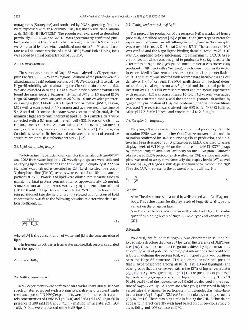
The structural features of Nogo-66 wild type (WT) have been de-scribed by circular dichroism (CD) and NMR spectroscopies [20].These experiments were performed in the aqueous phase where func-tional assays have shown Nogo-66 is active as an axonal growth inhib-itor, and in the presence of lipid or detergent. We mutated Glu26 toalanine and compared CD spectra of this sample to Nogo-66 WT inboth the aqueous phase and in the presence of DMPC lipid vesicles.We assessed the secondary structure of E26A using CD in Fig. 3A. TheCD spectrum of Nogo-66 WT in water features little helical content(15%) and a substantial contribution of random coil below 205 nm. Incontrast, the CD spectrum of themutant E26A shows a canonical helicalsignature, estimated to be 38% helix (Fig. 3A blue). Previously, we
Fig. 2. A) Model of a single DPC molecule docked into the Nogo-66 Glu26 cavity by manually apreviously determined between protein and DPC guide placement of the DPC phosphocholineknown to bind the choline cation of a phosphocholine group via interaction with either a GluID: 1B09) binds a PC at the protein surface through ionic interactions forming a salt bridge bbinds the choline using aromatic ring π–cation interactions. E) The enzyme PfPMT (PDB ID: 3Uand ionic interactions.
demonstrated that addition of lipid vesicles to Nogo-66 WT induces adramatic increase in helical content (to 85% helix) [20]. We performeda similar measurement for E26A but found a more modest increase inhelical content when lipid was added, E26A is estimated to be 64% heli-cal in DMPC. Based on these CDdata, E26A is ~23%more helical inwaterbut ~21% less structured in the presence of lipid compared toWT; trans-fer of E26A into the lipid phase only increases the helical content by~16%.
Since the lipid bilayer is a liquid phase, the partition coefficient, KX
is the correct term to compare the affinities of Nogo-66 WT and E26Awith the lipid phase [23]. This can be calculated from a change in ellip-ticity (θ, mdeg) if a peptide or protein shows substantial folding in thepresence of lipid. We measured CD spectra of WT and E26A withincreasing DMPC lipid concentration and normalized the change in el-lipticity at 222 nm between the two samples to determine the fractionof protein (fp) partitioned into lipid. Fig. 3B shows the partitioning ofE26A compared to WT. Notably, the two proteins transfer favorablyinto the lipid phase with very similar partition coefficients (WT: KX =1.8 × 10⁵; E26A: KX = 1.2 × 10⁵). The corresponding free energiesof transfer are virtually identical: ΔG = −5.8 kcal/mol for WT andΔG = −5.6 kcal/mol for E26A.
3.2. NMR characterization of E26A
We prepared a 15N-labeled sample of Nogo-66 E26A and collected15N-HSQC NMR spectra as a measure of tertiary structure. AlthoughCD data described above indicate that E26A is more helical in an
djusting the position of DPC and cationic side chains. Helices 1–5 are numbered. B) NOEsin contact with helices 1 and 5 and in proximity to Glu26 [20]. Several other proteins areor Asp carboxylate and/or aromatic π–cation bonding. C) Human C-reactive protein (PDBetween choline and Glu81. D) The antibody MC/Pc603 Fab–PC complex (PDB ID: 2MCP)JC) produces PC as a product. In this case, the protein binds choline using both π–cation
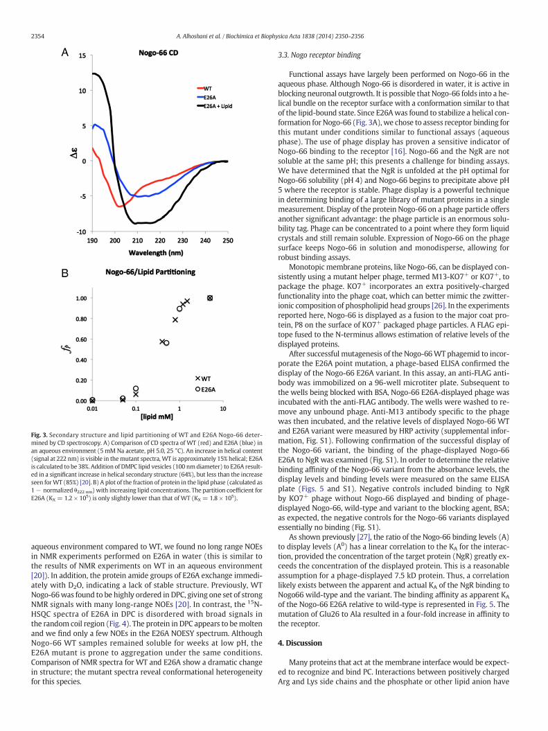
Fig. 3. Secondary structure and lipid partitioning of WT and E26A Nogo-66 deter-mined by CD spectroscopy. A) Comparison of CD spectra of WT (red) and E26A (blue) inan aqueous environment (5 mM Na acetate, pH 5.0, 25 °C). An increase in helical content(signal at 222 nm) is visible in themutant spectra, WT is approximately 15% helical; E26Ais calculated to be 38%. Addition of DMPC lipid vesicles (100 nmdiameter) to E26A result-ed in a significant increase in helical secondary structure (64%), but less than the increaseseen forWT (85%) [20]. B) A plot of the fraction of protein in the lipid phase (calculated as1− normalized θ222 nm) with increasing lipid concentrations. The partition coefficient forE26A (KX = 1.2 × 10⁵) is only slightly lower than that of WT (KX = 1.8 × 10⁵).
2354 A. Alhoshani et al. / Biochimica et Biophysica Acta 1838 (2014) 2350–2356
aqueous environment compared to WT, we found no long range NOEsin NMR experiments performed on E26A in water (this is similar tothe results of NMR experiments on WT in an aqueous environment[20]). In addition, the protein amide groups of E26A exchange immedi-ately with D2O, indicating a lack of stable structure. Previously, WTNogo-66was found to be highly ordered in DPC, giving one set of strongNMR signals with many long-range NOEs [20]. In contrast, the 15N-HSQC spectra of E26A in DPC is disordered with broad signals inthe random coil region (Fig. 4). The protein in DPC appears to bemoltenand we find only a few NOEs in the E26A NOESY spectrum. AlthoughNogo-66 WT samples remained soluble for weeks at low pH, theE26A mutant is prone to aggregation under the same conditions.Comparison of NMR spectra for WT and E26A show a dramatic changein structure; the mutant spectra reveal conformational heterogeneityfor this species.
3.3. Nogo receptor binding
Functional assays have largely been performed on Nogo-66 in theaqueous phase. Although Nogo-66 is disordered in water, it is active inblocking neuronal outgrowth. It is possible that Nogo-66 folds into a he-lical bundle on the receptor surface with a conformation similar to thatof the lipid-bound state. Since E26Awas found to stabilize a helical con-formation for Nogo-66 (Fig. 3A), we chose to assess receptor binding forthis mutant under conditions similar to functional assays (aqueousphase). The use of phage display has proven a sensitive indicator ofNogo-66 binding to the receptor [16]. Nogo-66 and the NgR are notsoluble at the same pH; this presents a challenge for binding assays.We have determined that the NgR is unfolded at the pH optimal forNogo-66 solubility (pH 4) and Nogo-66 begins to precipitate above pH5 where the receptor is stable. Phage display is a powerful techniquein determining binding of a large library of mutant proteins in a singlemeasurement. Display of the protein Nogo-66 on a phage particle offersanother significant advantage: the phage particle is an enormous solu-bility tag. Phage can be concentrated to a point where they form liquidcrystals and still remain soluble. Expression of Nogo-66 on the phagesurface keeps Nogo-66 in solution and monodisperse, allowing forrobust binding assays.
Monotopic membrane proteins, like Nogo-66, can be displayed con-sistently using a mutant helper phage, termed M13-KO7+ or KO7+, topackage the phage. KO7+ incorporates an extra positively-chargedfunctionality into the phage coat, which can better mimic the zwitter-ionic composition of phospholipid head groups [26]. In the experimentsreported here, Nogo-66 is displayed as a fusion to the major coat pro-tein, P8 on the surface of KO7+ packaged phage particles. A FLAG epi-tope fused to the N-terminus allows estimation of relative levels of thedisplayed proteins.
After successful mutagenesis of the Nogo-66WT phagemid to incor-porate the E26A point mutation, a phage-based ELISA confirmed thedisplay of the Nogo-66 E26A variant. In this assay, an anti-FLAG anti-body was immobilized on a 96-well microtiter plate. Subsequent tothe wells being blocked with BSA, Nogo-66 E26A-displayed phage wasincubated with the anti-FLAG antibody. The wells were washed to re-move any unbound phage. Anti-M13 antibody specific to the phagewas then incubated, and the relative levels of displayed Nogo-66 WTand E26A variant were measured by HRP activity (supplemental infor-mation, Fig. S1). Following confirmation of the successful display ofthe Nogo-66 variant, the binding of the phage-displayed Nogo-66E26A to NgR was examined (Fig. S1). In order to determine the relativebinding affinity of the Nogo-66 variant from the absorbance levels, thedisplay levels and binding levels were measured on the same ELISAplate (Figs. 5 and S1). Negative controls included binding to NgRby KO7+ phage without Nogo-66 displayed and binding of phage-displayed Nogo-66, wild-type and variant to the blocking agent, BSA;as expected, the negative controls for the Nogo-66 variants displayedessentially no binding (Fig. S1).
As shown previously [27], the ratio of the Nogo-66 binding levels (A)to display levels (A0) has a linear correlation to the KA for the interac-tion, provided the concentration of the target protein (NgR) greatly ex-ceeds the concentration of the displayed protein. This is a reasonableassumption for a phage-displayed 7.5 kD protein. Thus, a correlationlikely exists between the apparent and actual KA of the NgR binding toNogo66 wild-type and the variant. The binding affinity as apparent KA
of the Nogo-66 E26A relative to wild-type is represented in Fig. 5. Themutation of Glu26 to Ala resulted in a four-fold increase in affinity tothe receptor.
4. Discussion
Many proteins that act at the membrane interface would be expect-ed to recognize and bind PC. Interactions between positively chargedArg and Lys side chains and the phosphate or other lipid anion have
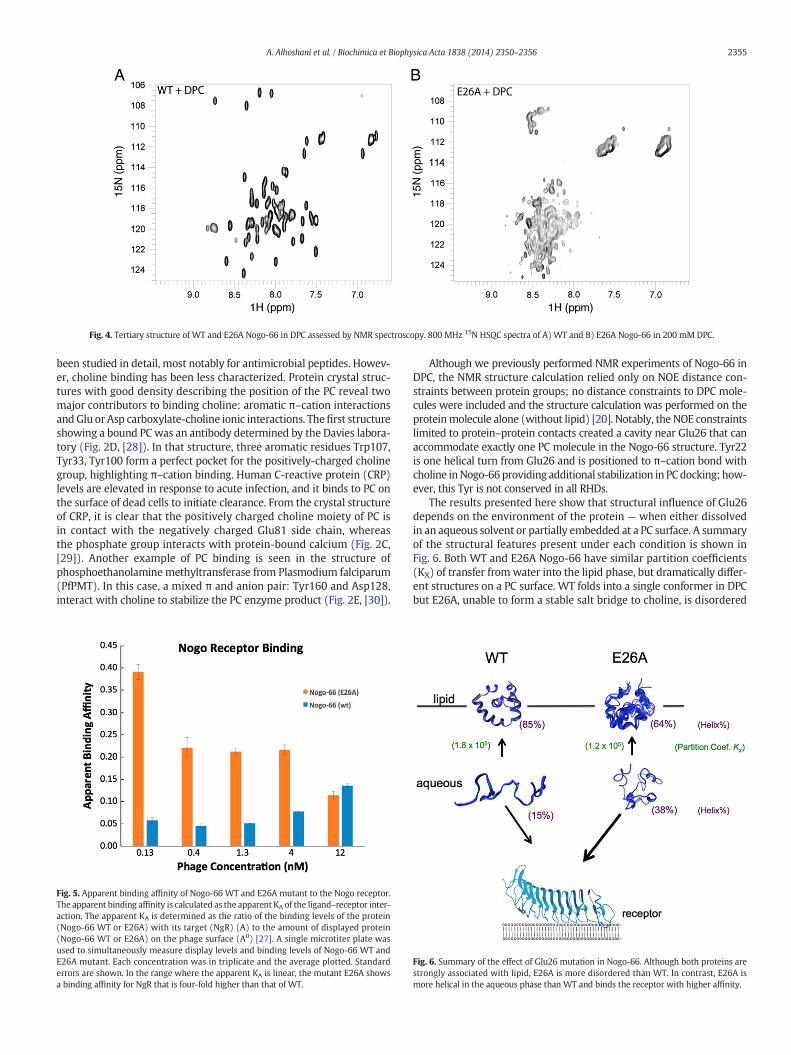
Fig. 4. Tertiary structure of WT and E26A Nogo-66 in DPC assessed by NMR spectroscopy. 800 MHz 15N HSQC spectra of A) WT and B) E26A Nogo-66 in 200 mM DPC.
2355A. Alhoshani et al. / Biochimica et Biophysica Acta 1838 (2014) 2350–2356
been studied in detail, most notably for antimicrobial peptides. Howev-er, choline binding has been less characterized. Protein crystal struc-tures with good density describing the position of the PC reveal twomajor contributors to binding choline: aromatic π–cation interactionsandGlu or Asp carboxylate-choline ionic interactions. Thefirst structureshowing a bound PC was an antibody determined by the Davies labora-tory (Fig. 2D, [28]). In that structure, three aromatic residues Trp107,Tyr33, Tyr100 form a perfect pocket for the positively-charged cholinegroup, highlighting π–cation binding. Human C-reactive protein (CRP)levels are elevated in response to acute infection, and it binds to PC onthe surface of dead cells to initiate clearance. From the crystal structureof CRP, it is clear that the positively charged choline moiety of PC isin contact with the negatively charged Glu81 side chain, whereasthe phosphate group interacts with protein-bound calcium (Fig. 2C,[29]). Another example of PC binding is seen in the structure ofphosphoethanolamine methyltransferase from Plasmodium falciparum(PfPMT). In this case, a mixed π and anion pair: Tyr160 and Asp128,interact with choline to stabilize the PC enzyme product (Fig. 2E, [30]).
Fig. 5. Apparent binding affinity of Nogo-66 WT and E26A mutant to the Nogo receptor.The apparent binding affinity is calculated as the apparent KA of the ligand–receptor inter-action. The apparent KA is determined as the ratio of the binding levels of the protein(Nogo-66 WT or E26A) with its target (NgR) (A) to the amount of displayed protein(Nogo-66 WT or E26A) on the phage surface (A0) [27]. A single microtiter plate wasused to simultaneously measure display levels and binding levels of Nogo-66 WT andE26A mutant. Each concentration was in triplicate and the average plotted. Standarderrors are shown. In the range where the apparent KA is linear, the mutant E26A showsa binding affinity for NgR that is four-fold higher than that of WT.
Although we previously performed NMR experiments of Nogo-66 inDPC, the NMR structure calculation relied only on NOE distance con-straints between protein groups; no distance constraints to DPC mole-cules were included and the structure calculation was performed on theproteinmolecule alone (without lipid) [20]. Notably, the NOE constraintslimited to protein–protein contacts created a cavity near Glu26 that canaccommodate exactly one PC molecule in the Nogo-66 structure. Tyr22is one helical turn from Glu26 and is positioned to π–cation bond withcholine inNogo-66providing additional stabilization in PCdocking; how-ever, this Tyr is not conserved in all RHDs.
The results presented here show that structural influence of Glu26depends on the environment of the protein — when either dissolvedin an aqueous solvent or partially embedded at a PC surface. A summaryof the structural features present under each condition is shown inFig. 6. Both WT and E26A Nogo-66 have similar partition coefficients(KX) of transfer fromwater into the lipid phase, but dramatically differ-ent structures on a PC surface. WT folds into a single conformer in DPCbut E26A, unable to form a stable salt bridge to choline, is disordered
Fig. 6. Summary of the effect of Glu26 mutation in Nogo-66. Although both proteins arestrongly associated with lipid, E26A is more disordered than WT. In contrast, E26A ismore helical in the aqueous phase than WT and binds the receptor with higher affinity.
2356 A. Alhoshani et al. / Biochimica et Biophysica Acta 1838 (2014) 2350–2356
and less helical. The similarity in transfer energy betweenWT and mu-tant when partitioning into lipid could be accounted for by compensat-ing effects; an energetically favorable increase in entropy of disorderedE26A in DPC could compensate for the destabilization resulting from alost salt bridge between Glu and the lipid headgroup.
In an aqueous environment, Nogo-66 WT is active in functional as-says but maintains a very disordered structure. In contrast, mutationof the buried Glu26 charge to Ala stabilizes helical conformations andincreases helical content by ~23% in E26A. Moreover, Nogo-66 E26Ahas a much higher affinity for the receptor thanWT in an aqueous envi-ronment; this effect ismost likely a consequence of stabilized secondarystructure in the lipid-free state. Although a complete structure of thecomplex between NgR, Nogo-66 and lipid has not been determined,these studies provide insight into the state of Nogo-66 when bindingthe receptor. We observe that the interaction of Glu26 with lipid stabi-lizes theWT protein fold. In contrast, when lipid is absent Glu26 desta-bilizes both the helical fold and receptor binding. The region 31–55 isknown to contain residues sufficient for Nogo-66 function; Glu26 is po-sitioned on the opposite protein face compared to the functional groups.The ability of a position within the interior of a compact helical bundle(Glu26) to affect receptor binding when it is not among the residuesexpected to make up the binding interface suggests a model whereNogo-66 folds on the receptor into a structure similar to that inducedby lipid.
The RHD-66 domain isflanked by twomembrane-embedded helicesthat are believed to induce curvature of the ER membrane. These struc-tures would be expected to stabilize the RHD-66 structure, both inanchoring the protein to the membrane and in extending the N- andC-terminal helices. It is also expected that the RHD-66 would be morestable in a natural membrane than in DPCmicelles. Compared to planarbilayers, micelles are highly curved andmuchmore dynamic. It is possi-ble that the loss in structure seen in the lipid-associated state for theE26Amutant is not as pronounced in the context of the full-length pro-tein and natural cell membranes. Nevertheless, a system that is on thecusp of stability is actually very useful in understanding protein folding.Proteins that are extremely stable will often not show a measurable ef-fect from a single mutation; this can make it difficult to determine therelative contributions of individual amino acids to the overall proteinstability. In deconstructing the RHD, we have found that Nogo-66 is anexcellent model for testing the forces that drive protein folding at alipid interface as well as providing specific detail on the RHD-66 fold.The structure of Nogo-66 in DPC is entirely consistent with functionalassays and these studies show that Nogo-66 is an autonomous foldingunit that uses a negative charge at position 26 to anchor the protein tothe membrane surface. Nearby aromatic rings can also anchor Nogo-66 in the membrane and contribute to the lipid-induced fold. In addi-tion, there are several other Glu/Asp side chains conserved in highervertebrates that could interact with choline and drive docking intothe membrane leaflet. Studies are ongoing to extend the structure ofNogo-66 with the flanking membrane embedded regions and define acomplete RHD structure.
Acknowledgements
We thank Jessica Schulz for sub-cloning the NgR gene, and DeepthiTummala for assisting in the preparation of Nogo receptor. We aregrateful to SudiptaMajumdar for expert advice and assistance on the re-ceptor binding experiments and to Antje Pokorny for the stimulatingconversation and advice on phosphocholine–protein interactions. Wethank Sean Moro for critically reading this manuscript. This work wassupported by the National Institutes of Health (GM069783), NationalInstitute of General Medical Sciences (R01-GM078528-0, R01-GM069783-06 and R01 GM100700-01), the Roman Reed ResearchFund (RR05-155) and the Ministry of Higher Education, King Saud Uni-versity, Riyadh, Saudi Arabia.
Appendix A. Supplementary data
Supplementary data to this article can be found online at http://dx.doi.org/10.1016/j.bbamem.2014.05.021.
References
[1] T. Oertle,M. Klinger, C.A. Stuermer,M.E. Schwab, A reticular rhapsody: phylogenic evo-lution and nomenclature of the RTN/Nogo gene family, FASEB J. 17 (2003) 1238–1247.
[2] G.K. Voeltz, W.A. Prinz, Y. Shibata, J.M. Rist, T.A. Rapoport, A class of membraneproteins shaping the tubular endoplasmic reticulum, Cell 124 (2006) 573–586.
[3] E. Kiseleva, K.N. Morozova, G.K. Voeltz, T.D. Allen, M.W. Goldberg, Reticulon 4a/NogoAlocates to regions of high membrane curvature and may have a role in nuclear enve-lope growth, J. Struct. Biol. 160 (2007) 224–235.
[4] Y.S. Yang, S.M. Strittmatter, The reticulons: a family of proteins with diverse functions,Genome Biol. 8 (2007) 234.
[5] A. Diaz, P. Ahlquist, Role of host reticulon proteins in rearranging membranes forpositive-strand RNA virus replication, Curr. Opin. Microbiol. 15 (2012) 519–524.
[6] S. Tagami, Y. Eguchi, N. Kinoshita, Y. Takeda, Y. Tsujimoto, A novel protein, RTN-XS,interacts with both Bcl-XL and Bcl-2 on endoplasmic reticulum and reduces theiranti-apoptotic activity, Oncogene 19 (2000) 5736–5746.
[7] L. Zhu, R. Xiang,W. Dong, Y.L. Liu, Y.P. Qi, Anti-apoptotic activity of Bcl-2 is enhancedby its interaction with RTN3, Cell Biol. Int. 31 (2007) 825–830.
[8] W.He, Y. Lu, I. Qahwash, X.Y. Hu, A. Chang, R. Yan, Reticulon familymembersmodulateBACE1 activity and amyloid-beta peptide generation, Nat. Med. 10 (2004) 959–965.
[9] W. He, Q. Shi, X. Hu, R. Yan, The membrane topology of RTN3 and its effect on bind-ing of RTN3 to BACE1, J. Biol. Chem. 282 (2007) 29144–29151.
[10] L. Acevedo, J. Yu, H. Erdjument-Bromage, R.Q. Miao, J.E. Kim, D. Fulton, P. Tempst, S.M. Strittmatter, W.C. Sessa, A new role for Nogo as a regulator of vascular remodel-ing, Nat. Med. 10 (2004) 382–388.
[11] T.Walchli, V. Pernet, O.Weinmann, J.Y. Shiu, A. Guzik-Kornacka, G. Decrey, D. Yuksel,H. Schneider, J. Vogel, D.E. Ingber, V. Vogel, K. Frei, M.E. Schwab, Nogo-A is a negativeregulator of CNS angiogenesis, Proc. Natl. Acad. Sci. U. S. A. 110 (2013) E1943–E1952.
[12] S.Y. Chong, S.S. Rosenberg, S.P. Fancy, C. Zhao, Y.A. Shen, A.T. Hahn, A.W. McGee, X.Xu, B. Zheng, L.I. Zhang, D.H. Rowitch, R.J. Franklin, Q.R. Lu, J.R. Chan, Neurite out-growth inhibitor Nogo-A establishes spatial segregation and extent of oligodendro-cyte myelination, Proc. Natl. Acad. Sci. U. S. A. 109 (2011) 1299–1304.
[13] V. Pernet, M.E. Schwab, The role of Nogo-A in axonal plasticity, regrowth and repair,Cell Tissue Res. 349 (2012) 97–104.
[14] A. Schmandke, A. Schmandke, M.E. Schwab, Nogo-A: multiple roles in CNS develop-ment, maintenance, and disease, Neuroscientist (2014), http://dx.doi.org/10.1177/1073858413516800 PMID: 24402613.
[15] F. Di Sano, P. Bernardoni, M. Piacentini, The reticulons: guardians of the structureand function of the endoplasmic reticulum, Exp. Cell Res. 318 (2012) 1201–1207.
[16] M.S. Chen, A.B. Huber, M.E. van der Haar, M. Frank, L. Schnell, A.A. Spillmann, F.Christ, M.E. Schwab, Nogo-A is a myelin-associated neurite outgrowth inhibitorand an antigen for monoclonal antibody IN-1, Nature 403 (2000) 434–439.
[17] T. GrandPre, F. Nakamura, T. Vartanian, S.M. Strittmatter, Identification of the Nogoinhibitor of axon regeneration as a reticulon protein, Nature 403 (2000) 439–444.
[18] R. Prinjha, S.E. Moore, M. Vinson, S. Blake, R. Morrow, G. Christie, D.Michalovich, D.L.Simmons, F.S. Walsh, Inhibitor of neurite outgrowth in humans, Nature 403 (2000)383–384.
[19] A.E. Fournier, T. GrandPre, S.M. Strittmatter, Identification of a receptor mediatingNogo-66 inhibition of axonal regeneration, Nature 409 (2001) 341–346.
[20] S.V. Vasudevan, J. Schulz, C. Zhou, M.J. Cocco, Protein folding at themembrane inter-face, the structure of Nogo-66 requires interactions with a phosphocholine surface,Proc. Natl. Acad. Sci. U. S. A. 107 (2010) 6847–6851.
[21] L. Whitmore, B.A. Wallace, DICHROWEB, an online server for protein secondarystructure analyses from circular dichroism spectroscopic data, Nucleic Acids Res.32 (2004) W668–W673.
[22] J.G. Lees, A.J. Miles, R.W. Janes, B.A. Wallace, Novel methods for secondary structuredetermination using low wavelength (VUV) circular dichroism spectroscopic data,BMC Bioinforma. 7 (2006) 507.
[23] S.H. White, W.C. Wimley, A.S. Ladokhin, K. Hristova, Protein folding in membranes:determining energetics of peptide-bilayer interactions, Methods Enzymol. 295(1998) 62–87.
[24] F. Delaglio, S. Grzesiek, G.W. Vuister, G. Zhu, J. Pfeifer, A. Bax, NMRPipe: a multidi-mensional spectral processing system based on UNIX pipes, J. Biomol. NMR 6(1995) 277–293.
[25] X.L. He, J.F. Bazan, G. McDermott, J.B. Park, K. Wang, M. Tessier-Lavigne, Z. He, K.C.Garcia, Structure of the Nogo receptor ectodomain: a recognitionmodule implicatedin myelin inhibition, Neuron 38 (2003) 177–185.
[26] R. Vithayathil, R.M. Hooy, M.J. Cocco, G.A. Weiss, The scope of phage display formembrane proteins, J. Mol. Biol. 414 (2011) 499–510.
[27] S. Rossenu, S. Leyman, D. Dewitte, D. Peelaers, V. Jonckheere, M. Van Troys, J.Vandekerckhove, C. Ampe, A phage display-based method for determination of rel-ative affinities of mutants. Application of the actin-binding motifs in thymosin beta4 and the villin headpiece, J. Biol. Chem. 278 (2003) 16642–16650.
[28] Y. Satow, G.H. Cohen, E.A. Padlan, D.R. Davies, Phosphocholine binding immunoglobu-lin Fab Mcpc603. An X-ray-diffraction study at 2.7 A, J. Mol. Biol. 190 (1986) 593–604.
[29] D. Thompson, M.B. Pepys, S.P. Wood, The physiological structure of human C-reactive protein and its complex with phosphocholine, Structure 7 (1999) 169–177.
[30] S.G. Lee, Y. Kim, T.D. Alpert, A. Nagata, J.M. Jez, Structure and reaction mechanism ofphosphoethanolamine methyltransferase from the malaria parasite Plasmodiumfalciparum: an antiparasitic drug target, J. Biol. Chem. 287 (2012) 1426–1434.