biodegradation of di-n-butyl phthalate by bacterial...
TRANSCRIPT

RESEARCH ARTICLE
Biodegradation of di-n-butyl phthalate by
bacterial consortium LV-1 enriched from river
sludge
Yangyang Wang1,2,3, Fangfang Li3, Xinling Ruan2,3, Jian Song3, Lv Lv3, Liyuan Chai4,
Zhihui Yang4, Lin Luo1*
1 School of Resources and Environment, Hunan Agricultural University, Changsha, China, 2 Key Research
Institute of Yellow River Civilization and Sustainable Development & Collaborative Innovation Center on
Yellow River Civilization of Henan Province, Henan University, Kaifeng, China, 3 Institute of Natural
Resources and Environment, Henan University, Kaifeng, China, 4 School of Metallurgical & Environment,
Central South University, Changsha, China
Abstract
A stable bacterial consortium (LV-1) capable of degrading di-n-butyl phthalate (DBP) was
enriched from river sludge. Community analysis revealed that the main families of LV-1 are
Brucellaceae (62.78%) and Sinobacteraceae (14.83%), and the main genera of LV-1 are
Brucella spp. (62.78%) and Sinobacter spp. (14.83%). The optimal pH and temperature for
LV-1 to degrade DBP were pH 6.0 and 30˚C, respectively. Inoculum size influenced the deg-
radation ratio when the incubation time was < 24 h. The initial concentration of DBP also
influenced the degradation rates of DBP by LV-1, and the degradation rates ranged from
69.0–775.0 mg/l/d in the first 24 h. Degradation of DBP was best fitted by first-order kinetics
when the initial concentration was < 300 mg/l. In addition, Cd2+, Cr6+, and Zn2+ inhibited
DBP degradation by LV-1 at all considered concentrations, but low concentrations of Pb2+,
Cu2+, and Mn2+ enhanced DBP degradation. The main intermediates (mono-ethyl phthalate
[MEP], mono-butyl phthalate [MBP], and phthalic acid [PA]) were identified in the DBP deg-
radation process, thus a new biochemical pathway of DBP degradation is proposed. Fur-
thermore, LV-1 also degraded other phthalates with shorter ester chains (DMP, DEP, and
PA).
1. Introduction
Phthalate esters (PAEs) are a group of refractory organic compounds that are widely-used in
coatings, medical product packaging, plastics, cosmetics, and building materials [1–2]. PAEs
can easily migrate into the environment from plastic products and other related products
because PAEs are not chemically bound to the polymeric matrix [3]. Numerous studies have
revealed that PAEs and their metabolites can impact reproduction and behavior of mammals,
even at very low concentrations [4–5]. Therefore, the environmental protection agency of
many countries has classified most PAEs as top priority pollutants [3,6].
PLOS ONE | https://doi.org/10.1371/journal.pone.0178213 May 25, 2017 1 / 13
a1111111111
a1111111111
a1111111111
a1111111111
a1111111111
OPENACCESS
Citation: Wang Y, Li F, Ruan X, Song J, Lv L, Chai
L, et al. (2017) Biodegradation of di-n-butyl
phthalate by bacterial consortium LV-1 enriched
from river sludge. PLoS ONE 12(5): e0178213.
https://doi.org/10.1371/journal.pone.0178213
Editor: Andrea Franzetti, Universita degli Studi di
Milano-Bicocca, ITALY
Received: December 15, 2016
Accepted: May 9, 2017
Published: May 25, 2017
Copyright: © 2017 Wang et al. This is an open
access article distributed under the terms of the
Creative Commons Attribution License, which
permits unrestricted use, distribution, and
reproduction in any medium, provided the original
author and source are credited.
Data Availability Statement: All relevant data are
within the paper and its Supporting Information
file.
Funding: This work was supported by a grant from
the National Natural Science Foundation of China
(41430637); Program for Innovative Research
Team (in Science and Technology) in University of
Henan Province (16IRTSTHN012); Science and
technology development project of Henan Province
(152102310297); Opening Foundation of the
Chinese National Engineering Research Center for
Control and Treatment of Heavy Metal Pollution,

Di-n-butyl phthalate (DBP), which belongs to the class of PAEs, has been detected in vari-
ous environments, such as soils, sediments, water, air, landfill leachates, plants, gas, and indoor
dust [7–9]. In addition, many crops can absorb and store DBP, which leads to DBP entry into
the food chain and threatens the health of mammals [6]. Therefore, it is critically important to
efficiently remove DBP from the environment.
Indeed, it has been shown that several natural processes can remove DBP from the environ-
ment, including photolysis, hydrolysis, and biodegradation [10–11]. Due to the low rate of
hydrolysis and photolysis, metabolic breakdown of DBP by microorganisms is regarded as
a major process in the environmental degradation of DBP [12–13]. Many DBP-degrading bac-
terial strains have been isolated from various environmental samples [7,14–15], including
approximately 25 genera, such as Pseudomonas fluorescens [16], Rhodococcus spp. [17], Sphin-gomonas spp. [18], and Bacillus subtilis [19]. However, discussions about biodegradation of
DBP by bacterial consortia are limited [20]. It has been shown that high diversity can enhance
survival of a bacterial consortium in different environments, and increase the biodegradation
efficiency of organic pollutions [20–21]. Therefore, investigating the biodegradation of DBP
by bacterial consortia is warranted.
In the present study a DBP-degrading bacterial consortium (LV-1) was enriched from river
sludge, and the DBP degradation potential of LV-1 was investigated. The influence of pH, tem-
perature, inoculum size, and heavy metal ions on DBP degradation by LV-1 was examined.
GC-MS and Illumina sequencing technology were used to analyze the degradation intermedi-
ates of DBP and the bacterial community structure.
2. Materials and methods
2.1 Chemicals
Di-n-butyl phthalate (DBP; 99% purity) used in this research was purchased from Aladdin-
reagent Co. (Shanghai, China). Methanol (HPLC grade), ethyl acetate (analytical grade), and
other chemical reagents (analytical grade) were purchased from the Chinese Medicine Group
(Shanghai, China).
2.2 Media and enrichment of the bacterial consortium
The bacterial consortium was enriched from river sludge collected from Kaifeng, Henan Prov-
ince, China (114˚35’E, 34˚79’N). The river sludge was contaminated with domestic waste and
sewage seriously over a long period of time. The medium used in the experiments was mini-
mum salt medium (MSM), as previously described by Wu et al. [22].
The enrichment procedure for the DBP-degrading bacteria consortium was similar to our
previous report [22]. 5g of river sludge were added to a 250-ml Erlenmeyer flask containing
100 ml of MSM and DBP (50 mg/l). The resulting suspension was cultured at 30˚C and 175
rpm in the dark for 7 days. Then, 1 ml of the enrichment culture was serially transferred to
fresh MSM containing 100, 200, 300, 400, 500, 1000, 1000, and 1000 mg/l of DBP (each enrich-
ment step lasted 7 days). The final enrichment culture was designated as LV-1, and used for
Illumina sequencing and further degradation experiments.
2.3 DNA extraction, amplification, and Illumina sequencing
Total genomic DNA of LV-1 was extracted using an EZ-10 Spin Column Genomic DNA
Minipreps Kit (Bio Basic Inc., Markham, Ontario, Canada) following the protocol provided
by the manufacturer. The primers and adapter sequences used for amplification of the V4
Biodegradation of DBP
PLOS ONE | https://doi.org/10.1371/journal.pone.0178213 May 25, 2017 2 / 13
Changsha, 410083, China (No. 2015CNERC-
CTHMP-).
Competing interests: The authors have declared
that no competing interests exist.

hypervariable region of the 16S rRNA gene were described by Caporaso et al. [23]. The PCR
reactions and data processing were performed according to the description of Xiao et al. [24].
2.4 DBP degradation experiments using the bacterial consortium
LV-1 was grown in MSM with DBP (500 mg/l) as the sole source of carbon and energy, har-
vested after 48 h, thrice-washed with 0.05 mol/l of potassium phosphate buffer (pH 7.5). The
washed cells were re-suspended in the same phosphate buffer (OD600 = 1.0) for application in
the following degradation experiments.
Various amounts of DBP dissolved in methanol were added to 50-ml Erlenmeyer flasks,
then incubated in a water bath rocker at 60˚C to evaporate the methanol. Then, 1 ml of cell
suspension and 19 ml of MSM were added to the Erlenmeyer flasks. The influence of the fol-
lowing environmental factors on DBP degradation were investigated within 48 h of incubation
in MSM containing 500 mg/l of DBP at 175 rpm: temperature (15, 20, 25, 30, 35, 40, and
45˚C); pH (4.0, 5.0, 6.0, 7.0, 8.0, 9.0, and 10.0); inoculum size (1.25, 2.5, 3.75, 5.00, 6.25, 7.5,
8.75, and 10.0%); Cd2+(2, 5, 10, 15, and 20 mg/l); Cr6+(10, 20, 30, 40, and 50 mg/l); and Pb2+,
Mn2+, Zn2+, and Cu2+(50, 100, 200, 300, and 400 mg/l). When one parameter changed, other
parameters are set as the commonly used condition in degrading of organic pollutant (pH 7.0,
temperature 30˚C, inoculum size 5%, and 175 rpm). After 48-h incubation, samples were with-
drawn from the rocker platform, and the residual DBP in Erlenmeyer flasks was detected by
ultra-performance liquid chromatography (UPLC). All experiments were performed in
triplicate.
2.5 Effect of the initial DBP concentration on DBP biodegradation
Flasks containing cells suspension of LV-1 and DBP (initial concentration: 50, 100, 200, 300,
400, 500, and 1000 mg/l) were removed from the rocking platform (30˚C and 175 rpm) at 12-h
intervals. The samples were stored at 4˚C for further UPLC analysis.
2.6 Analysis of DBP degradation intermediates
To analyze the biodegradation intermediates of DBP, samples were concentrated approxi-
mately 10-fold before gas chromatography–mass spectrometry (GC–MS; Agilent, USA) analy-
sis. The detection procedure and operating conditions for GC-MS were according to He et al.
[20].
2.7 Growth on other aromatic compounds
LV-1 was inoculated in MSM medium supplemented with other aromatic compounds (200
mg/l), including phthalic acid (PA), dimethyl phthalate (DMP), diethyl phthalate (DEP), dioc-
tyl phthalate (DOP), and bisphenol A (BPA), to assess the potential ability to degrade these
compounds. Cell growth was monitored turbidometrically at 600 nm after 48-h incubation.
All experiments and controls were performed in triplicate.
2.8 Analytical methods
Extraction of residual DBP and degradation intermediates from the liquid was following the
procedure described in our previous report [13]. The chromatographic conditions for detect-
ing DBP by UPLC were as follows: mobile phase, methanol: water (90:10 [v/v]); the flow rate
(0.5 ml/min); and UV wave-length (254 nm).
Biodegradation of DBP
PLOS ONE | https://doi.org/10.1371/journal.pone.0178213 May 25, 2017 3 / 13
2.9 Data analysis
The influence of heavy metals on DBP degradation was statistically analyzed by SPSS 13.0, and
the significant tests were conducted with ANOVA and multiple comparison.
3. Results and discussion
3.1 Enrichment and community analysis of LV-1
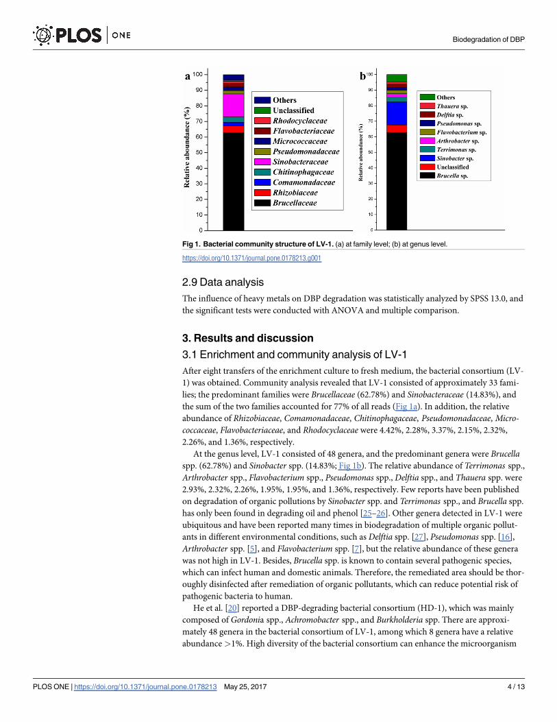
After eight transfers of the enrichment culture to fresh medium, the bacterial consortium (LV-
1) was obtained. Community analysis revealed that LV-1 consisted of approximately 33 fami-
lies; the predominant families were Brucellaceae (62.78%) and Sinobacteraceae (14.83%), and
the sum of the two families accounted for 77% of all reads (Fig 1a). In addition, the relative
abundance of Rhizobiaceae, Comamonadaceae, Chitinophagaceae, Pseudomonadaceae, Micro-coccaceae, Flavobacteriaceae, and Rhodocyclaceae were 4.42%, 2.28%, 3.37%, 2.15%, 2.32%,
2.26%, and 1.36%, respectively.
At the genus level, LV-1 consisted of 48 genera, and the predominant genera were Brucellaspp. (62.78%) and Sinobacter spp. (14.83%; Fig 1b). The relative abundance of Terrimonas spp.,
Arthrobacter spp., Flavobacterium spp., Pseudomonas spp., Delftia spp., and Thauera spp. were
2.93%, 2.32%, 2.26%, 1.95%, 1.95%, and 1.36%, respectively. Few reports have been published
on degradation of organic pollutions by Sinobacter spp. and Terrimonas spp., and Brucella spp.
has only been found in degrading oil and phenol [25–26]. Other genera detected in LV-1 were
ubiquitous and have been reported many times in biodegradation of multiple organic pollut-
ants in different environmental conditions, such as Delftia spp. [27], Pseudomonas spp. [16],
Arthrobacter spp. [5], and Flavobacterium spp. [7], but the relative abundance of these genera
was not high in LV-1. Besides, Brucella spp. is known to contain several pathogenic species,
which can infect human and domestic animals. Therefore, the remediated area should be thor-
oughly disinfected after remediation of organic pollutants, which can reduce potential risk of
pathogenic bacteria to human.
He et al. [20] reported a DBP-degrading bacterial consortium (HD-1), which was mainly
composed of Gordonia spp., Achromobacter spp., and Burkholderia spp. There are approxi-
mately 48 genera in the bacterial consortium of LV-1, among which 8 genera have a relative
abundance >1%. High diversity of the bacterial consortium can enhance the microorganism
Fig 1. Bacterial community structure of LV-1. (a) at family level; (b) at genus level.
https://doi.org/10.1371/journal.pone.0178213.g001
Biodegradation of DBP
PLOS ONE | https://doi.org/10.1371/journal.pone.0178213 May 25, 2017 4 / 13

survival ratio in different environments [20,28]. Bacterial consortium is more suitable for bio-
remediation than pure bacterial strains, suggesting that LV-1 is a suitable bacterial consortium
in the bioremediation of DBP contamination.
3.2 Effects of temperature and pH on DBP degradation
Bacterial growth is pH- and temperature-sensitive. The effect of pH on DBP degradation by
LV-1 is shown in Fig 2(a). The degradation ratio increased rapidly from 58.75% to 93.22%
when the initial pH of the medium increased from 4.0 to 6.0. When the pH exceeded 6.0, the
degradation ratio decreased slowly, indicating that the optimal pH for LV-1 degrading DBP
was 6.0. In addition, the degradation ratio could be maintained at 89.07% and 87.16% when
the initial pH increased to 7.0 and 8.0, respectively. DBP degradation ratio was > 58.75% in all
considered initial pH values, indicating that LV-1 has a broad pH value in degrading DBP.
The influence of temperature on degradation of DBP is shown in Fig 2(b). The DBP degra-
dation ratio increased from 14.43% to 93.68% as the temperature increased from 15˚C to
30˚C. However, the DBP degradation ratio decreased when the temperatures was higher than
30˚C, indicating that the optimum temperature was 30˚C. The degradation ratio could be
maintained > 42.07% when the temperature is between 20˚C to 40˚C, indicating that LV-1
also has a relatively broad temperature in the degradation of DBP.
In previous reports, bacteria can degrade DBP at certain temperature or pH with a high
degradation efficiency, such as 30˚C [5], 35˚C [3], pH 7.0 [29], pH 9.0 [5] etc. But the degrada-
tion efficiency decrease rapidly with the changes of pH and temperature. However, LV-1 can
degrade DBP efficiently at a broad range of temperature and pH, which may directly relate to
the high diversity of LV-1. LV-1 includes about 48 genera of bacteria, which made LV-1 more
easily to adapt to different environments and degrade DBP with relatively higher efficiency.
3.3 Effect of inoculum size on DBP degradation
The influence of inoculum size on the DBP degradation ratio is shown in Fig 3. Within 24 h of
incubation, the DBP degradation ratio was greatly influenced by the inoculum size. Nearly no
DBP was degraded in the control experiments (the inoculum size at 0%). The degradation
ratio just reached 55.28% with the inoculum size at 1.25%, and the degradation ratio increased
quickly with the increase of the inoculum size. When the inoculum size was higher than
Fig 2. Effects of pH (a) and temperature (b) on degradation of DBP by LV-1.
https://doi.org/10.1371/journal.pone.0178213.g002
Biodegradation of DBP
PLOS ONE | https://doi.org/10.1371/journal.pone.0178213 May 25, 2017 5 / 13
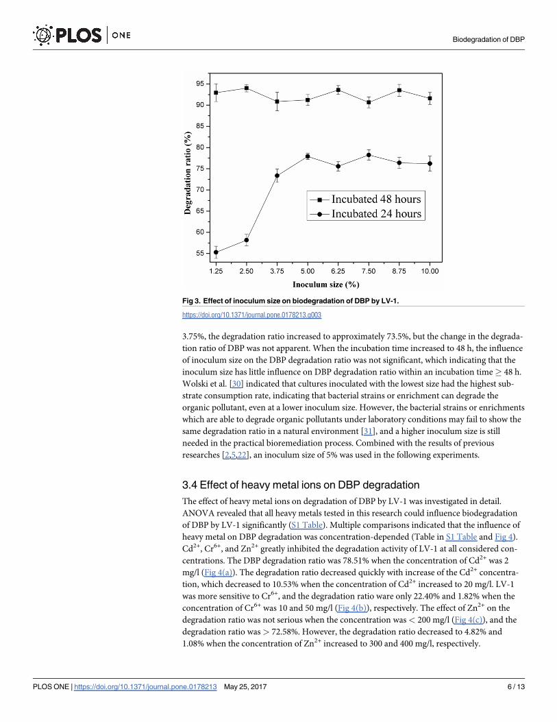
3.75%, the degradation ratio increased to approximately 73.5%, but the change in the degrada-
tion ratio of DBP was not apparent. When the incubation time increased to 48 h, the influence
of inoculum size on the DBP degradation ratio was not significant, which indicating that the
inoculum size has little influence on DBP degradation ratio within an incubation time� 48 h.
Wolski et al. [30] indicated that cultures inoculated with the lowest size had the highest sub-
strate consumption rate, indicating that bacterial strains or enrichment can degrade the
organic pollutant, even at a lower inoculum size. However, the bacterial strains or enrichments
which are able to degrade organic pollutants under laboratory conditions may fail to show the
same degradation ratio in a natural environment [31], and a higher inoculum size is still
needed in the practical bioremediation process. Combined with the results of previous
researches [2,5,22], an inoculum size of 5% was used in the following experiments.
3.4 Effect of heavy metal ions on DBP degradation
The effect of heavy metal ions on degradation of DBP by LV-1 was investigated in detail.
ANOVA revealed that all heavy metals tested in this research could influence biodegradation
of DBP by LV-1 significantly (S1 Table). Multiple comparisons indicated that the influence of
heavy metal on DBP degradation was concentration-depended (Table in S1 Table and Fig 4).
Cd2+, Cr6+, and Zn2+ greatly inhibited the degradation activity of LV-1 at all considered con-
centrations. The DBP degradation ratio was 78.51% when the concentration of Cd2+ was 2
mg/l (Fig 4(a)). The degradation ratio decreased quickly with increase of the Cd2+ concentra-
tion, which decreased to 10.53% when the concentration of Cd2+ increased to 20 mg/l. LV-1
was more sensitive to Cr6+, and the degradation ratio ware only 22.40% and 1.82% when the
concentration of Cr6+ was 10 and 50 mg/l (Fig 4(b)), respectively. The effect of Zn2+ on the
degradation ratio was not serious when the concentration was < 200 mg/l (Fig 4(c)), and the
degradation ratio was > 72.58%. However, the degradation ratio decreased to 4.82% and
1.08% when the concentration of Zn2+ increased to 300 and 400 mg/l, respectively.
Fig 3. Effect of inoculum size on biodegradation of DBP by LV-1.
https://doi.org/10.1371/journal.pone.0178213.g003
Biodegradation of DBP
PLOS ONE | https://doi.org/10.1371/journal.pone.0178213 May 25, 2017 6 / 13

Low concentrations of Pb2+, Cu2+, and Mn2+ enhanced biodegradation of DBP by LV-1.
The degradation ratio reached 95.04% and 98.17%, respectively, when the concentration of
Pb2+ and Cu2+ was 50 mg/l, which was higher than the value in control experiments (Fig 4(d)
and 4(e)). The degradation ratio decreased quickly with an increase in the concentrations of
Pb2+ and Cu2+, and the degradation of DBP was completely inhibited when the concentration
of Cu2+ was 400 mg/l. Mn2+ enhanced the biodegradation of DBP by LV-1 when the concen-
tration was < 200 mg/l (Fig 4(f)), but the degradation was inhibited when the concentration of
Mn2+ was� 300 mg/l. In fact, previous reports have shown that Mn2+ is an activator of differ-
ent enzymes and can enhance the enzymatic reactions, such as protease [32], β-xylosidase [33],
α-amylase [34], and leucine-rich repeat kinase 2 [35]. MnCl2 was also added in the MSM
(0.0015 mg/l), but the concentration was too low for LV-1 to degrade DBP, indicating that the
DBP degradation efficiency by LV-1 can be further improved through optimization of the
composition of MSM.
Many researchers have reported the influences of heavy metals on bacteria growth [36–38].
Mrvčić et al. [37] found that the addition of Zn and Mn have no effect on the bacteria growth,
while copper ion was highly toxic. Admas and Ghiorse [39]reported that Mn2+ have various
effects on the growth of Leptohrix discophora strain SS-1 in batch cultures depending on the
concentration added to the medium. Ravikumar et al. [40] found all identified bacterial species
in their experiment are sensitive to Hg and Zn. In fact, the influence of heavy metals on the
growth of bacteria is mainly affected by its influence on enzymes activity. Yu and Cheng [41]
found that urease activity firstly increased with the addition of Cu, Pb and Cd, then showed
declined trends. Zhang et al [42] reported that U has negative effects on all kinds of soil
enzymes in their experiments, but Mn can promote the activity of peroxidase, and different
Fig 4. Effect of various concentrations of heavy metal ions on biodegradation of DBP by LV-1. (a) Cd2+; (b) Cr6+; (c) Zn2+; (d) Pb2+; (e) Cu2+; (f) Mn2+.
The difference lowercases above the column indicate the influence of heavy metals on DBP degradation with significant differences.
https://doi.org/10.1371/journal.pone.0178213.g004
Biodegradation of DBP
PLOS ONE | https://doi.org/10.1371/journal.pone.0178213 May 25, 2017 7 / 13
concentration of Mn has different influence on the activities of sucrose. Besides, Pb also pro-
moted the activity of peroxidase when its concentration is low. In our research, Pb2+, Cu2+ and
Mn2+ promoted the growth of LV-1 at low concentration, whereas they inhibited the growth
of LV-1 when their concentration increased. Cd2+, Cr6+ and Zn2+ inhibited the growth of LV-
1 even at low concentration. That is to say, the influence of heavy metals on LV-1 is concentra-
tion-dependent, which is consistent well with previous researches [37,39,41–42].
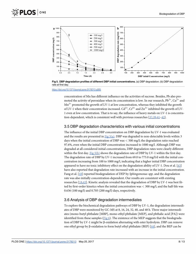
3.5 DBP degradation characteristics with various initial concentrations
The influence of the initial DBP concentration on DBP degradation by LV-1 was evaluated
and the results are presented in Fig 5(a). DBP was degraded to non-detectable levels within 3
days when the initial concentration of DBP was < 500 mg/l; the degradation ratio reached
97.6%, even when the initial DBP concentration increased to 1000 mg/l. Although DBP was
degraded at all considered initial concentrations, DBP degradation rates were clearly different
within the first day. Fig 5(b) shows the degradation rate of DBP by LV-1 within the first day.
The degradation rate of DBP by LV-1 increased from 69.0 to 775.0 mg/l/d with the initial con-
centration increasing from 100 to 1000 mg/l, indicating that a higher initial DBP concentration
appeared to have no toxic inhibitory effect on the degradation ability of LV-1. Dou et al. [43]
have also reported that degradation rate increased with an increase in the initial concentration.
Fang et al. [18] reported biodegradation of DEP by Sphingomonas spp. and the degradation
rate was also initially concentration-dependent. Our results are consistent with existing
researches [18,43]. Kinetic analysis revealed that the degradation of DBP by LV-1 was best fit-
ted by first-order kinetics when the initial concentration was < 300 mg/l, and the half-life was
0.636 (100 mg/l) and 0.705 (200 mg/l) days, respectively.
3.6 Analysis of DBP degradation intermediates
To explore the biochemical degradation pathways of DBP by LV-1, the degradation intermedi-
ates of DBP were monitored by GC-MS at 8, 16, 24, 32, 40, and 48 h. Three major intermedi-
ates (mono-butyl phthalate [MBP], mono-ethyl phthalate [MEP], and phthalic acid [PA]) were
identified from these samples (Fig 6). The existence of the MEP suggests that the biodegrada-
tion of DBP by LV-1 might be β-oxidation alternating with ester hydrolysis. DBP can remove
one ethyl group by β-oxidation to form butyl ethyl phthalate (BEP) [44], and the BEP can be
Fig 5. DBP degradation profiles of different DBP initial concentrations. (a) DBP degradation; (b) DBP degradation
rate at first day.
https://doi.org/10.1371/journal.pone.0178213.g005
Biodegradation of DBP
PLOS ONE | https://doi.org/10.1371/journal.pone.0178213 May 25, 2017 8 / 13
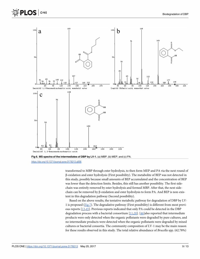
transformed to MBP through ester hydrolysis, to then form MEP and PA via the next round of
β-oxidation and ester hydrolysis (First possibility). The metabolite of BEP was not detected in
this study, possibly because small amounts of BEP accumulated and the concentration of BEP
was lower than the detection limits. Besides, this still has another possibility. The first side-
chain was entirely removed by ester hydrolysis and formed MBP. After that, the next side-
chain can be removed by β-oxidation and ester hydrolysis to form PA. And BEP is non-exis-
tent in this degradation pathway (Second possibility).
Based on the above results, the tentative metabolic pathway for degradation of DBP by LV-
1 is proposed (Fig 7). The degradative pathway (First possibility) is different from most previ-
ous reports [13,45]. Previous reports indicated that only PA could be detected in the DBP
degradation process with a bacterial consortium [11,20]. [46]also reported that intermediate
products were only detected when the organic pollutants were degraded by pure cultures, and
no intermediate products were detected when the organic pollutants were degraded by mixed
cultures or bacterial consortia. The community composition of LV-1 may be the main reason
for these results observed in this study. The total relative abundance of Brucella spp. (62.78%)
Fig 6. MS spectra of the intermediates of DBP by LV-1. (a) MBP, (b) MEP, and (c) PA.
https://doi.org/10.1371/journal.pone.0178213.g006
Biodegradation of DBP
PLOS ONE | https://doi.org/10.1371/journal.pone.0178213 May 25, 2017 9 / 13

and Sinobacter spp. was about 78% of LV-1, but few of them have been reported in degrading
organic pollutant. Therefore, the enriched Brucella spp. and Sinobacter spp. might partially
explain why the degradative pathway of LV-1 is different with previous reports. Besides, [22]
reported that bacteria can degrade DOP through β-oxidation, ester hydrolysis, and trans-ester-
ification, which are consistent well with our results. But the second possible degradative path-
way is similar with most previous reports [7,13,45].
3.7 Substrate utilization
MSM supplemented with different organic pollutants (each 200 mg/l) was used to determine
the range of substrate utilization by LV-1. The results (Table 1) showed that phthalates with
shorter ester chains were readily degraded by LV-1 (DMP, DEP, DBP, and PA), whereas those
with longer ester chains were only marginally degraded (DOP), which is consistent with our
previous report [2].
4. Conclusion
In summary, a bacterial consortium was enriched from river sludge and the bacterial commu-
nity structure was analyzed. Sinobacter spp. and Terrimonas spp. were reported to degrade
Fig 7. Proposed biochemical degradation pathway for DBP by LV-1. The dashed box indicates the
inferred intermediate that was not detected in this study.
https://doi.org/10.1371/journal.pone.0178213.g007
Table 1. Growth profile of LV-1 on various of organic pollutions.
Substrates
Control PA DMP DEP DBP DOP BPA
OD600 0 0.133±0.008 0.159±0.002 0.128±0.007 0.191±0.008 0.021±0.009 0.017±0.002
https://doi.org/10.1371/journal.pone.0178213.t001
Biodegradation of DBP
PLOS ONE | https://doi.org/10.1371/journal.pone.0178213 May 25, 2017 10 / 13
DBP for the first time. The influence of pH, temperature, initial substrate concentration, and
heavy metals on degradation of DBP by LV-1 was analyzed. The intermediates of DBP biodeg-
radation were analyzed by GC-MS, and the degradation pathway is proposed.
Supporting information
S1 Table. Variance analyze of the influence of heavy metal ions on biodegradation of DBP
by LV-1. (a) Cd2+; (b) Cr6+; (c) Zn2+; (d) Pb2+; (e) Cu2+; (f) Mn2+.
(DOCX)
Author Contributions
Conceptualization: LC ZY L. Luo.
Data curation: XR YW.
Formal analysis: FL JS L. Lv.
Funding acquisition: YW.
Investigation: FL L. Lv.
Methodology: YW.
Project administration: FL L. Lv.
Resources: FL L. Lv.
Software: XR.
Supervision: LC ZY L. Luo.
Validation: LC ZY L. Luo.
Visualization: YW.
Writing – original draft: YW.
Writing – review & editing: LC ZY L. Luo.
References1. Zeng F, Cui KY, Xie ZY, Liu M, Li YJ, Lin YJ, et al. Occurrence of phthalate esters in water and sediment
of urban lakes in a subtropical city, Guangzhou, South China. Environ Int. 2008; 34: 372–380. https://
doi.org/10.1016/j.envint.2007.09.002 PMID: 17915327
2. Wu XL, Wang YY, Liang RX, Dai QY, Chao WL. Degradation of di-n-butyl phthalate by newly isolated
Ochrobactrum sp. B Environ Contam Tox. 2010a; 85: 235–237.
3. Fang CR, Yao J, Zheng YG, Jiang CJ, Hu LF, Wu YY, et al. Dibutyl phthalate degradation by Enterobac-
ter sp. T5 isolated from municipal solid waste in landfill bioreactor. Int Biodeterior Biodegrad. 2010; 64:
442–446.
4. Schug TT, Janesick A, Blumberg B, Heindel JJ. Endocrine disrupting chemicals and disease suscepti-
bility. J Steroid Biochem. 2011; 127: 204–215.
5. Wang YY, Miao B, Hou DM, Wu XL, Peng B. Biodegradation of di-n-butyl phthalate and expression of
the 3, 4-phthalate dioxygenase gene in Arthrobacter sp. ZH2 strain. Process Biochem. 2012; 47: 936–
940.
6. Liao CS, Chen LC, Chen BS, Lin SH. Bioremediation of endocrine disruptor di-n-butyl phthalate ester
by Deinococcus radiodurans and Pseudomonas stutzeri. Chemosphere. 2010; 78: 342–346. https://
doi.org/10.1016/j.chemosphere.2009.10.020 PMID: 19959202
7. Liang DW, Zhang T, Fang H, He J. Phthalates biodegradation in the environment. Appl Microbiol Biot.
2008; 80: 183–198.
Biodegradation of DBP
PLOS ONE | https://doi.org/10.1371/journal.pone.0178213 May 25, 2017 11 / 13

8. Weschler CJ, Salthammer T, Fromme H. Partitioning of phthalates among the gas phase, airborne par-
ticles and settled dust in indoor environments. Atmos Environ. 2008; 42: 1449–1460.
9. Schossler P, Schripp T, Salthammer T, Bahadir M. Beyond phthalates: Gas phase concentrations and
modeled gas/particle distribution of modern plasticizers. Sci Total Environ. 2011; 409: 4031–4038.
https://doi.org/10.1016/j.scitotenv.2011.06.012 PMID: 21764421
10. Chen YH, Chen LL, Shang NC. Photocatalytic degradation of dimethyl phthalate in an aqueous solution
with Pt-doped TiO2-coated magnetic PMMA microspheres. J Hazard Mater. 2009; 172: 20–29. https://
doi.org/10.1016/j.jhazmat.2009.06.122 PMID: 19632042
11. Yuan SY, Huang IC, Chang BV. Biodegradation of dibutyl phthalate and di-(2-ethylhexyl) phthalate and
microbial community changes in mangrove sediment. J Hazard Mater. 2010; 184: 826–831. https://doi.
org/10.1016/j.jhazmat.2010.08.116 PMID: 20875923
12. Lertsirisopon R, Soda S, Sei K, Ike M. Abiotic degradation of four phthalic acid esters in aqueous phase
under natural sunlight irradiation. J Environ Sci. 2009; 21: 285–290.
13. Wu XL, Wang YY, Liang RX, Dai QY, Jin DC, Chao WL. Biodegradation of an endocrine-disrupting
chemical di-n-butyl phthalate by newly isolated Agrobacterium sp. and the biochemical pathway. Pro-
cess Biochem. 2011; 46: 1090–1094.
14. Hashizume K, Nanya J, Toda C, Yasui T, Nagano H, Kojima N. Phthalate esters detected in various
water samples and biodegradation of the phthalates by microbes isolated from river water. Biol Pharm
Bull. 2002; 25: 209–214. PMID: 11853168
15. Xu G, Li FS, Wang QH. Occurrence and degradation characteristics of dibutyl phthalate (DBP) and di-
(2-ethylhexyl) phthalate (DEHP) in typical agricultural soils of China. Sci Total Environ. 2008; 393: 333–
340. https://doi.org/10.1016/j.scitotenv.2008.01.001 PMID: 18258283
16. Xu XR, Li HB, Gu JD. Biodegradation of an endocrine-disrupting chemical di-n-butyl phthalate ester by
Pseudomonas fluorescens B-1. Int Biodeterior Biodegrad. 2005; 55: 9–15.
17. Lu Y, Tang F, Wang Y, Zhao JH, Zeng X, Luo QF, et al. Biodegradation of dimethyl phthalate, diethyl
phthalate and di-n-butyl phthalate by Rhodococcus sp. L4 isolated from activated sludge. J Hazard
Mater. 2009; 168: 938–943. https://doi.org/10.1016/j.jhazmat.2009.02.126 PMID: 19342169
18. Fang HHP, Liang D, Zhang T. Aerobic degradation of diethyl phthalate by Sphingomonas sp. Bioresour
Techno. 2007; 198: 717–720.
19. Navacharoen A, Vangnai AS. Biodegradation of diethyl phthalate by an organic-solvent-tolerant Bacil-
lus subtilis strain 3C3 and effect of phthalate ester coexistence. Int Biodeterior Biodegrad. 2011; 65:
818–826.
20. He Z, Xiao H, Tang L, Lu Z. Biodegradation of di-n-butyl phthalate by a stable bacterial consortium, HD-
1, enriched from activated sludge. Bioresour Technol. 2013; 128: 526–532. https://doi.org/10.1016/j.
biortech.2012.10.107 PMID: 23201908
21. Saratale R, Saratale G, Kalyani D, Chang J, Govindwar S. Enhanced decolorization and biodegradation
of textile azo dye Scarlet R by using developed microbial consortium-GR. Bioresour Technol. 2009;
100: 2493–2500. https://doi.org/10.1016/j.biortech.2008.12.013 PMID: 19157864
22. Wu XL, Liang RX, Dai QY, Wang YY, Chao WL. Complete degradation of di-n-octyl phthalate by bio-
chemical cooperation between Gordonia sp. strain JDC-2 and Arthrobacter sp. strain JDC-32 isolated
from activated sludge. J Hazard Mater. 2010b; 176: 262–268.
23. Caporaso JG, Lauber CL, Walters WA, Berg-Lyons D, Fierer N, Owens SM, et al. Ultra-high-throughput
microbial community analysis on the Illumina HiSeq and MiSeq platforms. ISME J. 2012; 6: 1621–
1624. https://doi.org/10.1038/ismej.2012.8 PMID: 22402401
24. Xiao YH, Xu YD, Dong WL, Liang YL, Fan FL, Zhang XX, et al. The complicated substrates enhance
the microbial diversity and zinc leaching efficiency in sphalerite bioleaching system. Appl Microbiol Biot.
2015; 99: 10311–10322.
25. Muthukumar N, Mohanan S, Maruthamuthu S, Subramanian P, Palaniswamy N, Raghavan M. Role of
Brucella sp. and Gallionella sp. in oil degradation and corrosion. Electrochem Commun. 2003; 5: 421–
425.
26. Zhou J, Uddin M, Guan X, Yuan Y. On characteristics and kinetics of phenol degradation by immobilized
Brucella sp. GXY-1. Journal of Liaoning Normal University. 2008; 31: 343–346.
27. Patil NK, Kundapur R, Shouche YS, Karegoudar TB. Degradation of plasticizer di-n-butylphthalate by
Delftia sp. TBKNP-05. Curr Microbio. 2006; 152: 369–374.
28. Herrmann S, Kleinsteuber S, Chatzinotas A, Kuppardt S, Lueders T, Richnow HH, et al. Functional
characterization of an anaerobic benzene- degrading enrichment culture by DNA stable isotope prob-
ing. Environ Microbiol. 2010; 12: 401–411. https://doi.org/10.1111/j.1462-2920.2009.02077.x PMID:
19840104
Biodegradation of DBP
PLOS ONE | https://doi.org/10.1371/journal.pone.0178213 May 25, 2017 12 / 13

29. Wu Q, Liu H, Ye LS, Li P, Wang YH. Biodegradation of Di-n-butyl phthalate esters by Bacillus sp.
SASHJ under simulated shallow aquifer condition. Int Biodeterior Biodegrad. 2013; 76: 102–107.
30. Wolski EA, Murialdo SE, Gonzalez JF. Effect of pH and inoculum size on pentachlorophenol degrada-
tion by Pseudomonas sp. Water SA. 2007; 32: 93–98.
31. Ramadan MA, Eltayeb OM, Alexander M. Inoculum size as a factor limiting success of inoculation for
biodegradation. Appl Environ Microb. 1990; 56: 1392–1396.
32. Sevinc N, Demirkan E. Production of Protease by Bacillus sp. N-40 isolated from soil and its enzymatic
properties. J Biol Environ Sci. 2011; 5: 95–103.
33. Guerfali M, Maalej I, Gargouri A, Belghith H. Catalytic properties of the immobilized Talaromyces ther-
mophilus β-xylosidase and its use for xylose and xylooligosaccharides production. J Mol Catal B-
Enzym. 2009; 57: 242–249.
34. Prakash O, Jaiswal N. A highly efficient and thermostable α-amylase from soya bean seeds. Biotechnol
Appl Bioc. 2010; 57: 105–110.
35. Lovitt B, Vanderporten EC, Sheng Z, Zhu H, Drummond J, Liu Y. Differential effects of divalent manga-
nese and magnesium on the kinase activity of leucine-rich repeat kinase 2 (LRRK2). Biochem. 2010;
49: 3092–3100.
36. Hattori H. Influence of heavy metals on soil microbial activities. Soil Sci Plant Nutr. 1992; 38: 93–100.
37. Mrvčić J, Solić E, Butorac A, Stanzer D, Bačun-Druzina V, Stehlik-Tomas V. The effect of metal ions
supplementation on growth and binding capacity of lactic acid bacteria. 7th International congress of
food technologists, biotecnologists and nutritionist. 2011; 20–23.
38. Velacano M, Castellanohinojosa A, Vivas AF, Toledo MVM. Effect of Heavy Metals on the Growth of
Bacteria Isolated from Sewage Sludge Compost Tea. Adv Appl Microbiol. 2014; 4: 644–655.
39. Adams LF, Ghiorse WC. Influence of Manganese on Growth of a Sheath less Strain of Leptothrix disco-
phora. Appl Environ Microb. 1985; 49: 556–562.
40. Ravikumar S, Williams GP, Shanthy S, Gracelin NA, Babu S, Parimala PS. Effect of heavy metals (Hg
and Zn) on the growth and phosphate solubilising activity in halophilic phosphorbacteria isolated from
Manakudi mangrove. J Environ Biol. 2007; 28: 109–114. PMID: 17717995
41. Yu L, Cheng JM. Effect of Heavy Metals Cu, Cd, Pb and Zn on Enzyme Activity and Microbial Biomass
Carbon in Brown Soil. Adv Mater Res. 2014; 1073–1076: 726–730.
42. Zhang F, Luo X, Wang J. Effects of Uranium and Associated Heavy Metals Mn and Pb on Soil Enzyme
Activities. Environ Sci Technol. 2015; 3: 44–49.
43. Dou J, Liu X, Ding A. Anaerobic degradation of naphthalene by the mixed bacteria under nitrate reduc-
ing conditions. J Hazard Mater. 2009; 165: 325–331. https://doi.org/10.1016/j.jhazmat.2008.10.002
PMID: 19013017
44. Amir S, Hafidi M, Merlina G, Hamdi H, Jouraiphy A, Gharous ME, et al. Fate of phthalic acid esters dur-
ing composting of both lagooning and activated sludges. Process Biochem. 2005; 40: 2183–2190.
45. Wang Y, Yin B, Hong Y, Yan Y, Gu JD. Degradation of dimethyl carboxylic phthalate ester by Burkhol-
deria cepacia DA2 isolated from marine sediment of South China Sea. Ecotoxicology. 2008; 17: 845–
852. https://doi.org/10.1007/s10646-008-0247-4 PMID: 18651216
46. Hudcova T, Halecky M, Kozliak E, Stiborova M, Paca J. Aerobic degradation of 2,4-dinitrotoluene by
individual bacterial strains and defined mixed population in submerged cultures. J Hazard Mater. 2011;
192:605–613. https://doi.org/10.1016/j.jhazmat.2011.05.061 PMID: 21665364
Biodegradation of DBP
PLOS ONE | https://doi.org/10.1371/journal.pone.0178213 May 25, 2017 13 / 13