biofiles - solutions for detection
TRANSCRIPT

Solutions for
Detection
Biofiles | Your Biology Resource

INTRODUCING
FULL-TEXT, PEER-REVIEWED PAPERSAccess your papers on sigma-aldrich.com, powered by Pubget.
Features include:
• Read open-access papers and journal subscriptions on Sigma-Aldrich.com
• Verify the effectiveness of Sigma-Aldrich products in an application performed by your peers
• Add products to your shopping cart while you read peer-reviewed papers
Watch our video online to learn how it works sigma.com/searchsigma
Accessing full-text papers has never been easier
©2013 Sigma-Aldrich Co. LLC. All rights reserved. SIGMA and SIGMA-ALDRICH are trademarks of Sigma-Aldrich Co. LLC, registered in the US and other countries. Where bio begins is a trademark of Sigma-Aldrich Co. LLC.
Powered by

sigma.com/detection
IntroductionFluorescent techniques are widespread and fast-growing analytical methods used in life science. They allow sensitive and selective investigation of biological processes, diagnostic screening, kinetics and conformational studies. Research is evolving from identification of a large number of target molecules to isolation and investigation at the level of a single molecule. Key innovations for microscopy and nanoscopy in the last decade have been:
• Confocal laser scanning microscopy (CLSM)
• Fluorescence correlation spectroscopy (FCS)
• Total internal reflection of fluorescence (TIRF)
• Stimulated emission depletion (STED) microscopy
• Ground State Depletion (GSD) Microscopy
• Spectral precision distance microscopy (SPDM)
• Scanning nearfield optical microscopy (SNOM)
• Fluorescence photoactivation localization microscopy (FPALM)
• Stochastic optical reconstitution microscopy (STORM)
These super-resolution microscopic and spectroscopic techniques allow a tremendous increase in lateral resolution and enable scientists to have a new perspective of cellular studies. For example, STED microscopy, the revolutionary technique invented by Professor Stefan Hell, enables a microscopic resolution limit below the theoretical limit of resolution and permits more detailed studies in cellular processes.Sigma offers a comprehensive selection of reagents for superior application results including the fluorescent Atto dyes series, CF-Dyes and also anti-Stokes Sunstones® probes developed by Intelligent Material. Sigma has recently launched the new protein detection assay Duolink® developed by Olink Bioscience that allows precise detection and quantification of proteins, protein interactions and protein modifications in fixed cells and tissue samples.
Table of Contents
Duolink® Using PLA® Technology 4-5
Prestige Antibodies® 6
Sunstone® Luminescent UCP Nanocrystals 7-10
CF™ Dyes 11
Mix-n-Stain™ Antibody Labeling Kit 12
Fluorescent Dye Comparison 13-14
Atto™ Dyes 15-18
Abberior® Dyes 19-20
Spectral Fluorescence Standard Kit 21-22

Accelerate the Signaling Pathway Discovery ProcessDetect, quantify and determine cell localization of a specific protein complex in the same experiment. Duolink, based on in situ PLA, which is a proximity ligation assay, enables you to visualize protein interactions in fixed cells and tissue samples, all while under endogenous protein expression.
• Visualize individual interactions without having to overexpress proteins
• Gain high specificity with dual binding of primary antibodies
• Single molecule sensitivity due to signal amplification
• Analyze using standard immunofluorescence instruments
The Duolink kit series of optimized, simple to use reagents, allows the user to combine any pair of immunofluorescence or immunohistochemistry validated antibodies for direct in-cell detection of protein interaction events. Duolink read-out is performed either with a fluorescent label for fluorescence microscopy or HRP for brightfield detection.The resulting distinct spots are derived from individual protein interaction events, which are visualized using a standard microscope.
Preparation
STEP 1. Fix cells or tissues onto microscope slide or microplate.
STEP 2. Wash and add two primary antibodies.
STEP 3. Wash and add the PLUS and MINUS PLA probes.
Detection
STEP 4. Wash and add Ligation solution.
STEP 5. Wash and add Amplification solution.
STEP 6. Review and capture images.
Analysis
STEP 7. Single protein interactions visualized using fluorescence and brightfield, respectively.
STEP 8. Obtain objective quantification using Duolink Image Tool.
STEP 9. Data analysis.
Cells or tissue deposited on slides or in microplates are fixed (Step 1) to preserve activation status and transient interactions. Validated primary antibodies for the targets are added (Step 2) followed by binding of the PLA probes (Step 3). Hybridization of the oligonucleotide arms of the PLA probes will create a template for rolling circle amplification (RCA) only when the epitopes of the target proteins are in close proximity (<40 nm) (Step 4), followed by amplification and labeling of the RCA product by detection probes (Step 5). The result can be detected and visualized by standard microscopy (Step 5, 6 and 7).
Discover your pathway at
sigma.com/duolink
Duolink® Using PLA® Technology

sigma.com/detection
Cat. No. Product Description Size
PLA Probes PLUSDUO92001-30RXN Duolink In Situ PLA probe anti-Mouse PLUS 30
DUO92001-100RXN Duolink In Situ PLA probe anti-Mouse PLUS 100
DUO92002-30RXN Duolink In Situ PLA probe anti-Rabbit PLUS 30
DUO92002-100RXN Duolink In Situ PLA probe anti-Rabbit PLUS 100
DUO92003-30RXN Duolink In Situ PLA probe anti-Goat PLUS 30
DUO92003-100RXN Duolink In Situ PLA probe anti-Goat PLUS 100
PLA Probes MINUSDUO92004-30RXN Duolink In Situ PLA probe anti-Mouse MINUS 30
DUO92004-100RXN Duolink In Situ PLA probe anti-Mouse MINUS 100
DUO92005-30RXN Duolink In Situ PLA probe anti-Rabbit MINUS 30
DUO92005-100RXN Duolink In Situ PLA probe anti-Rabbit MINUS 100
DUO92006-30RXN Duolink In Situ PLA probe anti-Goat MINUS 30
DUO92006-100RXN Duolink In Situ PLA probe anti-Goat MINUS 100
ProbemakerDUO92009-1KT Duolink In Situ Probemaker PLUS 20 μg
DUO92010-1KT Duolink In Situ Probemaker MINUS 20 μg
Detection Reagents Orange
DUO92007-30RXN Duolink In Situ Detection Reagents Orange1 30
DUO92007-100RXN Duolink In Situ Detection Reagents Orange1 100
Detection Reagents RedDUO92008-30RXN Duolink In Situ Detection Reagents Red2 30
DUO92008-100RXN Duolink In Situ Detection Reagents Red2 100
Detection Reagents Far RedDUO92013-30RXN Duolink In Situ Detection Reagents Far Red3 30
DUO92013-100RXN Duolink In Situ Detection Reagents Far Red3 100
Cat. No. Product Description Size
Detection Reagents GreenDUO92014-30RXN Duolink In Situ Detection Reagents Green4 30
DUO92014-100RXN Duolink In Situ Detection Reagents Green4 100
Detection Reagents BrightfieldDUO92012-30RXN Duolink In Situ Detection Reagents Brightfield5
DUO92012-100RXN Duolink In Situ Detection Reagents Brightfield5
Accessories
DUO80102-40ML Duolink In Situ Brightfield Mounting Medium 40ML
DUO82040-5ML Duolink In Situ Mounting Medium with DAPI 5ML
DUO82047-4L Duolink In Situ Wash Buffers for Brightfield 4L
DUO82047-20L Duolink In Situ Wash Buffers for Brightfield 20L
DUO82049-4L Duolink In Situ Wash Buffers for Fluorescence 4L
DUO82049-20L Duolink In Situ Wash Buffers for Fluorescence 20L
DUO82064-1KT Duolink In Situ Microplate Nuclear Stain and Anti-Fade
2ML
DUO82065-1EA Duolink In Situ Microplate Heat Transfer Block 1 Block
Software
DUO90806-1EA Duolink ImageTool
These products are for research use only. 1 reaction is calculated on 40 μL of reagents to cover 1 square cm.1) Orange – PLA signals detected with the same filters as for e.g. Cy3.2) Red – PLA signals detected with the same filters as for e.g. TexasRed.3) Far Red – PLA signals detected with the same filters as for e.g. Cy5, suitable for
confocal microscopy.4) Green – PLA signals detected with the same filters as for e.g. GFP, suitable with
red counterstaining e.g. propidium iodide.5) Brightfield – PLA signals visualized by enzymatic conversion of HRP/NovaRED
substrate using brightfield microscope.
Duolink In Situ Product ListTo perform a Duolink assay a PLA probe PLUS, a PLA probe MINUS and a Detection Reagent are required. Recommended accessories are Wash Buffers and Mounting Medium.
If species X is:
Mouse
Goat
Rabbit
Goat or Rabbit
Mouse or Rabbit
Mouse or Goat
then species Y should be:
PLA probe PLUS anti-species X
PLA probe MINUS anti-species Y
Primary antibody species Y anti-protein B
Primary antibody species X anti-protein A
To generate a PLA signal, use primary antibodies from Mouse, Goat or Rabbit as shown above. Alternatively, build your own PLA probe using Probemaker kits DUO92009 and DUO92010.
5

Prestige Antibodies® Powered by Atlas Antibodies are the Most Highly Characterized in the Industry• More than 700 IHC, IF and Western blot images for each antibody
• Standardized in detailed universal protocols
• Developed by the Human Protein Atlas (HPA) Project
• Access all data via the Human Protein Atlas website
Since 2008, Sigma has partnered with Atlas Antibodies to provide Prestige Antibodies. Each Prestige Antibody, designed and validated by the Human Protein Atlas Project, is accompanied by 700 immunohistochemistry (IHC), immunofluorescence (IF) and Western blot images. The validation process is supported by publicly available data of the Human Protein Atlas, a part of HUPO’s Human Antibody Initiative. To ensure our Prestige Antibody collection has market leading specificity, each antibody is tested by multiple quality assurance steps.
The Human Protein Atlas (proteinatlas.org) displays a collection of more than 13 million images of normal and cancer tissues, cell lines and primary cells. The database delivers visible proof of antibody performance and also provides a knowledge base demonstrating structural and temporal expression of proteins in various cells and tissues.
For more information, visit sigma.com/prestige
Immunofluorescence
Anti-TJP1: (Cat. No. HPA001636) Immunofluorescent staining of human cell line U-2 OS shows positivity in cytoplasm & plasma membrane.
Immunohistochemistry
Anti-MTHFD1: (Cat. No. HPA000704) Immunoperoxidase staining of formalin-fixed, paraffin-embedded human liver tissue shows strong cytoplasmic and/or membranous staining of hepatocytes.

sigma.com/detection 7
Low Background Detection in Life Science Applications using Sunstone® Luminescent UCP NanocrystalsUpconversion Imaging BackgroundWhile fluorescent imaging applications are commonly used in life science research, inherent limitations to fluorescence, especially for cellular applications, have encouraged development of alternative techniques and compounds. Upconversion luminescence using rare-earth doped nanocrystals is increasingly being used in commercial and industrial applications and more recently has been used in life science applications to overcome these limitations found with quantum dots and other normal fluorophores (see Figure 1).
Upconversionnano-particle
emission excitation
Quantum dots
emissionexcitation
468 nm excitation
980 nm excitation
Fluorescenceof mouse body
F1
A
B
Figure 1. Illustration of fundamental difference between quantum dots with normal fluorescence and upconversion nanocrystals.
Normal fluorescence converts higher-energy (shorter wavelength) light to lower-energy (longer wavelength) emitted light. Upconversion luminescence is based on the absorption of two or more low-energy (longer wavelength, typically infrared) photons by a nanocrystal followed by the emission of a single higher-energy (shorter wavelength) photon (see Figure 2).
300 400 500 600 700 800
Infrared
Visible
Ultraviolet
Wavelength (nm)
Ener
gy
UCP
2γ
γ
Figure 2. Simplified principle of up-conversion luminescence. Two or more low-energy photons (longer infrared wavelength) are absorbed by an upconverting phosphor followed by the emission of a single higher-energy photon (shorter visible wavelength).
Sunstone Upconverting Nanocrystals (Sunstone UCP Nanocrystals)Sunstone Nanocrystals from Intelligent Material Solutions® Inc. are a proprietary, novel series of rare earth-doped nanocrystals of small size, high quantum efficiency, and high photoluminescent intensity functionalized for use in industrial and life sciences applications. These patented materials possess unique and inherent atomic states that allow the conversion of various wavelengths of light energy up and down the electromagnetic spectrum.
Eliminate Autofluorescence in Biological Samples Upconversion bioimaging has significantly lower autofluorescence (see Figure 3) and a higher signal to noise ratio when compared to single-photon excitation for fluorescence detection.
Figure 3. Comparison of Sunstone Upconversion Nanocrystals (left) to fluorescein (right) for imaging of prostate serum antigen (PSA) in human prostate cancer immunohistology sample. Notice the absence of tissue autofluorescence in the sample detected using Sunstone Upconversion nanocrystals (left). Figure provided by Intelligent Materials, Inc. (Dr. Sam Niedbala, Lehigh University)

Large Anti-Stokes Shift for Discrete Emission Signals in Multiplex TechniquesA large anti-Stokes shift (up to 500 nm) with upconversion phosphorescence means well-separated and discrete emission peaks from the infrared excitation source. For NIR emitting nanoparticles, the longer wavelength allows deeper penetration of the exciting light into biological tissues. Furthermore, by varying the concentration and ratio of lanthanides, the decay time of Sunstone nanocrystals can be modified from less than a femtosecond to a millisecond or longer, and these temporal properties are reproducible for a specific formulation, ensuring consistency in replicate tests. Standard detectors can easily differentiate these temporal signatures. A combination of Sunstone Nanocrystals can be applied in an assay using a single excitation source to produce different emission signals for multiplex applications.
Small Particle Size for Life Science Applications Sunstone Nanocrystals available through Sigma-Aldrich are 30 nm rods, which are small enough for life science applications without further processing (see Figure 4). Sunstone Nanocrystals have a high degree of structural homogeneity, allowing for finely tuned temporal properties and narrow spectral emission. The nanocrystals are synthesized in the β crystal phase (β-NaYF4), requiring low phonon energy for upconversion1. Sunstone Nanocrystals exhibit excellent size distribution, uniformity in shape, and high monodispersity.
Additional sizes and morphologies of Sunstone Nanocrystals will be made available in the future to support life science research.
Figure 4. Transmission electron microscope ~30-nm images of rod-shaped NaYF4 Sunstone Upconverting Nanocrystals. Sunstone Nanocrystals exhibit excellent size distribution, uniformity in shape, and high monodispersity.
Resistant to Photobleaching and Fading Sunstone Upconverting Nanocrystals do not photobleach and allow permanent excitation with simultaneous signal integration. They can be stored indefinitely without a decrease in light emitting efficiency for repeated irradiation and analysis.
Biocompatible and Non-Toxic Sunstone Nanocrystals are biocompatible and non-toxic, with no cytotoxicity against human osteosarcoma cells2. Upconverting nanocrystals have been used in in vivo experiments with C. elegans3
and mice4 without demonstrating toxicity.
Life Science ApplicationsUpconverting materials have been used in a broad variety of life science applications including:
• Immunohistochemistry2,5-7
• Immunocytochemistry5-7
• Multiplex immunoassays5,6
• Nucleic acid microarrays6,8,9
• In vivo, in situ, and ex situ biomedical imaging2,3,5,10,11
• Flow cytometry8,12
• Enzymatic assays6,8,12
• Fluorescence resonance energy transfer (FRET) bioanalytical assays12
Sunstone Nanocrystals are ideal for life science detection and imaging with broad applicability in many in vitro and in vivo techniques. Sunstone Nanocrystals are extremely robust and can be incorporated into many chemical and biochemical compositions without losing their phosphorescence as long as the medium in which they are dispersed is optically transparent to both the absorbed and emitted radiation. With sharp emission bands and large anti-Stokes shifts, Sunstone Nanocrystals may be used in multiplex techniques allowing identification of multiple emission spectra from a single sample (see Figure 5). The nanocrystals are also suitable for FRET based assay systems.
Figure 5. ( green) CD4+ lymphocytes labeled with CD4 UCP-conjugated antibody. (blue) calcein dye.

9sigma.com/detection
Sunstone Nanocrystals have been used for in vivo and in situ macroscopic lymphatic imaging in mice (see Figure 6). Multiplex analysis using 980 nm excitation was performed by using a combination of green-emitting and red-emitting upconversion particles.
Figure 6. Luminescent in vivo and in situ lymphatic imaging with infrared Sunstone Upconverting nanocrystals obtained with both spectral and single shot imaging. The nanocrystals depicted the draining lymph nodes during in vivo and in situ imaging in mice. Due to the minimal background, single shot luminescence images obtained at 800 nm were comparable to the spectrally unmixed images which were post processed to remove autofluorescence4. (Dr. Hisataka Kobayashi, National Institutes of Health)
Carboxylated Sunstone Nanocrystals may be attached to biomolecules via the carboxyl group after EDC activation. Carboxylated nanocrystals can also be conjugated via electrostatic binding. Carboxylated Sunstone Nanocrystals can be conjugated to a variety of biomolecules including:
• Antibodies
• Enzymes
• Substrates
• Small molecules and inhibitors
Avidin conjugated Sunstone Nanocrystals can be used in common biotin-avidin/streptavidin reporter systems.
Ordering InformationProduct No. Description Functional group λEx (nm) λEm max. (nm)* Crytsal morphology89157 Sunstone Upconverting nanocrystals UCP 804, carboxylated Carboxyl 976 804 10 - 60 nm, rods61334 Sunstone Upconverting nanocrystals UCP 545, carboxylated Carboxyl 976 545 11 - 60 nm, rods66698 Sunstone Upconverting nanocrystals UCP 538, carboxylated Carboxyl 976 538 12 - 60 nm, rods90838 Sunstone Upconverting nanocrystals UCP 475, carboxylated Carboxyl 976 475 13 - 60 nm, rods76736 Sunstone Upconverting nanocrystals UCP 804, avidin conjugated Avidin 976 804 14 - 60 nm, rods90992 Sunstone Upconverting nanocrystals UCP 545, avidin conjugated Avidin 976 545 15 - 60 nm, rods75207 Sunstone Upconverting nanocrystals UCP 538, avidin conjugated Avidin 976 538 16 - 60 nm, rods89043 Sunstone Upconverting nanocrystrals UCP 475, avidin conjugated Avidin 976 475 17 - 60 nm, rods67345 Sunstone Upconverting nanocrystals UCP 475 none 976 475 10 - 60 nm, rods57688 Sunstone Upconverting nanocrystals UCP 475, amin conjugated amine 976 475 10 - 60 nm, rods74932 Sunstone Upconverting nanocrystals UCP 538 none 976 538 10 - 60 nm, rods52498 Sunstone Upconverting nanocrystals UCP 538, amine conjugated amine 976 538 10 - 60 nm, rods73909 Sunstone Upconverting nanocrystals UCP 538, biotin conjugated biotin 976 538 10 - 60 nm, rods74344 Sunstone Upconverting nanocrystals UCP 545, amine conjugated amine 976 545 10 - 60 nm, rods79882 Sunstone Upconverting nanocrystals UCP 545, biotin conjugated biotin 976 545 10 - 60 nm, rods53367 Sunstone Upconverting nanocrystals UCP 804 none 976 804 10 - 60 nm, rods73702 Sunstone Upconverting nanocrystals UCP 804, amine conjugated amine 976 804 10- 60 nm, rods43177 Sunstone Upconverting nanocrystals UCP 804, biotin conjugated biotin 976 804 10- 60 nm, rods42923 Sunstone Upconverting nanocrystals UCP 475 none 976 545 10 - 60 nm59956 Sunstone Upconverting nanocrystals UCP 475, biotin conjugate biotin 976 475 10 - 60 nm
*Refers to strongest signal.
Sunstone is a registered trademark of Intelligent Material® Solutions Inc. Sunstone upconverting nanocrystals are produced under license from SRI International.

References1. Ye, X., Collins, J.E., Kang, Y., Chen, J., Chen, D.T., Yodh, A.G., and Murray, C.B. Morphologi-cally controlled synthesis of colloidal upconversion nanophosphors and their shape-directed self-assembly. Proc. Natl. Acad. Sci. USA, 107, 22430-5 (2010).2. Shan, J., Chen, J., Meng, J., Collins, J., Soboyejo, W., Friedberg, J.S., and Ju, Y. Biofunction-alization, cytotoxicity, and cell uptake of lanthanide doped hydrophobically ligated NaYF4 upconversion nanophosphors. J. Appl. Phys., 104, 094308 (2008).3. Lim, S.F., Riehn, R., Ryu, W.S., Khanarian, N., Tung, C.K., Tank, D., and Austin, R.H. In vivo and scanning electron microscopy imaging of upconverting nanophosphors in Cae-norhabditis elegans. Nano Lett., 6, 169-74 (2006).4. Kobayashi, H., Kosaka, N., Ogawa, M., Morgan, N., Smith, P.D., Murray, C.B., Ye, X., Collins, J., Kumar, G.A., Bell, H., and Choyke, P.L. In vivo multiple color lymphatic imaging using upconverting nanocrystals. J. Mater. Chem., 19, 6481–84 (2009).5. Bünzli, J-C.G. Lanthanide luminescence for biomedical analyses and imaging. Chem. Rev., 110, 2729–55 (2010).6. Corstjens, P., Chen, Z., Zuiderwijk, M., Bau, H.H., Abrams, W.R., Malamud, D., Sam Nied-bala, R., and Tanke, H.J. Rapid assay format for multiplex detection of humoral immune responses to infectious disease pathogens (HIV, HCV, and TB). Ann. N.Y. Acad. Sci., 1098, 437-445 (2007).7. Wang, M., Mi, C.-C., Wang, W.-X., Liu, C.-H., Wu, Y.-F., Xu, Z.-R., Mao, C.-B., and Xu, S.-K. Immunolabeling and NIR-excited fluorescent imaging of HeLa cells by using NaYF4:Yb,Er upconversion nanoparticles. J. Phys. Chem., 113, 19021-7 (2009).8. Abrams, W.R., Barber, C.G., McCann, K., Tong, G., Chen, Z., Mauk, M.G., Wang, J., Volkov, A., Bourdelle, P., Corstjens, P.L., Zuiderwijk, M., Kardos, K., Li, S., Tanke, H.J., Sam Niedbala, R., Malamud, D., and Bau, H. Development of a microfluidic device for detection of patho-gens in oral samples using upconverting phosphor technology (UPT). Ann. N.Y. Acad. Sci., 1098, 375-388 (2007).9. Corstjens, P.L.A.M., Li, S., Zuiderwijk, M., Kardos, K., Abrams, W.R., Niedbala, R.S. and Tanke, H.J. Infrared up-converting phosphors for bioassays. IEE Proc. Nanobiotechnol., 152, 64–72 (2005).
10. Xue, X., Wang, F., and Liu, X. Emerging functional nanomaterials for therapeutics. J. Mater. Chem., 21, 13107-13127 (2011).11. Chatterjee, D.K., and Yong, Z. Upconverting nanoparticles as nanotransducers for photodynamic therapy in cancer cells. Nanomedicine (Lond.), 3, 73 -82 (2008).12. Soukka, T., Kuningas, K., Rantanen, T., Haaslahti, V., and Lövgren, T. Photochemical characterization of up-converting inorganic lanthanide phosphors as potential labels. J. Fluoresc., 15, 513-28 (2005).13. Wang, M., Mi, C.-C., Wang, W.-X., Liu, C.-H., Wu, Y.-F., Xu, Z.-R., Mao, C.-B., and Xu, S.-K. Immunolabeling and NIR-excited fluorescent imaging of HeLa cells by using NaYF(4):Yb,Er upconversion nanoparticles. ACS Nano, 3, 1580-6 (2009).14. Kokko, T., Liljenback, T., Peltola, M.T., Kokko, L., and Soukka, T. Homogeneous dual-parameter assay for prostate-specific antigen based on fluorescence resonance energy transfer. Anal. Chem., 80, 9763–8 (2008).15. Soukka, T., Rantanen, T., and Kuningas, K. Photon upconversion in homogeneous fluorescence-based bioanalytical assays. Ann. N.Y. Acad. Sci., 1130, 188–200 (2008).16. Zijlmans, H.J.M.A.A., Bonnet, J., Burton, J., Kardos, K., Vail, T., Niedbala, R.S., and Tanke H.J. Detection of cell and tissue surface antigens using up-converting phosphors: A new reporter technology. Anal. Biochem., 267, 30–36 (1999).
For more information, visit sigma.com/upconversion

11sigma.com/detection
CF dyes are a series of highly water-soluble fluorescent dyes spanning the visible and near-infrared (near-IR) spectrum (Figure 1A and 1B) for labeling antibodies, proteins, nucleic acids, and other biomolecules. Developed by scientists using new breakthrough chemistries, the brightness, photostability, and color selection of CF dyes rival or exceed the quality of other commercial dyes as a result of rational dye design.
Sigma® Life Science currently offers 19 CF dyes spanning the visible and near-IR wavelengths, with additional colors in development.
The CF dye product line includes reactive CF dyes, labeling kits, CF-labeled secondary antibodies, and other bioconjugates. This collection further expands Sigma Life Science’s broad range of carefully selected secondary antibodies and conjugates, allowing scientists to achieve greater sensitivity, brighter results, and better photostability in immunoassays.
The CF dye quick reference guide will help you choose the correct dye for your application.
Absorption/Emission Spectra of Goat Anti-Mouse Antibody
CF ™ DyesAchieve greater sensitivity, brighter results, and better photostability with enhanced CF™ dye technology.
Absorption spectra of CF dyes conjugated to goat anti-mouse IgG
0
20
40
60
80
100
120
250 300 350 400 450 500 550 600 650 700 750 800 850 900Wavelength(nm)
Abs
orpt
ion
1:CF3502:CF405S3:CF405M4:CF488A5:CF5436:CF5557:CF5688:CF5949:CF620R10:CF63311:CF640R12:CF64713:CF660C14:CF660R15:CF68016:CF680R17:CF75018:CF77019:CF790
Emission spectra of CF dyes conjugated to goat anti-mouse IgG
0
20
40
60
80
100
120
350 400 450 500 550 600 650 700 750 800 850 900Wavelength(nm)
Fluo
resc
ence
1:CF3502:CF405S3:CF405M4:CF488A5:CF5436:CF5557:CF5688:CF5949:CF620R10:CF63311:CF640R12:CF64713:CF66014:CF660R15:CF68016:CF680R17:CF75018:CF77019:CF790
Figure 1A
Figure 2A
Figure 2B
Figure 2C
Figure 1B
Brightness, Photostability and Conjugate Specificity of CF Dyes Compared to Other Commercial Dyes
Relative fluorescence of CF543 and Alexa Fluor 546 (AF546) goat anti-mouse conjugates as a function of the number of dye molecules per protein (degree of labeling).
Near-IR CF dyes are highly water soluble without carrying excessive negative charge, which can increase non-specific binding of antibody conjugates. Near-IR Western blots imaged using the Odyssey system (Li-COR Biosciences). Two dilutions of HeLa cell lysate were probed with mouse anti-tubulin antibody followed by goat anti-mouse conjugated to Alexa Fluor 790 (AF790) (2, 3) or CF790 (5, 6).
Relative photostability of CF633 and Alexa Fluor 647 (AF647) goat anti-mouse conjugates. Jurkat cells were fixed, permeabilized and stained with rabbit anti-CD3 followed by CF633 or Alexa Fluor 647 goat anti-rabbit IgG conjugates. Cells were imaged using a mercury arc lamp microscope equipped with a Cy5 filter set and CCD camera. Sequential images were captured at 0, 1, and 5 minutes.
For more information, visit sigma.com/cfdyes

Mixing antibody and dye in buffer
Dye conjugated antibody
Label 5 to 100 μg of an Antibody with the Brightest Dyes Available in Just 30 MinutesMix-n-Stain™ CF™ dye antibody labeling kits dramatically simplify he process of preparing fluorescently labeled antibodies, particularly primary antibodies. Simply mix your antibody with the CF dye of your choice in the buffer provided, a step that takes less than 30 seconds of hands-on time. After a quick 30-minute incubation, you will have a ready-to-use fluorescent antibody conjugate.
Advantages for Using Mix-n-Stain™ Labeling Kits• No need to calculate how much dye you should use - just mix your
antibody with the entire amount of dye provided
• No purification of conjugate necessary
• The labeling reaction can tolerate the presence of common stabilizers, such as sodium azide, Tris, and low levels of glycerol, BSA or gelatin.
• Choose from more than 20 of the brightest and most photostable dyes commercially available.
Simple Antibody Labeling Protocol – 3 Easy Steps1. Mix antibody and dye in buffer – 30 seconds hands-on time
2. Incubate antibody/dye for 30 minutes
3. Conjugate is ready-to-use — no purification necessary!
For more information, visit sigma.com/mixnstain
Mix-n-Stain™ Antibody Labeling Kit

13sigma.com/detection
Fluorescent Dye Comparison
CF™ dye Reactive formsλEx
(nm)λEm
(nm) MW εExcitation
sourceDirect replacement for: Reactive forms
λEx (nm)
λEm (nm) MW ε Advantages of CF™ dye
CF™ 350amine, aminooxy, hydrazide, maleimide, SE
347 448 496 18,000 UV
AMCA NHS, sulfo-NHS, hydrazide, HPDP, SE 353 442 330 19,000 - Yields the brightest blue fluorescent antibody conjugates when excited at ~350 nm - Highly water-soluble and insensitive to pH
Cascade Blue® Acetyl azide, hydrazid, ethylenediame 400 420 596 28,000
DyLight™ 350 NHS, maleimide 353 432 899 15,000
CF™ 405Samine, aminooxy, maleimide, SE
404 431 1169 33,000 405 nm laserAlexa Fluor® 405 SE, cadaverine 400 424 1028 35,000
- Much brighter than Alexa Fluor® 405 due to better compatibility with excitation and emission windows on common instruments
DyLight™ 405 NHS, maleimide 400 420 793 30,000 - Highly water-soluble and insensitive to pH
CF™ 405Maminooxy, maleimide, SE
408 452 503 41,000 405 nm laser
BD Horizon™ V450
Conjugated 404 448 - More photostable than Pacific Blue® dye - As bright as Pacific Blue® dye in the blue channel - Less spill-over fluorescence in the green channel - Highly water-soluble
eFluor® 450 Conjugated 405 450
Pacific Blue® SE 405 455 406 46,000
CF™ 488Aamine, aminooxy, hydrazide, maleimide, SE
490 515 914 70,000 488 nm laser
Alexa Fluor® 4885-SDPE, 5-TFPE, maleimide, alkyne, azide, cadaverine, hydrazide, hydroxylamine, SE
494 517 643 73,000 - Minimal charge compared to Alexa Fluor® 488 reduces nonspecific antibody staining - Less spill-over fluorescence in the red channel than Alexa Fluor® 488 - Extremely photostable - Highly water-soluble and pH-insensitive
Cy® 2 Bis NHS 489 506 714 150,000
DY-495NHS, maleimide, carboxylic acid, amino derivative
493 521 623 70,000
DyLight™ 488 NHS, maleimide 493 518 1011 70,000
FAM™ SE 495 520 83,000
FITC 494 518 389 77,000
Fluorescein NHS, maleimide 490 514 332 93,000
CF™ 543amine, hydrazide, maleimide, SE
541 560 870 100,000532 nm or 543, 546
laserAlexa Fluor® 546 N/A 556 573 N/A 112,000
- Significantly brigher than Alexa Fluor® 546- Highly water-soluble and pH-insensitive
CF™ 555amine, hydrazide, maleimide, SE
555 565 810 150,000532 nm or
568 nm laser
Alexa Fluor® 555maleimide, alkyne, azide, cadaverine, hydrazide, SE
555 572 1250 155,000 - Brighter and more photostable than Cy 3 - Minimal charge compared to Alexa Fluor® 555 reduces nonspecific antibody staining
Cy3™ Bis NHS, hydrazide, maleimide, NHS 552 566 767 150,000
DY-547NHS, maleimide, carboxylic acid, amino derivative
557 574 739 150,000
DyLight™ 550 NHS, maleimide 562 576 1040 150,000
TRITC 557 576 479 100,000
CF™ 568amine, aminooxy, hydrazide, maleimide, SE
562 583 714 100,000532 nm or
568 nm laser
Alexa Fluor® 568 maleimide, cadaverine, hydrazide, SE 578 602 792 88,000 - Optimal for the 568 nm line of the Ar-Kr mixed-gas laser - Brighter and more photostable than Alexa Fluor® 568
DY-560NHS, maleimide, carboxylic acid, amino derivative
559 578 769 120,000
Rhodamine Red™ NHS 570 590 654 129,000
CF™ 594amine, aminooxy, hydrazide, maleimide, SE
593 614 729 115,000532 nm or
594 nm laser
Alexa Fluor® 594maleimide, alkyne, azide, cadaverine, hydrazide, SE
590 617 820 92,000 - Yields the brightest antibody conjugates among spectrally similar dyes - Extremely photostableDY-594 NHS, maleimide 589 614 92,000
DyLight™ 594 NHS, maleimide 593 618 1078 80,000
Texas Red® SE 595 615 702 116,000
CF™ 620R maleimide, SE 617 639 738 115,000633 nm or
635 nm laserLightCycler® Red 640
NHS 625 640 758
- Excellent energy acceptor for FRET - Highly fluorescent - Extremely photostable - Highly water-soluble
CF™ 633amine, aminooxy,
hydrazide, maleimide, SE
630 650 821 100,000633 nm or
635 nm laser
Alexa Fluor® 633 maleimide, hydrazide, SE 621 639 1200 159,000 - Yields the brightest antibody conjugates among spectrally similar dyes Optimal for 633 nm He-Ne laser or the 635 nm red diode laser - Far more photostable than Alexa Fluor® 647 - Highly water-soluble
DY-630NHS, maleimide, carboxylic acid, amino derivative
636 657 732 200,000
DyLight™ 633 NHS, maleimide 638 658 1066 170,000
CF™ 640Ramine, aminooxy, hydrazide, maleimide, SE
642 662 832 105,000633 nm,
635 nm, or 640 nm laser
Alexa Fluor® 647maleimide, alkyne, azide, cadaverine, hydrazide, SE
651 672 1250 270,000 - Most photostable among spectrally similar dyes - Yields highly fluorescent protein conjugates - Very water-soluble and pH-insensitive
Cy® 5 Bis NHS, hydrazide, maleimide, NHS 649 666 792 250,000

CF™ dye Reactive formsλEx
(nm)λEm
(nm) MW εExcitation
sourceDirect replacement for: Reactive forms
λEx (nm)
λEm (nm) MW ε Advantages of CF™ dye
CF™ 647hydrazide, maleimide, SE
650 665 836 240,000633 nm,
635 nm, or 640 nm laser
Alexa Fluor® 647maleimide, alkyne, azide, cadaverine, hydrazide, SE
651 672 1250 270,000 - Antibody conjugates have the best signal-to-noise ratio compared to Alexa Fluor® 647, DyLight™ 649 and Cy 5 - Highly fluorescent - Highly water-soluble and pH-insensitive
Cy® 5 Bis NHS, hydrazide, maleimide, NHS 649 666 792 250,000
DY- 647NHS, maleimide, carboxylic acid, amino derivative
653 672 762 250,000
DyLight™ 650 NHS, maleimide 652 672 1066 250,000
CF™ 660C maleimide, SE 667 685 3112 200,000633 nm,
635 nm, or 640 nm laser
Alexa Fluor® 660 maleimide, SE 668 698 1100 132,000 - Much brighter than Alexa Fluor® 660 - More photostable than Alexa Fluor® 660 - Highly water-soluble
CF™ 660Raminooxy, maleimide, SE
663 682 888 100,000633 nm,
635 nm, or 640 nm laser
Alexa Fluor® 660 maleimide, SE 668 698 1100 132,000
- Brighter than Alexa Fluor® 660 - The most photostable 660 nm dye, ideal for confocal microscopy - Highly water-soluble
CF™ 680 maleimide, SE 681 698 3241 210,000680 nm or
685 nm laser
Alexa Fluor® 680 maleimide, SE 684 707 1150 183,000 - The brightest among spectrally similar dyes - Superior signal-to-noise ratio in immunostaining - Highly water-soluble and pH-insensitive
Cy® 5.5 Bis NHS, hydrazide, maleimide, NHS 675 695 1128 250,000
DyLight™ 680 NHS, maleimide 692 712 950 140,000
IRDye® 680 NHS, maleimide 676 693 1402 250,000
CF™ 680Raminooxy, maleimide, SE
680 701 912 140,000680 nm or
685 nm laser
Alexa Fluor® 680 maleimide, SE 684 707 1150 183,000 - The most photostable 680 nm dye - Suitable for labeling nucleic acids and small bio-molecules - Highly water-soluble and pH-insensitive - CF680/CF770 double labeling compatible with Li-COR Odyssey System
Cy® 5.5 Bis NHS, hydrazide, maleimide, NHS 675 695 1128 250,000
DyLight™ 680 NHS, maleimide 692 712 950 140,000
IRDye® 680 NHS, maleimide 676 693 1402 250,000
CF™ 750 SE 755 777 3009 250,000680 nm,
685 nm, or 785 nm laser
Alexa Fluor® 750 maleimide, SE 753 782 1300 290,000 - Exceptionally bright and stable - Less immunogenic than competing dyes - Better signal-to-noise ratio compared to APC- Alexa Fluor® 750 tandem dye with 633 nm excitation
Cy® 7 Bis NHS, NHS 743 767 818 250,000
DyLight™ 750 NHS, maleimide 752 778 1034 210,000
APC- Alexa Fluor® 750
Conjugated 650 779 240,000
CF™ 770 SE 770 797 3138 220,000 785 nm laser
DyLight™ 800 NHS, maleimide 777 794 899 270,000 - Exceptionally bright and stable- Less immunogenic than competing dyes- CF680/CF770 double labeling compatible with Li-COR Odyssey System
IRDye® 800CW NHS, maleimide, carboxylate 778 794 1166 300,000
CF™ 790 SE 784 806 3267 210,000 785 nm laser Alexa Fluor® 790 SE 785 810 1750 260,000- Exceptionally bright and stable- Less immunogenic than competing dyes
Fluorescent Dye Comparison cont’d
15sigma.com/detection
Atto™ Dyes for Superior Fluorescent Imaging
Activated fluorescent dyes are routinely used to tag proteins, nucleic acids, and other biomolecules for use in life science applications including fluorescence microscopy, flow cytometry, fluorescence in situ hybridization (FISH), fluorescence resonance energy transfer (FRET) techniques, receptor binding assays, and enzyme assays. The Atto dyes are a series of fluorescent dyes that meet the critical needs of modern fluorescent technologies:
• Enhanced photostability and ozone resistance
• Long signal lifetimes
• Reduced background for greater sensitivity
• Extensive selection of alternatives to common dyes
• Recommended for multiplex applications
Longer Excitation Wavelengths for Reduced Background Diode laser excitation at 635 nm and red-absorbing fluorescent dyes were shown to reduce autofluorescence of biological samples sufficiently so that individual antigen and antibody molecules could be detected in human serum samples.1,2 Excitation in the red spectral region also reduces cell damage when working with live cells.3
• Many Atto dyes (Atto 590 and above) can be excited using wavelengths greater than 600 nm.
• Using long-wavelength activated Atto dyes with the appropriate excitation wavelength reduces autofluorescence due to sample, solvent, glass, or polymer support.
• Background due to Rayleigh and Raman scattering can be dramatically reduced.
• Improved overall sensitivity in biological analysis and imaging techniques can be obtained since Atto dyes have less interference from fluorophores with shorter lifetimes.
• Fluorescent signals are stronger with the same molar amount of Atto 655 or Atto 680 since less dye is lost to inactivation. Other Atto dyes also have low triplet formation.
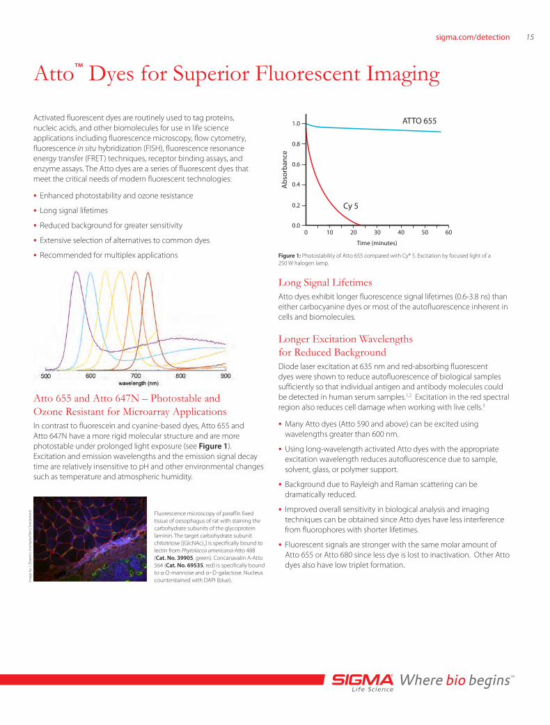
ATTO 655
Cy 5
0
1.0
0.8
0.6
0.4
0.2
0.010 20 30 40 50 60
Time (minutes)
Abs
orba
nce
Long Signal LifetimesAtto dyes exhibit longer fluorescence signal lifetimes (0.6-3.8 ns) than either carbocyanine dyes or most of the autofluorescence inherent in cells and biomolecules.
Figure 1: Photostability of Atto 655 compared with Cy® 5. Excitation by focused light of a 250 W halogen lamp.
Fluorescence microscopy of paraffin fixed tissue of oesophagus of rat with staining the carbohydrate subunits of the glycoprotein laminin. The target carbohydrate subunit chitotriose [(GlcNAc)3] is specifically bound to lectin from Phytolacca americana-Atto 488 (Cat. No. 39905, green). Concanavalin A-Atto 564 (Cat. No. 69535, red) is specifically bound to α-D-mannose and α−D-galactose. Nucleus counterstained with DAPI (blue).
Atto 655 and Atto 647N – Photostable and Ozone Resistant for Microarray ApplicationsIn contrast to fluorescein and cyanine-based dyes, Atto 655 and Atto 647N have a more rigid molecular structure and are more photostable under prolonged light exposure (see Figure 1). Excitation and emission wavelengths and the emission signal decay time are relatively insensitive to pH and other environmental changes such as temperature and atmospheric humidity.
Imag
e by
J. Z
baer
en, I
nsel
spita
l Ber
n, S
witz
erla
nd

Atto™ Dyes - Superior Tool for Super-Resolution Microscopy ApplicationRecent developments in microscopy application, like e.g. STED microscopy enables resolutions down to 10 nm. These applications require fluorescent dyes, that fulfill superior photo-physical criteria. Some Atto-dyes. Atto 488, Atto 647N, and Atto 655, have proven a suitable for techniques such as PALM, dSTORM, STED, etc..
Atto 655 and Atto 680 — Less Molecular Inactivation for Greater SignalNon-fluorescent triplet states and cis-conformations result in fluctuations that interfere with fluorescent signal yield.4 Dyes such as Cy®5, Cy5.5, or Alexa Fluor® 647 may form cis-isomers and triplet states, which precludes their usage in demanding techniques including fluorescence correlation spectroscopy (FCS), single molecule detection (SMD), and as acceptors in fluorescence resonance energy transfer (FRET).5 Atto 655 and Atto 680 have
low intersystem crossing and lack an isomeric bond so they cannot undergo configuration isomerization.
Recommended for Fluorescent Multiplex DetectionAtto dyes can be used to conjugate probes and biomolecules for multiplex applications. Selection of two Atto dyes with separated emission signals supports multiple excitation and measurement results from a single experiment.
Alternatives to Common FluorophoresWith the extensive selection available, Atto dyes can replace commonly used fluorescent dyes. There are Atto dyes suitable for use with any common excitation light source.
Immunoblot detection of Protein 1 and Protein 2 using two primary antibodies and two anti-IgG-Atto dye conjugates .Imaging was done sequentially using a FLA-3000 Fuji® laser scanner, first at an excitation wavelength of 532 nm with a 580 nm emission filter to detect Atto 550, then at an excitation wavelength of 633 nm with a 675 nm emission filter to detect Atto 633. The image overlay was done using a software tool.
Confocal microscopy (CLSM) image of paraffin fixed tissue of rat stomach. Actin stained with mouse anti–smooth muscle α-actin antibody and Atto 488 anti-mouse IgG (Cat. No. 62197, green), cytokeratin stained with polyclonal rabbit anti-cytokeratin and Atto 647N anti-rabbit IgG (Cat. No. 40839, red).
Fluorophore Recommended Atto dye alternativeAlexa Fluor® 488
Atto 488 FITC
FAM™
JOE™Atto 520
TET™
Alexa Fluor 532 Atto 532
HEX™ Atto 532, Atto Rho6G
TAMRA™ Atto 550
Cy®3 Atto 550
Cy3.5 Atto 565
ROX™ Atto 565, Atto Rho11
Alexa Fluor 594 Atto 590 , Atto 594
Texas Red® Atto 590
Alexa Fluor 633 Atto 633, Atto Rho14
Cy5 Atto 647, Atto 647N , Atto 655
Alexa Fluor 647 Atto 647, Atto 647N, Atto 655
Cy5.5 Atto 680, Atto 700
Light source Main lines (nm) Recommended Atto dyesMercury arc lamp 365, 405, 436, 546 Atto 390, Atto 425, Atto
465, Atto 550, Atto 565
Mercury arc lamp 577 Atto 590, Atto Rho101, Atto 594; Atto Rho13, Atto 610, Atto 611x
Xenon arc lamp Continuum and peaks >800 nm
Atto 610, Atto 620, Atto 647, Atto 647N, Atto 655, Atto 680
Halogen lamp Little UV and violet emission; Higher intensity toward longer wavelengths
Atto 610, Atto 620, Atto 647, Atto 647N, Atto 655, Atto 680
Argon ion laser 488, 514 Atto 488, Atto 520, Atto 532, Atto 550
Argon-krypton laser 488, 514, 647, 676 Atto 520, Atto 647, Atto 647N, Atto 655, Atto 680
Krypton laser 647,676 Atto 647, Atto 647N , Atto 655, Atto Oxa12, Atto 665, Atto 680, Atto 700, Atto 725, Atto 740
He-Ne laser 633 Atto Rho14, Atto 633, Atto 647, Atto 647N
Nd-NAG laser 532 Atto 532, Atto Rho6G, Atto 550, Atto 565, Atto Rho11, Atto Rho 12
Common diode laser 635, 650, 670 Atto 633, Atto 647, Atto 647N, Atto 655, Atto 680

17sigma.com/detection
Reactive Atto DyesAtto dyes produce intense fluorescent signals due to strong absorbance and high quantum yields.
• Strong signal intensity — Most Atto dyes have εmax values >100,000.
• Ideal for multiplex techniques using visible and near-IR emission wavelengths — Low excitation/emission overlap and good Stokes’ shift separation.
• Selection and versatility — There is an Atto dye suitable to use with any common excitation light source.
Atto dyes are available as:
• Free acid dyes for all routine staining applications.
• NHS-esters for use in common conjugation protocols.
• Maleimides for use in coupling to thiol-containing groups such as cysteine residues and thiol (-SH) tags added during automated synthesis.
Atto 655, Atto 680, and Atto 700 are quenched by guanosine, tryptophan and related compounds through direct contact between the dye and the quenching agent and using an electron transfer process. Fluorescent quenching of dyes by tryptophan residues in proteins has been used to differentiate unbound (non-fluorescent) protein from protein-antibody (fluorescent) interactions.1
Atto Dyeλabs
[nm]ε max
[m-1 cm-1]λem
[nm]ηem [%]
τem [ns]
Catalog NumberFree Acid NHS Ester Maleimide
Atto 390 390 24,000 479 90 3.8 89313 89204 89740
Atto 425 436 45,000 484 90 3.5 n/a 16805 49349
Atto 465 453 75,000 508 55 2.2 50712 53404 55607
Atto 488 501 90,000 523 80 3.2 41051 41698 28562
Atto 495 495 80,000 527 45 2.4 16951 00379 41022
Atto 520 516 110,000 538 90 3.8 70706 77810 16590
Atto 532 532 115,000 553 90 3.8 06699 88793 68499
Atto 550 554 120,000 576 80 3.2 42424 92835 30730
Atto 565 563 120,000 592 90 3.4 75784 72464 18507
Atto 590 594 120,000 624 80 3.7 70425 79636 39887
Atto 594 601 120,000 627 85 3.5 08637 08741 08717
Atto 610 615 150,000 634 70 3.3 78493 93259 41061
Atto 611X 611 100,000 681 35 2.5 40049 18708 n/a
Atto 620 619 120,000 643 50 2.9 92716 67351 n/a
Atto 633 629 130,000 657 64 3.2 18620 01464 n/a
Atto 647 645 120,000 669 20 2.3 97875 07376 41784
Atto 647N 644 150,000 669 65 3.4 04507 18373 05316
Atto 655 663 125,000 684 30 1.9 93711 76245 80661
Atto 665 663 160,000 684 60 16851 04022 01407
Atto 680 680 125,000 700 30 1.8 94875 75999 04971
Atto 700 700 120,000 719 25 1.5 30674 16986 50611
Atto 725 729 120,000 752 10 0.5 47156 93725 n/a
Atto 740 740 120,000 764 10 0.6 91394 59808 n/a
Fluorescent Signal Information for Atto Dyes
λabs - longest-wavelength absorption maximum εmax - molar extinction coefficient at the longest-wavelength absorption maximumλem - fluorescence maximum ηem - fluorescence quantum yield τem - fluorescence decay time

Convenient Atto™ Dye ConjugatesAn extensive selection of Atto dye conjugates and kits are available, including:
• Protein Labeling Kits
- Atto 488 is a superior alternative to fluorescein and Alexa Fluor® 488, producing conjugates with more photostability and brighter fluorescence.
- Atto 550 is an alternative to rhodamine dyes, Cy®3, and Alexa Fluor 550, offering more intense brightness and increased photostability.
- Atto 594 is an alternative to Alexa Fluor 594 and Texas Red®.
- Atto 647N, an extraordinary highly fluorescent dye, and Atto 655 are alternatives to Cy5 and Alexa Fluor 647.
- Atto 633 is an alternative to Alexa Fluor 633.
• Lectins for carbohydrate binding studies.
• Primary and secondary antibodies for direct and indirect ELISA, immunoblotting, immunohistochemistry, and other protein identification applications.
• Biotin and Streptavidin for avidin/streptavidin/biotin conjugation in applications including ELISA, immunohistochemistry, in situ hybridization, and flow cytometry.
• NTA Nickel conjugates for direct detection of polyhistidine-tagged recombinant proteins.
For more information and a comprehensive list of products, visit sigma.com/atto
References1. Neuweiler, H. et al., Detection of individual p53-autoantibodies by using quenched peptide-based
molecular probes. Angew. Chemie, 41, 4769-73 (2002).
2. Sauer, M. et al., Detection and identification of individual antigen molecules in human serum with pulsed semiconductor lasers. Appl. Phys. B, 65, 427-31 (1997).
3. Terasaki, M., and Dailey, M. E. , Confocal microscopy on living cells. In Handbook of biological confocal microscopy. Pawley, J. B., Ed. 2nd ed., pp 327-346, Plenum Press, New York (1995).
4. Widengren, J., and Schwille, P., Characterization of photoinduced isomerization and back-isomerization of the cyanine dye Cy5 by fluorescence correlation spectroscopy. J. Phys. Chem. A, 104, 6416-28 (2000).
5. Widengren, J. et.al., Two new concepts to measure fluorescence resonance energy transfer via fluorescence correlation spectroscopy: theory and experimental realizations. J. Phys. Chem. A, 105, 6851-66 (2001).
6. Buschmann, V., Weston, K.D., and Sauer, M., Spectroscopic study and evaluation of red-absorbing fluorescent dyes. Bioconjugate Chem., 14, 195-204 (2003).
Fluorescent microscopy of human skin tissue section (paraffin fixation) with fungal infection. The target carbohydrate chitotriose of the pathogenic fungi are specifically bound to lectin from Phytolacca americana Atto 488 conjugate (Cat. No. 39905) (green). The nuclei are counterstained with DAPI (blue). Image by J. Zbären, Inselspital, Bern.
His-tagged p38 MAPK protein (500 ng – 25 ng) was separated on a 4-20% Tris-glycine SDS-PAGE gel. After fixing and washing, the gel was incubated with Ni-NTA-Atto 647N (Cat. No. 02175) (1:1000) in the dark. The gel was washed and then imaged using a FLA-3000 Fuji® laser scanner with 633 nm excitation and a 675 nm emission filter for Ni-NTA-Atto 647N (λex 647 nm, λem 669 nm). The 50 ng band of His-tagged p38-MAPK is observed using fluorescence imaging.

19sigma.com/detection
Microscopic methods in life sciences are of tremendous importance for visualization cellular and tissue structures.
In recent years, development has reached a revolution in order to overcome the resolution barrier given by the diffraction limit. New microscopy concepts that enable resolution limits down to about 10 nm and the visualization of cellular structures and molecular interactions reveal new understanding in biological processes.
These super-resolution microscopy principles are based on several technological approaches. Conventional light microscopy enables a resolution limit of about 250 nm in the x- and y- direction and 450 – 700 nm in the z –direction. Super-resolution techniques have overcome the resolution-limit (Point-spread function), by at least a factor of 2. The resolution of super-resolution microscopy depends on the number of points that can be resolved on the structure of interest. Crucial for successful super-resolution imaging is the choice of fluorescent probe. Brightness and high contrast ratio between the states are of great importance. In most super-resolution methods, the states of the probe must be controllable, reversible or irreversible, switchable between a light or a dark state. Depending on the super-resolution method, further photo physical criteria the probe must be fulfilled. Established techniques are:
• STED (Stimulated emission depletion)
• GSDIM (Ground State Depletion)
• PALM (Photoactivated localization microscopy)
• STORM (Stochastic optical reconstruction microscopy)
• RESOLFT [Reversible saturable optical (flurorescence) transitions]
Sigma now offers the superior series of Abberior dyes that are especially designed and tested for super-resolution microscopy such as STED, RESOLFT, PALM, STORM, GSDIM and others. Abberior STAR, Abberior CAGE, Abberior FLIP and Abberior RSFP – the specific requirements of the super-resolution techniques are served with dedicated dye series.
Super-resolution microscopy depends on fluorescent labels more than any other fluorescence imaging technique. Manufactured by Abberior, the STAR, CAGE and FLIP dyes as well as RSFPs are exceptionally bright and photostable and provide optimized photoswitching for RESOLFT and PALM/STORM imaging. They are the only commercially available dyes tailored specifically to the needs of super-resolution microscopy.
Abberior dyes are also exceptionally well suited for confocal microscopy, epifluorescence imaging and single molecule applications. Basically all fluorescence applications which depend on a good signal to noise ratio and low background benefit from the novel Abberior dyes.
Benefits• Optimized for brightness and very low background
• Optimized switching behavior being the key for super-resolution
• All markers are tested for different super-resolution methods
ū Abberior STAR for STED, confocal and epifluorescence imaging
ū Abberior CAGE & FLIP for PALM, STORM and GSDIM
• Abberior dyes are recommended by renowned microscope vendors
• Proprietary, IP protected products
• Detailed characteristics of the dyes provided, e.g. optimal STED wavelength
Superior Super-Resolution Microscopy Application using Abberior® Dyes
Confocal STED
AbberiorSTAR series
Comparison of confocal (left) and STED (right) microscopy application.

Overview of Abberior® dyes
Dyes DescriptionAbsorption Maximum/ λmax
Extinction Coefficient, ε(λ)
Fluorescence Maximum, λfl
Recommended STED
Cat No., NHS activated
Cat No., maleimid activated
Abberior® CAGE 500 for single-molecule switching microscopy (e.g. PALM, STORM, GSDIM)
300 nm (caged,pH 7); 501 nm (uncaged, pH 7)
85,000 — 88,000 m-1cm-1 (pH 7, uncaged)
524 nm (pH 7), 523 nm (MeOH)
595-615 nm 44254 92546
Abberior® CAGE 532 for single-molecule switching microscopy (e.g. PALM, STORM, GSDIM)
304 nm (caged, PBS, pH 7); 518 nm (uncaged, pH 7)
29,000 m-1cm-1
(pH 7, uncaged)541 nm (pH 7) 610 - 640 nm 38977 95705
Abberior® CAGE 552 for single-molecule switching microscopy (e.g. PALM, STORM, GSDIM)
300 nm (caged,pH 7); 552 nm (uncaged, pH 7)
66,000 m-1cm-1
(pH 7, uncaged)574 nm (pH 7) 650-670 nm 94822 92545
Abberior® FLIP 565 for single-molecule switching microscopy (e.g. PALM, STORM, GSDIM)
314 nm (PBS, pH 7.4) 51,000 m-1cm1
(MeOH)580 nm (PBS, pH 7.4) 79189 92544
Abberior® STAR 440SX for long Stokes STED and 2-color STED application
430 nm (MeOH), 437 nm (PBS, pH 7.4)
30,800 m-1cm1 (MeOH), 22,700 m-1cm1 (PBS, pH 7.4)
501 nm (MeOH), 515 nm (PBS, pH 7.4)
590-620 nm 68221 38361
Abberior® STAR 470SX for long Stokes STED and 2-color STED application
475 nm (MeOH), 477 nm (PBS, pH 7.4)
30,400 m-1cm1 (MeOH), 22,700 m-1cm1 (PBS, pH 7.4)
609 nm (MeOH), 627 nm (PBS, pH 7.4)
740 - 770 nm 95348
Abberior® STAR 488 for STED application 501 nm (PBS, pH 7.4) 86,000 m-1cm1 (MeOH)
524 nm (PBS, pH 7.4) 585 - 605 nm 61048
Abberior® STAR 512 for STED application 517 nm (MeOH), 512 nm (PBS, pH 7.4)
74,000 m-1cm1 (MeOH)
536 nm (MeOH), 530 nm (PBS, pH 7.4)
590 - 620 nm 38922 03004
Abberior® STAR 580 for STED application 587 nm (MeOH), 583 nm (PBS, pH 7.4)
64,300 m-1cm1 (MeOH)
609 nm (MeOH), 605 nm (PBS, pH 7.4)
690 - 720 nm 38377
Abberior® STAR 635 for STED application 639 nm (MeOH), 634 nm (PBS, pH 7.4)
63,000 m-1cm1 (MeOH)
659 nm (MeOH), 654 nm (PBS, pH 7.4)
740 - 770 nm 30558 96013
Abberior® STAR 635P for STED application 635 nm (MeOH), 634 nm (PBS, pH 7.4)
80,000 m-1cm1 (water)
655 nm (MeOH), 654 nm (PBS, pH 7.4)
740 - 770 nm no, only Azide derivative available
References1. T. Müller, C. Schumann, A. Kraegeloh, "STED Microscopy and its Applications: New Insights into
Cellular Processes on the Nanoscale", ChemPhysChem 13, 1986–2000 (2012).
2. Diffraction-unlimited all-optical imaging and writing with a photochromic GFP
3. Tim Grotjohann, Ilaria Testa, Marcel Leutenegger, Hannes Bock, Nicolai T. Urban, Flavie Lavoie-Cardinal, Katrin I. Willig, Christian Eggeling, Stefan Jakobs and Stefan W. Hell. Nature 478, 204-208 (13 October 2011)
For a list of Abberior dyes, visitsigma.com/abberior

21sigma.com/detection
Spectral Fluorescence Standard KitThe Spectral Fluorescence Standard Kit (Cat. No. 69336) enables a simple characterization of the relative spectral responsivity and the long-term stability of the emission channel of fluorescence instruments under routine measurement conditions and provides the basis for an improved comparability of fluorescence measurement and eventual standardization.
• CD with Improved Software LINKCORRWIN V.1.1.0.0. developed by BAM for the data evaluation. Version1.1.0.0 only performs well in conjunction with BAM-F002a. The CD also contains instructions for use of BAM-F001 – BAM-F005 and LINKCORRWIN.
• Five Spectral Florescence Standards Ready-made from Sigma-Aldrich GmbH, which cover the spectral region of 300 nm to 770 nm as a set.
• Ethanol, absolute (Cat. No. 34923) Addition of aliquots of 10 mL of ethanol to each solid dye yields a solution that can be measured without additional dilution steps.
• BAM Certificate of the normalized corrected emission spectra of BAM-F001 – BAM-F005 (see also CD).
Application of the Emission Correction CurveBottom: Certified normalized corrected emission spectra (solid lines; lc(λem)) of the kit components and uncorrected, i.e. instrument-dependent emission spectra (dashed lines; lu (λem)), measured with the instrument to be calibrated.
Middle: Individual quotient QF00x = lcF00x (λem)/luF00x (λem) for each kit dye equaling 1/s(λ) of the instrument to be calibrated within the spectral region of the respective fluorescence standard.
Top: Combined emission correction curve 1/s(λ) (solid pink line) calculated from the statistically weighted QF00x as well as its reciprocal s(λ) (black solid line).
Corrected emission spectra are obtained by multiplication of measured spectra by the output of LINKCORR, i.e., 1/s(λ).
Determination of the relative spectral responsivity s(λ) of a fluorescence instrument with the Spectral Fluorescence Standard Kit and LINKCORRWIN.

Certified Reference Materials• For the determination of the relative spectral responsivity s (λ) of
fluorescence instruments. 1/s (λ) is termed emission correction curve. Both can be determined by LINKCORRWIN.
• For the determination of corrected, i.e., instrument-independent emission spectra that are comparable across instruments.
• For the characterization of the long-term stability of the emission channel of fluorescence instruments. Fluorescence spectra that are corrected accordingly do not contain contributions from aging of optical components in the emission channel.
Certified PropertiesNormalized corrected emission spectra of BAM-F001 – BAM-F005 in ethanol for T=25 °C. The emission spectra are traceable to the spectral radiance realized and disseminated in Germany by the Physikalisch-Technische Bundesanstalt (PTB).
The Calibration Kit has been tested in an interlaboratory comparison by the National Metrological Institutes NIST (National Institutes of Standards and Technology, USA), NRC (National Research Council, Canada), PTB (Physikalisch-Technische Bundesanstalt, Germany), and BAM (Federal Institute for Material Research and Testing) employing two different dye concentrations and two different measurement geometries, i.e., 0°/90° and 45°/0°.
For more information, please visit sigma.com/spectral
References1. Hollandt, J.; Taubert, R.D.; Seidel, J.; Resch-Genger, U.; Gugg-Helminger, A.; Pfeifer, D.; Monte, C.,
J. Fluoresc. 2005, 15, 311.
2. Resch-Genger, U.; Pfeifer, D.; Pilz, W.; Monte, C.; Hoffmann, A.; Spieles, M.; Rurack, K.; Hollandt, J.; Taubert, D.; Schönenberger, B.; Nording, P., J. Fluoresc. 2005, 15, 325.
3. Resch-Genger, U.; Hoffmann, K.; Nietfeld, W.; Engel, A.; Neukammer, J.; Nietschke, R.; Ebert, B.; Macdonald, R., J. Fluoresc. 2005, 15, 347.
4. Hoffmann, K.; Monte, C.; Pfeifer, D.; Resch-Genger, U., G.I.T. Laboratory Journal 6, 2005, 3, 18.
5. Monte, C.; Pilz, W.; Resch-Genger, U., Proc. SPIE 2005, 5880, 588019-1.
6. Hoffmann, K.; Monte, C.; Pfeifer, D.; Resch-Genger, U., G.I.T. Laboratory Journal 2005, 6, 29.
7. Monte, C.; Resch-Genger, U.; Pfeifer, D.; Taubert, R.D.; Hollandt, J., Metrologia 2009, 16, 589.
8. Monte, C.; Hoffmann, K.; Pfeifer, D.; Hoffmann, A.; Resch-Genger, U., J Fluoresc. 2006, 16, 441.

Sigma-Aldrich® Worldwide Offices
ArgentinaFree Tel: 0810 888 7446 Tel: (+54) 11 4556 1472 Fax: (+54) 11 4552 1698
AustraliaFree Tel: 1800 800 097 Free Fax: 1800 800 096 Tel: (+61) 2 9841 0555 Fax: (+61) 2 9841 0500
AustriaTel: (+43) 1 605 81 10 Fax: (+43) 1 605 81 20
BelgiumTel: (+32) 3 899 13 01 Fax: (+32) 3 899 13 11
BrazilFree Tel: 0800 701 7425 Tel: (+55) 11 3732 3100 Fax: (+55) 11 5522 9895
CanadaFree Tel: 1800 565 1400 Free Fax: 1800 265 3858 Tel: (+1) 905 829 9500 Fax: (+1) 905 829 9292
ChileTel: (+56) 2 495 7395 Fax: (+56) 2 495 7396
People’s Republic of ChinaFree Tel: 800 819 3336 Tel: (+86) 21 6141 5566 Fax: (+86) 21 6141 5567
Czech RepublicTel: (+420) 246 003 200 Fax: (+420) 246 003 291
DenmarkTel: (+45) 43 56 59 00 Fax: (+45) 43 56 59 05
FinlandTel: (+358) 9 350 9250 Fax: (+358) 9 350 92555
FranceFree Tel: 0800 211 408 Free Fax: 0800 031 052 Tel: (+33) 474 82 28 88 Fax: (+33) 474 95 68 08
GermanyFree Tel: 0800 51 55 000 Free Fax: 0800 64 90 000 Tel: (+49) 89 6513 0 Fax: (+49) 89 6513 1169
HungaryTel: (+36) 1 235 9055 Fax: (+36) 1 235 9068
IndiaTelephone Bangalore: (+91) 80 6621 9400 New Delhi: (+91) 11 4358 8000 Mumbai: (+91) 22 4087 2364 Pune: (+91) 20 4146 4700 Hyderabad: (+91) 40 3067 7450 Kolkata: (+91) 33 4013 8000
Fax Bangalore: (+91) 80 6621 9550 New Delhi: (+91) 11 4358 8001 Mumbai: (+91) 22 2579 7589 Pune: (+91) 20 4146 4777 Hyderabad: (+91) 40 3067 7451 Kolkata: (+91) 33 4013 8016
IrelandFree Tel: 1800 200 888 Free Fax: 1800 600 222 Tel: +353 (0) 402 20370 Fax: + 353 (0) 402 20375
IsraelFree Tel: 1 800 70 2222 Tel: (+972) 8 948 4222 Fax: (+972) 8 948 4200
ItalyFree Tel: 800 827 018 Tel: (+39) 02 3341 7310 Fax: (+39) 02 3801 0737
JapanTel: (+81) 3 5796 7300 Fax: (+81) 3 5796 7315
KoreaFree Tel: (+82) 80 023 7111 Free Fax: (+82) 80 023 8111 Tel: (+82) 31 329 9000 Fax: (+82) 31 329 9090
LuxembourgTel: (+32) 3 899 1301 Fax: (+32) 3 899 1311
MalaysiaTel: (+60) 3 5635 3321 Fax: (+60) 3 5635 4116
MexicoFree Tel: 01 800 007 5300 Free Fax: 01 800 712 9920 Tel: (+52) 722 276 1600 Fax: (+52) 722 276 1601
The NetherlandsTel: (+31) 78 620 5411 Fax: (+31) 78 620 5421
New ZealandFree Tel: 0800 936 666 Free Fax: 0800 937 777 Tel: (+61) 2 9841 0555 Fax: (+61) 2 9841 0500
NorwayTel: (+47) 23 17 60 00 Fax: (+47) 23 17 60 10
PolandTel: (+48) 61 829 01 00 Fax: (+48) 61 829 01 20
PortugalFree Tel: 800 202 180 Free Fax: 800 202 178 Tel: (+351) 21 924 2555 Fax: (+351) 21 924 2610
RussiaTel: (+7) 495 621 5828 Fax: (+7) 495 621 6037
SingaporeTel: (+65) 6779 1200 Fax: (+65) 6779 1822
SlovakiaTel: (+421) 255 571 562 Fax: (+421) 255 571 564
South AfricaFree Tel: 0800 1100 75 Free Fax: 0800 1100 79 Tel: (+27) 11 979 1188 Fax: (+27) 11 979 1119
SpainFree Tel: 900 101 376 Free Fax: 900 102 028 Tel: (+34) 91 661 99 77 Fax: (+34) 91 661 96 42
SwedenTel: (+46) 8 742 4200 Fax: (+46) 8 742 4243
SwitzerlandFree Tel: 0800 80 00 80 Free Fax: 0800 80 00 81 Tel: (+41) 81 755 2511 Fax: (+41) 81 756 5449
ThailandTel: (+66) 2 126 8141 Fax: (+66) 2 126 8080
United KingdomFree Tel: 0800 717 181 Free Fax: 0800 378 785 Tel: (+44) 01747 833 000 Fax: (+44) 01747 833 574
United StatesToll-Free: 800 325 3010 Toll-Free Fax: 800 325 5052 Tel: (+1) 314 771 5765 Fax: (+1) 314 771 5757
VietnamTel: (+84) 8 3516 2810 Fax: (+84) 8 6258 4238
Internet sigma-aldrich.com
803281073
Order/Customer Service: sigma-aldrich.com/order Technical Service: sigma-aldrich.com/techservice Development/Custom Manufacturing Inquiries [email protected] Safety-related Information: sigma-aldrich.com/safetycenter
World Headquarters 3050 Spruce St.
St. Louis, MO 63103 (314) 771-5765
sigma-aldrich.com
Enabling Science to Improve the Quality of Life
©2013 Sigma-Aldrich Co. LLC. All rights reserved. SIGMA and SIGMA-ALDRICH are trademarks of Sigma-Aldrich Co. LLC, registered in the US and other countries. FLUKA is a trademark of Sigma-Aldrich GmbH, registered in the US and other countries. MISSION, Stemline, Prestige Antibodies, Imprint, LOPAC, and CompoZr is a registered trademark of Sigma-Aldrich Co. LLC. Where bio begins, ReadyMix, N-TER and ReadyScript is a trademark of Sigma-Aldrich Co. LLC. Duolink and PLA are registered trademarks of Olink AB. Sunstone and Intelligent Material are registered trademarks of Intelligent Material Solutions, Inc. Abberior is a registered trademark of Abberior GmbH. DyLight is a registered trademark of Pierce Biotechnology. eFluor is a registered trademark of eBioscience, Inc. LightCycler is a registered trademark of Roche Molecular Systems, Inc. IRDYE is a registered trademark of LI-COR, Inc. Fuji is a registered trademark of Fujifilm Corp. Cy is a registered trademark of GE Healthcare. Cy3 is a trademark of GE Healthcare. CASCADE BLUE, ALEXA FLUOR, and TEXAS RED are registered trademarks of Life Technologies. Pacific Blue and Rhodamine Red are trademarks of Life Technologies. CF and Mix-n-Stain are trademarks of Biotium, Inc. Atto is a trademark of Atto-Tec GmbH. BD Horizon is a trademark of Becton, Dickinson and Company. FAM, TET, HEX, ROX, TAMRA and JOE are trademarks of Applera Corporation or its subsidiaries in the US and/or certain other countries. Suprasil is a registered trademark of HERAEUS QUARZGLAS GMBH & CO. Hellma is a registered trademark of Hellma GmbH. PlastiBrand is a registered trademark of Brand GmbH & Co. KG. Eppendorf and UVette are registered trademarks of Eppendorf AG. Sigma-Aldrich, Sigma, and Fluka brand products are sold by affiliated Sigma-Aldrich distributors. Purchaser must determine the suitability of the product(s) for their particular use. Additional terms and conditions may apply. Please see product information on the Sigma-Aldrich website at www.sigmaaldrich.com and/or on the reverse side of the invoice or packing slip.