biofilm structures in a mono-associated mouse model of...
TRANSCRIPT

Biofilm Structures in a Mono-AssociatedMouse Model of Clostridium difficile Infection
Item Type Article
Authors Soavelomandroso, Anna P.; Gaudin, Françoise; Hoys, Sandra;Nicolas, Valérie; Vedantam, Gayatri; Janoir, Claire; Bouttier,Sylvie
Citation Biofilm Structures in a Mono-Associated Mouse Model ofClostridium difficile Infection 2017, 8 Frontiers in Microbiology
DOI 10.3389/fmicb.2017.02086
Publisher FRONTIERS MEDIA SA
Journal Frontiers in Microbiology
Rights © 2017 Soavelomandroso, Gaudin, Hoys, Nicolas, Vedantam,Janoir and Bouttier. This is an open-access article distributedunder the terms of the Creative Commons Attribution License (CCBY).
Download date 07/06/2018 01:22:14
Link to Item http://hdl.handle.net/10150/626057

fmicb-08-02086 October 23, 2017 Time: 15:56 # 1
ORIGINAL RESEARCHpublished: 25 October 2017
doi: 10.3389/fmicb.2017.02086
Edited by:Hui Wu,
University of Alabama at Birmingham,United States
Reviewed by:Xin Fang,
Columbia University Medical Center,United States
Christiane Gerke,Institut Pasteur, France
*Correspondence:Sylvie Bouttier
Specialty section:This article was submitted to
Infectious Diseases,a section of the journal
Frontiers in Microbiology
Received: 17 March 2017Accepted: 11 October 2017Published: 25 October 2017
Citation:Soavelomandroso AP, Gaudin F,Hoys S, Nicolas V, Vedantam G,
Janoir C and Bouttier S (2017)Biofilm Structures in a
Mono-Associated Mouse Modelof Clostridium difficile Infection.
Front. Microbiol. 8:2086.doi: 10.3389/fmicb.2017.02086
Biofilm Structures in aMono-Associated Mouse Model ofClostridium difficile InfectionAnna P. Soavelomandroso1, Françoise Gaudin2, Sandra Hoys1, Valérie Nicolas3,Gayatri Vedantam4, Claire Janoir1 and Sylvie Bouttier1*
1 EA4043, Unité Bactéries Pathogènes et Santé (UBaPS), Univ. Paris-Sud, Université Paris-Saclay, Châtenay-Malabry,France, 2 Institut Paris Saclay d’Innovation Thérapeutique (IPSIT), UMS IPSIT Université Paris-Sud – US 31 INSERM – UMS3679 CNRS, Plateforme d’Histologie souris Immunopathologie de Clamart – PHIC, Clamart, France, 3 Institut Paris Saclayd’Innovation Thérapeutique (IPSIT), UMS IPSIT Université Paris-Sud – US 31 INSERM – UMS 3679 CNRS, Plateformed’Imagerie cellulaire – MIPSIT, Châtenay-Malabry, France, 4 School of Animal and Comparative Biomedical Sciences,University of Arizona, Tucson, AZ, United States
Clostridium difficile infection (CDI) is a major healthcare-associated disease with highrecurrence rates. Host colonization is critical for the infectious process, both in firstepisodes and in recurrent disease, with biofilm formation playing a key role. The abilityof C. difficile to form a biofilm on abiotic surfaces is established, but has not yet beenconfirmed in the intestinal tract. Here, four different isolates of C. difficile, which arein vitro biofilm producers, were studied for their ability to colonize germ-free mice.The level of colonization achieved was similar for all isolates in the different partsof the murine gastrointestinal tract, but pathogen burden was higher in the cecumand colon. Confocal laser scanning microscopy revealed that C. difficile bacteria weredistributed heterogeneously over the intestinal tissue, without contact with epithelialcells. The R20291 strain, which belongs to the Ribotype 027 lineage, displayed aunique behavior compared to the other strains by forming numerous aggregates. Byimmunochemistry analyses, we showed that bacteria were localized inside and outsidethe mucus layer, irrespective of the strains tested. Most bacteria were entrapped in 3-Dstructures overlaying the mucus layer. For the R20291 strain, the cell-wall associatedpolysaccharide PS-II was detected in large amounts in the 3-D structure. As thiscomponent has been detected in the extrapolymeric matrix of in vitro C. difficile biofilms,our data suggest strongly that at least the R20291 strain is organized in the mono-associated mouse model in glycan-rich biofilm architecture, which sustainably maintainsbacteria outside the mucus layer.
Keywords: C. difficile, biofilm, mono-associated mouse model, colonization, gut, mucus, immunochemistry,CLSM
INTRODUCTION
The obligate anaerobe Clostridium difficile is the leading cause of healthcare-associated diarrhea.C. difficile infection (CDI) represents 15–25% of antibiotic-associated diarrhea and clinicalsymptoms range from mild diarrhea to life-threatening pseudomembranous colitis (Evans andSafdar, 2015).
Frontiers in Microbiology | www.frontiersin.org 1 October 2017 | Volume 8 | Article 2086

fmicb-08-02086 October 23, 2017 Time: 15:56 # 2
Soavelomandroso et al. Clostridium difficile Biofilm in Monoxenic Mouse Model
CDI usually appears after ingestion of spores, which arethe contaminating forms of C. difficile. Concomitantly, thedisruption of the intestinal microbiota due to antibiotic therapysupports germination of spores as well as colonization ofoutgrowing vegetative cells. Vegetative cells express differentfactors involved in virulence such as several colonization factorsand two main virulence factors: the TcdA and TcdB toxins thatlead to actin cytoskeleton disorganization and cell death (Janoir,2016). A third toxin called the binary toxin (CdtAB) has beenfound in 23% of toxigenic strains in Europe in 2008 (Gerdinget al., 2014). It is assumed that CDT potentiates the toxicity ofTcdA and TcdB.
Recurrence is a major challenge encountered in themanagement of CDI, since as many as 35% of patients mayundergo a recurrence after the first episode, and this rateincreases after further episodes (Marsh et al., 2012). Recurrencecorresponds either to a relapse with the same strain or toa re-infection with another strain, which occurs in 38–56%of cases (Barbut et al., 2000). The causes of relapses are notclearly determined, but undoubtedly involve host and bacterialfactors. One of the main bacterial features involved in relapsingdisease is the ability of C. difficile to re-sporulate and persistin the gastrointestinal tract, resisting antibiotic exposure(Deakin et al., 2012). Discrepant results have been reported inthe correlation between recurrences and virulence factors ofbacteria. Stewart et al. (2013) found that the binary toxin isa predictor of recurrent infection. In contrast, a recent studydemonstrated that the outcome of CDI correlates neitherwith the virulence features nor with the in vitro level ofsporulation (Plaza-Garrido et al., 2015). Another hypothesisis that relapses may be associated with the persistence ofC. difficile in a biofilm. Indeed, it is well known that mono-or polymicrobial biofilm formation contributes to chronicityof several infections, such as chronic wounds, cystic fibrosisand periodontitis (Mihai et al., 2015). The persistence ofbacteria in the host is supported by biophysical characteristicsof biofilms, which result in avoidance of host immunity andresistance to environmental stresses, such as antibiotic therapy.Biofilm is defined in stricto sensu by a 3-D organization ofa bacterial community adherent to an abiotic or a bioticsupport, and embedded in an extracellular polymeric substance(Costerton et al., 1978). In addition to its role in chronicinfections, biofilms may participate in colonization of bothcommensal and pathogenic bacteria. Asymptomatic carriageof C. difficile has been described, and is now recognized as areservoir for C. difficile transmission (Donskey et al., 2015),and C. difficile biofilm could also be involved in asymptomaticcarriage.
The ability of C. difficile to form a biofilm in vitrowas first described 5 years ago (Donelli et al., 2012). Sincethen, the biofilm-producing phenotype of C. difficile has beenconfirmed, and several factors that modulate the biofilmformation have been identified. In particular, we have previouslyshown that the cell wall-associated cysteine protease Cwp84,involved in the S-layer maturation, negatively modulates biofilmformation by a still unknown mechanism (Pantaléon et al.,2015). In contrast, the regulator of quorum sensing LuxS and
the key regulator of sporulation Spo0A positively modulatebiofilm formation (Dawson et al., 2012; Ðapa et al., 2013).As observed for other species, the extrapolymeric matrix ofC. difficile in vitro biofilm is composed of polysaccharidesincluding cell wall-associated PSII, proteins and DNA (Ðapaet al., 2013; Semenyuk et al., 2014; Pantaléon et al., 2015).The relationship between the ability of C. difficile to forma biofilm and its capacity to colonize and to persist in ahost is not yet known. Moreover, how C. difficile associateswith biotic surfaces is still poorly understood. Lawley et al.(2009) reported individual or grouped C. difficile cells observedclosely associated with damaged epithelia in conventional mice.A recent work suggests that in the conventional murine modelof CDI, C. difficile is a minority member of the microbialcommunities during infection and is associated with the outermucus layer (Semenyuk et al., 2015). C. difficile was alsoshown to associate to polymicrobial biofilms in an in vitrohuman chemostat gut model particularly in spore form, whileplanktonic bacteria were primarily vegetative cells (Crowtheret al., 2014a,b).
To date, there are no data on the ability of C. difficileto form a mono-microbial biofilm in vivo. The aim of ourstudy was therefore to investigate the level of colonizationachieved by different in vitro biofilm-producing C. difficilestrains in the intestinal tract of germ-free mice, and define thespatial organization of bacteria associated with gut tissues. TheC. difficile mono-associated mouse model was chosen becausethis is a simplified model of colonization, devoid of competitiveinteraction; in addition this is an easy tool for the observation ofbiofilm structure.
MATERIALS AND METHODS
Bacterial Strains and MediaThe C. difficile strains used in this study are described in Table 1.Bacteria were grown at 37◦C, under anaerobic conditions (90%N2, 5% CO2 and 5% H2), in fresh BHISG (Brain Heart infusionbroth [Difco, United States] supplemented with 1.8% Glucose,0.1% L-Cysteine and 0.5% yeast extract) (Pantaléon et al., 2015).This sugar-rich medium is commonly used for C. difficile inin vitro biofilm assays (Dawson et al., 2012; Ðapa et al., 2013;Semenyuk et al., 2014) because the presence of glucose in theculture is essential for optimal biofilm formation (Ðapa et al.,2013).
In Vitro BiofilmBiofilm assays were performed in 24-well polystyrene plates(Costar, United States). Overnight suspensions of each C. difficilestrain in BHISG broth were diluted in fresh BHISG and 106
bacteria were added to each well. Plates were incubated at37◦C under anaerobic conditions. After 72 h of incubation,the supernatant was removed carefully, and the wells wererinsed twice with PBS. The biofilm biomass was thereafterquantified by crystal violet staining, and enumeration ofviable cells. Crystal violet (ACROS OrganicsTM, United States)quantification was performed as previously described (Dawson
Frontiers in Microbiology | www.frontiersin.org 2 October 2017 | Volume 8 | Article 2086

fmicb-08-02086 October 23, 2017 Time: 15:56 # 3
Soavelomandroso et al. Clostridium difficile Biofilm in Monoxenic Mouse Model
TABLE 1 | Characteristics of Clostridium difficile strains used in this study.
Clostridium difficile strains Ribotype Toxinotype Origin
R20291 RT 027 III Stoke Mandeville epidemic 027 strain (United Kingdom)
P30 RT 014/020 0 Strain isolated from poultry
6301erm RT 012 0 Erythromycin sensitive derivative of the 630 strain (Switzerland)
6301erm cwp84::erm (named thereafter cwp84 mutant) RT 012 0 Mutant strain derived from the 6301erm (Clostron)
et al., 2012; Ðapa et al., 2013; Pantaléon et al., 2015). Forviable cell enumeration, 1 ml of sterile pre-reduced PBSwas added to each well, the biofilm formed in the bottomwas scraped, diluted and plated on BHI agar supplementedwith 3% defibrinated horse blood. For each quantificationmethod, experiments were done on two wells from the sameplate, and biological replicates were performed at least threetimes.
Biofilm architecture was then analyzed by confocal laserscanning microscopy (CLSM). Prior to aerobic transfer,the plate was covered with parafilm to minimize oxygenexposure. The biofilm was stained 3 days post-incubationwith the LIVE/DEAD R© BacLightTM Bacterial Viability Kit(Thermo Fisher Scientific, United States): 200 µl of thediluted mixture (1:1000) was added per well and incubated15 min at 37◦C under anaerobic conditions. Samples werevisualized with a LSM 510 microscope (Carl Zeiss Inc.,Germany). SYTO R© 9 and propidium iodide exhibit anexcitation at 483 and 535 nm and a fluorescence emissionat 503 and 617 nm, respectively. Horizontal plane images witha z-step of 0.98 µm were acquired for each strain at threedifferent areas in each well. The thickness were determineddirectly from the confocal stack images using the softwareimage J.
The Mann–Whitney U Test was used for statistical analyses.P < 0.05 was considered statistically significant.
Animal ModelSix to 8 week-old germ-free C3H/HeN female mice werepurchased from CDTA (CNRS Orléans, France). All animalexperiments were performed according to European Unionguidelines for the handling of laboratory animals and allprocedures were approved by the Ethics Committee CAPSUD(Protocol 2012-109). Mice were housed in sterile isolators withad libitum access to food and water. Before experiments, eachanimal was confirmed to be germ-free by Gram staining offeces and by inoculating feces into BHI broth and incubatingthe broth for 48 h, either aerobically or anaerobically.Mice were challenged by oral gavage with 106 bacteria in0.5 ml volume. This inoculum was prepared as follows: anovernight culture in BHISG was pelleted, washed twice withPBS, and then re-suspended in PBS to a final concentrationof approximately 2 × 10⁶ vegetative cells/ml, estimated bymicroscopic cell counting. This bacterial concentration waschecked thereafter by enumerating both vegetative cells andspores. Seven days post-infection, mice were sacrificed anddifferent parts of the intestinal tract (jejunum, ileum, cecum,and colon) as well as fecal samples were collected, either
for enumeration of bacteria, confocal microscopy analyses orimmunohistochemistry analyses.
Intestinal Colonization LevelsTo determine the mean level of colonization of the fourC. difficile strains, enumeration of bacteria in the digestivetract of mono-associated mice was performed from three micefor each strain tested. First, the contents of the differentintestinal parts were collected and used for enumeration ofluminal bacteria (LB). Second, after three PBS rinses, themucosal tissues were homogenized for 1 min with Ultra-TurraxT25 (IKA R©, Labortechnik, Germany), and tissue-associatedbacteria (TAB) enumerated. Both vegetative cells and sporeswere enumerated in all samples. Vegetative cells were countedby plating serial dilutions onto BHI agar supplemented with3% horse blood. Then, samples were treated with ethanol,as previously described (Pantaléon et al., 2015) and sporeswere counted by plating serial dilutions onto BHI agarsupplemented with 3% horse blood and 0.1% taurocholate(Sigma, United States).
Confocal Laser Scanning Microscopy(CLSM)The spatial organization of tissue-associated bacteria wasdetermined by CLSM analysis of mouse mucosa from at leastthree mice for each strain. Seven days post-infection, the differentparts of the intestine were collected (jejunum, ileum, cecum,and colon). After removal of intestinal content, the tissueswere washed three times in 10 ml of PBS, spread on a glassslide and stained with the LIVE/DEAD R© BacLightTM BacterialViability Kit. Samples were visualized as described above withthe same parameters defined as for in vitro assay. During theZ-stack acquisition, an average of five areas on tissue samplewas analyzed. In addition, three-dimensional projections werereconstructed from x to z stacks using the software Imaris(Bitplane, United Kingdom).
Immunohistochemistry AnalysesImmunochemistry was performed on three mice orallychallenged with either the R20291, the P30 or the 6301ermstrain. As a negative control, a germ-free mouse was labeledsimilarly. Cecum and colon were sampled after the sacrifice ofmice, fixed in Carnoy’s solution and paraffin sections (7 µm)obtained.
Bacterial staining was performed with two fluorescent dyesthat stain both eukaryotic and prokaryotic double-strandedDNA. Samples were stained either with SYTO R© 9 at 1:1,000
Frontiers in Microbiology | www.frontiersin.org 3 October 2017 | Volume 8 | Article 2086

fmicb-08-02086 October 23, 2017 Time: 15:56 # 4
Soavelomandroso et al. Clostridium difficile Biofilm in Monoxenic Mouse Model
dilution, or Hoechst at 1:500 dilution, and incubated 1 h at 37◦C,before washing with PBS.
For mucus straining, rabbit antisera against Mucin 2 (H-300):sc-15334 (anti-Muc2, Santa Cruz Biotechnology, Inc.)were used at 1:100 dilution for 1 h at room temperature.After washing, the sections were incubated with a secondaryantibody for 1 h at room temperature before detection. Twodifferent secondary antibodies were used, depending on theobjective of the labeling. For detection of mucus only, we useda biotinylated antibody that was detected with streptavidin-HRP complex followed by 3,3′-diaminobenzidine or 3-amino-9-ethylcarbazole detection (LSAB kit, Dako, United States);sections were then counterstained with hematoxylin. In someexperiments, we performed a double-staining of mucus andbacteria to study the localization of bacteria with respect tomucus. In this case, we used Alexa Fluor 594 secondaryantibody (A-11007, Life Technologies, United States), detectedby immunofluorescence. A slide stained only with the secondaryantibody was used as an additional control for specificity of thestaining.
In a final set of experiments, the bacterial polysaccharide PS-IIwas stained by immunochemistry using rabbit antibodies againstPS-II at 1:1000 dilution for 1 h at room temperature. The sectionswere washed and incubated with a secondary antibody AlexaFluor 594 for 1 h at room temperature before detection.
Slides were scanned by the digital slide scanner NanoZoomer2.0-RS (Hamamatsu, Japan), which allowed an overall view ofthe samples. Images were digitally captured from the scan slidesusing the NDP.view2 software (Hamamatsu).
RESULTS
Biofilm Formation Varies betweenC. difficile Isolate in VitroWe questioned whether the ability of strains to form a biofilmcould predict their ability to colonize the gut. We studied fourstrains that were chosen according to their ability to form abiofilm on abiotic surfaces, and their level of colonization wasinvestigated in the mono-associated mouse model.
Using crystal violet staining, we were able to classify the strainsin three categories that display significant differences in theirbiofilm-producing abilities (Supplementary Figure S1A): onehigh-biofilm former (cwp84 mutant) (mean absorbance > 15),two moderate-biofilm formers (R20291 and P30) (5 < meanabsorbance < 15) and one low-biofilm former (meanabsorbance < 5) (6301erm). Overall, the biomass quantificationby crystal violet staining was proportional to the viable cell count(Supplementary Figure S1B). By confocal microscopy analysis(Supplementary Figure S1C) we confirmed the ability of thecwp84 mutant to form the most significant biofilm, as illustratedby the mean thickness of each biofilm (Supplementary FigureS1D). In a previous study (Pantaléon et al., 2015), we showedthat the surface of the cwp84 mutant was more hydrophobicthan the parental strain but the initial adhesion was not altered.Therefore, the increased biofilm is not related to an increase inadhesion in the early stages.
C. difficile Colonization Burden of theGerm-Free Murine Intestine IsLocation-SpecificThe ability to colonize the intestine of germ-free mice wasstudied for each of the strains described in Table 1. Bacterialcolonization was determined by the amount of both luminal (LB)and tissue-associated bacteria (TAB); vegetative cells and sporeswere enumerated.
Overall, titers of luminal bacteria of all four C. difficile strainswere similar, and indistinguishable in the different parts ofthe gut (jejunum, ileum, cecum, and colon) at 7 days post-infection (Figure 1). However, overall colonization levels were100-fold lower in the jejunum and ileum as compared to thececum and colon (Figure 1). We compared the amount ofadherent bacteria in the different parts of the gut. Titers of tissue-associated bacteria were slightly different according to strains inall parts of the gut and except for the ileum, the strains could beclassified as follows: P30 > cwp84 mutant > 6301erm > R20291.Gram staining was performed on bacterial suspension afterhomogenization and vortexing to ensure that bacteria no longerformed clusters.
Luminal bacteria and TAB spores were present in smalleramounts as compared to vegetative cells and represented less than15% of total population, except for the R20291 strain in jejunum.Surprisingly, in cecum and colon, the ratio of spores/vegetativecells seemed to be lower for R20291 strain compared to the P30and the cwp84 mutant strains.
Distribution of Bacteria Over EpithelialTissuesIn accordance to the very low amount of bacteria associatedwith the mucosa in jejunum and ileum (103–106 CFU/g oftissue), we rarely detected bacteria in these tissues by CLSM(data not shown) in contrast to what was observed in the cecum(Figure 2A) and the colon (data not shown). In both cecumand colon, irrespective of strain, we observed areas without andwith bacteria associated with tissues. When present, bacteria wereobserved both in cecum and colon, but the bacterial distributionwas different according to strain. While bacterial distribution forthe P30, 6301erm and cwp84 mutant was mostly as single cells,the R20291 was organized as bacterial aggregates (Figure 2A,Panel c).
Therefore, as observed by 3-D visualization (Figure 2A,projections at the bottom), bacteria were organized at theepithelial surface as a 3-D structure, and were never in directcontact with the epithelial cells. With the four strains, we foundrandomly distributed areas either with a high or a low thicknessof the C. difficile community and this was particularly observedfor R20291 and cwp84 mutant strains. Therefore, no significantdifference in mean thickness of the 3-D structure was observedbetween strains in the cecum or colon (Figure 2B).
This organization of C. difficile as a bacterial communityoverlaying the mucosa without contact with epithelium cellswas confirmed by Hoechst staining of histological sections ofmouse cecum and colon for the R20291 strain (SupplementaryFigure S2).
Frontiers in Microbiology | www.frontiersin.org 4 October 2017 | Volume 8 | Article 2086

fmicb-08-02086 October 23, 2017 Time: 15:56 # 5
Soavelomandroso et al. Clostridium difficile Biofilm in Monoxenic Mouse Model
FIGURE 1 | Intestinal colonization of monoxenic mice by Clostridium difficile strains. Quantitation of viable vegetative cells (luminal bacteria, LB andtissue-associated bacteria, TAB) in the different parts of the intestine (jejunum, ileum, cecum, and colon). The data represent the average of three independentassays. No statistical analysis was performed because the number of mice tested was limited to three per strain.
Murine C. difficile Communities AreAssociated with Glycopolymers andMucusThe establishment of the C. difficile community as a 3-Dstructure overlaying epithelial cells in rinsed mucosal tissuescould correspond either to bacteria entrapped in the mucus layer,or outside the mucus layer and maintained entrapped by anotherunknown structure. To address this question, we analyzed thelocalization of bacteria with respect to the mucus layer.
Mucus was detected in goblet cells, as well as outside of theepithelial cells (Supplementary Figure S4A). In the colon, twodistinct layers were clearly detected: the inner layer, which isfirmly adherent to the intestinal tissue and the outer layer whichis looser and thicker. In both infected and non-infected animals,twice the numbers of goblet cells were detected in the colonas compared with the cecum (Supplementary Figure S4B). Theuneven surface of mucus layer could be partially explained bytissue treatment including three rinses. As shown for R20291strain, villi were more developed and more mature in miceinfected by C. difficile than in germ-free mice (SupplementaryFigure S4A).
Histological sections were further double-stained with SYTO R©
9, which labels bacterial, eukaryotic and extracellular double-stranded DNA, and with anti-Muc2 for immunodetection of
mucus. A few bacteria were observed on these sections: some ofthem were embedded in the mucus (Figure 3 and SupplementaryFigure S3B), but most of them were localized outside of the mucuslayer, at the interface of the mucus and lumen (Figure 3). It isimportant to note that these structures were observed after threerinses of the cecal and colonic mucosa, suggesting that bacteriawere firmly associated with the mucus. However, some of themwere present as individual cells, but a majority were entrappedin a defined 3-D structure. Similar images were observed forthe R20291, the 6301erm and the P30 strains (Figures 3a–f).In contrast, we did not observe similar structures in the axenicmouse (Figures 3g,h).
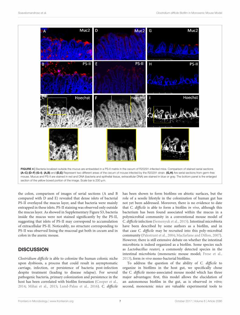
In order to determine the nature of the structure in whichbacteria seem to be entrapped, we performed double stainingof two serial sections, one with anti-Muc2/Hoechst and thesecond one with anti-PS-II/Hoechst. This was performed oncecal and colonic sections of animals infected with the R20291strain (Figures 4, 5). Visualization of multiple areas of thesections in Figure 4 (cecum) and Figure 5 (colon), confirmedthat bacteria were not in contact with the epithelial cells, andthat they were separated by the mucus layer lining the epitheliumtissue. However, some differences were observed between cecumand colon. The cecal sections observed showed that bacteriawere distributed in equivalent manner in and outside the mucusand that PS-II labeling was observed accordingly (Figure 4). In
Frontiers in Microbiology | www.frontiersin.org 5 October 2017 | Volume 8 | Article 2086

fmicb-08-02086 October 23, 2017 Time: 15:56 # 6
Soavelomandroso et al. Clostridium difficile Biofilm in Monoxenic Mouse Model
FIGURE 2 | Heterogeneous distribution of bacteria over the tissue in a mono-associated mouse model. Images are representative of at least five fields. (A) Confocallaser-scanning microscopy (CLSM) images (Z-stacks (above) and 3-D projection (below)] of tissue-associated bacteria obtained from cecum for the 6301erm (a), thecwp84 mutant (b), the R20291 (c), the P30 (d) strains and the germ-free mouse (e). Live cells [bacterial (rod) or epithelial] are labeled in green, dead cells are labeledin red. EC, epithelial cells. Scale bars (red): 50 µm. (B) Thickness of bacterial 3-D structure in cecum and colon. The thickness of the bacterial 3-D structure isdefined by the height on which bacteria are distributed. The thickness was determined directly from confocal Z-stack images. At least three mice were used forCLSM analyses for each strain, and at least eight fields per sample were observed. Data are presented as boxplots with median and minimum-maximum whiskers.No significant difference was observed between strains (Mann–Whitney test).
FIGURE 3 | Clostridium difficile cells are mainly localized at the surface of the mucus layer. Immunodetection of mucus and fluorescent labeling of bacteria by SYTO R©
9 in the cecum (a,c,e,g) and in the colon (b,d,f,h) of mice infected with strains 6301erm (a,b), R20291 (c,d), P30 (e,f), or in axenic mouse (g,h). Mucus is stainedin red and DNA (bacterial, eukaryotic intracellular and, extracellular DNA) is stained in green. The right panel is the enlarged section of the yellow boxed portion of theimage. Yellow arrows indicate position of bacteria outside the mucus layer. Blue arrows indicate position of bacteria in the outer layer of mucus. Scale bar is 200 µm.
Frontiers in Microbiology | www.frontiersin.org 6 October 2017 | Volume 8 | Article 2086
fmicb-08-02086 October 23, 2017 Time: 15:56 # 7
Soavelomandroso et al. Clostridium difficile Biofilm in Monoxenic Mouse Model
FIGURE 4 | Bacteria localized outside the mucus are embedded in a PS-II matrix in the cecum of R20291-infected mice. Comparison of stained serial sections(A–C) (D–F) (G–I). (A,B) and (D,E) Represent two different areas of the cecum of mouse infected by the R20291 strain. (G,H) Are serial sections from germ-freemouse. Mucus and PS-II are stained in red and DNA (bacteria and epithelial tissue, extracellular DNA) are stained in blue or gray. The bottom panel is the enlargedsection of the yellow boxed portion of the image. Scale bar is 200 µm.
the colon, comparison of images of serial sections (A and Bcompared with D and E) revealed that dense islets of bacterialPS-II overlayed the mucus layer, and that bacteria were mainlyentrapped in these islets. PS-II staining was observed only outsidethe mucus layer. As showed in Supplementary Figure S3, bacteriainside the mucus were not stained significantly by the PS-II,suggesting that islets of PS-II may correspond to accumulationof extracellular PS-II. Noticeably, no structure corresponding toPS-II was observed lining the mucosal gut both in cecum and incolon in the axenic mouse.
DISCUSSION
Clostridium difficile is able to colonize the human colonic nicheupon dysbiosis, a process that could result in asymptomaticcarriage, infection, or persistence of bacteria post-infectiondespite treatment (leading to disease relapse). For severalpathogenic bacteria, primary colonization and persistence in thehost has been correlated with biofilm formation (Cooper et al.,2014; Mihai et al., 2015; Lund-Palau et al., 2016). C. difficile
has been shown to form biofilms on abiotic surfaces, but therole of a sessile lifestyle in the colonization of human gut hasnot yet been addressed. Moreover, there is no evidence to datethat C. difficile is able to form a biofilm in vivo, although thisbacterium has been found associated within the mucus in apolymicrobial community in a conventional mouse model ofC. difficile infection (Semenyuk et al., 2015). Intestinal microbiotahave been described by some authors as a biofilm, and inthat case C. difficile may be recruited into this poly-microbialcommunity (Palestrant et al., 2004; Macfarlane and Dillon, 2007).However, there is still extensive debate on whether the intestinalmicrobiota is indeed organized as a biofilm. Some species suchas Lactobacillus reuteri, a commonly detected species in theintestinal microbiota (monoxenic mouse model; Frese et al.,2013), form in vivo mono-bacterial biofilms.
To address the question of the ability of C. difficile toorganize in biofilms in the host gut, we specifically chosethe C. difficile mono-associated mouse model which has threemajor advantages: first, this model allows the elucidation ofan autonomous biofilm in the gut, as is observed in vitro;second, monoxenic mice are valuable experimental tools to
Frontiers in Microbiology | www.frontiersin.org 7 October 2017 | Volume 8 | Article 2086

fmicb-08-02086 October 23, 2017 Time: 15:56 # 8
Soavelomandroso et al. Clostridium difficile Biofilm in Monoxenic Mouse Model
FIGURE 5 | Bacteria localized outside the mucus are embedded in a PSII matrix in the colon of R20291-infected mice. Comparison of stained serial sections (A–C)(D–F) (G–I). (A,B) and (D,E) Represent two different areas of the colon of mouse infected by the R20291 strain. (G,H) Are serial sections from germ-free mouse.Mucus and PS-II are stained in red and DNA (bacteria and epithelial tissue, extracellular DNA) are stained in blue or gray. The bottom panel is the enlargement of theyellow boxed portion of the image. Scale bar is 200 µm.
investigate host–bacterial interactions in an environment devoidof competitive interactions and to study easily both the locationof bacteria in the gut and the structure of biofilms, if any; third,this model allows C. difficile colonization without occurrence ofclinical signs, and therefore allowed us to test our hypothesisC. difficile biofilms could be involved in (human) asymptomaticcarriage and/or asymptomatic persistence. In contrast, in theconventional mouse model, colonization by C. difficile afterstrong antibiotherapy leads to gut lesions. In our model, however,no histological lesions were seen on gut sections of differentstrains (Figure 3).
As a first approach, we assessed the ability of four biofilm-producing strains (Pantaléon et al., 2015) to colonize germ-freemice. 6301erm is a modified strain derived from a humanclinical isolate, and the cwp84 mutant is further derived fromthis strain. The P30 strain has been isolated from poultry andmay not been considered as a clinical strain. This strain has beenshown to display an ecological advantage to colonize the mouseintestinal niche (Spigaglia et al., 2013). Thus, a human-origin,clinically relevant strain is R20291 isolated from an epidemic in
United Kingdom, and belonging to the 027 lineage. This lineage isknown to be associated particularly with a high rate of recurrence.All in vivo studies were carried out 7 days post-infection becausewe previously performed in vivo experiments in mono-associatedmice 3 days post-infection but only few bacteria could be detectedby confocal microscopy. Although the four strains displayeddifferent abilities to form biofilm on polystyrene plates, they wereable to achieve similar levels of colonization along the intestinaltract in our monoxenic mouse model. Therefore, we did not findany correlation between the ability of strains to form biofilmin vitro and their ability to associate with the mouse gut. Indeed,the poor biofilm-producing 6301erm strain colonizes the gut aswell as the other strains. This lack of correlation is undoubtedlyexplained by the different environmental conditions prevailingin vivo and in vitro, in particular the nature of the surface whichinfluences the biofilm formation. Of note, we observed that CDstimulates the maturation of villi and the production of mucusby goblet cells and to our knowledge, it is the first time thatthis feature is reported. However, several publications showedthat commensal bacteria favor the development of mucus and
Frontiers in Microbiology | www.frontiersin.org 8 October 2017 | Volume 8 | Article 2086

fmicb-08-02086 October 23, 2017 Time: 15:56 # 9
Soavelomandroso et al. Clostridium difficile Biofilm in Monoxenic Mouse Model
vascular networks in the gut, and this may be correlated withmaturation of villi (Stappenbeck et al., 2002).
The mucosa-associated bacteria were organized as a 3-Dbacterial community, as observed by CLSM analysis in boththe cecum and the colon of mice infected with the fourstrains. However, the organization of the bacterial communitiesin these parts of the mouse gut were different accordingto strains. Whereas the P30, the 6301erm and the cwp84mutant displayed mainly isolated bacteria, the R20291 strainformed numerous aggregates. These aggregates could correspondto microcolonies, which may result either from in situmultiplication of bacteria, or from mucosal reassociation ofplanktonic bacteria living in the luminal environment. Thisresult is reminiscent with those obtained by Lawley et al.(2009) in a conventional mouse model: they observed mats ofbacilli overlaying microvilli (Lawley et al., 2009), likely to beC. difficile.
One important objective of our study was to clarify thelocalization of tissue-associated bacteria with respect to theintestinal mucus layer. Many studies show that the inner layer ofmucus is devoid of bacteria, and that the outer layer is associatedwith bacteria (Hansson and Johansson, 2010; Johansson et al.,2011). In accordance with Semenyuk et al. (2015), we foundfew bacteria localized in the mucus (see Figure 3). However,we also visualized several C. difficile vegetative cells localizedoutside the outer layer of mucus (Figures 3–5). This discrepancymay be related to the different animal models used. Indeed,the microbiota influence the composition and physicochemicalproperties of the mucus, and may result in a potentially modifiedpenetration of bacteria in conventional as well as monoxenic mice(Johansson et al., 2015).
In our model, and following efficient rinsing, bacteria werestill entrapped in 3-D structures supported at the mucus layer.Interestingly, in addition to bacterial cells, we also observeddiffuse labeling with SYTO R© 9 reminiscent of extracellular DNA,a matrix component found in in vitro C. difficile biofilms (Ðapaet al., 2013; Semenyuk et al., 2014). These structures wereobserved for the three strains tested.
To further analyze the possibility that tissue-associatedC. difficile cells were encased in an extrapolymeric matrix, welabeled gut sections of a mouse infected with the R20291 withantibodies recognizing the cell wall-associated polysaccharideII of C. difficile, another component of the matrix of in vitrobiofilm (Ðapa et al., 2013; Semenyuk et al., 2014). We showedthat the bacteria overlaying the mucus layer are surroundedby a large amount of PS-II. In planktonic mode, PS-II is themain surface-associated polysaccharide and it is ubiquitous inall C. difficile strains (Danieli et al., 2011; Chu et al., 2016;
Monteiro, 2016). Nevertheless, the intensity and distribution oflabeling as compared to the distribution of bacteria in the samelocation are in accordance with extracellular PS-II entrappingthe bacteria. In addition, bacteria present in the mucus layer(Supplementary Figure S3C) are not labeled by the anti-PS-IIin the same conditions. As those structures were not observedin gut section of the axenic mice, we hypothesized that bacteriaoverlaying the mucus layer are organized as a biofilm entrappedin a glycan matrix composed at least of PS-II and possibly alsoDNA. Of note, these structures seem to be smaller in spatialextension than in vitro biofilms, but discrepancies between sizesof in vitro and in vivo biofilm have been already observed(Bjarnsholt et al., 2013). Indeed, the in vivo model is a dynamicmodel subject to various environmental stresses such as intestinalperistalsis, continuous flow, passage of bolus, in contrast to thestatic in vitro model which provides a stable environment butwith a decreased nutrient availability over time (Lebeaux et al.,2013). This could undoubtedly contribute to the small size ofin vivo biofilm structures. As revealed by immunochemistry(Figures 3–5), biofilm structures were present as small isletsirregularly distributed over the mucosa, and this is relevant withthe large-scale observations made by CLSM on the heterogeneousdistribution of bacteria over the gut mouse tissues. This couldbe explained either by (i) the detachment of mature biofilm,(ii) removal of biofilm due to the natural mucus renewal (Freseet al., 2013) or (iii) by a specific interaction with an underlyingintestinal tissue (Sommer et al., 2015).
To our knowledge, this report is the first description of thedevelopment in vivo of a stricto sensu biofilm of C. difficile. Moreinvestigations are now necessary to validate this biofilm-structurein other clinically relevant C. difficile strains, and to elucidate itsputative role in the colonization and persistence of C. difficile.
AUTHOR CONTRIBUTIONS
Conceived and designed the experiments: SB, CJ, andGV. Performed the experiments: AS, SB, SH, FG, andVN. Analyzed the data: AS, SB, CJ, and GV. Contributedreagents/materials/analysis tools: GV. Wrote the paper: AS, SB,CJ, and GV.
SUPPLEMENTARY MATERIAL
The Supplementary Material for this article can be foundonline at: https://www.frontiersin.org/articles/10.3389/fmicb.2017.02086/full#supplementary-material
REFERENCESBarbut, F., Richard, A., Hamadi, K., Chomette, V., Burghoffer, B., and Petit, J. C.
(2000). Epidemiology of recurrences or reinfections of Clostridium difficile-associated diarrhea. J. Clin. Microbiol. 38, 2386–2388.
Bjarnsholt, T., Alhede, M., Alhede, M., Eickhardt-Sørensen, S. R., Moser, C.,Kühl, M., et al. (2013). The in vivo biofilm. Trends Microbiol. 21, 466–474.doi: 10.1016/j.tim.2013.06.002
Chu, M., Mallozzi, M. J. G., Roxas, B. P., Bertolo, L., Monteiro, M. A.,Agellon, A., et al. (2016). A Clostridium difficile cell wall glycopolymerlocus influences bacterial shape, polysaccharide production andvirulence. PLOS Pathog. 12:e1005946. doi: 10.1371/journal.ppat.1005946
Cooper, R. A., Bjarnsholt, T., and Alhede, M. (2014). Biofilms in wounds: areview of present knowledge. J. Wound Care 23, 570, 572–574, 576–580 passim.doi: 10.12968/jowc.2014.23.11.570
Frontiers in Microbiology | www.frontiersin.org 9 October 2017 | Volume 8 | Article 2086

fmicb-08-02086 October 23, 2017 Time: 15:56 # 10
Soavelomandroso et al. Clostridium difficile Biofilm in Monoxenic Mouse Model
Costerton, J. W., Geesey, G. G., and Cheng, K. J. (1978). How bacteria stick. Sci.Am. 238, 86–95. doi: 10.1038/scientificamerican0178-86
Crowther, G. S., Chilton, C. H., Todhunter, S. L., Nicholson, S., Freeman, J.,Baines, S. D., et al. (2014a). Comparison of planktonic and biofilm-associatedcommunities of Clostridium difficile and indigenous gut microbiota in a triple-stage chemostat gut model. J. Antimicrob. Chemother. 69, 2137–2147. doi: 10.1093/jac/dku116
Crowther, G. S., Chilton, C. H., Todhunter, S. L., Nicholson, S., Freeman, J., Baines,S. D., et al. (2014b). Development and validation of a chemostat gut model tostudy both planktonic and biofilm modes of growth of Clostridium difficile andhuman microbiota. PLOS ONE 9:e88396. doi: 10.1371/journal.pone.0088396
Danieli, E., Lay, L., Proietti, D., Berti, F., Costantino, P., and Adamo, R. (2011).First synthesis of C. difficile PS-II cell wall polysaccharide repeating unit. Org.Lett. 13, 378–381. doi: 10.1021/ol1026188
Ðapa, T., Leuzzi, R., Ng, Y. K., Baban, S. T., Adamo, R., Kuehne, S. A., et al.(2013). Multiple factors modulate biofilm formation by the anaerobic pathogenClostridium difficile. J. Bacteriol. 195, 545–555. doi: 10.1128/JB.01980-12
Dawson, L. F., Valiente, E., Faulds-Pain, A., Donahue, E. H., and Wren, B. W.(2012). Characterisation of Clostridium difficile biofilm formation, a role forSpo0A. PLOS ONE 7:e50527. doi: 10.1371/journal.pone.0050527
Deakin, L. J., Clare, S., Fagan, R. P., Dawson, L. F., Pickard, D. J., West, M. R., et al.(2012). The Clostridium difficile spo0A gene is a persistence and transmissionfactor. Infect. Immun. 80, 2704–2711. doi: 10.1128/IAI.00147-12
Donelli, G., Vuotto, C., Cardines, R., and Mastrantonio, P. (2012). Biofilm-growingintestinal anaerobic bacteria. FEMS Immunol. Med. Microbiol. 65, 318–325.doi: 10.1111/j.1574-695X.2012.00962.x
Donskey, C. J., Kundrapu, S., and Deshpande, A. (2015). Colonization versuscarriage of Clostridium difficile. Infect. Dis. Clin. North Am. 29, 13–28.doi: 10.1016/j.idc.2014.11.001
Evans, C. T., and Safdar, N. (2015). Current trends in the epidemiology andoutcomes of Clostridium difficile infection. Clin. Infect. Dis. 60(Suppl. 2),S66–S71. doi: 10.1093/cid/civ140
Frese, S. A., Mackenzie, D. A., Peterson, D. A., Schmaltz, R., Fangman, T., Zhou, Y.,et al. (2013). Molecular characterization of host-specific biofilm formation in avertebrate gut symbiont. PLOS Genet. 9:e1004057. doi: 10.1371/journal.pgen.1004057
Gerding, D. N., Johnson, S., Rupnik, M., and Aktories, K. (2014). Clostridiumdifficile binary toxin CDT: mechanism, epidemiology, and potential clinicalimportance. Gut Microbes 5, 15–27. doi: 10.4161/gmic.26854
Hansson, G. C., and Johansson, M. E. (2010). The inner of the two Muc2 mucin-dependent mucus layers in colon is devoid of bacteria. Gut Microbes 105,15064–15069. doi: 10.4161/gmic.1.1.10470
Janoir, C. (2016). Virulence factors of Clostridium difficile and their role duringinfection. Anaerobe 37, 13–24. doi: 10.1016/j.anaerobe.2015.10.009
Johansson, M. E. V., Jakobsson, H. E., Holmén-Larsson, J., Schütte, A., Ermund, A.,Rodríguez-Piñeiro, A. M., et al. (2015). Normalization of host intestinalmucus layers requires long-term microbial colonization. Cell Host Microbe 18,582–592. doi: 10.1016/j.chom.2015.10.007
Johansson, M. E. V., Larsson, J. M. H., and Hansson, G. C. (2011). The twomucus layers of colon are organized by the MUC2 mucin, whereas the outerlayer is a legislator of host-microbial interactions. Proc. Natl. Acad. Sci. U.S.A.108(Suppl. 1), 4659–4665. doi: 10.1073/pnas.1006451107
Lawley, T. D., Clare, S., Walker, A. W., Goulding, D., Stabler, R. A., Croucher, N.,et al. (2009). Antibiotic treatment of clostridium difficile carrier mice triggersa supershedder state, spore-mediated transmission, and severe disease inimmunocompromised hosts. Infect. Immun. 77, 3661–3669. doi: 10.1128/IAI.00558-09
Lebeaux, D., Chauhan, A., Rendueles, O., and Beloin, C. (2013). From in vitroto in vivo models of bacterial biofilm-related infections. Pathogens 2, 288–356.doi: 10.3390/pathogens2020288
Lund-Palau, H., Turnbull, A. R., Bush, A., Bardin, E., Cameron, L., Soren, O., et al.(2016). Pseudomonas aeruginosa infection in cystic fibrosis: pathophysiological
mechanisms and therapeutic approaches. Expert Rev. Respir. Med. 10, 685–697.doi: 10.1080/17476348.2016.1177460
Macfarlane, S., and Dillon, J. F. (2007). Microbial biofilms in the humangastrointestinal tract. J. Appl. Microbiol. 102, 1187–1196. doi: 10.1111/j.1365-2672.2007.03287.x
Marsh, J. W., Arora, R., Schlackman, J. L., Shutt, K. A., Curry, S. R., andHarrison, L. H. (2012). Association of relapse of Clostridium difficile diseasewith BI/NAP1/027. J. Clin. Microbiol. 50, 4078–4082. doi: 10.1128/JCM.02291-12
Mihai, M. M., Holban, A. M., Giurcaneanu, C., Popa, L. G., Oanea, R. M., Lazar, V.,et al. (2015). Microbial biofilms: impact on the pathogenesis of periodontitis,cystic fibrosis, chronic wounds and medical device-related infections.Curr. Top. Med. Chem. 15, 1552–1576. doi: 10.2174/1568026615666150414123800
Monteiro, M. A. (2016). The design of a Clostridium difficile carbohydrate-basedvaccine. Methods Mol. Biol. 1403, 397–408. doi: 10.1007/978-1-4939-3387-7_21
Palestrant, D., Holzknecht, Z. E., Collins, B. H., Parker, W., Miller, S. E., andBollinger, R. R. (2004). Microbial biofilms in the gut: visualization by electronmicroscopy and by acridine orange staining. Ultrastruct. Pathol. 28, 23–27.doi: 10.1080/01913120490275196
Pantaléon, V., Soavelomandroso, A. P., Bouttier, S., Briandet, R., Roxas, B.,Chu, M., et al. (2015). The Clostridium difficile protease Cwp84 modulatesboth biofilm formation and cell-surface properties. PLOS ONE 10:e0124971.doi: 10.1371/journal.pone.0124971
Plaza-Garrido, Á., Miranda-Cárdenas, C., Castro-Córdova, P., Olguín-Araneda, V., Cofré-Araneda, G., Hernández-Rocha, C., et al. (2015). Outcomeof relapsing Clostridium difficile infections do not correlate with virulence-,spore- and vegetative cell-associated phenotypes. Anaerobe 36, 30–38.doi: 10.1016/j.anaerobe.2015.09.005
Semenyuk, E. G., Laning, M. L., Foley, J., Johnston, P. F., Knight, K. L., Gerding,D. N., et al. (2014). Spore formation and toxin production in Clostridiumdifficile biofilms. PLOS ONE 9:e87757. doi: 10.1371/journal.pone.0087757
Semenyuk, E. G., Poroyko, V. A., Johnston, P. F., Jones, S. E., Knight, K. L.,Gerding, D. N., et al. (2015). Analysis of bacterial communities duringClostridium difficile infection in the mouse. Infect. Immun. 83, 4383–4391.doi: 10.1128/IAI.00145-15
Sommer, F., Nookaew, I., Sommer, N., Fogelstrand, P., and Bäckhed, F. (2015).Site-specific programming of the host epithelial transcriptome by the gutmicrobiota. Genome Biol. 16, 62. doi: 10.1186/s13059-015-0614-4
Spigaglia, P., Barketi-Klai, A., Collignon, A., Mastrantonio, P., Barbanti, F.,Rupnik, M., et al. (2013). Surface-layer (S-layer) of human and animalClostridium difficile strains and their behaviour in adherence to epithelial cellsand intestinal colonization. J. Med. Microbiol. 62(Pt 9), 1386–1393. doi: 10.1099/jmm.0.056556-0
Stappenbeck, T. S., Hooper, L. V., and Gordon, J. I. (2002). Developmentalregulation of intestinal angiogenesis by indigenous microbes via Paneth cells.Proc. Natl. Acad. Sci. U.S.A. 99, 15451–15455. doi: 10.1073/pnas.202604299
Stewart, D. B., Berg, A., and Hegarty, J. (2013). Predicting recurrence of C. difficilecolitis using bacterial virulence factors: binary toxin is the key. J. Gastrointest.Surg. 17, 118–124. doi: 10.1007/s11605-012-2056-6
Conflict of Interest Statement: The authors declare that the research wasconducted in the absence of any commercial or financial relationships that couldbe construed as a potential conflict of interest.
Copyright © 2017 Soavelomandroso, Gaudin, Hoys, Nicolas, Vedantam, Janoir andBouttier. This is an open-access article distributed under the terms of the CreativeCommons Attribution License (CC BY). The use, distribution or reproduction inother forums is permitted, provided the original author(s) or licensor are creditedand that the original publication in this journal is cited, in accordance with acceptedacademic practice. No use, distribution or reproduction is permitted which does notcomply with these terms.
Frontiers in Microbiology | www.frontiersin.org 10 October 2017 | Volume 8 | Article 2086