biol 202 lab 7 c-fern investigations - genetics in action...
TRANSCRIPT

C-Fern® Lab Part 2
BIOL 202 LAB 7 C-Fern Investigations - Genetics in Action Mendelian Genetics Part 2.docx page 1 of 7
BIOL 202 LAB 7: C-Fern® Investigations: Genetics in Action Mendelian Genetics (Note that this document contains instructions and information for observing the C-Fern® spores, which were sown in the previous lab.) Equipment, Supplies & Materials:
• Petri dishes containing nutrient agar upon which presterilized C-Fern® spores of an F1 hybrid were sown
• C-Fern® culture instructions and background information (Hickok & Warne, 1998a)—instructor's copy
• C-Fern® Investigations: Genetics in Action Mendelian Genetics Student Instructions (Hickok & Warne, 1998b)—one copy per student
• Compound and dissecting stereomicroscopes Activities:
• Overview of Lab 7: Visualization of basic principles of Mendelian inheritance in C-Fern® by following the segregation of a visible marker, polka dot, in both the F1 gametophyte and F2 sporophyte generations. Students sow spores of an F1 hybrid (wild type x polka dot) to produce F1 gametophytes. Sporophytes also may be observed.
• Follow procedure as described in lab instructions (and as indicated by instructor) Assignment:
• Lab Report #7 (final version to be submitted to instructor the Friday following conclusion of this lab at 5:00 pm, following editorial review by the Science Writing Mentor)
C-Fern® Lab Part 2
BIOL 202 LAB 7 C-Fern Investigations - Genetics in Action Mendelian Genetics Part 2.docx page 2 of 7
Procedure (Adapted from Hickok & Warne, 1998b, pp. H-3-H-4.): 1. Observation of C-Fern® Cultures—Observe the cultures using both dissecting and
compound microscopes at various levels of magnification as necessary to view the C-Fern® spores. (Note that your instructor can demonstrate how to successfully observe the cultures in Petri dishes using the compound microscope at low and medium magnifications. This requires placing a blank microscope slide in the slide holder of the mechanical stage, placing the Petri dish on the slide, and using the stage control to move both the slide and the dish to observe the spores. Also note that the compound microscope will utilize transmitted light—light from below and transmitted through a specimen—rather than reflected light—light from above and reflected from a specimen; both types of illumination are possible using dissecting microscopes, and may be useful in viewing your specimens.)
2. Make Labelled Sketches of Your Observations—Sketch and label each different thing that you can observe in your Petri dish, including spores (both ungerminated and germinated), and any other structures that you may observe emerging from the spores. Based on your observations, labelled sketches, and the lecture content associated with this lab, please answer the following questions:
Q1: What do you observe? Q2: Do some of the spores show signs of germination? What is the first visible
structure in germinated spores? Q3: What type of nuclear replication and division, and cell division is occurring
during the germination process? Q4: What type of nuclear replication and division, and cell division took place in the
F1 sporophyte to produce these spores? Q5: Are the spores haploid or diploid? Q6: If the F1 hybrid sporophytes were heterozygous for a single mutant trait, what
genotypes would be present in the spores? What would be the expected ratio of genotypes?
Q7: What will develop from the spores as they germinate? Will they be haploid or diploid?
Q8: Would these be expected to show the mutant trait (i.e., Polka Dot)? 3. After Observation of C-Fern® Cultures—When you are finished with your
observations, place the cultures back in the culture dome under the lights. Figures:
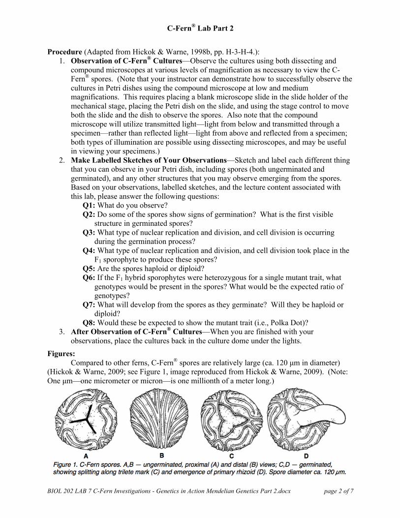
Compared to other ferns, C-Fern® spores are relatively large (ca. 120 µm in diameter) (Hickok & Warne, 2009; see Figure 1, image reproduced from Hickok & Warne, 2009). (Note: One µm—one micrometer or micron—is one millionth of a meter long.)

C-Fern® Lab Part 2
BIOL 202 LAB 7 C-Fern Investigations - Genetics in Action Mendelian Genetics Part 2.docx page 3 of 7
The images below show C-Fern® spores 24 hours after having been spread on the surface of basic C-Fern® medium in Petri dishes. The spore at the center of Figure 4 appears to possibly be germinating, but compare Figures 5 and 6, which show spores actually germinating on Day 5.
Figure 1. C-Fern® spores, 80x magnification Figure 2. C-Fern® spores, 150x magnification
Figure 3. C-Fern® spores, 250x magnification Figure 4. C-Fern® spores, 350x magnification
Figure 5. C-Fern® spores, 400x magnification Figure 6. C-Fern® spores, 1000x magnification (showing primary rhizoids in two spores) (showing primary rhizoid in one spore)
C-Fern® Lab Part 2
BIOL 202 LAB 7 C-Fern Investigations - Genetics in Action Mendelian Genetics Part 2.docx page 4 of 7
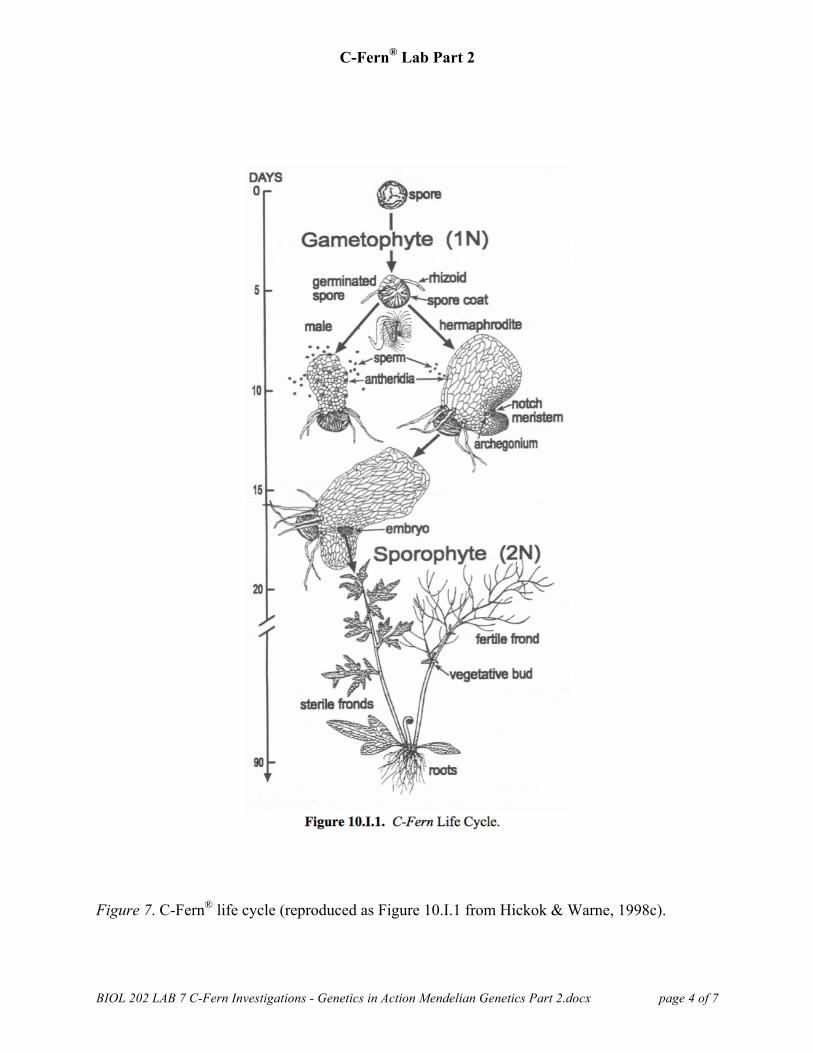
Figure 7. C-Fern® life cycle (reproduced as Figure 10.I.1 from Hickok & Warne, 1998c).

C-Fern® Lab Part 2
BIOL 202 LAB 7 C-Fern Investigations - Genetics in Action Mendelian Genetics Part 2.docx page 5 of 7
C-Fern® Polka Dot Mutation Notes: 1. C-Fern® Polka Dot Mutant (gene symbol cp [or dd, as indicated below])—"This is a
very striking visual mutant that exhibits a distinct green polka dot appearance in cells of both gametophytes and homozygous sporophytes when viewed with a low power microscope. In sporophytes, older leaves of individuals homozygous for this mutation have an attractive silver-green appearance. Although there is a slight reduction in growth and a decrease in spore viability* in spores produced from homozygotes, gametophytes carrying the polka dot mutant and homozygous polka dot sporophytes grow nearly as well as the wild type. This recessive trait has been observed in several independent selections that have been generated using both X-rays and EMS as a chemical mutagen. A study of the trait, using transmission electron microscopy, was not successful in discerning the structural basis of the phenotype. The phenotype is associated with a clumping of chloroplasts and other organelles around the nucleus. It may involve some disruption of the cytoskeleton. The pleiotropic effect (when one gene influences two or more seemingly unrelated phenotypic traits) of reduced spore viability is associated with somewhat fragile spore walls. Different degrees or strengths of the clumping phenotype are positively associated with increased spore wall weakness. *NOTE: Because of the decreased spore viability associated with spores produced by cp/cp [or dd/dd, using our allele symbols as described below] homozygotes, presterilized and bulk spores are supplied in larger quantities than other stocks. Although inviable spores will be apparent in cultures, the increased number of spores provided will produce adequate numbers of gametophytes" ("C-Fern Sport Report: Polka," n.d.).
2. C-Fern® Polka Dot Mutant Gene Alleles—The strain of C-Fern® used in this laboratory has both normal and mutant alleles associated with the Polka Dot mutant as follows ("Are There Different Kinds," n.d.; "You Will Be Following," n.d.): Allele Contribution to Phenotype
normal: D normal distribution of chloroplasts – dominant phenotype mutant: d chloroplasts clumped (polka dot) – recessive phenotype
The Polka Dot phenotype is visible in both haploid and diploid forms of the fern (except in the spores, eggs, and sperms). As a result:
For haploids: Genotype Phenotype (F1 gametophytes) D normal d polka dot
For diploids: Genotype Phenotype (F2 sporophytes) DD normal Dd normal dd polka dot
4. Phenotype Considerations—Based on the normal (wild type) and mutant (Polka Dot) alleles in the strain of C-Fern® used in this laboratory, and given what is know about dominant and recessive alleles as related to basic Mendelian genetics, we may expect the following: The F1 sporophyte will produce gametophytes in a 1:1 ratio of Polka Dot mutants to normal (wild) type. The recessive mutation results in a 3:1 ratio of the F2

C-Fern® Lab Part 2
BIOL 202 LAB 7 C-Fern Investigations - Genetics in Action Mendelian Genetics Part 2.docx page 6 of 7
sporophyte generation ("C-FERN Spores, F1 Polka," 2016). Based on your observations and the lecture content associated with this lab, please answer the following questions:
Q9: If you were to count 50 individual F1 gametophytes, how many normal and how many mutant individuals would you expect to count? Why?
Q10: If you were to count 50 individual F1 sporophytes, how many normal and how many mutant individuals would you expect to count? Why?

C-Fern® Lab Part 2
BIOL 202 LAB 7 C-Fern Investigations - Genetics in Action Mendelian Genetics Part 2.docx page 7 of 7
References Are there different kinds of C-Fern? (n.d.). Retrieved from C-Fern Project website: http://c-
fern.org/index.php?option=com_content&view=article&id=25:are-there-different-kind.. C-FERN spores, F1 polka dot, presterilized vial, kit-sized. (2016). Retrieved from Carolina
Biological Supply Company website: http://www.carolina.com/c-fern-spores/c-fern-spores-f1-polka-dot-presterilized-vial-kit-sized/156760.pr
C-Fern sport report: Polka dot (cp). (n.d.). Retrieved from Learning Online Network with CAPA website: https://s10.lite.msu.edu/res/msu/botonl/b_online/library/cfern/cfern.bio.utk.edu/resource/CBSSportPolka.html
Hickok, L. G., & Warne, T. R. (1998a). C-Fern® culture instructions and background information [Pamphlet]. Burlington, NC: Carolina Biological Supply Company.
Hickok, L. G., & Warne, T. R. (1998b). C-Fern® investigations: Genetics in action Mendelian genetics student instructions [Pamphlet]. Burlington, NC: Carolina Biological Supply Company.
Hickok, L. G., & Warne, T. R. (1998c). Laboratory investigations with C-Fern® (Ceratopteris richardii). Retrieved from Association for Biology Laboratory Education website: http://www.ableweb.org/volumes/vol-19/10-hickok.pdf
Hickok, L. G., & Warne, T. R. (2009). C-Fern web manual. Retrieved from Sunnyside website: http://learn.susd12.org/pluginfile.php/104322/mod_resource/content/1/C-Fern_Web_Manual_080409bb.pdf
You will be following the development of a fern. (n.d.). Retrieved from Course Hero, Inc. website: https://www.coursehero.com/file/p2jtddj/You-will-be-following-the-development-of-a-fern-Ceratopteris-richardii-c-fern/