biol. jb. dodonaea, 44, 1976, 345-352. - vliz · biol. jb. dodonaea, 44, 1976, 345-352. a study of...
TRANSCRIPT
Biol. Jb. Dodonaea, 44, 1976, 345-352.
A STUDY OF THE CHROMATOPHORE PIGMENTS IN THE SKIN OF THE CEPHALOPOD
SEPIA OFFICINAL/S L.
by
C. VAN DEN BRANDEN and W. DECLEIR
ABSTRACT.- In the chromatophores of the dorsal skin of the cuttlefish Sepia officiruJ!is L. at least three different pigments can be found. They belang to the ommochromes and they differ from each other by their color, solubility and redox behavior.
INTRODUC'TION
Cephalopods possess a unique system of chromatophare organs and iridophores which allow very rapid physiological color changes. The iridophores form a layer of immobile reflector cells which is only exposed when the chromatophare cells are absent or fully contracted (HoLMES, 1940 and MIROW, 1972, a) .
The morphology and structure of the chromatophare organ has been studiedindetail (MIROW, 1972, b- SERENI, 1930- BovcoTT, 1948). It is composed of a central pigment containing cell, several radially arranged obliquely striated muscle fibers, nerve cells, Schwann cells and sheath cells. The eelt bodies of the fibres innervating the chromatophare musdes are concentrated in anterior and posterior chromatophare lobes, which were classified as lower motor eentres (BovcoTT and YouNG, 1950). A possible endocrine regulation of chromatophores has been proposed by KAHR ( 1958 and 1959).
The different color changes and color patterns of cephalopods and the conesponding complex behaviaral patterns have been the subject of several studies (WELLS, 1962- KuHN, 1950- HoLMES, 1940- SERENI, 1930).
The cuttlefish, Sepia officinalis L. shows extreme rapid color changes as compared to mostother cephalopods. HILL and SüLANDT ( 1935) show that the change from complete contraction to complete expansion in a chromato-
345
phore can take place in two- thirds of a second. Moreover the chromatophare system of Sepia o.fficinalis and Octopus vulgaris is organized in such a way that a much wider variety of complicated color patterns can be produced than in any other cephalopod ( Bovcon , 1948). According to WELLS ( 1962) one out of every three hundred cells in the brain of these animals is involved in color displays.
The chromatophores in Sepia o.fficinalis lie in three layers parallel with the outer surface of the anima!. Those of the outer layer contain a bright yellow pigment, in the middle layer the pigment is orange red and in the basal layer the pigment is brown violet (HoLMES, 1940). The chromatophare pigments were studied by ScHWINCK (I 953 and 1955). She demonstraled that the pigments in the chromatophores and in theeyes of Sepia offlcinalis, Octopus vulgaris and Eledone moschata are ommochromes.
This paper describes our first efforts to isolate and characterize the different chromatophare pigments from the skin of Sepia of/icina/is.
MATERIAL AND METHOOS
We used adult specimens of Sepia o.fficinalis L. They were obtained either from the culture of RICHARD (1976) or from the "Expedition Cephalomanche". For our experiments only the dorsal skin of the cuttlefish was taken . After washing with tap water it was extracted with consecutively 0,05 M TRIS-HCI buffer pH=7,2, methanoi-HCI 0,1 N and formic acid (99-1 00% ). Each extraction was repeated until no more pigment was released. The extracts were centrifuged at 30.000 g for 20 minutes at 4°C. The resultant supernatants were used for spectrophotometric analyses or freeze-dried. The dry powders were partitioned into different fractions for solubility tests and further study.
Absorption spectra were obtained using a Cary 118 spectrophotometer over the range 650-300 nm. Redox reactions were tested by adding H20 2
(3 % final) or excess ascorbic acid or sodium thiosulphate to the measuring vessel. Formation of a purple halochrome was tested by addition of concentraled sulpuric acid (BECKER, 1941 ).
RESULTS
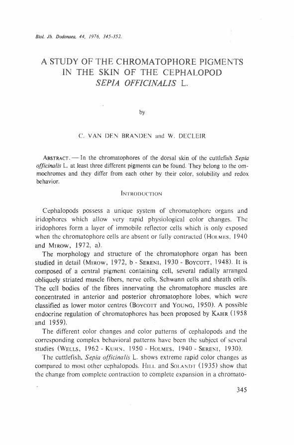
With our extraction procedure we succeeded in isolating three differently colored pigments. We cal! them pigment A (buffer extraction), pigment B (acid methanol extraction) and pigment C (formic acid extraction). They all show a typ i cal redox co lor and halochrome (Table I) .
346
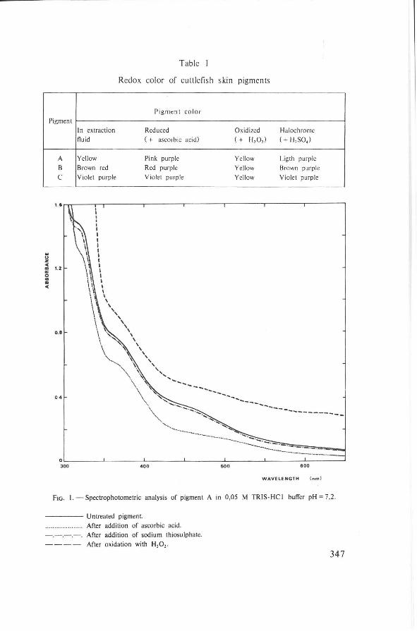
Table I
Redox color of cuttlefïsh skin pigments
... u z <C
Pigment
A B c
• 1.2 0: 0 .. • <C
0.8
0.4
In extraction fluid
Yellow Brown red Violet purple
I I I I I I I I I I I I I I I I I I \
' \ ' ',
' \ ' \ ' '
Pigment co lor
Reduced ( + ascorb ic acid)
Pink pu rple Red purple Violet purple
', ' ' ',,
....
........
...... ...... --
.............
------
-···-·············-
Oxidized Halochrome ( + H,O, ) ( + H, SO,)
Ye llow Ligth purp le Yellow Brown purple Yell ow Violet purple
------.... ___ _ ------------
OL---------L-------~L-------~L-------~L-------~--------~--------~ 300 400 500 800
WAVElENGTH (nml
FIG. I.-- Spectrophotometric analysis of pigment A in 0,05 M TRIS-HCI butTer pH = 7,2 .
------- Untreated pigment After actdition of ascorbic acid.
- .- .-.- . After actdition of sodium thiosulphate. After oxidation with H20 2•
347
1.0
0.8
w <.> z .. ., a: 0 "' 0.6 ., ..
0.4
WAVELENGTH ( nm I
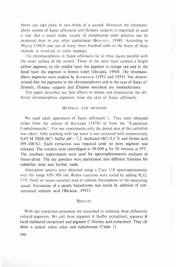
FrG. 2.- Spectrophotometric a na lysis of pigment B in metha nol- HC I 0, I N.
Untreated pigment.
After addition of asco rbic acid. -.-.-.-. After addition of sodium thiosulphate.
After oxidation with H,O, .
u z <( ., a: 0
"' "' <(
Q.8
0.6
___ ,... ....
0.4
······ ...................... . 0.2
----------------.,.__ ·· .. ,'-.... ••..... •,,
•,, ', ', --........ _ ... ___________ _
OL-------~---------L--------L-------~L--------L------~~·=·=·=--=-=-=-=-=-~ 300 400 500
FrG. 3.- Spec tropho to metric analysis of pigment C in formic acid.
Untreated pigmen t.
After addition of ascorbic acid.
After oxidation with H20 2.
600
WAVELENGTH I nm )
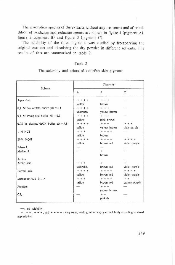
The absorption spectra of the extracts without any treatment and after addition of oxidizing and reducing agents are shown in figure I (pigment A), fïgure 2 (pigment B) and figure 3 (pigment C).
The solubility of the three pigments was studied by freezedrying the original extracts and dissolving the dry powder in different solvents. The results of this are summarized in table 2.
Table 2
The solubility and colors of cuttlefish skin pigments
Pigments
Solvent A B c
Aqua dest. + + + + + + + -
yellow brown
0,1 M Na acetate butTer pH = 4,4 + + + + + + + -
yellowish yellow brown
0,1 M Phosphate butTer pH = 6,3 + + + + + + + -
yellow pink brown
0,05 M glycine/NaOH butTer pH= 9,8 + + + + + + + + + + yellow yellow brown pink purple
I N HCI + + + + + + + -
yellow brown
20% KOH + + + + + + + + + + + + yellow brown red violet purple
Ethanol - - -Methanol - + -
brown
Aceton - - -
Acetic acid + + + + + yellowish brown red violet purple
Formic acid + + + + ++++ + + + + yellow brown red violet purple
Methanol/ HC I 0,1 N + + + + + + + + + yellow brown red orange purple
Pyridine - + + + -yellow brown
es, - + + -pinkish
- : no solubility. + , + +, + + + , and + + + + : very weak, weak, good or very good solubility according to visual
appreciation.
349
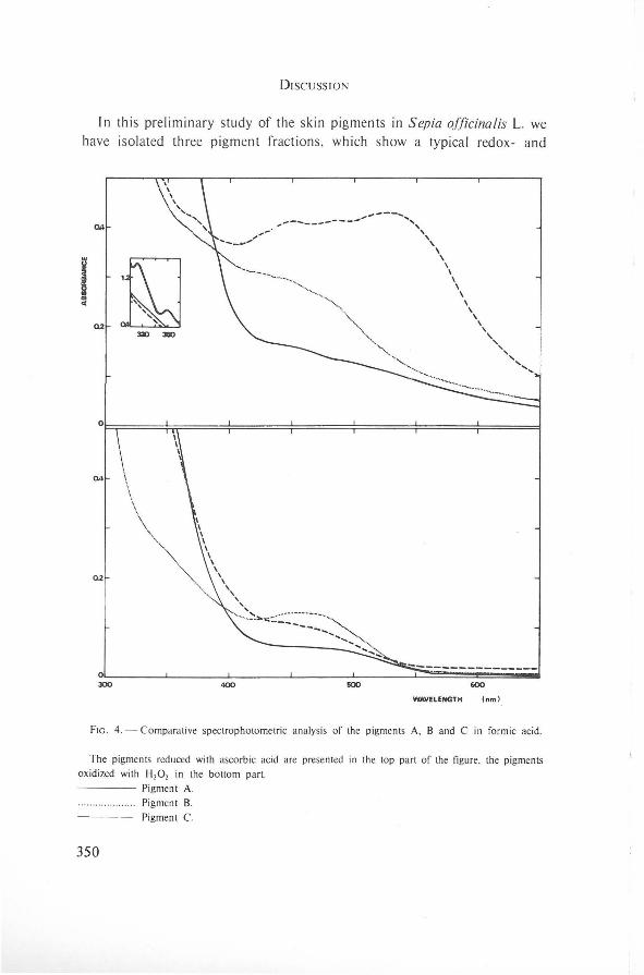
DISCUSSION
In this preliminary study of the skin pigments in Sepia officinalis L. we have isolated three pigment fractions , which show a typical redox- and
Qll
i ~
~~ ....
I .. Q2
3Z) 3110
,-----------""----......... ,, ',,
\ \
\ I I \ \
\ \
\ \
' '',,
WAVELENGTH I nm I
',,,
FtG. 4.- Comparative spectrophotomelric analysis of the pigments A, B and C in formic acid.
The pigments reduced with ascorbic acid are presenled in the top part of the figure, the pigmenls
oxidized wilh H,O, in the bollom part.
350
Pigment A.
Pigment B.
Pigment C.
halochrome reaction. They are not dissolved by neutral organic solvents but are soluble in alkali , acid methanol and formic acid. All these properties correspond with the definition of ommochromes as given by BECKER ( 1941) and LINZEN (1967) . This confirms the publications of ScHWINCK (1953 and 1955) who found that the chromatophare pigments in the skin of cephalopods are ommochromes. Moreover we were able to demonstrate that at least three different pigments are present in the chromatophores of Sepia officina/is. They differ by their color (table 1), solubility (table 2) and redoxreaction. A comparison of the oxidized and reduced absorption curves of these pigments in the same solvent (formic acid) is presented in figure 4.
As our pigment fractions are respectively yellow (fraction A), brown red (fraction B) and violet purple (fraction C) it is plausible to put forward a correspondence of these pigments with the three chromatophare layers which were described as yellow, orange red and brown violet. However this is a subject for further research. We also plan to study the further purification and identification of the three pigments and the way in which they are bound to their substrates.
SAMENVATTING
In de chromatophoren van de rughuid van de zeekat, Sepia officina/is L. komen ten minste drie verschillende pigmenten voor. Het zijn ommochromen en ze verschillen onderling in kleur, oplosbaarheid en redoxreaktie.
RÉSUMÉ
Dans les chromatophores de la seiche, Sepia officinalis L. on peut démontrer la présence d'au moins trois pigments difTérents. Ce sont des ommochromes et ils se distinguent par leur couleur, leur solubilité et leurs propriétés d'oxydoréduction.
ACKNOW LEDGEM ENTS
We are very indebted to Professor RICHARD and Professor DuRCHON (Scientific and Teehoical University of LilleI and lnstitute of Marine Biology of WimereuxFrance) for offering ideal working conditions. We also want to thank Dr. G. WoLF for her help in collecting the cuttlefish during the " Expédition Cephalomanche" aboard the " La Pelagia", vessel belonging to the l.S.T.P.M.
Finally one of us (C.Vd.B.) is very indebted to the " Ministerie van Nationale Opvoeding" for a grant (beurs aan jonge navorsers) covering a stay of one month at the Marine Biology lnstitute of Wimereux.
351

LITERATURE
BECKER, E. ( 1941 ). Die Pigmente der Ommin- und Ommatingruppe, eine neue Klasse von Naturfarbstoffe. Naturwiss., T. 29, pp. 237-238 .
Bovcon, B. B. (1948) . The chromatophore system of ct:phalopods. Proc. Linn. Soc. London, Vol. 164, pp. 235-240.
Bovcon, B. B. and YouNG, J. Z. (1950). The comparative study of learning. Symp. Soc. Exp. Biol., IV, pp. 432-453 .
HrLL and SoLANDT (1935). Myograms from the chromatophores of Sepia. Proc. Physiol. Soc., 13.
HoLMES, W. (1940). The color changes and color patterns of Sepia officinalis L. Proc. Roy. Soc. London, Ser. A, Vol. 110, pp. 17-36.
KAHR, H. (1958). Die Bedeutung des Serotonins für die Melanophorenreaktion des Octopus vulgaris. Naturwiss., 45, Heft 10, pp. 243.
KAHR, H. ( 1959). Zur endokrinen Steuerung der Melanophorenreaktion bei Octopus vulgaris. Zeitschr. Verg!. Physiol., Bd. 41, pp. 435-448.
KuHN, A. ( 1950). Ueber Farbwechsel und Farbensinn von Cephalopoden. Zeilschr. Verg!. Physiol., Bd. 32, pp. 572-598.
LINZEN, B. ( 1967). Zur Biochemie der Ommochrome. Naturwiss., 54, Heft 11, pp. 259-267.
MIROW, S. ( 1972, a) . Skin co lor in the squid Loligo pealii and Loligo opalescens. 11. Iridophores. Zeitschr. Zellforsch. 125, pp. 176-190.
MJROW, S. ( 1972, b). Skin co lor in tne squid Loligo pealii and Loligo opalescens. I. Chromatophores. Zeitsch. Zellforsch. 125, pp. 143-17 5.
RICHARD, A. (1976). L'élevage de la seiche (Sepia officinalis L.). Proc. lOth Eur. Symp. Mar. Biol., Vol. I, pp. 359-380. Universa Press, Wetteren (Belgium), 1976.
ScHWINCK , I. (1953). Ueber den Nachweiss eines Redoxpigmentes (ommochrom) in der Haut van Sepia officinalis. Naturwiss. , 40, pp. 365.
ScHWINCK, I. (1955). Vergleich des Redoxpigmentes aus Chromatophoren und Retina von Sepia officinalis mit lnsektenpigmenten der Ommochromgruppe. Zool. Anz. Suppl., Band 19, pp. 71-74.
SERENI, E. ( 1930). The chromatophores of cephalopods. Biol. Bull., Vol. LIX, n• 3, pp. 247-268.
WELLS, M. (1962). Brain and behavior in cephalopods. Stanford University Press - Stanford.
352
University of Antwerp R.U.C.A. Department of Biochemistry. (Oir. Prof. Dr. W. Decleir).
Slachthuislaan 68, B-2000 Antwerpen (Belgium).