biological markers for pulpal inflammation: a systematic ... · pdf filebiological markers for...
TRANSCRIPT
RESEARCH ARTICLE
Biological Markers for Pulpal Inflammation:
A Systematic Review
Dan-Krister Rechenberg1*, Johnah C. Galicia2, Ove A. Peters2
1 Department of Preventive Dentistry, Periodontology and Cariology, Center of Dental Medicine, University of
Zurich, Zurich, Switzerland, 2 Department of Endodontics, Arthur A. Dugoni School of Dentistry, University of
the Pacific, San Francisco, California, United States of America
Abstract
Background and Objective
Pulpitis is mainly caused by an opportunistic infection of the pulp space with commensal
oral microorganisms. Depending on the state of inflammation, different treatment regimes
are currently advocated. Predictable vital pulp therapy depends on accurate determination
of the pulpal status that will allow repair to occur. The role of several players of the host
response in pulpitis is well documented: cytokines, proteases, inflammatory mediators,
growth factors, antimicrobial peptides and others contribute to pulpal defense mechanisms;
these factors may serve as biomarkers that indicate the status of the pulp. Therefore, the
aim of this systematic review was to evaluate the presence of biomarkers in pulpitis.
Methods
The electronic databases of MEDLINE, EMBASE, Scopus and other sources were
searched for English and non-English articles published through February 2015. Two inde-
pendent reviewers extracted information regarding study design, tissue or analyte used, out-
come measures, results and conclusions for each article. The quality of the included studies
was assessed using a modification of the Newcastle-Ottawa-Scale.
Results and Conclusions
From the initial 847 publications evaluated, a total of 57 articles were included in this review.
In general, irreversible pulpitis was associated with different expression of various biomark-
ers compared to normal controls. These biomarkers were significantly expressed not only in
pulp tissue, but also in gingival crevicular fluid that can be collected non-invasively, and in
dentin fluid that can be analyzed without extirpating the entire pulpal tissue. Such data may
then be used to accurately differentiate diseased from healthy pulp tissue. The interplay of
pulpal biomarkers and their potential use for a more accurate and biologically based diag-
nostic tool in endodontics is envisaged.
PLOS ONE | DOI:10.1371/journal.pone.0167289 November 29, 2016 1 / 24
a11111
OPENACCESS
Citation: Rechenberg D-K, Galicia JC, Peters OA
(2016) Biological Markers for Pulpal Inflammation:
A Systematic Review. PLoS ONE 11(11):
e0167289. doi:10.1371/journal.pone.0167289
Editor: Irina Kerkis, Instituto Butantan, BRAZIL
Received: September 9, 2016
Accepted: November 11, 2016
Published: November 29, 2016
Copyright: © 2016 Rechenberg et al. This is an
open access article distributed under the terms of
the Creative Commons Attribution License, which
permits unrestricted use, distribution, and
reproduction in any medium, provided the original
author and source are credited.
Data Availability Statement: All relevant data are
within the paper and its Supporting Information
files.
Funding: This research was supported by
institutional funds of the Department of Preventive
Dentistry, Periodontology, and Cariology,
University of Zurich Center for Dental Medicine.
The funders had no role in study design, data
collection and analysis, decision to publish, or
preparation of the manuscript.
Competing Interests: The authors have declared
that no competing interests exist.

Introduction
The dental pulp is equipped to express numerous mediators of inflammation, which can com-
bat irritating factors [1–4]. Its mechanistic response begins with vascular changes mediated by
Toll-like receptors (TLR) 4/2-positive cells and includes release of measurable inflammatory
mediators such as IL-8, IL-6, IL-1 and others [4–7]. Under normal physiologic conditions (left
in Fig 1), the vasculature consists of central vessels that branch out into a plexus towards the
periphery and specifically the pulp horns. An important difference from soft tissue-enclosed
portions of the body is that dental hard tissues enclose the pulp creating a low compliance
environment. Dental blood vessels are mainly under control by local metabolites and less by
sympathetic innervation. The main cellular components of the pulp are peripherally located
odontoblasts and stromal fibroblasts. There are also undifferentiated mesenchymal cells found
mainly in the paravascular niche and immune cells (Fig 1). In health, neutrophils predominate
but dendritic cells and occasional macrophages are also found.
Inflammation of the dental pulp (pulpitis) has been viewed as a tightly regulated sequence
of vascular and cellular events mediated by molecular factors [8]. Pulpitis is typically caused by
an opportunistic infection of the pulp space by commensal oral microorganisms [9]. The most
common route of entry for the microorganisms is dental caries. Other potential pathways for
pulpal microbial infection include trauma, dentinal cracks, exposed dentinal tubules or the
main apical foramen [10]. Cells in human dental pulp that express TLR contribute trigger
immune responses to microorganisms and their by-products [2–4]. This group includes odon-
toblasts [11], endothelial cells [12] as well as macrophages and dendritic cells [13]. Some of
these cells may form mechanical barriers (i.e. odontoblasts), detect and transmit sensations
(nerve fibers) or differentiate (i.e. dental pulp stem cells) to limit infection, signal injury and
promote repair, respectively.
Based on the patients’ signs, symptoms, and examination, four clinical pulpal conditions
are described: normal, reversibly inflamed, irreversibly inflamed or necrotic [14]. Histology
represents the gold standard to determine the inflammatory state of pulp tissue [15, 16]; how-
ever, it is generally agreed that histologic and clinical classification of pulpal diagnosis still
needs to be improved and refined. Normal and necrotic pulps have straightforward histologi-
cal presentation. The conundrum lies in differentiating reversible and irreversible pulpitis.
Fig 1. Schematic illustration of a tooth with a healthy pulp (left panel) and an inflamed pulp (right
panel) subjacent to a caries lesion. Involved factors and biological effectors are indicated for both pulpal
conditions.
doi:10.1371/journal.pone.0167289.g001
Biomarkers for Pulp Diagnostics
PLOS ONE | DOI:10.1371/journal.pone.0167289 November 29, 2016 2 / 24

Based on histological reports, reversible pulpitis is characterized by the absence of bacteria and
by localized coagulation and liquefaction necrosis immediately surrounding the irritant,
whereas irreversible pulpitis is characterized by the presence of the bacteria or their by-prod-
ucts in the dental pulp and by preponderance of acute inflammatory cells predominantly neu-
trophils in the tissue beneath the lesion suggesting chemotactic activity. Lysosomal enzymes
discharged by neutrophils result in widespread tissue damage and suppuration [16–18]. Acute
pulpitis (reversible, and irreversible) can be an extremely painful condition and is believed to
be one of the main causes for patients to seek emergency dental treatment during or after office
hours [19, 20]. The main clinical difference between reversible and irreversible pulpitis is in
the pulp’s response to thermal stimulus. Reversible pulpitis presents an exaggerated yet non-
lingering response to cold stimulus. Irreversible pulpitis on the other hand is characterized by
constant, spontaneous pain with exaggerated and lingering response to cold stimulus. How-
ever, forty percent of teeth with irreversible pulpitis can be painless [21]. In reversible pulpitis,
the pulp is expected to recover after removal of the causative stimulus. In contrast, if the pulp
is irreversibly inflamed, healing is not expected and pulpectomy (i.e., full removal of the dental
pulp) is indicated.
The succession of signaling events resulting from dental pulp stimulation by microorgan-
isms to the release of an array of immune mediators that in turn may cause pulpal or odonto-
genic pain, pulpitis, or in advanced stages, pulpal necrosis and finally apical periodontitis have
been well described in the past [4–7]. Detailed discussion of these mechanisms is beyond the
scope of this article.
Currently, diagnostic procedures that aim to assess pulpal inflammation involve case his-
tory, as well as clinical and radiographic examination. Clinical examination includes different
procedures such as inspection, pulp sensitivity to thermal or electric stimuli, and pain on pal-
pation or percussion. These procedures apparently did not change much in the last century
[22]. However, the validity of the currently employed clinical tests to determine the actual or
histopathological status of the pulp remains controversial [15]. A recently performed literature
review summarized the available information on the diagnostic accuracy of signs/symptoms
and current tests used to determine the condition of the pulp [23]. These authors concluded
that the overall evidence was insufficient to support the accuracy of such test, even if the tests
are combined. Hence, the current diagnostic procedures do not reliably identify the inflamma-
tory status of the pulp. This is particularly unfortunate since decision making in this field, for
example differentiation between vital pulp therapy and root canal treatment, critically depends
on an accurate pulpal diagnosis.
According to the National Library of Medicines, the medical subject heading term (MeSH
term) definition for a biological marker is a measurable and quantifiable biological parameter
that serves as an indicator for health- and physiology-related assessments. Molecules expressed
in the cascade of tissue inflammation may serve as (diagnostic) biomarkers for the presence of
inflammation. Some research suggests that the dental pulp is not an isolated entity in an
encased, solid environment but a reactive tissue that extends its biological products into the
outside environment [24, 25]. In fact, studies have shown that pulpal events can be reflected
through measurable levels of protein markers that correlated with pulpal symptoms in pulpal
blood [26], dentinal fluid [27], periapical fluid [28], and gingival crevicular fluid (GCF; [1, 29]).
In the field of periodontology, biomarkers in oral fluids/saliva or gingival crevicular fluid
are used to detect the occurrence and progression of periodontitis [30, 31]. For example,
matrix metalloproteinases (MMPs) such as MMP-8 and -9 have been shown to be central bio-
markers of soft tissue breakdown in periodontal pockets [32]. Periodontal and pulpal inflam-
mation shares certain features: initially, both exhibit soft-tissue inflammation caused by
microbial infection. At a later stage, these pathologic processes culminate in bone resorption
Biomarkers for Pulp Diagnostics
PLOS ONE | DOI:10.1371/journal.pone.0167289 November 29, 2016 3 / 24

(vertical bone-loss or apical periodontitis, respectively). It is therefore possible that both
pathoses may express the same biomarkers. In this regard, MMPs were shown to be potential
biomarker for both pulpal [33] and periodontal disease [32]. However, the application of
molecular diagnostics in pulpal disease is as yet not used for clinical decision-making [34].
Previous studies have investigated the molecular regulatory pathways of pulpal inflamma-
tion employing explanted cell cultures in vitro [35–37]. However, the extrapolation of such
results to the clinical situation is difficult, perhaps due to the reductionist nature of such exper-
iments. In vivo, the presence of other cellular players (e.g. immune cells), inhibitory proteins
(e.g. protease inhibitors) and other molecules that modify the inflammatory response may
present a completely different inflammatory response and consequently, a different clinical
outcome compared with what may be suggested by in vitro experimental results. Studies
reporting clinical samples for the presence of potential biomarkers for pulpal inflammation are
still on the rise. The clinical importance of identifying these biomarkers that can be used to
diagnose or to stage pulpal inflammation warrants not only additional studies but also a critical
or systematic review and analysis of published reports. Therefore, the aim of this paper is to
systematically review the currently available information on biomarkers that were identified
from pulp tissues diagnosed as normal or inflamed.
Systematic Review
Eligibility Criteria and Literature Search
This systematic review was prepared in accordance with PRISMA (S1 Table) [38]. Studies
were eligible for inclusion to the review that clinically and/ or histologically differentiate
between a healthy and a irreversibly inflamed pulp in permanent human teeth, and analyzed
interstitial/ dentinal liquor, gingival crevicular fluid, pulpal tissue, dentin fluid or apical blood
for the presence of a biological marker. A biological marker is defined as measurable and
quantifiable biological molecule that theoretically can be present in those substrates and might
serve as an indicator for a healthy or diseased pulp (adapted from MeSH Unique ID:
D015415). An electronic search strategy with combined keywords and indexing vocabulary
(MeSH terms) was conducted in the Medline database of the US National Library of Medicine
employing the OvidSP interface. We used the following search terms and other subject head-
ings: ‘pulpitis’, ‘acute pulpitis’, ‘irreversible pulpitis’, ‘painful pulpitis’, ‘biological markers’,
‘inflammation mediators’, ‘dentinal fluid’, and ‘gingival crevicular fluid’. S2 Table lists the
detailed search strategy performed in Medline. The same electronic search strategy was used in
Biosis (OvidSP), the Cochrane library (Wiley), Embase (http://www.embase.com) and the
Web of Science (Thomson Reuters). The last date entered was February 19, 2015. No language
restrictions were imposed and all articles were included from the inception of the respective
database (S3 Table). To ensure the completeness of the search, one reviewer (DRK) conducted
a thorough search of the bibliographies of all included studies.
Study Selection and Quality Assessment
The search and selection process is summarized in Fig 2 [38]. A pool of 1733 records was ini-
tially identified using the electronic search strategy and other sources. After removal of dupli-
cates, 851 records remained. Two reviewers (DKR and JCG) independently screened the titles
and abstracts of the references collected. Communications not related to the topic were dis-
carded (n = 695). Communications deemed appropriate by one of the reviewers were assigned
for full text evaluation. One hundred and fifty-six records were identified using this approach
and reviewed as full texts. Articles were collected and evaluated independently by both review-
ers. Non-English abstracts or manuscripts were translated with the help of translators. Further
Biomarkers for Pulp Diagnostics
PLOS ONE | DOI:10.1371/journal.pone.0167289 November 29, 2016 4 / 24

articles (n = 99) were excluded for one of the following reasons: i) studies not on human teeth,
ii) cell culture study only, iii) no potential biomarker was investigated or the study was off
topic, iv) no clear distinction between reversible, irreversible or necrotic pulp, v) studies rather
on histologic features or presence of cells, bacteria or viruses than on quantification of a bio-
marker, vi) review articles, editorials, comments, abstract only or case reports (S4 Table). In
Fig 2. PRISMA flowchart depicting the systematic selection and exclusion of articles related to the topic. A detailed description of the
excluded articles with the respective reasons for exclusion is presented in the running text and S4 Table. From: Moher D, Liberati A, Tetzlaff J,
Altman DG, The PRISMA Group (2009). Preferred Reporting Iterns for Systematic Reviews and Meta-Analyses: The PRISMA Statement. PLoS
Med 6(7): e1000097. doi: 10.1371/journal.pmed1000097 For more information, visit www.prisma-statement.org.
doi:10.1371/journal.pone.0167289.g002
Biomarkers for Pulp Diagnostics
PLOS ONE | DOI:10.1371/journal.pone.0167289 November 29, 2016 5 / 24

case of disagreement consensus was achieved through discussion by third party arbitration
(OAP). Articles where no exclusion criteria applied were included to the review. There was
94.2% agreement prior to arbitration between both reviewers and finally 57 publications were
included to the review. The included articles were written in English (n = 54) or Chinese
(n = 3) language.
Quality Assessment
The quality of the included studies was assessed using a modification of the Newcastle-
Ottawa-Scale (NOS; [39, 40]). The NOS rates the 3 study domains ‘selection’, ‘comparability’
and ‘outcome’. Each positive rating was awarded with a star. The parameters recorded for
‘selection’ were: selection of the cohort (gender and age distribution reported) and condition
of the cohort (general health and medication reported). The parameters recorded for ‘compa-
rability’ were: diagnostics of cases and controls (anamnesis, clinical and radiological inspection
described in sufficient detail), histological confirmation of the diagnosis performed (yes/no),
quality of the controls (control sample from the same patient as the case sample) and the ratio
of the group size (cases:controls� 1:2). The parameters recorded for ‘outcome’ were: reported
blinding to the case/control status (yes/no) and that the same tests were performed with the
cases and control samples (yes/no). Consequently two stars could be awarded for ‘selection’,
four stars for ‘comparability’ and two stars for ‘outcome’. A value of ‘0’ represents the lowest
study quality and ‘8’ the highest possible quality rating of the modified NOS.
Data Extraction and Statistical Analysis
Quantitative data were collected from all studies included to the review. An electronic protocol
for data extraction was defined and piloted on several manuscripts before final completion.
Relevant information regarding reference name, publication date, substrate analyzed for the
presence of a biological marker, how was the substrate collected, number of specimens in
experimental group and control group, was the substrate pooled before analysis (yes/no),
name of the biological marker under investigation, what type of molecule is the biological
marker, what general function serves the biological marker, what was the molecular expression
level of the biological marker, which analyte was evaluated for the presence of the biological
marker, which method was used for analysis, and were statistically significant differences
between specimens of the irreversible pulpitis group compared to the control group (healthy
pulp) reported (yes/no), were collected. The synthesis of the data is presented in a descriptive
manner. Moreover, descriptive statistics were applied when deemed appropriate (JMP 10.0.0,
SAS Institute, Cary, N.C., USA).
Results
Study Characteristics and Quality Assessment
The studies excluded during full text evaluation (N = 99; Fig 2) are presented in S4 Table. The
studies included in the review are listed in Tables 1 and 2. Due to the heterogeneous nature of
the studies it was not possible to perform meta-analysis on their outcome. The quality ratings
of the included studies according to the modified NOS are presented, along with the full refer-
ence, in S5 Table. The average quality score was 3.9 ± 1.1 (mean ± SD). Fig 3 provides an over-
view on the total ratings for the respective parameters for the study domains selection,
comparability and outcome. Weaknesses were noticed for the parameters selection of the
cohort, quality of the controls and reported blinding to the case/control status.
Biomarkers for Pulp Diagnostics
PLOS ONE | DOI:10.1371/journal.pone.0167289 November 29, 2016 6 / 24

Table 1. Studies assessing pulp tissue for the presence of biomarkers associated with pulpal condition.
Reference n per group
(irreversibly
inflamed/
non-inflamed)
Biomarker Function Target Analyte Method Significant
difference
between
groups
Cytokines
Zehnder et al. 2003
[48]
11/13 IL-1α Regulates immune and inflammatory
reactions; stimulates bone resorption
mRNA PTS RT-PCR n
Abd-Elmeguid et al.
2013* [49]
12/30 Protein PTS Multiplex
assay
y
Abd-Elmeguid et al.
2013* [49]
12/30 IL-1rα Acute phase protein that increases
neutrophil presence of
Protein PTS Multiplex
assay
y
Zehnder et al. 2003
[48]
11/13 IL-1β Regulates immune and inflammatory
reactions; stimulates bone resorption
mRNA PTS RT-PCR n
Paris et al. 2009 [50] 10/7 mRNA PTS RT-PCR y
Silva et al. 2009* [51] 5/5 Protein PTS ELISA n
Abd-Elmeguid et al.
2013* [49]
12/30 Protein PTS Multiplex
assay
y
Rauschenberger et al.
1997* [52]
15/17 IL-2 Regulates activities of leukocytes Protein PTS ELISA y
Anderson et al. 2002
[53]
32/24 Protein PTS ELISA n
Abd-Elmeguid et al.
2013* [49]
12/30 IL-4 Key regulator in humoral and adaptive
immunity; stimulates activated B cells,
T-cell proliferation, and the
differentiation of B-cells into plasma
cells
Protein PTS Multiplex
assay
y
Zehnder et al. 2003
[48]
11/13 IL-6 Regulator of T- and B-cell growth,
acute phase protein production
mRNA PTS RT-PCR y
Abd-Elmeguid et al.
2013* [49]
12/30 Protein PTS Multiplex
assay
y
Abd-Elmeguid et al.
2013* [49]
12/30 IL-7 Stimulates proliferation and maturation
of B and T cells
Protein PTS Multiplex
assay
y
Huang et al. 1999*[54]
14/15 IL-8 Recruitment and activation of
neutrophils
Protein PTS ELISA y
Zehnder et al. 2003
[48]
11/13 mRNA PTS RT-PCR y
Silva et al. 2009* [51]
*5/5 Protein PTS ELISA n
Abd-Elmeguid et al.
2013* [49]
12/30 Protein PTS Multiplex
assay
y
Abd-Elmeguid et al.
2013* [49]
12/30 IL-12p40 Subunit of IL-12; acts on T- and natural
killer cells
Protein PTS Multiplex
assay
y
Abd-Elmeguid et al.
2013* [49]
12/30 IL-13 Mediator of allergic inflammation and
disease
Protein PTS Multiplex
assay
y
Abd-Elmeguid et al.
2013* [49]
12/30 IL-15 Induces proliferation of natural killer
cells
Protein PTS Multiplex
assay
n
Zehnder et al. 2003
[48]
11/13 IL-18 Pro-inflammatory cytokine involved in
cell mediated immunity
mRNA PTS RT-PCR y
Pezelj-Ribaric et al.
2002 [55]
19/18 TNF-α Delays neutrophil apoptosis Protein PTS ELISA y
Kokkas et al. 2007 [43] 6/6 mRNA PTS RT-PCR y
Keller et al. 2009 [56] 5/5 mRNA PTS RT-PCR y
Paris et al. 2009 [50] 10/7 mRNA PTS RT-PCR y
Abd-Elmeguid et al.
2013* [49]
12/30 Protein PTS Multiplex
assay
y
(Continued )
Biomarkers for Pulp Diagnostics
PLOS ONE | DOI:10.1371/journal.pone.0167289 November 29, 2016 7 / 24

Table 1. (Continued)
Reference n per group
(irreversibly
inflamed/
non-inflamed)
Biomarker Function Target Analyte Method Significant
difference
between
groups
Abd-Elmeguid et al.
2013* [49]
12/30 TNF-β Mediates a large variety of
inflammatory, immunostimulatory, and
antiviral responses
Protein PTS Multiplex
assay
n
Li et al. 2011* [57] 4/4 MIP-1α Mediate immune responses towards
infection and inflammation; activation
of granulocytes
mRNA PTS RT-PCR y
Abd-Elmeguid et al.
2013* [49]
12/30 Protein PTS Multiplex
assay
y
Abd-Elmeguid et al.
2013* [49]
12/30 MIP-1β Mediate immune responses towards
infection and inflammation; activation
of granulocytes
Protein PTS Multiplex
assay
y
Nakanishi et al. 2005*[58]
8/5 MIP-3α Chemoattractant for lymphocytes and
neutrophils
Protein FPT IHC n/a
Nakanishi et al. 2005*[58]
8/5 CCR6 MIP-3αReceptor on memory T-cells,
dendritic cells and Th17 cells
Protein FPT IHC n/a
Abd-Elmeguid et al.
2013* [49]
12/30 TGF-α Induces epithelial development and
wound healing
Protein PTS Multiplex
assay
y
Piattelli et al. 2004*[59]
20/23 TGF-β1 Modulates pro-inflammatory cytokine
production, inhibits mitogenic effects of
IL-2 on T and B lymphocytes, blocks
activity of other immunocompetent
cells
Protein FPT IHC y
Adachi et al. 2007*[60]
9/4 CXCL10 Chemoattractant for monocytes/
macrophages, T cells, NK cells, and
dendritic cells
Protein PTS RT-PCR y
5/4 Protein FPT IHC n/a
Jiang et al. 2008* [61] 6/5 SDF-1 Chemotactic for lymphocytes mRNA PTS RT-PCR y
4/4 Protein FPT IHC n/a
Huang et al. 2009*[62]
15/15 Oncostatin M Involved in hematopoiesis, tissue
remodelling processes and
inflammation
mRNA PTS RT-PCR y
Protein FPT IHC y
Abd-Elmeguid et al.
2013* [49]
12/30 GM-CSF Stimulates production of granulocytes
and monocytes
Protein PTS Multiplex
assay
y
Abd-Elmeguid et al.
2013* [49]
GRO Neutrophil chemoattractant. Involved in
angiogenesis, inflammation, wound
healing and tumorigenesis
Protein PTS Multiplex
assay
y
Abd-Elmeguid et al.
2013* [49]
MCP-1 Chemoattractant for monocytes,
recruits memory T cells, and dendritic
cells to the sites of inflammation
Protein PTS Multiplex
assay
y
Abd-Elmeguid et al.
2013* [49]
MCP-3 Chemoattractant for monocytes;
regulates macrophage function
Protein PTS Multiplex
assay
y
Abd-Elmeguid et al.
2013* [49]
MDC Chemotactic for monocytes, dendritic
cells and natural killer cells
Protein PTS Multiplex
assay
y
Abd-Elmeguid et al.
2013* [49]
INF-α Antiviral agents, modulate functions of
the immune system
Protein PTS Multiplex
assay
y
Abd-Elmeguid et al.
2013* [49]
G-CSF Stimulates proliferation and
differentiation of granulocytes
Protein PTS Multiplex
assay
y
Abd-Elmeguid et al.
2013* [49]
Eotaxin-1 Recruits eosinophils by inducing their
chemotaxis
Protein PTS Multiplex
assay
y
(Continued )
Biomarkers for Pulp Diagnostics
PLOS ONE | DOI:10.1371/journal.pone.0167289 November 29, 2016 8 / 24

Table 1. (Continued)
Reference n per group
(irreversibly
inflamed/
non-inflamed)
Biomarker Function Target Analyte Method Significant
difference
between
groups
Abd-Elmeguid et al.
2013* [49]
flt3ligand Stimulates proliferation and
differentiation of various blood cell
progenitors
Protein PTS Multiplex
assay
y
Abd-Elmeguid et al.
2013* [49]
Fractalkine Chemoattractant for T cells and
monocytes; promotes strong adhesion
of leukocytes to activated endothelial
cells
Protein PTS Multiplex
assay
y
Abd-Elmeguid et al.
2013* [49]
CD40L Co-stimulatory molecule for T cells;
promotes B cell maturation and
function
Protein PTS Multiplex
assay
n
Abd-Elmeguid et al.
2013* [49]
sIL-2rα Receptor that mediates IL-2 activities;
increased levels biological fluids
correlate with increased immune
system activation
Protein PTS Multiplex
assay
n
Abd-Elmeguid et al.
2013* [49]
IP-10 Chemoattractant for monocytes/
macrophages, T cells, NK cells, and
dendritic cells
Protein PTS Multiplex
assay
n
Abd-Elmeguid et al.
2013* [49]
PDGF-AA Receptor that regulates cell
proliferation, cellular differentiation, cell
growth and development
Protein PTS Multiplex
assay
n
Abd-Elmeguid et al.
2013* [49]
PDGF-AB/BB Receptor that regulates cell
proliferation, cellular differentiation, cell
growth and development
Protein PTS Multiplex
assay
n
Abd-Elmeguid et al.
2013* [49]
RANTES Chemoattractant for leukocytes to
inflammatory sites; proliferation and
activation of natural-killer cells
Protein PTS Multiplex
assay
n
Abd-Elmeguid et al.
2013* [49]
Osteocalcin Regulation of bone mineralization Protein PTS Multiplex
assay
y
Protein FPT IHC n/a
Proteases and other enzymes
Gusman et al. 2002
[63]
17/18 MMP-1 Regulator of connective tissue
remodeling
Protein PTS ELISA Not detected
Gusman et al. 2002
[63]
17/18 MMP-2 Hydrolysis of intercellular matrix Protein PTS ELISA y
Accorsi-Mendonca
et al. 2013 [64]
10/10 Protein PTS Zymography y
Accorsi-Mendonca
et al. 2013 [64]
10/10 pro-MMP-2 Pro-form of MMP-2 Protein PTS Zymography n
Gusman et al. 2002
[63]
17/18 MMP-3 Hydrolysis of intercellular matrix Protein PTS ELISA y
Tsai et al. 2005* [65] 14/14 mRNA PTS RT-PCR y
Protein FPT IHC y
Gusman et al. 2002
[63]
17/18 MMP-9 Hydrolysis of intercellular matrix;
regulatory factor for neutrophil
migration across basement membrane
Protein PTS ELISA y
Suwanchai et al. 2012
[66]
7/18 Protein PTS Western
Blot
y
Accorsi-Mendonca
et al. 2013 [64]
10/10 Protein PTS Zymography n/a
Huang et al. 2005*[67]
17/13 t-PA Involved in soft-tissue breakdown;
catalyzes the conversion of
plasminogen to plasmin
mRNA PTS RT-PCR y
(Continued )
Biomarkers for Pulp Diagnostics
PLOS ONE | DOI:10.1371/journal.pone.0167289 November 29, 2016 9 / 24

Table 1. (Continued)
Reference n per group
(irreversibly
inflamed/
non-inflamed)
Biomarker Function Target Analyte Method Significant
difference
between
groups
Protein FPT IHC n/a
Huang et al. 2007*[68]
22/9 Protein PTS Zymography y
22/9 Protein PTS ELISA y
Ge et al. 1996 [69] 12/9 SOD Antioxidant Protein
activity
PTS Enzyme
assay
y
Tulunoglu et al. 1998
[70]
10/7 Protein
activity
PTS Enzyme
assay
n
Bodor et al. 2007 [71] 16/10 Cu, ZN-SOD Protection against reactive oxygen
species
mRNA PTS RT-PCR y
Varvara et al. 2005
[72]
13/12 Protein
activity
PTS Enzyme
assay
y
Bodor et al. 2007 [71] 16/10 Mn-SOD Protection against reactive oxygen
species
mRNA PTS RT-PCR y
Ge et al. 1996 [69] 12/9 MDA Oxidative stressor Protein
activity
PTS Enzyme
assay
y
Cootauco et al. 1993*[73]
5/8 Elastase Cleavage of elastin, collagen,
proteoglycans
Protein FPT IHC y
Cathepsin-G Proteolysis Protein FPT IHC y
Spoto, Fioroni, Rubini,
Tripodi, Di Stilio, et al.
2001 [74]
10/10 Alkaline
phosphatase
Hydrolysis of phosphate ester-bonds Protein
activity
PTS Enzyme
assay
n
Spoto, Fioroni, Rubini,
Tripodi, Perinetti, et al.
2001 [75]
20/20 Aspartate
Aminotransferase
Catalyzes transfer of aminotransferase
amino group of aspartate to α-
ketoglutarat
Protein
activity
PTS Enzyme
assay
n
Esposito, Varvara,
Caputi, et al. 2003 [76]
15/18 Catalase Catalyzes the breakdown of hydrogen
peroxide
Protein
activity
PTS Enzyme
assay
y
Esposito, Varvara,
Murmura, et al. 2003
[77]
12/11 Protein
activity
PTS Enzyme
assay
y
da Silva et al. 2008*[78]
6/6 NADPH-
diaphorase
Detoxification to produce ROS Protein FPT IHC y
Di Nardo Di Maio et al.
2004* [79]
10/10 eNOS Nitric oxide synthase mRNA PTS RT-PCR y
Protein PTS Western blot y
Protein FPT IHC y
10/10 iNOS Nitric oxide synthase mRNA PTS RT-PCR y
Protein PTS Western blot y
Protein FPT IHC y
Spoto, Ferrante, et al.
2004 [80]
6/12 cGMP PDE Hydrolysis of cyclic nucleotide Protein
activity
PTS Enzyme
assay
y
Spoto, Menna, et al.
2004 [81]
6/12 cAMP PDE Hydrolysis of cyclic nucleotide Protein
activity
PTS Enzyme
assay
y
Accorsi-Mendonca
et al. 2013 [64]
10/10 TIMP-2 Inhibits MMP-2 Protein PTS ELISA y
MPO Generation of reactive oxygen species Protein
activity
PTS Enzyme
assay
y
Inflammatory mediators
Bolanos and Seltzer
1981 [82]
17/7 cAMP Activation of protein kinases Protein PTS RIA n
(Continued )
Biomarkers for Pulp Diagnostics
PLOS ONE | DOI:10.1371/journal.pone.0167289 November 29, 2016 10 / 24

Table 1. (Continued)
Reference n per group
(irreversibly
inflamed/
non-inflamed)
Biomarker Function Target Analyte Method Significant
difference
between
groups
cGMP Activation of protein kinases Protein PTS RIA n
Cohen et al. 1985 [47] 13/20 PGE2 Multiple pro-inflammatory and
immunomodulatory effects
Protein PTS† RIA y
PGF2α Multiple pro-inflammatory and
immunomodulatory effects
Protein PTS† RIA y
Cootauco et al. 1993*[73]
5/8 α-2M Neutralization of proteinases Protein IHC n/a
Dong et al. 1999 [83] 9/11 6-K-PGF1α Vasodilators; inhibits the aggregation
of blood platelets; involved in
inflammation
Protein PTS RIA y
TXB2 Involved in platelet aggregation,
vasoconstriction and reproductive
functions
Protein PTS RIA y
Khabbaz et al. 2001
[84]
15/5 Endotoxins Induces strong immune response Protein
activity
PTS LAL y
Nakanishi et al. 2001*[85]
10/5 COX-2 Prostaglandin synthesis Protein IHC n/a
Guven et al. 2007*[86]
12/12 Protein FPT IHC n/a
Awawdeh et al. 2002
[87]
46/20 Substance P Vasoactive mediator, immune mediator Protein PTS RIA y
Caviedes-Bucheli et al.
2006 [88]
6/6 Protein PTS RIA y
Awawdeh et al. 2002
[87]
46/20 Neurokinin A Generates three different
preprotachykinins
Protein PTS RIA y
Caviedes-Bucheli et al.
2006 [88]
6/6 Protein PTS RIA y
Awawdeh et al. 2002
[87]
46/20 CGRP Vasodilation and increased
microvascular permeability
Protein PTS RIA y
Caviedes-Bucheli et al.
2004 [89]
5/5 Protein Pulp cells
in
suspension
Flow
cytometry
y
Caviedes-Bucheli et al.
2005 [90]
6/4 Protein PTS RIA y
Caviedes-Bucheli et al.
2006 [88]
6/6 Protein PTS RIA y
Caviedes-Bucheli et al.
2006 [88]
6/6 Neuro-peptide Y Potent vasoconstrictor,
parasympathetic nervous system
Protein PTS RIA y
VIP Vasodilator, parasympathetic nervous
system
Protein PTS RIA n
da Silva et al. 2008*[78]
6/6 NOD2 Involved in host response against
bacteria
mRNA PTS RT-PCR y
Keller et al. 2009 [56] 5/5 mRNA PTS RT-PCR y
Growth Factors
Artese et al. 2002*[91]
25/25 VEGF Stimulates vasculogenesis and
angiogenesis
Protein FPT IHC n/a
Guven et al. 2007*[86]*
12/12 Protein FPT IHC n/a
Abd-Elmeguid et al.
2013* [49]
12/30 Protein PTS Multiplex
assay
n
(Continued )
Biomarkers for Pulp Diagnostics
PLOS ONE | DOI:10.1371/journal.pone.0167289 November 29, 2016 11 / 24

Tissues Studied
Eighty-eight percent of the studies included (50/57; Table 1) analyzed pulp tissue for the pres-
ence of a biomarker either collected via pulpectomy (N = 5), tooth extraction and fracturing
(N = 25), or a combination of both (N = 20). Twelve percent (7/57; Table 2) of the studies
included analyzed substrates other than pulp tissue: pulpal blood (N = 2), peripheral blood
serum (N = 1), GCF (N = 1), dentinal fluid (N = 1), or extracellular pulpal fluid (N = 2). Pulpal
blood, GCF and dentinal fluid were collected using absorbable membranes, blood serum via
peripheral blood collection and extracellular pulpal fluid by inserting microdialysis mem-
branes into vital pulp tissue [41, 42]. Eighty-two percent of the studies analyzing pulp tissue
Table 1. (Continued)
Reference n per group
(irreversibly
inflamed/
non-inflamed)
Biomarker Function Target Analyte Method Significant
difference
between
groups
Abd-Elmeguid et al.
2013* [49]
12/30 FGF Involved in angiogenesis, wound
healing, embryonic development and
various endocrine signaling pathways
Protein PTS Multiplex
assay
n
Antimicrobial peptides
Paris et al. 2009 [50] 10/7 hBD-1 Activates the innate and adaptive
immune system. Chemotactic for
monocytes, T-lymphocytes, dendritic
cells and mast cells
mRNA PTS RT-PCR y
hBD-2 mRNA PTS RT-PCR n
hBD-3 mRNA PTS RT-PCR n
hBD-4 mRNA PTS RT-PCR y
Others
Caviedes-Bucheli et al.
2007 [92]
5/5 Substance P
receptor
Vasoactive mediator, immune mediator Protein PTS RIA y
Caviedes-Bucheli,
Moreno, et al. 2008
[93]
13/13 AAMø CD163
+ expressing
CGRPr
Alternatively activated polarized
monocyte/macrophage; different
phenotype compared to the classical
ones. Then expressing CD163+
Protein Pulp cells
in
suspension
Flow
cytometry
See text
Suwanchai et al. 2012
[66]
7/18 NaV 1.8 Initiation and propagation of action
potentials; involved in pain perception
Protein PTS Western blot y
NaV 1.9 Initiation and propagation of action
potentials; involved in pain perception
Protein PTS Western blot y
Zhong et al. 2012 [94] 18/12 miRNAs Regulators of post-transcriptional gene
expression in biological processes like
inflammation, immune response, and
osteoclastic bone resorption
mRNA PTS Microarray See text
Dong et al. 2013* [95]
*21/12 EphA7 Involved in embryonic development,
angiogenesis, tumorigenesis,
inflammation & pain
Protein FPT IHC y
mRNA PTS RT-PCR y
* Pulpal inflammation confirmed histologically;† Substrate pooled before analysis;
y: Yes; n: No; n/a: Not applicable.
Analytes were mostly either pulp tissue supernatant (PTS) or fixed pulp tissue (FPT). One study used pulp cells in suspension, another one pulpal fluid.
Analytical methods used included reverse transcription polymerase chain reaction (RT-PCR), multiplex assay, microarray, Western Blot,
radioimmunoassay (RIA), immunohistochemistry (IHC), enzyme-linked immunosorbent assay (ELISA), zymography, flow cytometry, limulus amoebocyte
assay (LAL), and specific enzyme assays.
doi:10.1371/journal.pone.0167289.t001
Biomarkers for Pulp Diagnostics
PLOS ONE | DOI:10.1371/journal.pone.0167289 November 29, 2016 12 / 24

(41/50) used tissue collected from extracted healthy, non-carious permanent, or wisdom teeth
as their control. Fourteen percent (7/50) used healthy pulp tissue collected via pulpectomy
because of elected root canal treatment for prosthetic reasons as their control. One study used
tissues from extraction and pulpectomy as control [43], another one did not state precisely
how they collected control tissue [2]. Of the 7 studies evaluating substrates other than pulp tis-
sue, two sampled blood [44, 45], and another one extracellular fluid [41] from healthy teeth
that were assessed but subsequently planed for extraction because of prosthetic or orthodontic
reasons as control. One study sampled venous (peripheral) blood during pulp inflammation
and used a consecutive peripheral blood sample after treatment as control [46]. One further
study sampled GCF from healthy contralateral or adjacent teeth as control [1], and another
one collected dentinal fluid from non-symptomatic teeth scheduled for replacement of a filling
as control [33]. The substrate in one study was pooled before performing the confirmatory test
[47].
Confirmatory Tests
Analytical methods used for the assessment of pulp tissue included reverse transcription poly-
merase chain reaction, multiplex assay, microarray, western blot, radioimmunoassay, immu-
nohistochemistry, enzyme-linked immunosorbent assay, zymography, flow cytometry,
limulus amoebocyte assay and specific enzyme assays (Table 1). Pulpal inflammation was con-
firmed by histology in 42% (21/50; Table 1 and S5 Table) of these studies. Substrates other
than pulp tissue were analyzed using radioimmunoassay, enzyme-linked immunosorbent
assay, specific serum, or enzyme assays (Table 2). Histology was not used to confirm pulpal
diagnosis in those studies. Seventy-four percent of the studies evaluating pulp tissue (37/50)
analyzed actual protein expression or protein activity, whereas 16% (8/50) analyzed the pulp
tissue on the DNA level. Five studies (10%) analyzed the pulp tissue substrates at both levels
(Table 1). All studies evaluating other substrates than pulp tissue evaluated protein expression
or protein activity (Table 2).
Markers Studied
Pulp tissue was assessed for a total of 89 biological markers. Statistical significant differences
between an irreversible inflamed and a healthy pulp could be detected for 64 biological mark-
ers (71.9%) by at least one study. Nineteen biological markers showed no statistically signifi-
cant differences between inflammation and health, whereas 6 biological markers were not
evaluated employing statistical tests (Table 1). Substrate other than pulp tissue was evaluated
for 16 biological markers. For twelve biological marker (75%) statistical significant differences
between irreversible inflammation and health could be detect by at least one study.
Discussion
The results point to a response in pulpitis by immunocompetent tissues that ultimately results
in the release of mediators, which in turn trigger a series of inflammatory events and an
attempt to initiate repair. Collectively, the data presented here demonstrate the involvement of
various TLR-induced chemotactic molecules (i.e. IL-8, CXCL-10, MIP family, GRO, MCP
family, RANTES, Eotaxin, IP10, and others).
TLRs have been shown to confer immunocompetence to the dental pulp [56]. They are
expressed by both immune and non-immune cells in the pulp including neurons, fibroblasts,
endothelial cells, epithelial cells and others, which recognize viral and microbial structures as
well as self molecules (such as single stranded RNAs) that may accumulate in non-physiologic
amounts or sites during inflammation [96–100].
Biomarkers for Pulp Diagnostics
PLOS ONE | DOI:10.1371/journal.pone.0167289 November 29, 2016 13 / 24

Table 2. Studies assessing other substrates than pulp tissue for the presence of a biomarker.
Reference n in group
(irreversibly
inflamed/ non-
inflamed)
Biomarker Function Target Analyte Analysis Significant
difference
between groups
Cytokines
Nakanishi
et al. 1995
[44]
27/9 IL-1α Regulates immune and inflammatory
reactions; stimulates bone resorption
Protein PB ELISA n
IL-1β Regulates immune and inflammatory
reactions; stimulates bone resorption
Protein PB ELISA n
Elsalhy et al.
2013 [45]
43/25 IL-2 Regulates the activities of leukocytes Protein PB ELISA n
Nakanishi
et al. 1995
[44]
27/9 IL-6 Regulator of T- and B-cell growth,
acute phase protein production
Protein PB ELISA Not detected
Elsalhy et al.
2013 [45]
43/25 Protein PB ELISA y
Karapanou
et al. 2008 [1]
17/17 IL-8 Recruitment and activation of
neutrophils
Protein GCF ELISA y
Elsalhy et al.
2013 [45]
43/25 Protein PB ELISA y
Elsalhy et al.
2013 [45]
43/25 IL-10 Multiple effects in immunoregulation
and inflammation; anti-inflammatory
Protein PB ELISA y
Nakanishi
et al. 1995
[44]
27/9 TNF-α Delays neutrophil apoptosis Protein PB ELISA n
Karapanou
et al. 2008 [1]
25/25 Protein GCF ELISA Not detected
Elsalhy et al.
2013 [45]
43/25 Protein PB ELISA y
Elsalhy et al.
2013 [45]
43/25 IFN-γ Cytokine that is critical for innate and
adaptive immunity
Protein PB ELISA y
Proteases and other enzymes
Zehnder et al.
2011 [33]
16/12 MMP-9 Hydrolysis of intercellular matrix;
regulatory factor for neutrophil
migration across basement
membranes
Protein
activity
Dentinal fluid Enzyme
assay
y
Nakanishi
et al. 1995
[44]
27/9 Elastase Cleavage of elastin, collagen,
proteoglycans
Protein PB ELISA y
Vasoactive agents
Lepinski et al.
2000 [41]
11/10 Bradykinin Vasodilator involved in pain and
inflammation mechanisms
Protein Extracellular
pulpal fluid
RIA y
Bowles et al.
2003 [42]
16/8 Protein Extracellular
pulpal fluid
RIA y
Others
Nakanishi
et al. 1995
[44]
27/9 IgG Antigen neutralization Protein PB ELISA y
Nakanishi
et al. 1995
[44]
27/9 IgA Antigen neutralization Protein PB ELISA y
Nakanishi
et al. 1995
[44]
27/9 IgM Antigen neutralization Protein PB ELISA y
(Continued )
Biomarkers for Pulp Diagnostics
PLOS ONE | DOI:10.1371/journal.pone.0167289 November 29, 2016 14 / 24

Under normal conditions, very few immune cells are present in the dental pulp [101]. In
the presence of infection (i.e. caries), immune cells are recruited to the pulp even in the absence
of direct bacterial contact on the pulp tissue itself. The permeability of dentin to soluble bacte-
rial products allows pulpal response to occur prior to carious pulpal exposure. These soluble
bacterial products, along with components of the complement system and products of the
lipoxygenase pathway of arachidonic acid metabolism are chemotactic for leukocytes [102].
The exponential increase in the number of infiltrating leukocytes brings with it a corre-
sponding increase in lysosomal enzymes that cause tissue damage. Proteases like elastase and
MMPs (Tables 1 and 2) cleave elastin and proteoglycans that destroy the pulp tissue resulting in
irreversible damage [33, 58, 63]. Furthermore, the accompanying spike in inflammatory media-
tors like PGE2, cAMP, COX-2, CGRP, neurokinins and others stimulate vasodilation and
microvascular permeability by binding into their respective receptors (i.e. EP2/3 receptor for
PGE2) and induce cytoskeletal rearrangement or contraction of vascular smooth muscle [103].
Table 2. (Continued)
Reference n in group
(irreversibly
inflamed/ non-
inflamed)
Biomarker Function Target Analyte Analysis Significant
difference
between groups
Nakanishi
et al. 1995
[44]
27/9 PGE2 Multiple pro-inflammatory and
immunomodulatory effects
Protein PB ELISA y
Evcil et al.
2006 [46]
16/16 Serum NO Cellular signaling molecule involved in
many physiological and pathological
processes
Protein
activity
Peripheral
blood serum
Serum
assay
n
y: Yes; n: No.
Analytes were mostly either pulpal blood (PB) or gingival crevicular fluid (GCF). Extracellular pulpal fluid and peripheral serum were used in one study each.
Analytical methods used included radioimmunoassay (RIA), enzyme-linked immunosorbent assay (ELISA), and specific serum or enzyme assays.
doi:10.1371/journal.pone.0167289.t002
Fig 3. Bar chart showing the quality ratings of the included studies based on a modified Newcastle-
Ottawa-Scale.
doi:10.1371/journal.pone.0167289.g003
Biomarkers for Pulp Diagnostics
PLOS ONE | DOI:10.1371/journal.pone.0167289 November 29, 2016 15 / 24
Equally as important is the action of neuropeptides (e.g. substance P, calcitonin-gene
related peptide) (Table 1). These neuropeptides typically reside in endings of afferent nerve
close to blood vessels but also associated with macrophages and odontoblasts [104]. As a
response to stimuli, afferent nerve sprouting has been demonstrated, and with it an increase in
neuropeptide concentration [105], which can cause spontaneous pain, allodynia or hyperalge-
sia in teeth with pulpitis.
Simultaneous to the destructive effects of leukocytic infiltration is the capability of these
cells to induce repair through the release of VEGF, TGF-B, GM-CSF and others (Tables 1 and
2) that induce alterations of the local extracellular matrix, promote induction of endothelial
cells to migrate or proliferate, and inhibition of vascular growth with formation of differenti-
ated capillaries [106]. The increased expression in inflamed pulp of toll-mediated human beta-
defensins (hBD) [50] that play an important role in the innate host defense against bacterial
invasion, contribute to promotion of adaptive immune responses, and show chemotactic activ-
ities further underscore the dynamic range of response of the dental pulp during inflamma-
tion. In addition, it can also be appreciated that during pulpal inflammation, the anti-
inflammatory effects of various mediators such as tissue inhibitors of matrix proteinases
(TIMPs), siRNA [94, 107] and others also come into play.
As a direct result of the release of inflammatory biomarkers, pulpal responses include classi-
cal signs of inflammation specifically a vascular response, along with changes in mediator pro-
files and cellular constituents. The transition from reversible to irreversible pulpitis has been
broadly characterized by a migration of dendritic cells towards odontoblasts and accumulation
of immune cells [108]. However, a more detailed analysis such in the majority of studies
included in this paper evaluating biomarkers of pulpal inflammation demonstrates (statistical)
significant differences between a clinically diagnosed healthy or irreversibly inflamed pulp at
the molecular level.
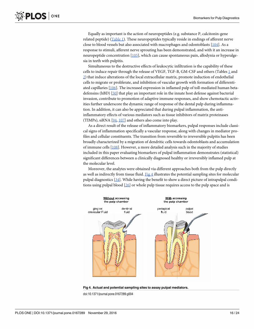
Moreover, the analytes were obtained via different approaches both from the pulp directly
as well as indirectly from tissue fluid. Fig 4 illustrates the potential sampling sites for molecular
pulpal diagnostics [34]. While having the benefit to show a direct picture of intrapulpal condi-
tions using pulpal blood [26] or whole pulp tissue requires access to the pulp space and is
Fig 4. Actual and potential sampling sites to assay pulpal mediators.
doi:10.1371/journal.pone.0167289.g004
Biomarkers for Pulp Diagnostics
PLOS ONE | DOI:10.1371/journal.pone.0167289 November 29, 2016 16 / 24
therefore not applicable as a chairside screening tool. Conversely, indirect methods such as
dentin fluid collection or assessment of mediators in GCF can be performed clinical in a less
invasive way. Dentin fluid is the extracellular fluid that is contained within dentinal tubules
[109]; its composition includes inflammatory mediators and vasoactive compounds associated
with inflammation. While initial evidence suggested that these mediators can be assayed [33]
problems exist with protein yield [27] and the need to remove the existing restoration or in
other cases to prepare an initial cavity deep in dentin.
GCF was used to sample mediators in one study included here (Table 2) [1]. This fluid is an
exudate that from the gingival crevice and it contains several host factors including antibodies,
bacterial antigens, proteins, and cytokines [110, 111]. GCF analyses may be promising due to
the ease of application. Moreover, it may be possible to assess the dynamics of apical periodon-
titis using GCF [34]. However, the major drawback in the evaluation of host mediators in GCF
is that tissue inflammation, independent of its cause, is a non-specific process of innate immu-
nity [112] and this makes it difficult to distinguish on a molecular level between a marginal
and apical periodontal inflammation. When sampling from the GCF for pulpal diagnostics,
this potential drawback could be overcome by (i) creating healthy periodontal conditions, (ii)
averaging out several sites on one or multiple teeth, (iii) combining clinical and radiographic
observations, or (iv) defining a specific pattern of metabolites relevant to the pulp and not the
periodontium, or possibly by other as yet unidentified means. Furthermore, the detection of
mediators of pulpitis in GCF is impacted by the need for these compounds to reach the peri-
odontal ligament and ultimately the gingival crevice in sufficient concentrations. Indeed, the
notion that mediators will diffuse from the pulp via dentinal tubules or accessory canals to the
periodontal ligament has been called into question [87]. Periapical fluid samples, while requir-
ing a direct access to the apical site, are of interest to determine the level of systemic inflamma-
tion [28].
Discovering an improved method to determine the present inflammatory condition of the
pulp could be of great value: on the one hand, pulp necrosis is one of the most frequent com-
plications after coronal restoration of assumed non-inflamed (vital) teeth, on the other per-
forming a full pulpectomy on teeth that could have been kept vital (at least in part) suggests
that overtreatment may occur in many cases [113]. Endodontic diagnosis should therefore
focus on either the extent of the microbial infection or the inflammatory reaction of the host
tissue; however, current methods do neither [14, 23, 34].
Keeping a pulp vital offers distinct advantages compared to root canal treatment: the pro-
tective immune capacity of the pulp remains preserved and the remaining tooth structure gets
not unnecessarily weakened by access cavity preparation and root canal enlargement. Unfortu-
nately, the only available long-term outcome studies on direct pulp capping procedures (i.e.
direct pulpal interventions), which attempt to maintain pulpal vitality, show unsatisfactory
success rates as low as 20% after ten years [114]. The development of biocompatible materials
facilitates a wound closure free of inflammation after pulpal capping procedures or partial pul-
potomy [108]. However, the likelihood of a pulp to survive such procedures remains question-
able using current schemes for assessment of pulpal inflammation.
One limitation of this systematic review is that merely 2 out of 57 studies [33, 45] were spe-
cifically designed to investigate potential biomarkers in the context of pulpal diagnostics. Most
of the studies analyzed here merely target the presence of molecules and their function in
pulpal inflammation. Nevertheless, based on the current state of knowledge this review pro-
vides an overview on molecules that are present and measurable during pulpal inflammation
and therefore potentially can serve as a biomarker for pulpal inflammation. This may provide
impulses for further research. This research needs to explore patient (age, gender, systemic
condition) and infection related factors (varying composition of the microbiological
Biomarkers for Pulp Diagnostics
PLOS ONE | DOI:10.1371/journal.pone.0167289 November 29, 2016 17 / 24
infection). Clinical investigations should be conducted that are specifically designed to confirm
the results collected from the research collected here. More specifically mediator profiles
should be assessed in defined clinical scenarios. In addition, the assays methodology should be
tested for their applicability with the possible substrates. The ultimate goal should then be to
develop an inexpensive chairside test for non-invasive molecular pulp diagnostics. In fact,
such a chair-side assay, based on the immunochromatographic detection of MMP-8 specific
antibodies, is already commercial available to diagnose periodontal inflammation [115]. For
endodontic procedures of the future, such as partial pulpotomies and pulp regeneration, a
comparable test will be of significant value.
Indeed, various biomarkers that are produced by cellular components of the dental pulp
can provide a snapshot of the biological mechanisms that propel this immunocompetent tissue
towards healing or necrosis. The imbalance between tissue destructive molecules like proteases
and tissue inductive molecules like VEGF may serve as a diagnostic or prognostic tool for end-
odontic intervention. The challenge remains on developing a method to make these biomark-
ers readily measureable in a clinical setting.
Conclusions
In the included studies, irreversible pulpitis was associated with different expression of various
biomarkers compared to non-inflamed controls. These biomarkers were significantly
expressed not only in pulp tissue, but also in gingival crevicular fluid that can be collected
non-invasively and in dentin fluid that can be analyzed without extirpating the pulpal tissue.
This may be used to accurately differentiate diseased from healthy pulp tissue. The main cur-
rent challenges in the clinical application of biomarkers lie in the identification of biomarkers
or biomarker subsets that reliably correlate with pulpal inflammation, the improvement of
sample collection (substrate and protein yields), and their analysis (interference of the bio-
markers with inflammation of other than pulpal origin). If these hurdles can be overcome, a
more accurate pulpal diagnosis and more predictable vital pulp treatment regime may create
better clinical outcomes.
Supporting Information
S1 Table. PRISMA Checklist.
(DOCX)
S2 Table. Example of search strategy employed for this literature review.
(DOCX)
S3 Table. Hits from the literature search obtained with the different databases.
(DOCX)
S4 Table. Studies excluded from the final analysis.
(DOCX)
S5 Table. Assessment of the study quality using the modified Newcastle Ottawa Scale.
(DOCX)
Acknowledgments
The authors want to thank Mrs. Monika Marending, Mrs. Sabine Groddeck, Mr. Kai Bo, Mr.
Manuel Sanchez, Mr. Valon Bejic and Mr. Amund Odin Asphaug for their help with transla-
tions of manuscripts.
Biomarkers for Pulp Diagnostics
PLOS ONE | DOI:10.1371/journal.pone.0167289 November 29, 2016 18 / 24
Author Contributions
Conceptualization: DKR.
Data curation: DKR JCG OAP.
Formal analysis: DKR JCG OAP.
Funding acquisition: DKR.
Investigation: DKR JCG.
Methodology: DKR.
Project administration: DKR JCG OAP.
Resources: DKR JCG OAP.
Visualization: DKR JCG OAP.
Writing – original draft: DKR JCG OAP.
Writing – review & editing: DKR JCG OAP.
References1. Karapanou V, Kempuraj D, Theoharides TC. Interleukin-8 is increased in gingival crevicular fluid from
patients with acute pulpitis. J Endod. 2008; 34: 148–151. doi: 10.1016/j.joen.2007.10.022 PMID:
18215670
2. Keller JF, Carrouel F, Staquet MJ, Kufer TA, Baudouin C, Msika P, et al. Expression of NOD2 is
increased in inflamed human dental pulps and lipoteichoic acid-stimulated odontoblast-like cells.
Innate Immun. 2011; 17: 29–34. doi: 10.1177/1753425909348527 PMID: 19880660
3. Smith AJ. Pulpal responses to caries and dental repair. Caries Res. 2002; 36: 223–232. PMID:
12218270
4. Staquet MJ, Carrouel F, Keller JF, Baudouin C, Msika P, Bleicher F, et al. Pattern-recognition recep-
tors in pulp defense. Adv Dent Res. 2011; 23: 296–301. doi: 10.1177/0022034511405390 PMID:
21677082
5. Graves DT, Oates T, Garlet GP. Review of osteoimmunology and the host response in endodontic
and periodontal lesions. J Oral Microbiol. 2011; 3.
6. Nakanishi T, Takegawa D, Hirao K, Takahashi K, Yumoto H, Matsuo T. Roles of dental pulp fibroblasts
in the recognition of bacterium-related factors and subsequent development of pulpitis. Jpn Dent Sci
Rev. 2011; 47: 161–166.
7. Stashenko P. Role of immune cytokines in the pathogenesis of periapical lesions. Endod Dent Trau-
matol. 1990; 6: 89–96. PMID: 2079017
8. Ahlquist ML, Franzen OG. Inflammation and dental pain in man. Endod Dent Traumatol. 1994; 10:
201–209. PMID: 7843060
9. Hahn CL, Falkler WA Jr, Minah GE. Microbiological studies of carious dentine from human teeth with
irreversible pulpitis. Arch Oral Biol. 1991; 36: 147–153. PMID: 2059163
10. Bergenholtz G. Inflammatory response of the dental pulp to bacterial irritation. J Endod. 1981; 7: 100–
104. doi: 10.1016/S0099-2399(81)80122-7 PMID: 6938628
11. Horst O, Tompkins K, Coats S, Braham P, Darveau R, Dale B. TGF-beta1 Inhibits TLR-mediated
odontoblast responses to oral bacteria. J Dent Res. 2009; 88: 333–338. doi: 10.1177/
0022034509334846 PMID: 19407153
12. Jiang HW, Zhang W, Ren BP, Zeng JF, Ling JQ. Expression of toll like receptor 4 in normal human
odontoblasts and dental pulp tissue. J Endod. 2006; 32: 747–751. doi: 10.1016/j.joen.2006.01.010
PMID: 16861074
13. Mutoh N, Tani-Ishii N, Tsukinoki K, Chieda K, Watanabe K. Expression of toll-like receptor 2 and 4 in
dental pulp. J Endod. 2007; 33: 1183–1186. doi: 10.1016/j.joen.2007.05.018 PMID: 17889686
14. Levin LG, Law AS, Holland GR, Abbott PV, Roda RS. Identify and define all diagnostic terms for pulpal
health and disease states. J Endod. 2009; 35: 1645–1657. doi: 10.1016/j.joen.2009.09.032 PMID:
19932339
Biomarkers for Pulp Diagnostics
PLOS ONE | DOI:10.1371/journal.pone.0167289 November 29, 2016 19 / 24
15. Dummer PM, Hicks R, Huws D. Clinical signs and symptoms in pulp disease. Int Endod J. 1980; 13:
27–35. PMID: 6935168
16. Seltzer S, Bender IB, Ziontz M. The dynamics of pulp inflammation: correlations between diagnostic
data and actual histologic findings in the pulp. Oral Surg Oral Med Oral Pathol. 1963; 16: 846–871
contd. PMID: 13987830
17. Ricucci D, Loghin S, Siqueira JF Jr, Correlation between clinical and histologic pulp diagnoses. J
Endod. 2014; 40: 1932–1939. doi: 10.1016/j.joen.2014.08.010 PMID: 25312886
18. Trowbridge H. Pathogenesis of pulpitis resulting from dental caries. J Endod. 1981; 7: 52–60. PMID:
6938624
19. Allareddy V, Rampa S, Lee MK, Allareddy V, Nalliah RP. Hospital-based emergency department visits
involving dental conditions: profile and predictors of poor outcomes and resource utilization. J Am
Dent Assoc. 2014; 145: 331–337. doi: 10.14219/jada.2014.7 PMID: 24686965
20. The Pew Center on the States. A Costly Dental Destination. Pew Children’s Dental Campaign 2012.
http://www.pewtrusts.org/~/media/assets/2012/01/16/a-costly-dental-destination.pdf
21. Michaelson P, Holland G. Is pulpitis painful. Int Endod J. 2002; 35: 829–832. PMID: 12406376
22. Baume R. Lehrbuch der Zahnheilkunde. 1st ed. Leipzig: Arthur Felix; 1877.
23. Mejare IA, Axelsson S, Davidson T, Frisk F, Hakeberg M, Kvist T, et al. Diagnosis of the condition of
the dental pulp: A systematic review. Int Endod J. 2012; 45: 597–613. doi: 10.1111/j.1365-2591.2012.
02016.x PMID: 22329525
24. Walton RE, Langeland K. Migration of materials in the dental pulp of monkeys. J Endod. 1978; 4: 167–
177. doi: 10.1016/S0099-2399(78)80171-X PMID: 106088
25. Vongsavan N, Matthews RW, Matthews B. The permeability of human dentine in vitro and in vivo.
Arch Oral Biol. 2000; 45: 931–935. PMID: 11000378
26. Mente J, Petrovic J, Gehrig H, Rampf S, Michel A, Schurz A, et al. A Prospective Clinical Pilot Study
on the Level of Matrix Metalloproteinase-9 in Dental Pulpal Blood as a Marker for the State of Inflam-
mation in the Pulp Tissue. J Endod. 2016; 42: 190–197. doi: 10.1016/j.joen.2015.10.020 PMID:
26725178
27. Zehnder M, Rechenberg DK, Bostanci N, Sisman F, Attin T. Comparison of vehicles to collect dentinal
fluid for molecular analysis. J Dent. 2014; 42: 1027–1032. doi: 10.1016/j.jdent.2014.01.014 PMID:
24681278
28. Rechenberg DK, Bostanci N, Zehnder M, Belibasakis GN. Periapical fluid RANKL and IL-8 are differ-
entially regulated in pulpitis and apical periodontitis. Cytokine. 2014; 69: 116–119. doi: 10.1016/j.cyto.
2014.05.014 PMID: 25022970
29. Avellan NL, Sorsa T, Tervahartiala T, Forster C, Kemppainen P. Experimental tooth pain elevates sub-
stance P and matrix metalloproteinase-8 levels in human gingival crevice fluid. Acta Odontol Scand.
2008; 66: 18–22. doi: 10.1080/00016350701810658 PMID: 18320414
30. Giannobile WV, Beikler T, Kinney JS, Ramseier CA, Morelli T, Wong DT. Saliva as a diagnostic tool
for periodontal disease: current state and future directions. Periodontol 2000. 2009; 50: 52–64. doi:
10.1111/j.1600-0757.2008.00288.x PMID: 19388953
31. Bostanci N, Ilgenli T, Emingil G, Afacan B, Han B, Toz H, et al. Gingival crevicular fluid levels of
RANKL and OPG in periodontal diseases: implications of their relative ratio. J Clin Periodontol. 2007;
34: 370–376. doi: 10.1111/j.1600-051X.2007.01061.x PMID: 17355365
32. Sorsa T, Hernandez M, Leppilahti J, Munjal S, Netuschil L, Mantyla P. Detection of gingival crevicular
fluid MMP-8 levels with different laboratory and chair-side methods. Oral Dis. 2010; 16: 39–45. doi:
10.1111/j.1601-0825.2009.01603.x PMID: 19627514
33. Zehnder M, Wegehaupt FJ, Attin T. A first study on the usefulness of matrix metalloproteinase 9 from
dentinal fluid to indicate pulp inflammation. J Endod. 2011; 37: 17–20. doi: 10.1016/j.joen.2010.10.003
PMID: 21146069
34. Rechenberg DK, Zehnder M. Molecular diagnostics in endodontics. Endod Topics. 2014; 30: 51–65.
35. Botero TM, Shelburne CE, Holland GR, Hanks CT, Nor JE. TLR4 mediates LPS-induced VEGF
expression in odontoblasts. J Endod. 2006; 32: 951–955. doi: 10.1016/j.joen.2006.03.018 PMID:
16982271
36. Carrouel F, Staquet MJ, Keller JF, Baudouin C, Msika P, Bleicher F, et al. Lipopolysaccharide-binding
protein inhibits toll-like receptor 2 activation by lipoteichoic acid in human odontoblast-like cells. J
Endod. 2013; 39: 1008–1014. doi: 10.1016/j.joen.2013.04.020 PMID: 23880268
37. Farges JC, Carrouel F, Keller JF, Baudouin C, Msika P, Bleicher F, et al. Cytokine production by
human odontoblast-like cells upon Toll-like receptor-2 engagement. Immunobiology. 2011; 216: 513–
517. doi: 10.1016/j.imbio.2010.08.006 PMID: 20850890
Biomarkers for Pulp Diagnostics
PLOS ONE | DOI:10.1371/journal.pone.0167289 November 29, 2016 20 / 24
38. Moher D, Liberati A, Tetzlaff J, Altman DG, Group P. Preferred reporting items for systematic reviews
and meta-analyses: the PRISMA statement. J Clin Epidemiol. 2009; 62: 1006–1012. doi: 10.1016/j.
jclinepi.2009.06.005 PMID: 19631508
39. Wells GA, Brodsky L, O’Connell D, Shea B, Henry D, Mayank S, et al. An evaluation of the Newcastle
Ottawa Scale: an assessment tool for evaluating the quality of non-randomized studies (abstract). XI
International Cochrane Colloquium Book of Abstracts, O-63. 2003: 26.
40. Gomes MS, Blattner TC, Sant’Ana Filho M, Grecca FS, Hugo FN, Fouad AF, et al. Can apical peri-
odontitis modify systemic levels of inflammatory markers? A systematic review and meta-analysis. J
Endod. 2013; 39: 1205–1217. doi: 10.1016/j.joen.2013.06.014 PMID: 24041380
41. Lepinski AM, Hargreaves KM, Goodis HE, Bowles WR. Bradykinin levels in dental pulp by microdialy-
sis. J Endod. 2000; 26: 744–747. doi: 10.1097/00004770-200012000-00020 PMID: 11471646
42. Bowles WR, Withrow JC, Lepinski AM, Hargreaves KM. Tissue levels of immunoreactive substance P
are increased in patients with irreversible pulpitis. J Endod. 2003; 29: 265–267. doi: 10.1097/
00004770-200304000-00009 PMID: 12701777
43. Kokkas AB, Goulas A, Varsamidis K, Mirtsou V, Tziafas D. Irreversible but not reversible pulpitis is
associated with up-regulation of tumour necrosis factor-alpha gene expression in human pulp. Int
Endod J. 2007; 40: 198–203. doi: 10.1111/j.1365-2591.2007.01215.x PMID: 17305696
44. Nakanishi T, Matsuo T, Ebisu S. Quantitative analysis of immunoglobulins and inflammatory factors in
human pulpal blood from exposed pulps. J Endod. 1995; 21: 131–136. PMID: 7561655
45. Elsalhy M, Azizieh F, Raghupathy R. Cytokines as diagnostic markers of pulpal inflammation. Int
Endod J. 2013; 46: 573–580. doi: 10.1111/iej.12030 PMID: 23240887
46. Evcil MS, Keles A, Uzun I, Demircan B, Koseoglu M. Nitric oxide levels in serum of patients with symp-
tomatic irreversible pulpitis. J Pain Palliat Care Pharmacother. 2006; 20: 15–19.
47. Cohen JS, Reader A, Fertel R, Beck M, Meyers WJ. A radioimmunoassay determination of the con-
centrations of prostaglandins E2 and F2alpha in painful and asymptomatic human dental pulps. J
Endod. 1985; 11: 330–335. PMID: 3863874
48. Zehnder M, Delaleu N, Du Y, Bickel M. Cytokine gene expression—part of host defence in pulpitis.
Cytokine. 2003; 22: 84–88. PMID: 12849707
49. Abd-Elmeguid A, Abdeldayem M, Kline LW, Moqbel R, Vliagoftis H, Yu DC. Osteocalcin expression in
pulp inflammation. J Endod. 2013; 39: 865–872. doi: 10.1016/j.joen.2012.12.035 PMID: 23791253
50. Paris S, Wolgin M, Kielbassa AM, Pries A, Zakrzewicz A. Gene expression of human beta-defensins
in healthy and inflamed human dental pulps. J Endod. 2009; 35: 520–523. doi: 10.1016/j.joen.2008.
12.015 PMID: 19345797
51. Silva AC, Faria MR, Fontes A, Campos MS, Cavalcanti BN. Interleukin-1 beta and interleukin-8 in
healthy and inflamed dental pulps. J Appl Oral Sci. 2009; 17: 527–532. doi: 10.1590/S1678-
77572009000500031 PMID: 19936537
52. Rauschenberger CR, Bailey JC, Cootauco CJ. Detection of human IL-2 in normal and inflamed dental
pulps. J Endod. 1997; 23: 366–370. doi: 10.1016/S0099-2399(97)80184-7 PMID: 9545944
53. Anderson LM, Dumsha TC, McDonald NJ, Spitznagel JK Jr. Evaluating IL-2 levels in human pulp tis-
sue. J Endod. 2002; 28: 651–655. doi: 10.1097/00004770-200209000-00006 PMID: 12236309
54. Huang GT, Potente AP, Kim JW, Chugal N, Zhang X. Increased interleukin-8 expression in inflamed
human dental pulps. Oral Surg Oral Med Oral Pathol Oral Radiol Endod. 1999; 88: 214–220. PMID:
10468466
55. Pezelj-Ribaric S, Anic I, Brekalo I, Miletic I, Hasan M, Simunovic-Soskic M. Detection of tumor necro-
sis factor alpha in normal and inflamed human dental pulps. Arch Med Res. 2002; 33: 482–484. PMID:
12459320
56. Keller JF, Carrouel F, Staquet MJ, Kufer TA, Baudouin C, Msika P, et al. Expression of NOD2 is
increased in inflamed human dental pulps and lipoteichoic acid-stimulated odontoblast-like cells.
Innate immunity. 2009; 17: 29–34. doi: 10.1177/1753425909348527 PMID: 19880660
57. Li NN, Zhang ZM, Wang CK, Wang JR, Meng XP. Expression of MIP-1(alpha) mRNA in inflammed
pulp tissue and its significance. J Jilin Univ Med. 2011; 37: 312–314.
58. Nakanishi T, Takahashi K, Hosokawa Y, Adachi T, Nakae H, Matsuo T. Expression of macrophage
inflammatory protein 3alpha in human inflamed dental pulp tissue. J Endod. 2005; 31: 84–87. PMID:
15671814
59. Piattelli A, Rubini C, Fioroni M, Tripodi D, Strocchi R. Transforming growth factor-beta 1 (TGF-beta 1)
expression in normal healthy pulps and in those with irreversible pulpitis. Int Endod J. 2004; 37: 114–
119. PMID: 14871177
Biomarkers for Pulp Diagnostics
PLOS ONE | DOI:10.1371/journal.pone.0167289 November 29, 2016 21 / 24
60. Adachi T, Nakanishi T, Yumoto H, Hirao K, Takahashi K, Mukai K, et al. Caries-related bacteria and
cytokines induce CXCL10 in dental pulp. J Dent Res. 2007; 86: 1217–1222. PMID: 18037659
61. Jiang HW, Ling JQ, Gong QM. The expression of stromal cell-derived factor 1 (SDF-1) in inflamed
human dental pulp. J Endod. 2008; 34: 1351–1354. doi: 10.1016/j.joen.2008.07.023 PMID: 18928845
62. Huang FM, Tsai CH, Yang SF, Chang YC. The upregulation of oncostatin M in inflamed human dental
pulps. Int Endod J. 2009; 42: 627–631. doi: 10.1111/j.1365-2591.2009.01567.x PMID: 19467046
63. Gusman H, Santana RB, Zehnder M. Matrix metalloproteinase levels and gelatinolytic activity in clini-
cally healthy and inflamed human dental pulps. Eur J Oral Sci. 2002; 110: 353–357. PMID: 12664465
64. Accorsi-Mendonca T, Silva EJ, Marcaccini AM, Gerlach RF, Duarte KM, Pardo AP, et al. Evaluation of
gelatinases, tissue inhibitor of matrix metalloproteinase-2, and myeloperoxidase protein in healthy and
inflamed human dental pulp tissue. J Endod. 2013; 39: 879–882. doi: 10.1016/j.joen.2012.11.011
PMID: 23791255
65. Tsai CH, Chen YJ, Huang FM, Su YF, Chang YC. The upregulation of matrix metalloproteinase-9 in
inflamed human dental pulps. J Endod. 2005; 31: 860–862. PMID: 16306818
66. Suwanchai A, Theerapiboon U, Chattipakorn N, Chattipakorn SC. NaV 1.8, but not NaV 1.9, is upregu-
lated in the inflamed dental pulp tissue of human primary teeth. Int Endod J. 2012; 45: 372–378. doi:
10.1111/j.1365-2591.2011.01986.x PMID: 22085016
67. Huang FM, Tsai CH, Chen YJ, Liu CM, Chou MY, Chang YC. Upregulation of tissue-type plasminogen
activator in inflamed human dental pulps. Int Endod J. 2005; 38: 328–333. doi: 10.1111/j.1365-2591.
2005.00951.x PMID: 15876297
68. Huang FM, Yang SF, Chen YJ, Tsai CH, Chang YC. Tissue type plasminogen activator level and
caseinolytic activity in clinically healthy and inflamed human dental pulp. J Dent Sci. 2007; 2: 152–156.
69. Ge J, Ji J, Wang T. Superoxide dismutase and malonyl dialdehyde in human pulp tissue. Chung Hua
Kou Chiang Hsueh Tsa Chih. 1996; 31: 201–203. PMID: 9592267
70. Tulunoglu O, Alacam A, Bastug M, Yavuzer S. Superoxide dismutase activity in healthy and inflamed
pulp tissues of permanent teeth in children. J Clin Pediatr Dent. 1998; 22: 341–345. PMID: 9796506
71. Bodor C, Matolcsy A, Bernath M. Elevated expression of Cu, Zn-SOD and Mn-SOD mRNA in inflamed
dental pulp tissue. Int Endod J. 2007; 40: 128–132. doi: 10.1111/j.1365-2591.2006.01196.x PMID:
17229118
72. Varvara G, Traini T, Esposito P, Caputi S, Perinetti G. Copper-zinc superoxide dismutase activity in
healthy and inflamed human dental pulp. Int Endod J. 2005; 38: 195–199. doi: 10.1111/j.1365-2591.
2005.00936.x PMID: 15743423
73. Cootauco CJ, Rauschenberger CR, Nauman RK. Immunocytochemical distribution of human PMN
elastase and cathepsin-G in dental pulp. J Dent Res. 1993; 72: 1485–1490. PMID: 7693782
74. Spoto G, Fioroni M, Rubini C, Tripodi D, Di Stilio M, Piattelli A. Alkaline phosphatase activity in normal
and inflamed dental pulps. J Endod. 2001; 27: 180–182. doi: 10.1097/00004770-200103000-00010
PMID: 11487147
75. Spoto G, Fioroni M, Rubini C, Tripodi D, Perinetti G, Piattelli A. Aspartate aminotransferase activity in
human healthy and inflamed dental pulps. J Endod. 2001; 27: 394–395. doi: 10.1097/00004770-
200106000-00005 PMID: 11487132
76. Esposito P, Varvara G, Caputi S, Perinetti G. Catalase activity in human healthy and inflamed dental
pulps. Int Endod J. 2003; 36: 599–603. PMID: 12950573
77. Esposito P, Varvara G, Murmura G, Terlizzi A, Caputi S. Ability of healthy and inflamed human dental
pulp to reduce hydrogen peroxide. Eur J Oral Sci. 2003; 111: 454–456. PMID: 12974692
78. da Silva LP, Issa JP, Del Bel EA. Action of nitric oxide on healthy and inflamed human dental pulp tis-
sue. Micron. 2008; 39: 797–801. doi: 10.1016/j.micron.2008.01.018 PMID: 18337111
79. Di Nardo Di Maio F, Lohinai Z, D’Arcangelo C, De Fazio PE, Speranza L, De Lutiis MA, et al. Nitric
oxide synthase in healthy and inflamed human dental pulp. J Dent Res. 2004; 83: 312–316. PMID:
15044505
80. Spoto G, Ferrante M, D’Intino M, Rega L, Dolci M, Trentini P, et al. Cyclic GMP phosphodiesterase
activity role in normal and inflamed human dental pulp. Int J Immunopathol Pharmacol. 2004; 17: 21–
24. PMID: 16857102
81. Spoto G, Menna V, Serra E, Santoleri F, Perfetti G, Ciavarelli L, et al. Cyclic AMP phosphodiesterase
activity in normal and inflamed human dental pulp. Int J Immunopathol Pharmacol. 2004; 17: 11–15.
82. Bolanos OR, Seltzer S. Cyclic AMP and cyclic GMP quantitation in pulp and periapical lesions and
their correlation with pain. J Endod. 1981; 7: 268–271. doi: 10.1016/S0099-2399(81)80005-2 PMID:
6265580
Biomarkers for Pulp Diagnostics
PLOS ONE | DOI:10.1371/journal.pone.0167289 November 29, 2016 22 / 24
83. Dong Y, Zhou J, Pan Y. Quantitive study of 6-keto-prostaglandin F1 alpha and thromboxane B2 in
human pulp. Chung Hua Kou Chiang Hsueh Tsa Chih. 1999; 34: 236–238. PMID: 11776916
84. Khabbaz MG, Anastasiadis PL, Sykaras SN. Determination of endotoxins in the vital pulp of human
carious teeth: association with pulpal pain. Oral Surg Oral Med Oral Pathol Oral Radiol Endod. 2001;
91: 587–593. doi: 10.1067/moe.2001.113831 PMID: 11346740
85. Nakanishi T, Shimizu H, Hosokawa Y, Matsuo T. An immunohistological study on cyclooxygenase-2
in human dental pulp. J Endod. 2001; 27: 385–388. doi: 10.1097/00004770-200106000-00003 PMID:
11487130
86. Guven G, Altun C, Gunhan O, Gurbuz T, Basak F, Akbulut E, et al. Co-expression of cyclooxygenase-
2 and vascular endothelial growth factor in inflamed human pulp: an immunohistochemical study. J
Endod. 2007; 33: 18–20. doi: 10.1016/j.joen.2006.07.014 PMID: 17185121
87. Awawdeh L, Lundy FT, Shaw C, Lamey PJ, Linden GJ, Kennedy JG. Quantitative analysis of sub-
stance P, neurokinin A and calcitonin gene-related peptide in pulp tissue from painful and healthy
human teeth. Int Endod J. 2002; 35: 30–36. PMID: 11853236
88. Caviedes-Bucheli J, Lombana N, Azuero-Holguin MM, Munoz HR. Quantification of neuropeptides
(calcitonin gene-related peptide, substance P, neurokinin A, neuropeptide Y and vasoactive intestinal
polypeptide) expressed in healthy and inflamed human dental pulp. Int Endod J. 2006; 39: 394–400.
doi: 10.1111/j.1365-2591.2006.01093.x PMID: 16640639
89. Caviedes-Bucheli J, Camargo-Beltran C, Gomez-la-Rotta AM, Moreno SC, Abello GC, Gonzalez-
Escobar JM. Expression of calcitonin gene-related peptide (CGRP) in irreversible acute pulpitis. J
Endod. 2004; 30: 201–204. PMID: 15085045
90. Caviedes-Bucheli J, Arenas N, Guiza O, Moncada NA, Moreno GC, Diaz E, et al. Calcitonin gene-
related peptide receptor expression in healthy and inflamed human pulp tissue. Int Endod J. 2005; 38:
712–717. doi: 10.1111/j.1365-2591.2005.01006.x PMID: 16164685
91. Artese L, Rubini C, Ferrero G, Fioroni M, Santinelli A, Piattelli A. Vascular endothelial growth factor
(VEGF) expression in healthy and inflamed human dental pulps. J Endod. 2002; 28: 20–23. doi: 10.
1097/00004770-200201000-00005 PMID: 11806643
92. Caviedes-Bucheli J, Gutierrez-Guerra JE, Salazar F, Pichardo D, Moreno GC, Munoz HR. Substance
P receptor expression in healthy and inflamed human pulp tissue. Int Endod J. 2007; 40: 106–111.
doi: 10.1111/j.1365-2591.2006.01189.x PMID: 17229115
93. Caviedes-Bucheli J, Moreno GC, Lopez MP, Bermeo-Noguera AM, Pacheco-Rodriguez G, Cuellar A,
et al. Calcitonin gene-related peptide receptor expression in alternatively activated monocytes/macro-
phages during irreversible pulpitis. J Endod. 2008; 34: 945–949. doi: 10.1016/j.joen.2008.05.011
PMID: 18634925
94. Zhong S, Zhang S, Bair E, Nares S, Khan AA. Differential expression of microRNAs in normal and
inflamed human pulps. J Endod. 2012; 38: 746–752. doi: 10.1016/j.joen.2012.02.020 PMID:
22595106
95. Dong Y, Lan W, Wu W, Huang Z, Zhao J, Peng L, et al. Increased expression of EphA7 in inflamed
human dental pulp. J Endod. 2013; 39: 223–227. doi: 10.1016/j.joen.2012.11.020 PMID: 23321235
96. Bhattacharyya S, Kelley K, Melichian DS, Tamaki Z, Fang F, Su Y, et al. Toll-like receptor 4 signaling
augments transforming growth factor-beta responses: a novel mechanism for maintaining and ampli-
fying fibrosis in scleroderma. Am J Pathol. 2013; 182: 192–205. doi: 10.1016/j.ajpath.2012.09.007
PMID: 23141927
97. Villalba M, Hott M, Martin C, Aguila B, Valdivia S, Quezada C, et al. Herpes simplex virus type 1
induces simultaneous activation of Toll-like receptors 2 and 4 and expression of the endogenous
ligand serum amyloid A in astrocytes. Med Microbiol Immunol. 2012; 201: 371–379. doi: 10.1007/
s00430-012-0247-0 PMID: 22622619
98. Shi B, Huang Q, Tak PP, Vervoordeldonk MJ, Huang CC, Dorfleutner A, et al. SNAPIN: an endoge-
nous Toll-like receptor ligand in rheumatoid arthritis. Ann Rheum Dis. 2012; 71: 1411–1417. doi: 10.
1136/annrheumdis-2011-200899 PMID: 22523426
99. Goh FG, Piccinini AM, Krausgruber T, Udalova IA, Midwood KS. Transcriptional regulation of the
endogenous danger signal tenascin-C: a novel autocrine loop in inflammation. J Immunol. 2010; 184:
2655–2662. doi: 10.4049/jimmunol.0903359 PMID: 20107185
100. Merline R, Moreth K, Beckmann J, Nastase MV, Zeng-Brouwers J, Tralhao JG, et al. Signaling by the
matrix proteoglycan decorin controls inflammation and cancer through PDCD4 and MicroRNA-21. Sci
Signal. 2011; 4: ra75. doi: 10.1126/scisignal.2001868 PMID: 22087031
101. Izumi T, Kobayashi I, Okamura K, Sakai H. Immunohistochemical study on the immunocompetent
cells of the pulp in human non-carious and carious teeth. Arch Oral Biol. 1995; 40: 609–614. PMID:
7575232
Biomarkers for Pulp Diagnostics
PLOS ONE | DOI:10.1371/journal.pone.0167289 November 29, 2016 23 / 24
102. Moreland LW. Rheumatology and Immunology Therapy. 1st ed. Springer Berlin Heidelberg New
York; 2004.
103. Omori K, Kida T, Hori M, Ozaki H, Murata T. Multiple roles of the PGE2 -EP receptor signal in vascular
permeability. British Journal of Pharmacology. 2014; 171: 4879–4889. doi: 10.1111/bph.12815 PMID:
24923772
104. Caviedes-Bucheli J, Munoz HR, Azuero-Holguin MM, Ulate E. Neuropeptides in dental pulp: the silent
protagonists. J Endod. 2008; 34: 773–788. doi: 10.1016/j.joen.2008.03.010 PMID: 18570980
105. Byers MR. Effects of inflammation on dental sensory nerves and vice versa. Proc Finn Dent Soc.
1992; 88 Suppl 1: 499–506.
106. Sunderkotter C, Steinbrink K, Goebeler M, Bhardwaj R, Sorg C. Macrophages and angiogenesis.
Journal of Leukocyte Biology. 1994; 55: 410–422. PMID: 7509844
107. Galicia JC, Naqvi AR, Ko CC, Nares S, Khan AA. MiRNA-181a regulates Toll-like receptor agonist-
induced inflammatory response in human fibroblasts. Genes Immun. 2014; 15: 333–337. doi: 10.
1038/gene.2014.24 PMID: 24848932
108. Goldberg M, Farges JC, Lacerda-Pinheiro S, Six N, Jegat N, Decup F, et al. Inflammatory and immu-
nological aspects of dental pulp repair. Pharmacol Res. 2008; 58: 137–147. doi: 10.1016/j.phrs.2008.
05.013 PMID: 18602009
109. Coffey CT, Ingram MJ, Bjorndal AM. Analysis of human dentinal fluid. Oral Surg Oral Med Oral Pathol.
1970; 30: 835–837. PMID: 5273868
110. Griffiths GS. Formation, collection and significance of gingival crevicular fluid Periodontol 2000. 2003;
31: 32–42. PMID: 12656994
111. Taba M Jr., Kinney J, Kim AS, Giannobile WV. Diagnostic biomarkers for oral and periodontal dis-
eases. Dent Clin North Am. 2005; 49: 551–571, vi. doi: 10.1016/j.cden.2005.03.009 PMID: 15978241
112. Farges JC, Alliot-Licht B, Renard E, Ducret M, Gaudin A, Smith AJ, et al. Dental pulp defence and
repair mechanisms in dental caries. Mediators Inflamm. 2015; 2015: 230251. doi: 10.1155/2015/
230251 PMID: 26538821
113. Simon S, Perard M, Zanini M, Smith AJ, Charpentier E, Djole SX, et al. Should pulp chamber pulpot-
omy be seen as a permanent treatment? Some preliminary thoughts. Int Endod J. 2013; 46: 79–87.
doi: 10.1111/j.1365-2591.2012.02113.x PMID: 22900881
114. Barthel CR, Rosenkranz B, Leuenberg A, Roulet JF. Pulp capping of carious exposures: treatment
outcome after 5 and 10 years: a retrospective study. J Endod. 2000; 26: 525–528. doi: 10.1097/
00004770-200009000-00010 PMID: 11199794
115. Mantyla P, Stenman M, Kinane DF, Tikanoja S, Luoto H, Salo T, et al. Gingival crevicular fluid collage-
nase-2 (MMP-8) test stick for chair-side monitoring of periodontitis. J Periodontal Res. 2003; 38: 436–
439. PMID: 12828663
Biomarkers for Pulp Diagnostics
PLOS ONE | DOI:10.1371/journal.pone.0167289 November 29, 2016 24 / 24