“biological markers in fundamental and clini-
TRANSCRIPT


Collection of abstracts “Biological Markers in Fundamental and Clini-cal Medicine” (official specialized scientific journal of The Czech Republic, registration number MK CR E 22955) by the publishing center of The ESCBM provides its lanes for information materials in the field of scientific research of modern biological markers in clinical and experimental medicine, pharma-cy, and fundamental biology. The collection publishes abstracts of scientific and practical conferences, seminars, symposia, dedicated to the study of molecular-biochemical and functional markers, playing a role in pathogen-esis, diagnosis, prognosis, as well as assessing the monitoring of the treat-ment effectiveness for the various systems and organs diseases. Among the priority topics of the journal there is the research of molecular mechanisms of diseases pathogenesis, the study of the structure and functions of pep-tides, nucleic acids, nucleotides, lipids and other biologically active com-ponents of body cells.
The collection is intended for fast and systematic publication of ab-stracts, containing the results of author’s research, reviews highlighting ma-jor developments in the field of biological markers, short messages, new experimental and clinical studies, which use biological markers, as well as proposing new principles and methods for the study of biological markers.
The collection is published 4 times a year. Circulation - 200 copies.
Editorial Office Address: ESCBM, Příkop 838/6, Zábrdovice, 60200 Brno, Česká republika.Tel. +420773530856
3
CONTENTS
Assessment of ischemia modified albumin serum level in isoproterenol-induced myocardial infarctionTimercan T., Timercan V.
Comparative evaluation of the dynamics of the size of the furuncle infiltrate of the maxillofacial area with different postoperative treatment Flerjanovich M.S., Pohodenko-Chudakova I.O.
Elemental composition of hair as an objective indicator for diagnostics, prevention and treatment of microelementosisSharipov K.O., Bulygin K.A., Sharipov D.K., Dosymbekova R.S.
Effect of the antioxidant – herbal derivate on biomarkers of oxidative stress and an-tioxidant systems on the model of gastric ulcer Zalyubovska O.I., Tiupka T.I., Minaieva A.O., Zlenko V.V., Litvinenko M.I., Avidzba Y.N.
Effect of freezing on anti-inflammatory efficiency of human placental extractsGovorova Yu.S., Bobrova O.M., Repina S.V., Rozanova E.D., Nardid О.А., Schetinskey M.I.
Substantiation of the study on prediction of surgical dental treatment for women in the period of menstruation on the basis of the frequency of this risk factor detecting based on anamnesisSudakova S. E., Pohodenko-Chudakova I. O.
Vitamin D receptor gene polymorphism in patients with juvenile rheumatoid arthritisMyslivets M., Paramonova N., Stepuro T.
Ceruloplasmin in experimental ovarian torsion/detorsionLazăr C.
Caries-preventive effect of lactobacillus reuteri in children with gastrointestinal pa-thologyDenysova E. G.
Loss of heterozygosity at the BRCA 1 locus as a marker of sensitivity for adjuvant che-motherapy in hereditary ovarian cancerSokolenko A.P., Savanevich A.L. , Stepuro T.L.2, Shulga A.V. , Berlev I.V. , Imyanitov E.N.
B-cell lymphoma-2 receptor expression in human breast cancerFulga V.
Correlation between molecular and clinical, histological characteristics of breast cancer in young women Bacalîm L., Sofroni L., Corobcean N., Chimencedji I., Odobescu Ox., Ghidirim N.
Сurrent aspects of odontogenic jaw cysts treatmentStoyan E. Y., Denysova E. G.
Atom-absortive spectrometry for determining of the elemental content of the glycyr-rhiza glabra rootsKasimova D.B, Abdukhalikova N.U, Gaibnazarova D.T.
4
7
10
13
16
19
22
24
26
29
32
36
41
44

Biological Markers in Fundamental and Clinical Medicine. – Vol.1, №3. – 2017. ISSN 2570-5911 (Print); ISSN 2570-5903 (On-Line) DOI: 10.29256/v.01.03.2017.escbm01-14
4
FORWARD SESSION
SELECTED ABSTRACTS OF THE ON-LINE CONFERENCES “Modern Technologies of Diagnostics and Monitoring of Therapy in Experimental, Clinical Medi-cine and Pharmacy” and “Biological markers in fundamental and applied
biology. From theory to practice”.
DOI: 10.29256/v.01.03.2017.escbm01
ASSESSMENT OF ISCHEMIA MODIFIED ALBUMIN SERUM LEVEL IN ISO-PROTERENOL-INDUCED MYOCARDIAL INFARCTION
Timercan T., Timercan V.Nicolae Testemitsanu State University of Medicine and Pharmacy
Kishinev, Moldova
Introduction: Acute coronary syndrome (ACS) remains the leading cause of mortality worldwide. Various epidemiologic studies demonstrate that the death rates due to cardiac diseases have been risen since 1990 from 28,9 % to 36,9 % (2020)[1]. Cardiac ischemia is known to be the key-mechanism un-derlying ACS [2]. Prolonged ischemia causes cellular necrosis and acute myo-cardial infarction (AMI)[3]. The everyday challenge for contemporary emer-gence medicine is to identify cardiac ischemia at the earliest stage before irreversible cell damage[4]. Ischemia modified albumin (IMA) is an early-rising serum biomarker of cardiac ischemia[5] [6]. IMA is produced as result of the damage in the N-terminal aspartate-alanine-histidine-lysine sequence of se-rum albumin[4] in ischemic conditions due to hypoxia, acidosis, and oxidative stress[7].
Aim: The statistical analysis was performed to identify correlations be-tween serum IMA and homogenate ischemia modified proteins (IMP) levels in experimental, isoproterenol induced myocardial infarction.
Materials and methods: Our study was carried out on 40 adult male rats (Ratta albicans) that were divided into five groups: L1 – intact (n=11); L2 – control animals which were administrated NaCl 0.9% (n=11); L3 (n=6), L4 (n=6) and L5 (n=6) included the animals with experimental myocardial infarction, which was induced by subcutaneous injection of isoproterenol 100 mg/kg (one dose) and sacrificed at 6 h, 24 h, and 7 days respectively. Serum IMA and homogenate IMP levels were assessed using the Gudumac et al. meth-

5
od[8]. The data were represented by median, 25 and 75 percentile, and inter-quartile range. For result comparison, the Kruskal-Wallis nonparametric test was performed using “Statistical Package for the Social Science” version 23.
Results: The investigated groups have presented statistically significant difference in serum IMA levels (χ2 = 9.918 df = 4 p = 0.042) (Table 1), and no statistically significant difference in homogenate IMP levels (χ2 = 6.524 df = 4 p = 0.163) (Table 2). There was not any statistically significant difference in serum and homogenate IMA values in L1 and control groups.
Table 1. Serum IMA levels (p < 0.05)Groups Median
(25 percentile – 75 percentile) % Interquartile range
L1 0.280 (0.263 – 0.288) 100 0.025L2 0.291 (0.275 – 0.303) 104 0.104L3 0.336 (0.276 – 0.362) 120 0.086L4 0.304 (0.272 – 0.318) 108.6 0.046L5 0.305 (0.286 – 0.342) 108.9 0.056
Table 2. Homogenate IMP levels (p < 0.2)Groups Median
(25 percentile – 75 percentile) % Interquartile range
L1 64.68 (54.92 – 67.78) 100 12.86L2 66.16 (57.32 – 70.23) 102.3 12.91L3 69.67 (63.30 – 92.51) 107.7 29.21L4 60.53 (58.59 – 66.31) 93.6 7.71L5 69.99 (65.21 – 76.34) 108.2 11.13

Biological Markers in Fundamental and Clinical Medicine. – Vol.1, №3. – 2017. ISSN 2570-5911 (Print); ISSN 2570-5903 (On-Line) DOI: 10.29256/v.01.03.2017.escbm01-14
6
Fig.1 Serum IMA and homogenate IMP contentSerum IMA value was higher in L3 by 120%, p < 0.05 (median 0.336; inter-
quartile range 0.086) compared to L1 by 100%, p < 0.05 (median 0.280; inter-quartile range 0.025), L2 by 104%, p < 0.05 (median 0.291; interquartile range 0.104), L4 by 108.6%, p < 0.05 (median 0.304; interquartile range 0.046) and L5 by 108.9%, p < 0.05 (median 0.305; interquartile range 0.056). Homogenate IMP value was higher in L3 by 107.7%, p < 0.2 (median 69.67; interquartile range 29.21) compared to L1 by 100%, p < 0.2 (median 64.68; interquartile range 12.86), L2 by 102.3%, p < 0.2 (median 66.16; interquartile range 12.91), and L4 by 93.6%, p < 0.2 (median 60.53; interquartile range 7.71), but lower compared to L5 by 108.2%, p < 0.2 (median 69.99; interquartile range 11.13). Homogenate IMP content in L4 was the lowest compared to all the groups. Observed power for the tests varies between 0,2 - 0,25.
Conclusion: Oxidative stress generates high levels of free radicals in isch-emic myocardium. As result of the N-terminal oxidative damage of serum albumin, increased content of IMA can be assessed in blood. Our research demonstrated that serum IMA and homogenate IMP levels rise at the onset of ischemic attack. It confirms the utility of usage IMA as a marker for iden-tification of cardiac ischemia. The results obtained in this study should be treated with caution, as a limited number of samples were assessed and the research is still carrying on.
References:1. Ahmet Hazini, et al Investigation of ischemia modified albumin, oxidant and antioxidant mark-
ers in acute myocardial infarction Advances in Interventional Cardiology, 2015; 11; 4 (42): 298-303.
2. M. K. Sinha, D. Roy, D. C. Gaze, P. O. Collinson, J.-C. Kaski Role of ‘‘Ischemia Modified Albumin’’, a new biochemical marker of myocardial ischaemia, in the early diagnosis of acute coronary syndromes Emerg Med J, 2004; 21: 29–34.
3. Chawla R., Navendu Goyal, Rajneesh Calton and Shweta Goyal Ischemia modified albumin: a novel marker for acute coronary syndrome Indian Journal of Clinical Biochemistry, 2006; 21 (1): 77-82.
4. David C. Gaze Ischemia Modified Albumin: A Novel Biomarker for the Detection of Cardiac Ischemia Drug Metab. Pharmacokinet., 2009; 24 (4): 333–341.
5. Muhip Kanko, Sadan Yavuz, Can Duman, Tulay Hosten, Emin Oner and Turan Berki Isch-emia-modified albumin use as a prognostic factor in coronary bypass surgery Journal of Car-diothoracic, 2012; 7:3.
6. Cui Liyan, Zhang Jie, Wu Yonghua, and Hu Xiaozhou Assay of Ischemia-Modified Albumin and C-Reactive Protein for Early Diagnosis of Acute Coronary Syndromes Journal of Clinical Labo-ratory Analysis, 2008; 22: 45–49.
7. Ramón Rodrigo, Matías Libuy, Felipe Feliú, and Daniel Hasson Oxidative Stress-Related Biomark-ers in Essential Hypertension and Ischemia-Reperfusion Myocardial Damage Disease Markers, 2013; 35 (6): 773–790.
8. Gudumac V., Tagadiuc O. Metodă de determinare a capacităii albuminei ischemic modifi-cate de legare a cobaltului. Brevet de invenie MD 4054. 2009.11.02, MD-BOPI 7/2010, p. 20 // Gudumac V., Tagadiuc O. Method for determining the capacity of ischemic modified albumin to cobalt binding. Patent MD 4054. 2009.11.02, MD-BOPI 7/2010, p. 20.
Accepted for printing on 27 Oct 2017

7
DOI: 10.29256/01.03.2017.escbm02
COMPARATIVE EVALUATION OF THE DYNAMICS OF THE SIZE OF THE FURUNCLE INFILTRATE OF THE MAXILLOFACIAL AREA WITH DIFFERENT
POSTOPERATIVE TREATMENT
Flerjanovich M.S., Pohodenko-Chudakova I.O.The Belarusian State Medical University,
Vitebsk State Medical University,Republic of Belarus
Introduction: Questions of the etiology and pathogenesis of furuncles and carbuncles of the maxillofacial area are still relevant in dentistry, der-matology and general surgery [1].
The presence of severe complications, speaks about the need to im-prove the existing method of complex treatment of this pathology. The timely correction of the homeostatic parameters of patients, the stimula-tion of protective forces, and restorative treatment with the aim of maxi-mizing the restoration of the aesthetics of the skin of the maxillofacial re-gion, is of particular importance for patients with furuncles and carbuncles of this region [1, 2].
Electroneurostimulation, a long-established method of treating inflam-matory processes, including the maxillofacial area, has a large number of advantages, in comparison with other methods and methods of therapeu-tic and rehabilitation effects. Its indisputable advantages include the ab-sence of allergic reactions, ease of use, the minimum number of contrain-dications, and the possibility of application as early as the first day after the initial surgical treatment of the infectious-inflammatory focus [3].
The aim of the work is to compare the changes in the area of infiltra-tion of boils of the maxillofacial area with standard complex treatment and complex treatment supplemented with the course of reflexotherapy.
Materials and methods of research. Eighteen patients with furuncles of the maxillofacial region were included into the study and divided into two groups. Group 1 included 10 patients who received standard treat-ment. Group 2 (8 patients) received standard course of treatment which was supplemented by a course of reflexotherapy.

Biological Markers in Fundamental and Clinical Medicine. – Vol.1, №3. – 2017. ISSN 2570-5911 (Print); ISSN 2570-5903 (On-Line) DOI: 10.29256/v.01.03.2017.escbm01-14
8
Owing to the fact that in the vast majority of observations, the infiltrate has a shape approaching the circle. In this connection, the area of the in-flammatory process was calculated according to the following formula: S = πr2, where S is the area of the circle (cm2), the number π is equal to 3, 14 and r is the radius of the circle (cm).
The measurements were carried out on the first, third and fifth days after the primary surgical treatment of the infectious inflammatory focus in dressings in patients with maxillofacial furuncles who received standard complex treatment in the dental department of the Vitebsk Regional Clin-ical Hospital. For this purpose, calipers had used, pretreated with an anti-septic solution.
The received data had processed using a personal computer and packages of applied “Statistica 10.0”.
Results. Measurements of infiltrates infectious inflammatory foci in pa-tients who received only standard complex therapy were as follows. In the first day, the average value of the infiltrate area for furuncles of the maxillo-facial region was 6.84 ± 0.35 cm2. The index on the third day of observation was 3.59 ± 0.21 cm2. The result on the fifth day of the study was 1.4 ± 1.74 cm2.
The results obtained indicate a decrease in the area of the inflamma-tory infiltrate from the first day to the third day by 47.51%. In the period from the third to the fifth, the area of the infiltration decreased by 61%. In gener-al, the reduction in the area of infiltration from the first to the fifth day of the maxillofacial furuncles with standard complex treatment was 79.53%.
The following results were obtained in patients who underwent a stan-dard complex treatment supplemented with a course of reflexotherapy. On the first day, the mean value of the infiltrate area for furuncles of the maxillofacial region was 6.31 ± 0.42 cm2. The index on the third day of ob-servation was 3.11 ± 1.52 cm2. The average size of the infiltration on the fifth day of the study was 0.89 ± 0.26 cm2.
The results indicated decrease in the area of the inflammatory infil-trate from the first day to the third day by 50.7%. In the period from the third to the fifth, the area of the infiltration decreased by 71.38%. In general, the reduction in the area of infiltration of boils in the maxillofacial area with standard complex treatment was 85.9%.

9
Conclusions.The obtained results indicated more efficient decrease in the aver-
age infiltrate area of the infectious-inflammatory lesion in the focus in pa-tients with maxillofacial furuncles who underwent complex treatment sup-plemented with a course of reflexotherapy.
References:1. Robustova, TG Surgical dentistry / TG Robustova. - M: Medicine, 2003. - 504 p.
2. Epidemiology of furuncles and carbuncles of the face, methodical approaches and treatment / NN Bazhanov [and others] // Stomatology 2007: materials of the annual scientific, forum of the CNIIS. - Moscow: Eslan, 2007. - P. 73-76.
3. Pohodenko-Chudakova, I.O. Reflexotherapy in the complex treatment of odontogenic abscess-es of the maxillofacial area: monograph / I.O. Pohodenko-Chudakov, Yu. M. Kazakova. - Minsk: BSU Publishing Center, 2011. - 133 pp.
Accepted for printing on 24 Oct 2017

Biological Markers in Fundamental and Clinical Medicine. – Vol.1, №3. – 2017. ISSN 2570-5911 (Print); ISSN 2570-5903 (On-Line) DOI: 10.29256/v.01.03.2017.escbm01-14
10
DOI: 10.29256/01.03.2017.escbm03
ELEMENTAL COMPOSITION OF HAIR AS AN OBJECTIVE INDICATOR FOR DIAGNOSICS, PREVENTION AND TREATMENT OF MICROELEMENTOSIS
Sharipov K.O., Bulygin K.A., Sharipov D.K., Dosymbekova R.S.Kazakh National Medical University named after S.D. Asfendiyarov,
Almaty, Kazakhstan
Introduction: Medicine of the XXI century is inextricably linked and in-creasingly uses biomarkers for the diagnosis and treatment of various dis-eases. The usage of biomarkers in clinic and biomedicine contributes to the formation of the innovative concept of an individual approach to each patient, i.e. personalized medicine [1].
A biomarker is a characteristic that can be objectively measured, and which can serve as an indicator of the metabolism or pharmacological responses to therapeutic intervention [2]. Nowadays, an increasing num-ber of biomarkers are determined relatively easily, and they are part of routine medical examinations. However, there is still an urgent need in de-velopment of new biomarkers for detecting a number of diseases, their prevention and monitoring of the effectiveness of treatment. Contempo-rary researchers use omics technologies to develop new biomarkers, which mean an in-depth study of the genome, metabolome, metabonome, “metallome” etc.[3].
Metallomics is a quantitative measurement of the metallome compo-nents - the products of interaction of the ionic and atomic forms of met-als with endogenous ligands (nucleotides, nucleosides, proteins, peptides, amino acids, carbohydrates and others). In recent time, the concept of metallomics includes not only the study of metals in the body, but also es-sential micronutrients [4,5].
Nowadays, the role of increasing number of macro- and micronutri-ents are identified in growth, differentiation, regeneration, apoptosis and necrosis of cells and in the pathogenesis of several diseases, accompanied by significant changes in the elemental status of the body [Oberlis, 2008, Panchenko, 2004, Skalnyi, 2003]. The imbalance of micro elements can be a triggering mechanism of the deregulate apoptosis. Either deficiency or

11
excess of some essential trace elements could have aggravated influence on the manifestation of genotoxic effects of some metals.
The purposes of the research are the determination of components of “metallome” i.e. micro- and macro elements in the hair of people and elaboration of biomarkers on the basis of revealed quantitive deviations of essential chemical elements from reference intervals.
Methods and materials. The object of investigations was the hair of the residents of Almaty city. The research were conducted by the methods of atomic emission spectrometry with inductively coupled argon plasma (ICP-AES) and mass spectrometry with inductively coupled plasma (ICP-MS). These methods are characterized by high informativeness, performance, sensitivity, and allows to identify simultaneously more than 25 chemical el-ements in the investigated objects (Aluminum (Al), Beryllium (Be) Boron (B), Vanadium (V), Iron (Fe), Iodine (I), Potassium (K), Cadmium (Cd) Calcium (Ca), Cobalt (Co), Silicon (Si), Lithium (Li), Magnesium (Mg), Manganese (Mn), Copper (Cu), Arsenic (As), Sodium (Na), Nickel (Ni), Tin (Sn), Mercury (Hg), Lead (Pb), Selenium (Se), Phosphorus (P), Chromium (Cr), Zinc (Zn)).
Results. The results of the instrumental study of the content of chemi-cal elements in hair (element graph) were mostly in the normal range and all insignificant deviations had multidirectional character, as were to be expected. It should be noted that the contents of calcium, phosphorus and potassium were above normal on a 10%, while chromium and sodium were above by 12% in the studied groups of population. There were more specific abnormalities of essential trace elements such as cobalt, zinc and copper which had downward trend. In contrast to these, silicon had an upward trend. The cobalt’s content was below reference intervals of vari-ation in 1/3 part of surveyed groups of people.
Discussion of results and conclusions. Elementogram of hair of partic-ularly patient represents individual elemental status – the condition of the components of “metallome”. Elemental composition of hair reflects the regional specificity of environmental and nutritional factors. On a slight change of content of calcium, phosphorus, potassium and sodium, we did not pay special attention, as this may be associated with age, lifestyle, so-cial status, nutrition and usually short-term. Multidirectional changes in the content of essential elements such as zinc, copper, chromium and espe-
Biological Markers in Fundamental and Clinical Medicine. – Vol.1, №3. – 2017. ISSN 2570-5911 (Print); ISSN 2570-5903 (On-Line) DOI: 10.29256/v.01.03.2017.escbm01-14
12
cially cobalt may lead to certain orphan diseases or alarming signal which require comprehensive research and personalized approach.
Thus, the stable level of the “metallome” is a crucial factor of cellular homeostasis, whilst the individual element graph is a dynamic indicator of the MLH and could serve both for preclinical diagnosis and subsequent planning of personalized treatment and prevention of microelementosis .
References1. Kewal K. Jain. Textbook of Personalized Medicine (Sec. Edition). 2017, 732 p.
2. Biomarkers are indicators of the health state // Medinteres.ru, 2 Mar, 2014.
3. Kamalidin O. Sharipov et al. «Possibilities of Multi-Omiks Technologies in Personalized Medicine». Medical- Clinical Research & Reviews 2017, volume 1, issue 3.
4. Kaletina N., Kaletin G. I., Skalny A., In Violation of metal-ligand homeostasis (LHS) as a possible cause of adverse side effects//trace elements in medicine -2005. – T. 5. –vol.4. – S. 64–69.
5. Orbelis D. the Biological role of macro - and microelements in humans and animals / D. Orbelis, B. Harland, A. Skalny; under the editorship of Professor A. V. Skalny. — S.Pb.: Science, 2008. — 543 p.
Accepted for printing on 30 Oct 2017

13
DOI: 10.29256/01.03.2017.escbm04
EFFECT OF THE ANTIOXIDANT – HERBAL DERIVATE ON BIOMARKERS OF OXIDATIVE STRESS AND ANTIOXIDANT SYSTEMS ON THE MODEL OF
GASTRIC ULCER
Zalyubovska O.I.1, Tiupka T.I.1, Minaieva A.O.2, Zlenko V.V.1, Litvinenko M.I.1, Avidzba Y.N.1
1 – Kharkiv National Medical University, Ukraine2 – National University of Pharmacy, Ukraine
Introduction: A definite value in the pathogenesis of peptic ulcer belongs to the activation of lipid peroxidation (LPO) processes. The ac-tivity of LPO processes is controlled by antioxidant defense factors, which should have cytoprotective properties. However, with their functional insuf-ficiency, oxidative stress develops, in which LPO products “attack” cellular structures, are accompanied by damage to the lipids of cell membranes, increased permeability and destruction of cells, and contributes to the for-mation of a ulcerative defect [1, 2].
Phenolic compounds – natural synergists of ascorbic acid, are widely represented in the plant world, they have significant place among a large number of antioxidants. Polyphenols of vegetable origin have a number of biological effects, in particular antioxidant and anti-inflammatory effects. Polyphenols of grape seeds are recognized as one of the most powerful among all known natural antioxidants. By activity, they are almost 50 times higher than vitamin E and 20 times – vitamin C. At the same time, these secondary metabolism substances have low bioavailability [3, 4].
The purpose of our study was to study the effect of the liposomal emul-sion of grape seed polyphenols (LEGSP) on the state of the system of LPO and antioxidant enzymes of erythrocytes on the model of chronic hydro-cortisone gastric ulcer.
Materials and methods. Peculiarities of the therapeutic effectiveness of LEGSP were studied in white rats with chronic ulcer caused by intramus-cular injection of hydrocortisone acetate 2.5% twice daily for 10 days at a dose of 15 mg/kg [5]. LEGSP was administered at a dose of 115 mg/kg with a prophylactic treatment regimen once daily intragastric.

Biological Markers in Fundamental and Clinical Medicine. – Vol.1, №3. – 2017. ISSN 2570-5911 (Print); ISSN 2570-5903 (On-Line) DOI: 10.29256/v.01.03.2017.escbm01-14
14
The state of lipid peroxidation (LPO) and the activity of antioxidant defense enzymes were determined on the eleventh day of the experiment. In erythrocytes, the content of diene conjugates (DC), lipid hydroperox-ides, glucose-6-phosphate dehydrogenase and catalase (CA) activity of malonic dialdehyde (MDA) and total antioxidant activity was determined; activity of superoxide dismutase (SOD) of glutathione peroxidase (HP) and the content of reduced glutathione (RG); activity of glutathione reductase (GR) [6].
Results. In the course of this experiment, it was found that in the control group, the level of DC increased 2.5 times, lipid hydroperoxides increased in 3.5 times and MDA - in 1.4 times, which indicated activation of this de-structive process in gastric ulcer.
ELPVN contributed to a decrease in the processes of intoxication with peroxides, as it was evidenced by a decrease in MDA content – 1.3, DC – 2.1, lipid hydroperoxides – 2.3 times, which was not significantly different from the indices of the intact group of animals and indicated a significant decrease in intoxication peroxides.
Increase of the antioxidant activity of blood, under the influence of LEGSP, was observed by 32.9% more in comparison with the control group.
All fat-soluble and water-soluble antioxidants neutralized free radicals either directly, or with the help of enzymes of CA, GR, HP, SOD. In the con-trol pathology group, the most pronounced changes were revealed in re-lation to SOD and CA: the activity of SOD was significantly increased twice, and the activity of the CA decreased 1.85 times compared to the intact group of animals. The RH level decreased 3.1 times in comparison with the intact group, indicating a significant decrease in the antioxidant potential in erythrocytes. The use of LEGSP in the experimental gastric ulcer was ac-companied by a tendency to normalize the activity of all antioxidant en-zymes studied. The level of SOD increased 1.3 times, CA – 1.6 times; GP – 1,1 times, GR – 1,7 times. The increase in the level of GH leads to the restoration of glutathione, the activity of which increased 2.8 times, which indicates its increased use to reduce the peroxide intoxication of the organism and update the activity of the glutathione antiperoxidase system.
Conclusions. On the model of chronic hydrocortisone gastric ulcer, the ability of emulsion of liposomes from polyphenols of grape seeds to
15
reduce the activity of lipid peroxidation of red blood cells (decrease in malonic dialdehyde content, diene conjugates and hydroperoxide lipids) and increase the activity of enzymes antioxidant protection of red blood cells (increase in superoxide dismutase, catalase, glutathione reductase and reduced glutathione) was studied.
Prospects for further research. Determination of biomarkers of oxida-tive stress and antioxidant state in patients with peptic ulcer and study of the effectiveness of the use of plant antioxidants in complex treatment.
References:1. Окислительный стресс. Прооксиданты и антиоксиданты / [Е. Б. Меньщикова [и др.]. – М.:
Фирма «Слово», 2006. – 556 с.
2. Окислительный стресс: Патологические состояния и заболевания / [Меньщикова Е. Б. [и др.]. – Новосибирск: АРТА, 2008. – 284 с.
3. Зенков, Н.К. Фенольные биоантиоксиданты / Н.К. Зенков, Н.В. Кандалинцева, В.З. Ланкин, Е.Б. Меньщикова, А.Е. Просенко. – Новосибирск: Изд-во СО РАМН, 2003. – 328 с.
4. Лапин А.А. Антиоксидантные свойства продуктов растительного происхождения / А.А. Ла-пин, М.Ф. Борисенков, А.П. Карманов и др. // Химия растительного сырья. 2007. - №2. – С. 79-83.
5. Стефанов О.В. Доклінічні дослідження лікарських засобів. Методичні рекомендіції / О.В. Стефанов // Київ.: Авіцена. – 2001. – 527 с.
6. Дослідження пероксидної оксидації ліпідів та антиоксидантного захисту організму в клінічній практиці (методичні рекомендації) /Львів – 2002. – 20 с.
Accepted for printing on 15 Nov 2017

Biological Markers in Fundamental and Clinical Medicine. – Vol.1, №3. – 2017. ISSN 2570-5911 (Print); ISSN 2570-5903 (On-Line) DOI: 10.29256/v.01.03.2017.escbm01-14
16
DOI: 10.29256/01.03.2017.escbm05
EFFECT OF FREEZING ON ANTI-INFLAMMATORY EFFICIENCY OF HUMAN PLACENTAL EXTRACTS
Govorova Yu.S., Bobrova O.M., Repina S.V., Rozanova E.D., Nardid О.А., Schetinskey M.I.Institute for Problems of Cryobiology and Cryomedicine of the National Academy of Sci-
ences of Ukraine, Kharkiv, Ukraine
Introduction: It is known that placental tissues contain a large number of biologically active substances that determines a wide use of placental extracts in medicine. Due to its high anti-inflammatory efficacy, the extracts are used in the treatment of wounds of various origins, pressure ulcers, in-flammatory processes of the oral cavity, etc [1, 2]. Placenta rapidly loses its unique properties during hypothermic storage. Low-temperature storage of placenta allows to ensure the availability of raw materials necessary for the preparation of extracts at any convenient time. In this work the influ-ence of freezing on an anti-inflammatory efficiency of human placental extracts (HPE) was investigated.
An increase in platelet aggregation and a decrease in erythrocyte membrane resistance are the inflammation biomarkers in human body [3-5]. Therefore researchers often evaluate an anti-inflammatory effect of drugs of various origins in vitro using these biomarkers. The aim of the research was to assess the effect of freezing on the ability of placenta extracts to reduce the induced aggregation of platelets and to increase the thermal stability of red blood cells.
Materials and methods. Placental specimens were frozen in plastic bags either down to – 196 °C (in liquid nitrogen) or down to –20 °C (in a freezer), then thawed in a water bath at 37 °C. Water-salt extracts were prepared by the standard method [1-3]. Fractions of HPE were obtained us-ing gel chromatography.
The ADP-induced platelet aggregation in platelet rich plasma (PRP) was investigated in this work [3]. To study the effect of HPE on this index, PRP was incubated for 15 minutes with HPE and its fractions at 37 °C. The per-centage of platelet aggregation was estimated by the change in the light scattering level at 650 nm spectrophotometrically.

17
To study the thermohemolysis, the washed red blood cells were resus-pended with phosphate- buffered saline solution in a 1:1 ratio, incubated with HPE and fractions of different molecular weights for one hour. Then the erythrocytes were washed from HPE and fractions, resuspended and ex-posed of hyperthermia (20 min in an adjustable water thermostat at 55°C). Afterwards, the hemolysis was evaluated by measuring of the optical den-sity in supernatant at a wavelength of 540 nm spectrophotometrically.
The temperature-dependent dynamics of the erythrocyte cytosol sta-tus was studied using the hydrophilic spin probe TEMPON (2,2,6,6-tetrameth-yl-4-oxo-piperidine-1-oxyls) in combination with a pericidal broadening agent potassium. After erythrocyte suspension heating in spectrometer’s resonator, there were also recorded the spectra at a fixed temperature of 55°C for 25-30 min every 5 min. Erythrocytes thermostability was evaluated by the rate of amplitude decrease of the mid-field component of TEMPON spectrum. The EPR spectra were recorded with EPR with a thermostatic de-vice.
Results: The HPE were shown to modify the behavior of structural-dy-namic state of erythrocyte cytosol within the range of + (40 ÷ 50) °C, and erythrocyte thermal stability, by reducing of the rate of barrier properties disturbance in erythrocyte membrane at + 55°C. Placental freezing down to -20°C and 196°C did not affect the HPE ability to modify the high-tem-perature changes in erythrocytes.
The HPE was found to reduce the ADP-induced platelet aggregation and the level of thermal hemolysis by (20.4 ± 2.1)% and (18.7 ± 1.9)%, re-spectively. The fractions obtained from HPE efficiently reduced the platelet aggregation and thermal hemolysis of erythrocytes. The highest percent-age of inhibition of platelet aggregation was found in the high molecular weight fraction (32.3 ± 3.5)%. Fractions of <4 kDa and 12-20 kDa are effi-cient in decreasing the thermal hemolysis of erythrocytes and platelet ag-gregation.
Placental cooling both down to -20 and -196°C and following thaw-ing did not lead to loss of the capability its extracts to reduce an induced platelet aggregation and thermohemolysis of red blood cells. Extract frac-tions from the frozen placenta preserved the ability to inhibit the platelet aggregation and thermal hemolysis of erythrocytes. After placental cool-
Biological Markers in Fundamental and Clinical Medicine. – Vol.1, №3. – 2017. ISSN 2570-5911 (Print); ISSN 2570-5903 (On-Line) DOI: 10.29256/v.01.03.2017.escbm01-14
18
ing down to - 20 °C, the percentage of inhibition of thermal hemolysis by fractions of HPE <4 kDa and 12-20 kDa was (29.4 ± 3.1)% and (26 ± 2.8)%, respectively. The percentage of inhibition of platelet aggregation in this case was (43.8 ± 4.5)% and (38.7 ± 4.1)%, respectively.
Thus, the placental freezing did not result in a significant decrease in anti-aggregation efficiency of the extracts and extract fractions and their ability to increase the thermal stability of red blood cells. According to the presented in vitro model for assessing an anti-inflammatory effect, using the presented biomarkers, it can be concluded that the anti-inflammatory properties of the extracts are preserved after placental freezing. A high ef-ficiency of fractions with weight <4 kDa and 12-20 kDa was demonstrated.
References:1. Tiwary S.K, Shukla D, Tripathi A.K. et al. Effect of placental-extract gel and cream on healing
wounds // J. Wound Care. – 2006. – Vol. 15, №7. – P. 325–328.
2. De D., Chakraborty P.D., Bhattacharyya D. Regulation of trypsin activity by peptide fraction of aqueous extract of human placenta used as wound healer // J Cell Physiol. – 2011. – Vol. 226, № 8. – P. 2033–2040.
3. Sur T.K., Biswas T.K., Ali L., Mukherjee B. Anti-inflammatory and anti-platelet aggregation activity of human placental extract // Acta Pharmacol Sin. – 2003. – Vol. 24, №2. – P. 187–192.
4. Nakamura H., Yokoyama Y., Motoyoshi S. et al. Effect of a nonsteroidal anti- inflammatory agent, tolmetin sodium on exudative inflammation in experimental animals / Nippon Yakuriga-ku Zasshi. – 1979. –Vol. 75, №5. – P. 447–458.
5. Okoli C.O., Akah P.A, Onuoha N.J. et al. Acanthus montanus: An experimental evaluation of the antimicrobial, anti-inflammatory and immunological properties of a traditional rem-edy for furuncles // BMC Complementary and Alternative Medicine. – 2008. – Vol. 8:27. doi:10.1186/1472-6882-8-2.
Accepted for printing on 16 Dec 2017

19
DOI: 10.29256/01.03.2017.escbm06
SUBSTANTIATION OF THE STUDY ON PREDICTION OF SURGICAL DENTAL TREATMENT FOR WOMEN IN THE PERIOD OF MENSTRUATION
ON THE BASIS OF THE FREQUENCY OF THIS RISK FACTOR DETECTING BASED ON ANAMNESIS
Sudakova S. E., Pohodenko-Chudakova I. O.Belarusian State Medical University, Minsk, Belarus
Introduction: The success of surgical treatment, especially when emer-gency surgical interventions are performed, largely depends on careful collection of anamnesis and identification of risk factors in each individual patient. One of such factors is menstruation [3]. This condition can be com-plicated by diseases which may cause deficiency of vitamin K, by oral an-ticoagulant which are accepted because of the risk of developing throm-bosis, in cardiovascular pathologies [4], and infectious and inflammatory processes of various genesis of the maxillofacial region and neck [1, 2, 9 ].
However, despite all of the above, at the moment there is no set of preventive measures aimed at reducing probability of development of complications in the patient, there is no system of measures for providing emergency outpatient surgical dental care for this category of persons.
The aim was to study prediction of the surgical dental treatment for women in the period of menstruation on the basis of the frequency of this risk factor detectiong based on anamnesis.
Materials and methods of research. Questioning of 120 patients aged between 20 and 55 years was conducted as the part of the study. The level of women’s awareness of the possibility of performing surgical interventions during menstruation and possible complications was assessed. Additionally 120 dentists in Minsk, Belarus, were interviewed in order to determine how often they are interested in the cycle phase of the patients and correct the treatment they are carrying out in connection with this information they received.
The data obtained during the questioning were subjected to statistical processing using the “Statistica 10.0” software package [5].
Results. Analysis of the questionnaires of dentists showed that 13.3% of them are not interested in the presence of menstruation in the patient, as

Biological Markers in Fundamental and Clinical Medicine. – Vol.1, №3. – 2017. ISSN 2570-5911 (Print); ISSN 2570-5903 (On-Line) DOI: 10.29256/v.01.03.2017.escbm01-14
20
they do not consider it necessary, 6.7% do not do it by reason of the lack of time and 40% are sure that patient must know necessary information and inform the doctor. At the same time, only 11.1% of patients do this.
Analysis of the questionnaires of the interviewed patients who applied for specialized medical care to the dentist surgeon found that the pro-portion of patients with an increased risk of complications from the total number of respondents, due to heavy menstruation was 26.7%. In addition, based on the data of laboratory studies, the proportion of people with the presence of associated pathological hemostasis disorders was 8.3%. This group of patients had pathology of the liver, biliary tract, gastrointestinal tract, in particular pathology of terminal ileum, autoimmune diseases. Also decline of fibrinogen, prothrombin index and an increase in thrombin time were diagnosed. It should be noted that 85.8% of the total number of re-spondents have a reduced coagulability of blood. It was found that 50% of interviewed women had taken estrogen-containing contraceptives, which reduce the risk of bleeding, this information is consistent with the reports of the special literature [7, 8]. At the same time, 10.8% of them used estro-gen-containing drugs to control benign formations, this was confirmed by the data of D. Casertaetal. (2014) [6].
The analysis of the questionnaires also made it possible to identify the number of patients who are aware of the possible risks of performing op-erative outpatient dental interventions in the first phase of the menstrual cycle, it was only 15%. Frequency of possible risks of bleeding during emer-gency operations in the maxillofacial area in women during menstruation was 56.7%. And the total percentage of operations conducted in the first phase of the menstrual cycle was 22.5%.
Prospects for further research. In the future, it is possible to develop an educational program for work with the female population on the risks as-sociated with performing interventions during menstruation, as well as cre-ating a system for providing emergency outpatient and inpatient surgical care to patients during menstruation.
References:1. Богатов, В. В. Новый подход к лечению фурункулов челюстно-лицевой области / В. В. Бога-
тов, Н. М. Бурова // Рос. стоматология. - 2012. - № 5 (2). - С. 14-17.

21
2. Кожуро, В. А. Оценка частоты встречаемости одонтогенного острого гнойного периостита (ООГП) челюстей и стандартного способа медикаментозного лечения в послеоперационном периоде / В. А. Кожуро // Актуальные проблемы современной ме-дицины и фармации - 2015: сб. тез. докл. 69-й нвуч.-практ. конф. студентов и молодых ученых с междунар. участием. - Минск: БГМУ, 2015. - С. 814.
3. Кулаков, А. А. Хирургическая стоматология и челюстно-лицевая хирургия: Национальное руководство / А. А. Кулаков, Т. Г. Робустова, Л. И. Неробеев. - М.: ГЭОТАР-Медиа, 2015. - 928 с.
4. Нарушения гемостаза, обусловленные дефицитом витамин К-зависимых факторов свер-тывания крови – патогенез, способы коррекции и рекомендации по лечению / Г. М. Галстян // Гематол. и трансфузиол. -2012. – Т. 57, № 2. – С. 7-21.
5. Реброва, О. Ю. Статистический анализ медицинских данных. Применение пакета при-кладных программ STATISTICA / О. Ю. Реброва. - М.: МедиаСфера, 2002. - 312 с.
6. Combined oral contraceptives: health benefits beyond contraception / D. Caserta[et al.] // Panminerva medica. -2014. - Vol. 56, № 3. - P. 233-244.
7. Oral versus vaginal combined hormonal contraceptives’ effect on coagulation and inflamma-tory biomarkers among young adult women / A. A. Divani [et al.] // Clin. and applied thrombo-sis-hemostasis. - 2015. - Vol. 18, № 5. - P. 487-494.
8. Thromboelastography identifies cyclic haemostatic variations in healthy women using oral contraceptives / E. K. Murray [et al.] // Thrombosisres. - 2015. - Vol. 136, № 5. - P. 1022-1026.
9. Watanabe, K. Furuncle of the nose / K. Watanabe, N. Toki // Acta otorrinolaringol. esp. - 2013. -Vol. 64, № 5. - P. 382.
Accepted for printing on 25 Oct 2017

Biological Markers in Fundamental and Clinical Medicine. – Vol.1, №3. – 2017. ISSN 2570-5911 (Print); ISSN 2570-5903 (On-Line) DOI: 10.29256/v.01.03.2017.escbm01-14
22
DOI: 10.29256/01.03.2017.escbm07
VITAMIN D RECEPTOR GENE POLYMORPHISM IN PATIENTS WITH JUVENILE RHEUMATOID ARTHRITIS
Myslivets M., Paramonova N., Stepuro T.Grodno State Medical University, Belarus
Introduction: Juvenile rheumatoid arthritis (JRA) is the most common rheumatic disease developing in childhood; its onset occurs before 16 years of age [1]. It is supposed that the heterogeneity of this disease is due to different factors contributed to its pathogenesis. The central role in the pathogenesis of JRA has been attributed to dysregulation of the adap-tive immune response, where regulatory T cells, effector T cells and an-tibody-producing B cells play the leading role. Further, activated B cells produce immunoglobulins, such as rheumatoid factor (RF) and antinuclear antibodies (ANAs). The information about the role of hereditary factors in the pathogenesis of juvenile rheumatoid arthritis has increased with the discovery of the growing number of genetic loci and single polymorphisms associated with RA susceptibility [2]. The most studied susceptibility locus for this disease is the major histocompatibility complex (MHC) located on chromosome 6p. Rheumatoid arthritis is the known risk factor of osteoporo-sis and bone fragility [3]. Vitamin D is one of the key point that interacts with both the immune system and bone tissue. The biological effects of vitamin D are mediated by the functional activity of its receptor. More than 200 sin-gle nucleotide polymorphisms were detected in vitamin D receptor (VDR) gene that regulates its activity. VDR polymorphic alleles are the risk factor of such diseases as rheumatoid arthritis, celiac disease, systemic lupus ery-thematosus and many others. Thus, the study of molecular-genetic markers of JRA is highly required to reveal the hereditary predisposition and early diagnosis of this pathology.
Objectives: The aim of the study was to evaluate the distribution of VDR gene polymorphic alleles BsmI in patients with JRA.
Materials and methods: One hundred and four children were enrolled in this study. The patients were divided into three groups: 37 patients had

23
JRA according to ILAR classification criteria, 33 children had articular syn-drome and 34 hospital controls without any signs of autoimmune or inflam-matory diseases. The method of patients genotyping: BsmI (c.IVS7 + 283 G>А) polymorphism of VDR was detected by allele-specific polymerase chain reaction method with the use of reagent kit and in accordance with manufacture’s instruction.
Results: It has been shown that VDR genotype GG was the most fre-quent among all three groups (18 (48,6%), 18 (54,5%), 20 (58,8%), respective-ly). In patients with JRA, it was revealed the increased frequency of allele A from 32,4% to 10,2% (p<0,05) as compared to the controls. In patients with articular syndrome frequency of allele A was 28,8%. In our study children with AA genotype had also earlier manifestation of articular syndrome, as compared to the patients with GA VDR gene polymorphism (8,0 (2,3–13,0) years and 13,0 (5,0–13,7) years, respectively (p<0,04).
Conclusions: Thus, minor A allele of polymorphic locus BsmI VDR gene is associated with JRA arising and with predisposition to earlier manifesta-tion of the joint syndrome in patients with JRA.
Prospects for further research. It remains to be further investigated how genetic variants VDR gene may be associated with the activity of the in-flammatory process in JRA patients. The finding of our future research will be allowed to answer the question if it is possible to predict complications of JRA and effectiveness of its treatment based on VDR gene polymorphism.
References1. Ravelli A., Martini A. (2007). Juvenile idiopathic arthritis. Lancet. 369 (9563), 767–778.
2. Suzuki A., Yamamoto K. (2015). From genetics to functional insights into rheumatoid arthritis. Clin Exp Rheumatol. 33 (Suppl. 92), S40–3.
3. Deal C. (2012). Bone loss in rheumatoid arthritis: systemic, periarticular, and focal. Curr Rheu-matol Rep. 14, 231–237.
4. Kim K., Jiang X., Cui J. et al. (2015). Interactions between amino acid defined major histocom-patibility complex class II variants and smoking in seropositive rheumatoid arthritis. Arthritis Rheumatol. 67, 2611–23.
Accepted for printing on 01 Nov 2017

Biological Markers in Fundamental and Clinical Medicine. – Vol.1, №3. – 2017. ISSN 2570-5911 (Print); ISSN 2570-5903 (On-Line) DOI: 10.29256/v.01.03.2017.escbm01-14
24
DOI: 10.29256/01.03.2017.escbm08
CERULOPLASMIN IN EXPERIMENTAL OVARIAN TORSION/DETORSIONLazăr C.
Department of Biochemistry and Сlinical Biochemistry, “Nicolae Testemita-nu” State University of Medicine and Pharmacy, Republic of Moldova
Introduction: Ceruloplasmin is a protein that has antioxidant proper-ties [1, 2]. This characteristic is due to the capacity of ceruloplasmin to bind myeloperoxidase [3] and also to the possibility to convert the ferrous iron to ferric form [2, 4], and in this manner to avoid Fenton reaction that produce a very reactive oxygen species as •OH (hydroxyl radical), that may damage cells components [2].
Ovarian torsion is a gynecological emergency. The treatment first im-plies the detorsion of the torsioned anexa, but reperfusion can aggravate the initial injuries due to oxidative stress [5]. We suppose that ceruloplasmin with its antioxidant properties may help the organ not to be damaged during the process of reperfusion.
The aim of our research was to examine ceruloplasmin levels variations in the serum samples of female rats which were exposed to different ovari-an torsion/detorsion models. The Ethics Committee of the “Nicolae Testem-itanu” State University of Medicine and Pharmacy, Republic of Moldova approved our study protocol.
Materials and methods. Seventy healthy female rats (Rattus albicans), weighing approximately
180-265 grams, were divided into seven experimental groups (n = 10): Group 1: The rats underwent no intervention. Group 2: The sham group: The rats underwent only laparotomy. Experimental group 3: The rats were exposed to ovarian torsion for 3 hours (ischemia). Group 4: The rats were exposed to ovarian torsion for 3 hours and 1 hour simple reperfusion. Group 5: The rats underwent 3 hours ischemia and 1 hour controlled “on-off“ reperfusion. This technique of reperfusion was performed by opening and closing the clips that were placed on the ovarian annexes in 10 seconds intervals, “on-off“, for 120 seconds, and then continued reperfusion up to 1 hour. Group 6: The

25
rats were exposed to ovarian torsion for 3 hours and 24 hours reperfusion. Group 7: The rats underwent 3 hours ischemia and 24 hours “on-off “ con-trolled reperfusion.
Ceruloplasmin levels were determined in blood serum samples by Colb V.G. and Camishnicov V.S. method (1982) [6].
The results were analyzed by Welch’s ANOVA with Games-Howell post hoc test.
Results.Differences in ceruloplasmin levels registered in our torsion, torsion/de-
torsion groups were statistically insignificant (p>0.05) compared to control group. Statistically significant high levels of ceruloplasmin were registered in experimental groups compared to no-intervention group. This result indi-cates that our intervention determines the activation of the processes that increase concentration of ceruloplasmin in blood samples.
The prospects for further research.Being an acute phase protein synthesized by the liver, ceruloplasmin
levels were supposed to be statistically significant higher in torsion, torsion/detorsion groups compared to sham group. We suppose that ceruloplas-min was consumed during antioxidant protection of cells exposed to isch-emia/reperfusion. Further research are required.
References:1. GOLDSTEIN, Ira M., et al. Ceruloplasmin: an acute phase reactant that scavenges oxygen‐
derived free radicals. Annals of the New York Academy of Sciences, 1982, 389.1: 368-379.
2. GUTTERIDGE, John MC. Inhibition of the Fenton reaction by the protein caeruloplasmin and other copper complexes. Assessment of ferroxidase and radical scavenging activities. Chemi-co-biological interactions, 1985, 56.1: 113-120.
3. CHAPMAN, Anna LP, et al. Ceruloplasmin is an endogenous inhibitor of myeloperoxidase. Jour-nal of biological chemistry, 2013, 288.9: 6465-6477.
4. HELLMAN, Nathan E.; GITLIN, Jonathan D. Ceruloplasmin metabolism and function. Annual review of nutrition, 2002, 22.1: 439-458.
5. LAGANÀ, Antonio Simone, et al. Oxidative stress during ovarian torsion in pediatric and ad-olescent patients: changing the perspective of the disease. International journal of fertility & sterility, 2016, 9.4: 416.
6. COLB V.G., CAMISHNICOV V.S. Справочник по клинической химии. Minsk, 1982.
Accepted for printing on 12 Dec 2017

Biological Markers in Fundamental and Clinical Medicine. – Vol.1, №3. – 2017. ISSN 2570-5911 (Print); ISSN 2570-5903 (On-Line) DOI: 10.29256/v.01.03.2017.escbm01-14
26
DOI: 10.29256/01.03.2017.escbm09
CARIES-PREVENTIVE EFFECT OF LACTOBACILLUS REUTERI IN CHILDREN WITH GASTROINTESTINAL PATHOLOGY
Denysova E. G.Kharkiv National Medical University, Ukraine
Introduction: Gastrointestinal diseases are the most common pathol-ogy compared with other diseases of the organs and systems in children; their incidence is 140 per 1000 children [1-4]. With such a somatic pathology, the risk of tooth caries is rather high. Thus, in patients with chronic gastrodu-odenitis, the prevalence of caries is 97%, and its intensity makes 5,80±0,54 [5]. Gastroesophageal reflux (GER) is one of the most frequent manifestations of motor disorders of the upper gastrointestinal tract. In 25% of cases, it ac-companies and complicates digestive disorders in children [6]. Acid reflux at the oral cavity level can cause various pathological conditions leading to deterioration in the quality of life of these patients. They include erosion and caries of the hard tissues of the teeth, dryness in the mouth, burning sensation of the oral mucosa, erythema of the mucous membrane of the palate and uvula, halitosis [7].
The choice of the drug for local prevention and treatment of the pa-thology of hard tooth tissues in children against a background of gastroin-testinal diseases is an urgent problem that is determined by compatibility of the drugs used to treat the underlying disease and caries and results in improvement in the quality of life of the patient. At present, a promising direction in dentistry is the use of probiotics in prevention and treatment of the oral cavity diseases, in particular tooth decay. This is determined by the fact that they are representatives of the normal microflora of the oral cavity, thus, do not initiate a carious process, but create an uncomfortable local environment for pathogens development [8-10].
Purpose of the study. To determine the effectiveness of pastilles con-taining Lactobacillus Reuteri in the prevention and treatment of dental caries in children with gastrointestinal diseases.
Methods of research. The study involved 16 children, aged 10-15 years,

27
who were registered with a gastroenterologist for gastroesophageal reflux disease (GERD). The clinical examination included assessment of the inten-sity of tooth and surface involvement by caries (indices DMFT and DMFS, where D - decayed, M –missing, F – filled, T - tooth decay and S -caries sur-faces).
The children were divided into 2 groups: basic (B) and controls (C). The treatment in the controls consisted of professional teeth cleaning, oral cavity sanitation, and individual hygiene. In addition to the above therapy, the children of the basic group were administered with pastilles containing Lactobacillus Reuteri, 1 - 2 pastilles for dissolving in the mouth after brush-ing the teeth. The drug reduces the number of bacteria that cause caries (Streptococcus mutans); selectively inhibits periodontal-associated bacte-ria (Porphyromonas gingivalis, Fusobacterium nucleatum, Prevotella inter-media, Aggregatibacter actinomycetemcomitans), reduces the plaque formation.
Statistical processing of the obtained data was carried out with the help of STATISTICA (StatSoft Inc. 1984 – 2011) software.
Results. The intensity of caries in the examined children was DMFT = 11 ± 0,65, DMFS = 14 ± 0,65, the difference between the controls and the basic group was statistically insignificant.
The carious process in the examined persons proceeded with the in-volvement of almost all teeth, including the immune zones. Such caries in-volvement of all functionally oriented groups of teeth indicated a very low level of resistance of hard tooth tissues.
Intensity of caries in the controls a year after the treatment and pre-ventive measures (according to DMFT index) increased (12,57 ± 0,65), while caries increase was 1,29 ± 0,61. In the basic group, increase in this indica-tor (11,56 ± 0,62) was also observed, but the difference was not statistical-ly significant, and intensity gain was only 0,22 ± 0,35. DMFS indices were somewhat better; despite the increase of this index in both groups, the difference was not statistically significant (C – 14,71 ± 0,98 and B – 14,22 ± 0,74). The increase in intensity according to DMFS in the controls was 0,85 ± 0,48, in the basic group – 0,22 ± 0,44, and this difference was statistically significant (p<0,05).
Thus, introduction to the complex of therapeutic and preventive of pastilles containing Lactobacillus Reuteri results in deceleration in the in-

Biological Markers in Fundamental and Clinical Medicine. – Vol.1, №3. – 2017. ISSN 2570-5911 (Print); ISSN 2570-5903 (On-Line) DOI: 10.29256/v.01.03.2017.escbm01-14
28
crease in caries intensity according to both DMFT and DMFS, which makes it possible to talk about the prospects of the chosen tactics of managing patients with tooth decay against a background of the gastrointestinal tract pathology.
References1. Епідеміологічні аспекти перебігу хронічної гастродуоденальної патології у дітей / В.І.
Боброва, О.В. П’янкова, Н.І. Надточій, В.В. Замула // Сучасна гастроентерологія. — 2010. — № 2(52). — С. 33-36.
2. Звягин А.А., Щербаков П.Л., Мошурова Л.В., Почивалова Е.А. Соотношение симптома-тики функциональной диспепсии и гастритических изменений // Вопр. соврем. педиа-трии. — 2005. — Т. 4, прил. № 1. — С. 187.
3. Імунозалежність та напрямки імунотерапії хронічних захворювань гастродуоденальної зони в дітей / М.І. Борисенко, Ю.Б. Чайковський.— К.: Здоров’я, 2005.— 114 с.
4. Наказ МОЗ України № 438 «Про затвердження Протоколів діагностики та лікування органів травлення у дітей» від 26.05.2010.
5. Романенко Е.Г. Характер и частота изменений в полости рта у детей с хроническим гастродуоденитом /Романенко Е.Г. // Здоровье ребенка № 1, 2012 р., С. 70-77
6. Алексеева О.П. Внепищеводные маски гастроэзофагеальной рефлюксной болезни: [учеб. пособ.] / О.П. Алексеева, Д.В. Пикулев, И.В. Долбин. — Н. Новгород: Издательство Нижегородской государственной медицинской академии, 2006. — С. 8-12.
7. Romano C. Gastroesophageal reflux disease and oral manifestations /C. Romano, S. Car-dile// Romano and Cardile Italian Journal of Pediatrics- 2014.- 40(Suppl 1):A73.- Режим досту-па: http://www.ijponline.net/content/40/S1/A73 )
8. Кисельникова Л.П. Современные возможности профилактики кариеса зубов у детей раннего возраста / Кисельникова Л.П., Вагеманс Н.В. // Педиатрия – 2010.-№ 5.- С.130-136.
9. Якубова І.І. Обгрунтування та розробка схеми профілактики карієсу тимчасових зубів у дітей до двох років / Якубова І.І. // Современная стоматология -2012.-№2.- С.1-4.
10. Пробиотики и пребиотики. Общие практические рекомендации Всемирной гастроэнтерологической организации / Ліки України.-2012.- №7 (163).- С. 34-43.
Accepted for printing on 25 Oct 2017

29
DOI: 10.29256/01.03.2017.escbm10
LOSS OF HETEROZYGOSITY AT THE BRCA1 LOCUS AS A MARKER OF SEN-SITIVITY FOR ADJUVANT CHEMOTHERAPY
IN HEREDITARY OVARIAN CANCER
Sokolenko A.P.1, Savanevich A.L. 2, Stepuro T.L. 2, Shulga A.V. 2, Berlev I.V. 1, Imyanitov E.N. 1
1N.N. Petrov Institute of Oncology, St-Petersburg, Russia2Grodno State Medical University, Belarus
Introduction: It was shown that at the initial stages of treatment of BRCA1-associated (inherited) ovarian carcinoma (OC), neoadjuvant che-motherapy (NACT) is rather effective type of therapy. The exposure to cy-totoxic drugs even for a short period leads to reduction of tumor size. How-ever, in such patients after 1 - 3 years it is observed a relapse of the disease despite surgical treatment followed by the administration of long courses of platinum-based therapy. It is assumed that the likely cause of carcinoma resistance to NACT may be a change in the status of the BRCA1 gene in tumor cells.
The aim was to study loss of heterozygosity at the BRCA 1 locus as a marker of sensitivity for adjuvant chemotherapy in hereditary ovarian can-cer.
Materials and methods: BRCA1 germ-line mutation carriers were iden-tified via the analysis of Slavic founder alleles in BRCA1 gene. Among them were the most frequently detected mutations 5382insC (c.5266dupC), 4153delA (c.4034delA) and C61G (c.181T > G).
Primary chemonaïve cancer cells were isolated from archival cytolog-ical slides (n = 21) or tumor biopsies (n = 2); post-NACT tumor samples were obtained from surgically removed material. The dissection of tumor cells and DNA extraction are described in Mitiushkina N.V. et al. [1].
BRCA1 loss-of-heterozygosity (LOH) was assessed by real-time allele-spe-cific polymerase chain reaction (PCR) using mutation- and wild-type-spe-cific primers [2]. Cases demonstrating the reversion of LOH status during neo-adjuvant therapy were additionally analyzed by at least one independent method. QX100™ Droplet Digital PCR System (Bio-Rad, USA) was utilized for the validation of LOH results obtained for BRCA1 5382insC (c.5266dupC)

Biological Markers in Fundamental and Clinical Medicine. – Vol.1, №3. – 2017. ISSN 2570-5911 (Print); ISSN 2570-5903 (On-Line) DOI: 10.29256/v.01.03.2017.escbm01-14
30
mutation carriers; the threshold for LOH was a two-fold difference in the count of wild-type and mutant-specific signals. Those samples, which failed droplet PCR amplification, were subjected to conventional allele-specific PCR with fluorescently-labeled primers; the intensity of peaks correspond-ing to the total amount of mutation- specific and wild-type-specific PCR products was measured by Nanophore-5 genetic analyzer (Syntol, Russia), and the ratio (R) between these values was calculated for pre-NACT (R1) and post-NACT (R2) tumors. LOH reversion status was assigned to the pairs with R1/R2 score equal or greater than 2. LOH reversion in the OC pairs ob-tained from BRCA1 4153delA (c.4034delA) or C61G (c.181T > G) mutation carriers was validated by Sanger sequencing. Search for single-nucleotide polymorphisms within BRCA1 gene was performed using high resolution melting analysis and Sanger sequencing [3].
Results: The loss-of-heterozygosity (LOH) at the BRCA1 locus was deter-mined for 23 paired tumor samples obtained from BRCA1 germ-line muta-tion carriers before and after NACT. First, we determined that 17/23 (74%) pretreatment samples contained LOH at the BRCA1 locus. All instances of LOH involved a loss of the wild-type allele.
Next, we analyzed the material surgically removed after NACT and revealed the retention of the wild-type BRCA1 copy in 11 (65%) of 17 tu-mors that have shown LOH before NACT. Furthermore, we tested 3 of these reversion samples for LOH at intragenic BRCA1 single nucleotide polymor-phisms and confirmed a complete restoration of the SNP heterozygosity in all instances.
Among 11 tumors with the restored BRCA1 heterozygosity, 8 (73%) were exposed to 3 or more cycles of NACT, while 3 (27%) underwent surgery after 2 cycles of systemic treatment. Patients with preserved LOH during NACT tended to have shorter duration of preoperative chemotherapy: 2 out of these 6 women received only 1 cycle of NACT and 1 additional patient underwent surgery after 2 cycles of treatment. An apparent gain of BRCA1 LOH after NACT was documented in 1 patient.
The prospects for further research: The neoadjuvant chemotherapy for BRCA1-associated OC is accompanied by a rapid expansion of pre-ex-isting BRCA1-proficient tumor clones suggesting that continuation of the

31
same therapy after NACT and surgery may not be justified even in patients initially experiencing a rapid tumor regression.
It remains to be further investigated, to what extent the reversion of the LOH status in BRCA1-mutated tumors after chemotherapy may influence their sensitivity to a subsequent treatment. In theory, a somatic inactiva-tion of the wild-type BRCA1 allele should correlate with the sensitivity to a platinum-based therapy, while restoration of the BRCA1 heterozygosity after NACT may call for alternative treatment options. In this respect, our study may be practice-changing and lead to a re-evaluation of post-NACT treatment options for hereditary BRCA1 mutation positive OC patients. Our results call for a separate clinical trial, in which the BRCA1 LOH status would be evaluated before and after NACT, on the one hand, and the efficien-cy of the same chemotherapeutic agents as before the surgery would be compared with alternative agents, to which the patients has not been ex-posed previously, on the other hand.
Scientific work is supported by the Russian and Belorussian Foundation for Basic Research [grant number 16-54-00055/M16P-214].
References: 1. N.V. Mitiushkina, A.G. Iyevleva, A.N. Poltoratskiy, A.O. Ivantsov, A.V. Togo, I.S. Polyakov, et al.,
Detection of EGFR mutations and EML4-ALK rearrangements in lung adenocarcinomas using archived cytological slides, Cancer Cytopathol. 121 (2013) 370-376.
2. A.P. Sokolenko, M.E. Rozanov, N.V. Mitiushkina, N.Y. Sherina, A.G. Iyevleva, E.V. Chekmariova, et al., Founder mutations in early-onset, familial and bilateral breast cancer patients from Russia, Fam. Cancer 6 (2007) 281-286.
3. A.G. Iyevleva, E.N. Suspitsin, K. Kroeze, T.V. Gorodnova, A.P. Sokolenko, K.G. Buslov, et al., Non-founder BRCA1 mutations in Russian breast cancer patients, Cancer Lett. 298 (2010) 258-263.
Accepted for printing on 30 Oct 2017

Biological Markers in Fundamental and Clinical Medicine. – Vol.1, №3. – 2017. ISSN 2570-5911 (Print); ISSN 2570-5903 (On-Line) DOI: 10.29256/v.01.03.2017.escbm01-14
32
DOI: 10.29256/01.03.2017.escbm11
B-CELL LYMPHOMA-2 RECEPTOR EXPRESSION IN HUMAN BREAST CANCER
Fulga V. Department of Histology, Cytology and Embryology, “Nicolae Testemitanu” State University of Medicine and Pharmacy, Republic of Moldova
Introduction: BCL2 is a member of regulator proteins that regulate cell death. It is considered as an important anti-apoptotic protein, which apart from its well-known function, apoptosis inhibition, can also block progres-sion of the cell cycle by delaying entry into the S phase and maintaining cells in the G0 phase. This marker can improve individualization of patient therapy management, by predicting response to chemotherapy, hormone therapy and radiotherapy. Due to its anti-apoptotic function BCL2 is con-sidered an important factor in the modulation of hormonal/anti-hormonal responsiveness exhibited by tumors. Patients with elevated BCL2 expression have the greatest benefit from endocrine therapy.
The aim of this study was to highlight BCL2 expression according to ER, PR, HER2, basal cytokeratin CK5 status and molecular subtype of breast cancer. As result was determined an affinity of BCL2 expression to Luminal tumors.
Materials and methods. Patients. There were analyzed breast carcino-mas of no special type (NST) from 84 patients of 33-86 years old (57.7±1.3, with a median of 58.5). In all cases patients underwent radical mastectomy and lymph nodes dissection, without prior chemo- and radiotherapy.
Specimen processing and immunohistochemistry. The specimens were fixed in 10% phosphate buffered formalin for 24-48h and paraffin embed-ded (Paraplast High Melt, Leica Biosystems). The immunohistochemical as-sessment were processed automatically with Leica Bond-Max autostainer and included 6 surrogate markers: ER (clone Er/6F11, Leica Biosystems), PR (clone Pr16, Leica Biosystems), human epidermal growth factor receptor 2 (HER2/polyclonal, DakoCytomation), marker of proliferation Ki67 (clone K2, Leica Biosystems), basal cytokeratin CK5 (clone XM26, Leica Biosystems) and BCL2 (clone BCL2/100/D5, Leica Biosystems). All cases were evaluated also by FISH as international rules recommended (PathVysion HER-2 DNA

33
Probe Kit II, Abbot). The Harris modified (HHS32, SigmaAldrich) hematoxylin solution was used for counterstaining.
Microscopic evaluation. The guidelines of ER and PR assessments pur-posed by Allred et al. were used [1]. The threshold of positivity was 10%. The HER2 assessments were done with LeicaBond Oracle HER2 IHC System (Lei-caBiosystem). The HER2 status was interpreted in accordance with Ameri-can Society of Clinical Oncology recommendations [2]. For Ki67 we used a 14% threshold. The basal cytokeratin CK5 was interpreted in accordance with Azoulay et al. recommendations [3]. Cases evaluated as +1 to +3 were considered positive. The BCL2 evaluation was based on Callagy et al. rec-ommendations [4]. Cases scored as +2 and +3 were considered positive.
Results. The most frequent histological grade was G2, determined in 45 cases (53.6%), followed by G3 with 40.5%/34 cases and G1 (6%/5 cases).
In relation to BCL2 expression, the majority of positive scores were no-ticed in cases with G2 (36 cases/42.9%) and G3 grades (23 cases/27.4%).
BCL2 was positive in 62 cases/73.8%. The positive BCL2 highest score was determined in cases with high expression of ER (56 cases/66.7%) and PR (44 cases/54.8%) receptors, and vice versa increasing of the HER2 (10 cases/11.9%) and CK5 (4 cases/4.8%) expressions led to BCL2 score dimin-ishing.
The proliferation marker Ki67 was considered positive (≥14) in 50 cas-es/59.52% of tumors. A positive BCL2 was followed by an elevated Ki67 (≥14) in 32 cases/38.10% and in 30 cases/35.71% with a Ki67<14. In 18 cas-es/21.43% of high Ki67 level, BCL2 was considered negative.
The most often determined subtypes were related to Luminal B group (45 cases/53.5%), structured as: 8 cases/9.5% of Luminal B/HER2 and 37 cas-es/44% of Luminal B/Ki67. Tumors described as Luminal A constituted 26 cases/31% followed by HER2 (8 cases/9.5%) and triple-negative subtypes (5 cases/6%).
By comparing the molecular profile with BCL2 score the highest rate of positivity was determined in Luminal B (39.29%) and Luminal A subtype (28.57%). The ratio of positive/negative BCL2 expression vs HER2+ subtype was 50/50 - (4.76% positive)/(4.76% negative). Appropriate ratio was de-termined also for Luminal B/HER2 – 4.76% positive/3.57% negative. A single triple-negative case of 5 expressed BCL2.

Biological Markers in Fundamental and Clinical Medicine. – Vol.1, №3. – 2017. ISSN 2570-5911 (Print); ISSN 2570-5903 (On-Line) DOI: 10.29256/v.01.03.2017.escbm01-14
34
Discussion. Breast carcinoma is the most common cause of death among women. Failure to undergo apoptosis is one of mechanisms of can-cerogenesis and chemoresistance.
BCL2 contributes to oncogenesis because in cooperation with c-myc, ras or viral genes it can transform and immortalize cells. Its expression was associated with a better differentiation of the cancers and particularly, G1 – 100% of BCL2-positive tumors, G2 – 81%, G3 – 60%. Contradictory, Binder et al. (1995) presented a significant inverse correlation between histologi-cal grading and immunoreactivity for BCL2, recently confirmed by Ermiah et al. too [5]. In the present study a statistical significant correlation between histological grade and BCL2 was not found.
BCL2 is considered as modulator of hormonal/anti-hormonal re-sponsiveness exhibited by tumors. Binder et al. (1995) supposed that loss of BCL2 expression induce the loss of hormonal regulation, increased de-differentiation and deregulated proliferation. Later, Linjawi et al. (2004) de-termined that expressions of hormone receptors were strongly associated with BCL2 score, results which are in line with present study too.
BCL2 expression was inversely related to c-erbB-2 oncoprotein. Petry et al. (2010) purposed the concept that ERBB2 influences the expression of BCL2 family members to induce an anti-apoptotic phenotype. Authors indicated that ERBB2 alters the expression of BCL2 in a way that leads to adverse prognosis. In our assays BCL2 correlated negatively with HER2 ex-pression.
The anti-apoptotic function of BCL2 should predispose tumor to high proliferation. No associations were observed with Ki67 proliferative status by some researches. More, a high proliferative activity assessed by Ki67 correlated inversely with BCL2 expression in primary tumor in Binder et al. (1995) experiments. The results of the present study are complementary to above mentioned.
CK5/6-positive breast carcinomas have a low BCL2 expression and highly proliferation rate. Same data arise from this study too.
Korsching et al. (2002) considered that different cellular subgroups in the female breast give rise to subgroups of breast carcinomas with differ-ent protein expression and cytogenetic alteration patterns that may be

35
related to clinical behavior. Approximately 80% of patients develop hor-mone positive tumors. Dawson et al. (2010) established that prognostic value of BCL2 was present across molecular subtypes (ER+/Luminal, HER2+, HER2− and triple negative), and was independent of tumor size, grade and stage. Cases with ER+/BCL2− pattern had a worse prognosis than those with ER−/BCL2+. The present study revealed the affinity of BCL2 expression to the ER, PR positive tumors.
Conclusion: The BCL2 expression is not dependent on carcinomas proliferative activity. The highest scores were determined in cases of BCL2 co-expression with ER and PR receptors. The HER2 and CK5 exhibition led to decrease BCL2 score. Luminal B and Luminal A carcinomas are leaders in BCL2 positivity.
References1. Allred D.C., Harvey J.M., Berardo M., Clark G.M. Prognostic and predictive factors in breast
cancer by immunohistochemical analysis. Mod. Pathol. 1998; 11:155-168.
2. Wolff A.C., Hammond M.E., Hicks D.G., et al. Recommendations for Human Epidermal Growth Factor Receptor 2 Testing in Breast Cancer: American Society of Clinical Oncology/College of American Pathologists Clinical Practice Guideline Update. J. Clin. Oncol. 2013; 31(31):3997-4013.
3. Azoulay S., Laé M., Fréneaux P., Merle S. et al. KIT is highly expressed in adenoid cystic carcinoma of the breast, a basal-like carcinoma associated with a favorable outcome. Mod. Pathol. 2005; 18(12):1623-31.
4. Callagy G.M., Pharoah P.D., Pinder S.E. et al. BCL2 is a prognostic marker in breast cancer independently of the Nottingham Prognostic Index. Clin. Cancer. Res. 2006; 12(8):2468–2475.
5. Ermiah E., Buhmeida A., Khaled B.R. et al. Prognostic value of BCL2 expression among women with breast cancer in Libya. Tumour Biol. 2013; 34(3):1569-78.
Accepted for printing on 23 Oct 2017

Biological Markers in Fundamental and Clinical Medicine. – Vol.1, №3. – 2017. ISSN 2570-5911 (Print); ISSN 2570-5903 (On-Line) DOI: 10.29256/v.01.03.2017.escbm01-14
36
DOI: 10.29256/01.03.2017.escbm12
CORRELATION BETWEEN MOLECULAR AND CLINICOHISTOLOGICAL CHAR-ACTERISTICS OF BREAST CANCER IN YOUNG WOMEN
Bacalîm L.¹, Sofroni L.², Corobcean N.¹, Chimencedji I.², Odobescu Ox.¹, Ghidirim N.¹¹Department of oncology, SMPhH “Nicolae Testemitsanu”,
²Oncological Institute, Chisinau, Republic of Moldova
Introduction: The most common type of cancer among women is breast cancer, with over 1.7 million women worldwide diagnosed with the disease annually, [1] and of these women, 35% will lose their lives to the dis-ease. [2,18,20] Although, the breast cancer in young women is encountered more rarely, tend to have a more aggressive evolution and consequently a poorer prognosis than older women, with specific morphologic and mo-lecular properties.
Molecular subtypes have been characterized in the last 20 years, than Perou and Sorlie proposed “Molecular Classification” terminology in breast cancer in 2000. [14,19,21-24] Currently there are 20 major types and 18 minor subtypes of breast cancer that have been defined and included in the re-cently published WHO classification. [9] The current molecular classification divides breast cancer into five groups (Table 1) in depends of ER, PR, HER-2/neu, Ki-67, epidermal growth factor receptor (EGFR) and basal cytokera-tins (CK14 and CK5/6, etc).
Table 1Molecular classification the breast cancer
Molecular subtypes Immunohistochemical profileLuminal A ER+ and/or PR+, HER-2/neu- and Ki-67 ˂14% (negative)Luminal B ER+ and/or PR+, HER-2/neu+ and Ki-67 ˂14% (negative)
ER+ and/or PR+, HER-2/neu- and high expression Ki-67 ˂14% (positive)
HER-2/neu + ER-, PR-, HER-2/neu+TNBC (triple negative breast
cancer)/basal-like ER-,PR-, HER-2/neu-
Normal-like ER-,PR-, HER-2/neu-, EGFR+, CK 5/6+
The Prospective study of Outcomes in Sporadic and Hereditary breast cancer is the largest to investigate factors affecting breast cancer progno-

37
sis. One-third of them were hormone receptor (HR) negative, 20% had TNBC, HER2/neu overexpression, almost 60% had poorly differentiated or undifferen iated tumors, anormal expression P53, and the presence of vascular inva-sion compared with older women (Colleoni et al., 2002; Dubsky et al., 2002; Kothari and Fentiman 2002) [3-5,7,8]. Numerous other studies have also suggest-ed more biologically aggressive cancers in younger women [6,10,11–17].
The aim was to estimate correlation between molecular, clinical and histological characteristics of breast cancer in young women.
Materials and methods. The study population consisted of 120 young patients with breast cancer treated in the Department of mamology On-cological Institute from 2012 until 2017 at the Oncological Institute in the Republic of Moldova.
Results. The distribution of patients by age classifications was as follows: 33 patients (27,5%) were younger than 35 years old, and 87 patients (72,5%) were 40 years and older (range: 26-47 years). Mean age at diagnosis in the younger women was 37,38 years. Molecular subtypes distribution of the tumors was as follows: luminal A: 43 patients (35,8%), luminal B: 35 patients (29,1%), HER-2/neu+: 27 patients (22,5%) and TNBC: 15 patients (12,5%). Tu-mor characteristics in younger women tend to have a more aggressive evolution and consequently a poorer prognosis than older women, with specific molecular and clinicohisthological properties. Tumor characteris-tics are summarized in Table 2.
Tabel 2Correlation between molecular and clinicohistological characteristics
CharacteristicsNr.
(%)Luminal A (%)
Luminal B (%)
HER-2/neu+ (%) TNBC (%)
Age (years) ≤35 >35
33(27,5)
87(72,5)
5(11,6)
38(88,3)
6(17,14)
29(82,8)
15(55,5)
12(44,4)
7(46,6)
8(53,4)
Histology invasive ductal carcino-
ma neinvasive carcinoma
lobular tubular infiltrativ
88(73,3)4(3,3)
22(18,3)2(1,6)4(3,3)
34(79,0)2(4,65)7(16,2)
--
28(80,0)2(5,7)
4(11,4)1(2,8)
-
18(66,6)-
6(22,2)1(3,7)2(7,4)
8(53,3)-
5(33,3)-
2(13,3)

Biological Markers in Fundamental and Clinical Medicine. – Vol.1, №3. – 2017. ISSN 2570-5911 (Print); ISSN 2570-5903 (On-Line) DOI: 10.29256/v.01.03.2017.escbm01-14
38
Tumor stage pT1 pT2 pT3 pT4
74(61,6)24(20,0)12(10,0)10(8,3)
35(81,3)7(16,2)
-1(2,3)
23(65,7)8(22,8)3(8,5)1(2,8)
14(51,8)6(22,2)4(14,8)3(11,1)
2(13,3)3(20,0)5(33,3)5(33,3)
Node status negative
positive
78(65,0)42(35,0)
36(83,7)7(16,2)
28(80,0)7(20,0)
12(44,4)15(55,5)
2(13,3)13(86,6)
Nodal stage pN0 pN1 pN2 pN3
78(65,0)33(27,5)
7(5,8)2(1,6)
36(83,7)5(11,6)2(4,6)
-
28(80,0)6(17,1)1(2,85)
-
12(44,4)13(48,1)
2(7,4)-
2(13,3)9(60,0)2(13,3)2(13,3)
Degree of differentiation G1 G2 G3 G4
12(10,0)61(50,8)45(37,5)2(1,66)
5(11,6)31(72,0)7(16,2)
-
2(5,7)23(65,7)10(28,5)
-
3(11,1)7(25,9)
15(55,5)2(7,4)
2(13,3)-
13(86,6)-
TNM stage I II III IV
19(15,8)68(56,6)18(15,0)15(12,5)
11(25,5)25(58,1)5(11,6)2(4,6)
5(14,2)22(62,8)4(11,4)4(11,4)
2(7,4)17(62,9)4(14,8)4(14,8)
1(6,6)4(26,6)5(33,3)5(33,3)
Lymphovascular invasion negative positive
54(45,0)66(55,0)
12(27,9)31(72,1)
22(62,8)13(37,1)
14(51,8)13(48,1)
6(40,0)9(60,0)
The most common histologic finding was invasive ductal carcinoma (especially poor differentiated or undifferentiated forms), involving 88 pa-tients (73.3%), predominant with luminal A and B subtypes. In depends of tumor stages, pT1 in 81,3% cases there are luminal A than pT3 and pT4 – HER-2/neu+ and TNBC. The highest rates of lymph node involvment (pN0-3) also were observed in the HER-2/neu+ and TNBC subtypes. 42 out of 120 patients (35,0%) had lymph node involvement. In depends of degree of differentiation, 86,6% of the women with G3 was TNBC, but TNM stage were uniform distribuited in the all molecular subtypes. In addition, 55,0% (66 out of 120) had lymphovascular invasion (Her-2/neu+ and TNBC) compared with 54 (45%) of 120 patients with negative lymphovascular invasion.

39
Future directions. Molecular subtyping developed in breast cancer emphasized biological heterogeneity, which has been histopathologically defined by pathologists for a long time. Our understanding of the correla-tion between molecular subtypes of breast cancer with clinicohistological characteristics has evolved exponentially since the discovery of the role of many new therapeutic targets specific to each tumor’s molecular patho-genesis.
References.1. Morandi, I. Plaza-Menacho, and C. M. Isacke, “RET in breast cancer: functional and therapeu-
tic implications, ”Trends in Molecular Medicine, vol. 17, no. 3, pp. 149–157, 2011
2. Bae SY, Kim S, Lee JH, et al. Poor prognosis of single hormone receptor-positive breast cancer: similar outcome as triple-negative breast cancer. BMC Cancer. 2015;15:138.
3. Bal O, Yalcintas Arslan U, Durnali A, et al. Progesterone receptor status in determining the prognosis of estrogen receptor positive/HER2 negative breast carcinoma patients. J BUON. 2015;20(1):28–34.
4. Cancello G, Maisonneuve P, Rotmensz N, et al. Progesterone receptor loss identifies luminal B breast cancer subgroups at higher risk of relapse. Ann Oncol. 2013;24(3):661–668
5. Coates AS, Winer EP, Goldhirsch A, et al. Tailoring therapies improving the management of early breast cancer: St Gallen International Expert Consensus on the Primary Therapy of Early Breast Cancer 2015. Ann Oncol. 2015;26(8):1533–1546.
6. Couch FJ, Hart SN, Sharma P, et al. Inherited mutations in 17 breast cancer susceptibility genes among a large triple-negative breast cancer cohort unselected for family history of breast cancer. J Clin Oncol. 2015;33:304–311.
7. Fattaneh A. Tavassoli, MD. Correlation Between Gene Expression Profiling-Based Molecular and Morphologic Classification of Breast Cancer. Internaţional Journal of Surgical Pathology Supplement to 18(3). 2010; 18: 167S-169S
8. Ferlay J, Soerjomataram I, Ervik M, et al. Cancer Incidence and Mortality Worldwide: IARC CancerBase No. 11 [Internet]. Lyon: International Agency for Research on Cancer; 2014.
9. Ellis IO, Cornelisse CJ, Schnitt SJ, Sasco AJ, Sastre-Garau X, Kaaks R. Invasive breast carcino-mas. In: Tavassoli FA, Devilee P, editors. WHO Classification of Tumours Pathology and Genetics of Tumours of the Breast and Female Genital Organs. Lyon: IARC Press; 2003. pp. 13–19.
10. Goldhirsch A, Winer EP, Coates AS, et al. Personalizing the treatment of women with early breast cancer: highlights of the St Gallen International Expert Consensus on the Primary Thera-py of Early Breast Cancer 2013. Ann Oncol. 2013;24(9):2206–2223.
11. Harris JR, Lippman ME, Morrow M and Kent Osborne C (2010) Diseases of the Breast, 4th edn. Philadelphia: Lippincott Williams & Wilkins.
12. J. Cheng, C. Zhang, and D. J. Shapiro, “A functional serine 118 phosphorylation site in estrogen receptor-α is required for down-regulation of gene expression by 17β-estradiol and 4-hydroxy-tamoxifen,” Endocrinology, vol. 148, no. 10, pp. 4634–4641, 2007.
13. Jemal A, Bray F, Center MM, Ferlay J, Ward E, Forman D. Global cancer statistics. CA Cancer J Clin. 2011;61:69–90.1.J. D. Marotti, L. C. Collins, R. Hu, and R. M. Tamimi, “Estrogen receptor-𝛽 expression in invasive breast cancer in relation to molecular phenotype: results from the nurs-es’ health study,” Modern Pathology, vol. 23, no. 2, pp. 197–20425.
14. Kennecke H, Yerushalmi R, Woods R, Cheang MC, Voduc D, Speers CH, Nielsen TO, Gelmon K. Metastatic behavior of breast cancer subtypes. J Clin Oncol. 2010;28:3271–3277. http://dx.doi.org/10.1200/JCO.2009.25.9820. [PubMed]
15. Perou CM, Sorlie T, Eisen MB, van de Rijn M, Jeffrey SS, Rees CA, Pollack JR, Ross DT, Johnsen H, Akslen LA, Fluge O, Pergamenschikov A, Williams C, Zhu SX, Lonning PE, Borresen-Dale AL,
Biological Markers in Fundamental and Clinical Medicine. – Vol.1, №3. – 2017. ISSN 2570-5911 (Print); ISSN 2570-5903 (On-Line) DOI: 10.29256/v.01.03.2017.escbm01-14
40
Brown PO, Botstein D. Molecular portraits of human breast tumours. Nature. 2000;406:747–752. http://dx.doi.org/10.1038/35021093. [PubMed]
16. Rakha EA, Reis-Filho JS, Baehner F, Dabbs DJ, Decker T, Eusebi V, Fox SB, Ichihara S, Jacquemi-er J, Lakhani SR, Palacios J, Richardson AL, Schnitt SJ, Schmitt FC, Tan PH, Tse GM, Badve S, Ellis IO. Breast cancer prognostic classification in the molecular era: the role of histological grade. Breast Cancer Res. 2010;12:207. [PMC free article] [PubMed]
17. Sasso Marianna, Bianchi Francesca, Ciravolo Valentina, Tagliabue Elda. Manuela Campiglio HER2 splice variants and their relevance in breast cancer. Journal of Nucleic Acids Investiga-tion. 2011;2:e9. http://dx.doi.org/10.4081/jnai.2011.e9.
18. Staaf J, Ringnér M, Vallon-Christersson J, Jönsson G, Bendahl PO, Holm K, Arason A, Gunnars-son H, Hegardt C, Agnarsson BA, Luts L, Grabau D, Fernö M, Malmström PO, Johannsson OT, Loman N, Barkardottir RB, Borg A. Identification of subtypes in human epidermal growth factor receptor 2-positive breast cancer reveals a gene signature prognostic of outcome. J Clin On-col. 2010; 28:1813–1820.
19. Nishimukai A, Yagi T, Yanai A, et al. High Ki-67 expression and low progesterone receptor ex-pression could independently lead to a worse prognosis for postmenopausal patients with estrogen receptor-positive and HER2-negative breast cancer. Clin Breast Cancer. 2015;15(3): 204–211.
20. Purdie CA, Quinlan P, Jordan LB, et al. Progesterone receptor expression is an indepen-dent prognostic variable in early breast cancer: a population-based study. Br J Cancer. 2014;110(3):565–572.
21. Weigelt B, Reis-Filho JS. Histological and molecular types of breast cancer: is there a unifying taxonomy? Nat Rev Clin Oncol. 2009;6:718–730.
22. Wu SG, He ZY, Li Q, et al. Predictive value of breast cancer molecular subtypes in Chi-nese patients with four or more positive nodes after postmastectomy radiotherapy. Breast. 2012;21(5):657–661.
23. Yip CH, Rhodes A. Estrogen and progesterone receptors in breast cancer. Future Oncol. 2014;10(14):2293–2301.
24. Zong Y, Zhu L, Wu J, et al. Progesterone receptor status and Ki-67 index may predict early relapse in luminal B/HER2 negative breast cancer patients: a retrospective study. PLoS One. 2014;9(8):e95629
Accepted for printing on 30 Oct 2017

41
DOI: 10.29256/01.03.2017.escbm13
CURRENT ASPECTS OF ODONTOGENIC JAW CYSTS TREATMENTStoyan E. Y., Denysova E. G.
Kharkiv National Medical University, Ukraine
Introduction: In the structure of benign neoplasms of the maxillofacial area, the number of patients with odontogenic cysts does not have any tendency to decrease. According to the recent literature, the incidence of periapical granuloma varies between 9,3 and 87,1%, and development of radicular cysts ranges from 6 to 55% [6].
It should be mentioned that among operations performed by maxil-lofacial surgeons in outpatient settings, surgical intervention for presence of jaw cysts takes second place after tooth extraction [4]. Traditional meth-ods of cystectomy involve mandatory resection of the root apex when the bone wound healing occurs under the blood clot [1- 3, 7].
The difference in approaches to conducting such an operation is de-termined by the material used to fill in the cyst cavity. To improve osteogenic regeneration, the use materials with osteoinduction and osteointegration properties is growing [5]. But even with the perfect preliminary endodontic treatment, regression of the pathological cells, which greatly increases the indications for resection of the root apex, is not uncommon.
Therefore, one of the topical problems of modern dentistry is the search for the most effective means and methods for treating the bone cavity and tooth root during the stage of cystectomy under the conditions of maintaining its length.
The purpose of the work was to improve the method of cystectomy, which preserves the length of the tooth root, effects bone repair and mini-mizes the allergic action of the drugs used during and after the surgery.
Materials and Methods. We examined and operated 15 patients aged 30-56 years with radicular cysts of the alveolar process of the upper or low-er jaw, who were divided into 2 groups: controls (C) - 7 persons and main group (MG) - 8 persons. Before the operation all patients underwent the necessary clinical and radiological examination. In C, the operation was carried out using the traditional method, with administration of a complex antibiotic therapy.

Biological Markers in Fundamental and Clinical Medicine. – Vol.1, №3. – 2017. ISSN 2570-5911 (Print); ISSN 2570-5903 (On-Line) DOI: 10.29256/v.01.03.2017.escbm01-14
42
MG patients underwent the root canal filling in the causative tooth (teeth) with a self-curing paste under local anesthesia followed by serpen-tine or trapezoidal incision in the projection of the root, separation of the mucoperiosteal flap, trepanation of the cortical plate (in the presence of its defect, the bone defect was expanded to the required sizes), cystec-tomy. The cyst bed was treated with a 36% solution of polycresulenic acid for 1 - 2 minutes, washed with a physiological solution. The bone cavity and root apex were treated by ultrasound using a ball-shaped diamond bur through a 0,05% solution of chlorhexedine bigliuconate for 5 minutes with ultrasonic apparatus UDS-L LED ULTRASONIC SKALER, an E9 handpiece with 3 - 20 W intensity and 28 ± 3 kHz frequency in the operating mode of 40-sec-ond action, 20-second pause. The bed of cyst (cystogranuloma) was filled with the gum “Stimul OSS” (solution of collagen, chlorhexidine, hydroxylap-atite), the mucoperiosteal flap was put in place, the wound was sutured tightly by catgut. In the postoperative period, resorption of Septefril 0,2 mg was administered 6 times a day for 4 - 7 days; Brustan 1 tablet for 7 - 8 hours, was administered as an analgesic agent when necessary.
Results In the postoperative period, MG patients did not have any complaints, the body temperature was 37,2 ± 0,3 0C, only 2 of them took analgesics once. Objectively, on day 2-3 in MG patients, swelling was mod-erate, palpation of the region of the intervention projection was painless, the suture was complete, the mucosa in the area of intervention did not demonstrated any pathology.
The controls complained of an increased body temperature on the day of the intervention to 37,6 ± 1,60C, and the pain intensity required pain relief 2 - 4 times per day in all patients. Objectively, on day 2 - 3, swelling of some tissues was expressed, palpation of the area of the intervention projection was painful, the suture was complete, the mucosa in the area of the intervention was hyperemic, edematous. On day 4 - 5 in the controls the edema was moderate, palpation of the area of the intervention pro-jection was painless, the suture was complete, the mucosa in the area of the intervention was without pathology.
Thus, MG patients on day 3 - 4 after the surgical intervention fully re-ceived food, resumed their work activities. In C only on day 7 the patients could lead a normal life.
Follow-up 3 months later consisted of computer tomography of the
43
intervention segment. The bone defect boundaries were determined in C patients, within which the bone tissue of the spongy structure with areas of heterogeneity was seen. In MG the bone tissue area had with a less distinct pattern compared with the surrounding tissue of the alveolar process, while there was no clear distinction between the structures, that is, it was not pos-sible to determine the location of the operated radicular cyst.
Conclusion Thus, the proposed method of cystotomy allows to improve the efficacy of the treatment of odontogenic cysts and cystogranulomas and to maintain functional value of the tooth due to preservation of the length of the root and accelerate the wound healing process by ensuring complete osteointegration and angioosteogenesis of the implanted mate-rial, which is a promising aspect in the process of the subsequent orthope-dic treatment and preservation of the physiological status of the dentofa-cial system.
References1. Ефимов, Ю.В. Хирургическое лечение нагноившихся околокорневых кист челюстей с
дооперационным прогнозом размеров остаточной косной полости / Ю. В. Ефимов, В. А. Семенов // Стоматология. 1994. - Т. 72, № 2. -С. 83
2. Кац, А. Г. Регенерация костной ткани после удаления кисты челюсти / А. Г. Кац // Стома-тология. 1965. - № 5. - С. 52-57
3. Кислых, Ф.И. Применение высокопористого ячеистого углерода «Углекон-М» при лече-нии больных с кистогранулёмами и радикулярными кистами / Ф.И. Кислых. — Наука практике. - 1998. - С. 179-181
4. Соловьев М.М., Семенов Г.М., Галецкий Д.В. Оперативное лечение одонтогенных кист. - СПБ., 2004
5. Хушвахтов Д. И. Усовершентвование хирургических методов лечения больных с одонтогенными кистами челюстей: [Электронный ресурс] / автореф. дис. на соискание уч. степени к. мед. н.: спец. 14.01.14 «Стоматология» / Д. И. Хушвахтов - Самара 2012. - Режим доступа: http://pandia.ru
6. Fernandes M. Nonsurgical management of periapical lesions /Marina Fernandes and Ida de Ataide//J Conserv Dent. 2010 Oct-Dec; 13(4): 240–245. Режим доступу: https://www.ncbi.nlm.nih.gov
7. Marx, R.E. Получение тромбоцитарного концентрата в амбулаторныхусловиях: сравнительный анализ мануальных и автоматизированных систем / R.E. Marx, S.V. Kevy, M.S. Jackobson // Dental market -2004. - №1. - C. 17-19.
Accepted for printing on 27 Oct 2017

Biological Markers in Fundamental and Clinical Medicine. – Vol.1, №3. – 2017. ISSN 2570-5911 (Print); ISSN 2570-5903 (On-Line) DOI: 10.29256/v.01.03.2017.escbm01-14
44
DOI: 10.29256/01.03.2017.escbm14
ATOM-ABSORTIVE SPECTROMETRY FOR DETERMINING OF THE ELEMENTAL CONTENT OF THE GLYCYRRHIZA GLABRA ROOTS
Kasimova D.B, Abdukhalikova N.U, Gaibnazarova D.T.
Tashkent Pharmaceutical institute, Uzbekistan
Introduction: As it was previously proved that nutrients are biological-ly meaningful substances and elements of human body which are divid-ed into macronutrients and micronutrients. Studies showed that number of essential nutrients, which is necessary for functioning of the human body cells, is about 600.
Derivate of the medicinal plant Glycyrrhiza is in the focus of the medi-cal interest and, for instance, is used as expectorant for treatment of the re-spiratory tract diseases, and light laxative for treatment of the chronic con-stipation. Herbal drug which is obtained from Glycyrrhiza roots is named glycyram. Glycyram can be used for asthma, eczema, allergic dermati-tis treatment. Other substance - glycerinate can be used for Trichomonas colpitis treatment. Glycyrrhizin and glycyrrhetin acids act as a regulator of water-salt metabolism and have effect similar to the dezoxycorticosteron.
Materials and methods. Mass-spectral analysis with ICP MS (Inductively coupled plasma mass-spectrometer) was used for qualitative and quanti-tative element analysis of substances during determination of the element concentration.
Parameters of the apparatus: power of plasma was 1200Vt, integrating time was 0, 1 second, rotation speed of peristaltic pump was 0, 1 rot/sec-ond. The other parameters of the apparatus were determined in the tuning process and they were invariable during periods of making technical ser-vice. As a standard, there has been used multi-elemental (27components) standard solution with principal components 1, 0 mg/l.
Samples passed to the mass-spectrometer with a speed 1ml/min by the peristaltic pump into the pulverizer (nebulizer). Particles of dispersed sample were insimminated into the central channel of inductively cou-pled plasma, where they were evaporated. Ions from plasma fall into the

45
mass-detector through the cone series, where ions were separated based on the ratio mass to the charge. Mass-detector took signal, which was pro-portional to particle concentration with the same ratio. Concentration was determined by calibration with multi-element standards. ICP MS analysis permited to determine elements with the atomic weight 7 to 250, that is Li to U.
ResultsThe range of the following elements were determined in the derivate
from Glycyrrhiza glabra (table 1).Table 1
Results of the mass-spectrometeryName Mass Cons. Counts(CPS) Time(sec)
Li 7 23.00mg/kg 76,890.00 0.1Be 9 0.1800 mg/kg 116.6667 0.1B 1 56.00 mg/kg 38,400.00 0.1
Na 23 3.700 mg/kg 16,307.150 0.1Mg 24 4.900 mg/kg 3,718,10 0.1Al 27 2.100 mg/kg 2,915,867 0.1Si 29 120.0 mg/kg 136,300.0 0.1P 31 100.0 mg/kg 103,620.0 0.1S 34 38.00 mg/kg 57,993.34 0.1K 39 14.00 mg/kg 24,008,580 0.1
Ca 43 1.600 mg/kg 12,016.67 0.1Sc 45 1.100 mg/kg 2,240.000 0.1Ti 47 8.600 mg/kg 1,116.667 0.1V 51 2.400 mg/kg 3,423.333 0.1Cr 53 12.00 mg/kg 1,820.000 0.1Mn 55 21.00 mg/kg 26,213.33 0.1Fe 57 450.0 mg/kg 16,460.00 0.1Co 59 3.600 mg/kg 3,616.667 0.1Ni 60 15.00 mg/kg 3,940.000 0.1Cu 63 210.0 mg/kg 112,970.0 0.1Zn 66 35.00 mg/kg 3,576.667 0.1As 75 2.500 mg/kg 233.3333 0.1Se 82 5.900 mg/kg 40.00000 0.1Br 79 21.00 mg/kg 286.6667 0.1Rb 85 8.000 mg/kg 6,153.333 0.1Sr 88 35.00 mg/kg 54,700.00 0.1Y 89 0.1600 mg/kg 176.6667 0.1Zr 90 0.4200 mg/kg 220.0000 0.1
Nb 93 0.1000 mg/kg 100.0000 0.1Mo 95 2.800 mg/kg 446.6667 0.1Rh 103 0.05400 mg/kg 303.3333 0.1
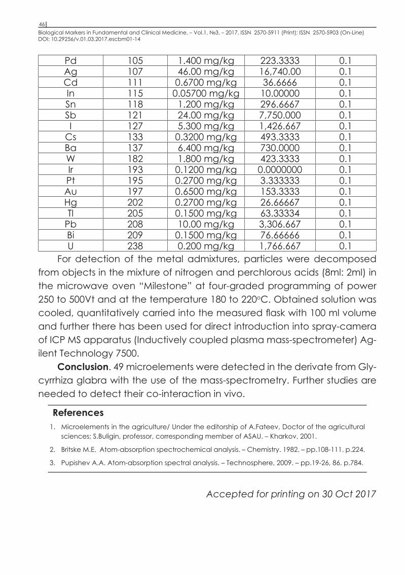
Biological Markers in Fundamental and Clinical Medicine. – Vol.1, №3. – 2017. ISSN 2570-5911 (Print); ISSN 2570-5903 (On-Line) DOI: 10.29256/v.01.03.2017.escbm01-14
46
Pd 105 1.400 mg/kg 223.3333 0.1Ag 107 46.00 mg/kg 16,740.00 0.1Cd 111 0.6700 mg/kg 36.6666 0.1In 115 0.05700 mg/kg 10.00000 0.1Sn 118 1.200 mg/kg 296.6667 0.1Sb 121 24.00 mg/kg 7,750.000 0.1I 127 5.300 mg/kg 1,426.667 0.1
Cs 133 0.3200 mg/kg 493.3333 0.1Ba 137 6.400 mg/kg 730.0000 0.1W 182 1.800 mg/kg 423.3333 0.1Ir 193 0.1200 mg/kg 0.0000000 0.1Pt 195 0.2700 mg/kg 3.333333 0.1Au 197 0.6500 mg/kg 153.3333 0.1Hg 202 0.2700 mg/kg 26.66667 0.1Tl 205 0.1500 mg/kg 63.33334 0.1
Pb 208 10.00 mg/kg 3,306.667 0.1Bi 209 0.1500 mg/kg 76.66666 0.1U 238 0.200 mg/kg 1,766.667 0.1
For detection of the metal admixtures, particles were decomposed from objects in the mixture of nitrogen and perchlorous acids (8ml: 2ml) in the microwave oven “Milestone” at four-graded programming of power 250 to 500Vt and at the temperature 180 to 220oC. Obtained solution was cooled, quantitatively carried into the measured flask with 100 ml volume and further there has been used for direct introduction into spray-camera of ICP MS apparatus (Inductively coupled plasma mass-spectrometer) Ag-ilent Technology 7500.
Conclusion. 49 microelements were detected in the derivate from Gly-cyrrhiza glabra with the use of the mass-spectrometry. Further studies are needed to detect their co-interaction in vivo.
References1. Microelements in the agriculture/ Under the editorship of A.Fateev, Doctor of the agricultural
sciences; S.Buligin, professor, corresponding member of ASAU. – Kharkov, 2001.
2. Britske M.E. Atom-absorption spectrochemical analysis. – Chemistry. 1982. – pp.108-111. p.224.
3. Pupishev A.A. Atom-absorption spectral analysis. – Technosphere, 2009. – pp.19-26, 86. p.784.
Accepted for printing on 30 Oct 2017

Publishing center ESCBM, Příkop 838/6, Zábrdovice, 60200 Brno, Česká republika.
