biological nanocrystallography using fels – an overview€¦ · particularly difficult to...
TRANSCRIPT

Biological nanocrystallographyusing FELs – an overview
Thomas Barends
MPI Medical Research HeidelbergMax Planck Advanced Study Group at CFEL, Hamburg

Cells are the basis of life
F1/F0 ATPsynthase
ATP synthesis
Transcribed byRNA polymerase
mRNA mRNA
Translated and synthesizedin protein: ribosome
DNA: storage of genetic info
Viral infection

Cells are the basis of life
F1/F0 ATPsynthase
ATP synthesis
Transcribed byRNA polymerase
mRNA mRNA
Translated and synthesizedin protein: ribosome
DNA: storage of genetic info
Viral infection
WE WANT TO KNOW:
-WHO IS DOING WHAT ?
-WHEN DO THEY DO IT ?
-WHERE DO THEY DO IT ?
-HOW DO THEY DO IT ?
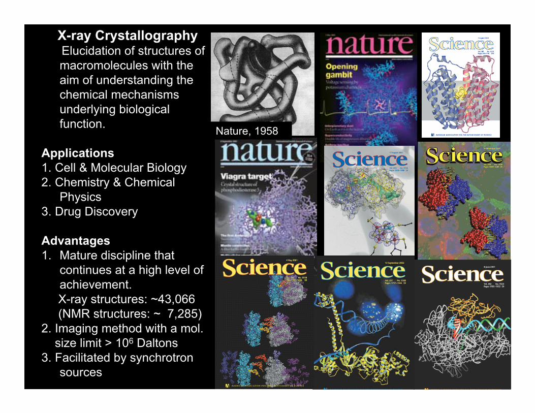
X-ray CrystallographyElucidation of structures of macromolecules with the aim of understanding the chemical mechanisms underlying biological function.
Applications1. Cell & Molecular Biology2. Chemistry & Chemical
Physics3. Drug Discovery
Advantages1. Mature discipline that
continues at a high level of achievement.X-ray structures: ~43,066 (NMR structures: ~ 7,285)
2. Imaging method with a mol. size limit > 106 Daltons
3. Facilitated by synchrotron sources
Nature, 1958
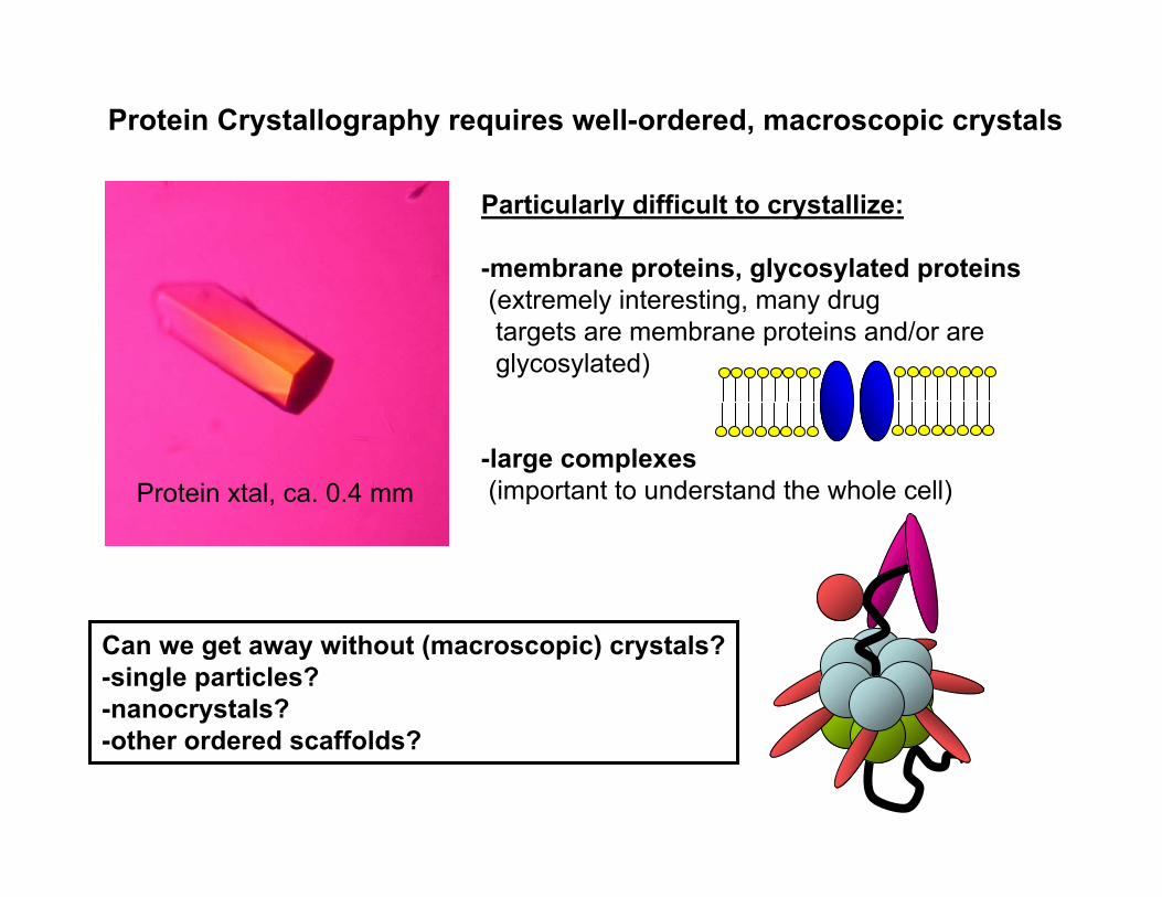
Protein Crystallography requires well-ordered, macroscopic crystals
Can we get away without (macroscopic) crystals?-single particles?-nanocrystals?-other ordered scaffolds?
Protein xtal, ca. 0.4 mm
Particularly difficult to crystallize:
-membrane proteins, glycosylated proteins(extremely interesting, many drugtargets are membrane proteins and/or areglycosylated)
-large complexes(important to understand the whole cell)

Emission ofAuger electron~ 30 eV
Fluorescence(>Z=30)
• 10% Rayleigh scattering•vvv
• 10% Compton effect•vvv
• 80% Photoelectric effect
X-raye-
At 12 keV (λ=1.03 Å)
e-
Nine out of ten X-ray photons cause radiation damage – can we reduce radiation
damage?sample

diffraction pattern
- 1012-13 photons - 10 keV- 10 fs pulse
particle injection
100 nm focus
~1021 W/cm2
- biological nano-crystals- virus structures, virus genomes- non-crystallisable proteins: 60 %
Measure data before Measure data before destuctiondestuction??
Coherent Diffractive Imaging
Calculations in vacuum, Neutze et al., Nature 2000

peak brilliance FEL
Synchrotron
Free Electron LasersFree Electron Lasers
• FLASH: 2005• Fermi: 2009• LCLS: 2009• SCSS: 2011…• XFEL: 2016• KVI, Shanghai, …
9 orders of magnitude!
•• 10101212--13 13 photons: ~ 10 photons: ~ 10 fsfs pulsespulses•• repetition rate: now 120 Hzrepetition rate: now 120 Hz•• photon energies: 10 photon energies: 10 keVkeV•• transversally: fully coherent transversally: fully coherent

FLASH – Free Electron Laser Hamburg
LCLS – Linear Coherent Light Source Stanford
Lasing at 1.5 keV observed in May 2009
0.2 keV, 6 nm wavelength

Some biological model systems for nanodiffraction/diffractive imaging:
lysozyme nanoxtals Photosystem I nanocrystals (Heidelberg) (Petra Fromme group,
U. of Arizona)
800 nm(200 unit cells)
Image: Prof. Didier Raoult, La Timone, Marseille, France
Various virus particles (Heidelberg) Mimivirus (Janos Hajdu group)
Magnetotactic bacteria(Heidelberg)

Gaffney & Chapman, Science 2007
Diffractive imagingof particles
Data evaluation3D reconstruction

Gaffney & Chapman, Science 2007
Diffractive imagingof particles
Data evaluation3D reconstruction
1.BRAGG PEAKS MAKE HIT FINDING EASY2.NOTHING BEATS A CRYSTAL IN TERMS OF SIGNAL/NOISE3.DISCRETE, LATTICE CHARACTER OF REC. SPACE SOLVES ORIENTATION PROBLEM
THE HUGE ADVANTAGE OF CRYSTALS:BRAGG PEAKS!

CFEL-ASG Multi PurposeCFEL-ASG Multi Purpose
FEL FEL
ion imaging
ion ion imaging imaging
electron imaging electron electron imaging imaging
scattered andfluorescence
photon imaging
scattered andscattered andfluorescence fluorescence
photon imaging photon imaging
90 eV – 25 keV90 eV 90 eV –– 25 25 keVkeV
1 Mega Pixels 1 Mega Pixels 1 Mega Pixels
λ
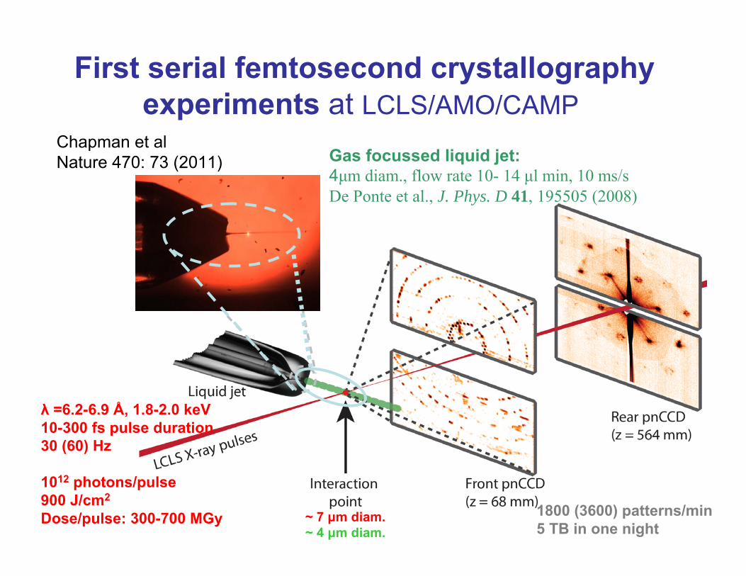
First serial femtosecond crystallographyexperiments at LCLS/AMO/CAMP
Gas focussed liquid jet:4μm diam., flow rate 10- 14 μl min, 10 ms/sDe Ponte et al., J. Phys. D 41, 195505 (2008)
λ =6.2-6.9 Å, 1.8-2.0 keV10-300 fs pulse duration30 (60) Hz
1012 photons/pulse900 J/cm2
Dose/pulse: 300-700 MGy ~ 7 μm diam.~ 4 μm diam.
1800 (3600) patterns/min5 TB in one night
Chapman et alNature 470: 73 (2011)

Indexing and integrating reflections: conventional methods
Rotation method-rotate xtal over finite range-calculate orientation matrixfrom observed spot positionsCan fully integrate whole reflections!
Laue method-use polychromatic radiation-calculate orientation matrix from observed spot positionsCan fully integrate whole reflections!
Powder method:-Rotate powder of many xtals-assign hkl from scattering angleof reflections (if unique!)Fully integrates whole reflections!

Serial femtosecond crystallography
- Numerous shots of different crystals with possibly different sizes
- No a priori control over orientation- Crystals effectively stand still during a 300 fs pulse-Only part of reflection intersects Ewald sphere (“partials”, no “fullies”)
- Fringes rather than neat spots
It is possible to do a Monte Carlo integration over multiple indexed femtosecond images and obtain a dataset of fully integrated reflections
Kirian et al (2010), Optics Express,18, 5713-5723:
6x6x6 200x200x200unit cells unit cells
(Simulation software by Wolfgang Kabsch)

N -2 fringes,
Intensity:
N unit cells
“Extra” features allow sizing and phasing of nanocrystals
Chapman et alNature 470: 73 (2011)
b
bNI
2
2
sin
sin

Chapman et alNature 470: 73 (2011)

So what’s the bad news?• Hit rates are low, + only a fraction of hits
indexable• the method needs:
– 1-to-several ml of highly concentrated (yoghurt-like!) suspension of microcrystals (hit rates are low, for high resolution many 10,000s images needed)
How do you make that much protein?(usual yields are in the 0.1-1mg range for membrane proteins..., very difficult to produce, not stable!)
Can you make nanocrystals of it?(how do you know you have them?how do you know they are any good?testing them can only be done at the FEL...)
(can you inject them?PEG/salts may clog the nozzle.....)
.......? DROPLET-ON-DEMANDTO SAVE SAMPLE ?HIGH PULSE RATE?

What if you want to do “normal”xtallography, e.g. for time-resolved studies?
-serial nanocrystal data processing is based on averaging large numbers of exposures
-Preliminary experience with larger xtals (fewer exposures, “regular”xtallography) shows:
PULSE-TO-PULSE REPRODUCIBILITY IS EVERYTHING!!!(Intensity, coherence, spectrum –seeding?)
CO-myoglobin xtalLCLS X-ray pump-probeDec. 2010

Conclusions for nanocrystallography:
-Sample consumption is high
-Sample preparation is non-trivial
-Good data can be collected from nano/microcrystals using femtosecond pulses
-FEL time structure may allow time-resolved studies MOLECULAR MOVIES ?
membrane protein diffractionon an FEL source

Nils Kimmel, Georg Weidenspointner, Daniel Pietschner, Günter Hauser, Sven Herrmann, Gerhard Schaller, Florian Schopper, Robert Andritschke, LotharStrüder
MPI Semiconductor Lab
Robert Hartmann, Peter Holl, Christian Reich, Heike SoltauPNSensor GmbH, Munich
Kai-Uwe Kühnel, Claus-Dieter Schröter, Joachim UllrichMPI for Nuclear Physics, Heidelberg
Sascha W. Epp, Daniel Rolles, Artem Rudenko, Lutz Foucar, Benedikt Rudek, Benjamin Erk, Carlo Schmidt, André Hömke, Faton Krasniqi
Max Planck Advanced Study Group, Center for Free Electron Laser Science (CFEL)
Filipe R.N.C. Maia, Nicusor Timneanu, M. Marvin Seibert, Jakob Andreasson, Olof Jönsson, Martin Svenda, Janos Hajdu
Molecular Biophysics, Uppsala University
Richard A. Kirian, Uwe Weierstall, R. Bruce Doak, John Spence
Depart. of Physics, Arizona State University, Petra Fromme, Raimund Fromme, Mark S. Hunter
Dept. of Chem & Biochem. Arizona State Univ.
Karol NassUniversity of Hamburg
Anton Barty, Thomas A. White, Andrew Aquila, Daniel P. DePonte, Andrew Martin, Nicola Coppola, Joachim Schulz, Mengning Liang, Lars Gumprecht, Carl Caleman, Stephan Stern, Henry N. Chapman
Center for Free-Electron Laser Science, DESY
Lukas Lomb, Stephan Kassemeyer, Robert L. Shoeman, Daniel Rolles, Anton Meinhart, Maike Gebhardt, Jan Steinbrenner, Mario Bott, Ulrike Mersdorf, Ilme Schlichting
MPI for Medical Research, Heidelberg
James Holton, Stefano MarchesiniAdvanced Light Source
Lawrence Livermore National Laboratory
PULSE Institute and SLAC
Christoph Bostedt, John D. Bozek, Garth J. Williams, Sébastien Boutet, JacekKrzywinski, Marc Messerschmidt, Marco Cammarato, David Fritz
LCLS
Miriam Barthelmess, Saša Bajt, Helmut Hirsemann, Guillaume Potdevin, Björn Nilsson, C. Wunderer, Heinz Graafsma
Photon Science, DESY,
Michael J. Bogan, Christina Y. Hampton, Raymond Sierra, Dmitri Starodub
Stefan P. Hau-Riege, Matthias Frank
Max Planck Group for Struct. Dynamics (CFEL) Arash Zarrine Afsar, Christina, Mueller, ,Dwayne Miller
NIH Friedrich Schotte, Hyun Sun Cho, Philip Anfinrud