biology diss - georgia state university scholarworks
TRANSCRIPT

Georgia State University Georgia State University
ScholarWorks @ Georgia State University ScholarWorks @ Georgia State University
Biology Dissertations Department of Biology
Spring 5-7-2011
Control of Secondary Granule Release in Neutrophils by Ral Control of Secondary Granule Release in Neutrophils by Ral
GTPase GTPase
Xiaojing Chen
Follow this and additional works at: https://scholarworks.gsu.edu/biology_diss
Recommended Citation Recommended Citation Chen, Xiaojing, "Control of Secondary Granule Release in Neutrophils by Ral GTPase." Dissertation, Georgia State University, 2011. https://scholarworks.gsu.edu/biology_diss/96
This Dissertation is brought to you for free and open access by the Department of Biology at ScholarWorks @ Georgia State University. It has been accepted for inclusion in Biology Dissertations by an authorized administrator of ScholarWorks @ Georgia State University. For more information, please contact [email protected].
CONTROL OF SECONDARY GRANULE RELEASE IN NEUTROPHILS BY RAL GTPASE
by
XIAOJING CHEN
Under the Direction of Yuan Liu, MD., Ph.D.
ABSTRACT
Neutrophil (PMN) inflammatory functions, including cell adhesion, diapedesis, and phagocyto-
sis, are dependent on the mobilization and release of various intracellular granules/vesicles. In
this study, I found that treating PMN with damnacanthal, a Ras family GTPase inhibitor, resulted
in a specific release of secondary granules, but not primary or tertiary granules, and caused dy-
sregulation of PMN chemotactic transmigration and cell surface protein interactions. Analysis of
the activities of Ras members identified Ral GTPase as a key regulator during PMN activation
and degranulation. In particular, Ral was active in freshly isolated PMN, while chemoattractant
stimulation induced a quick deactivation of Ral that correlated with PMN degranulation. Over-
expression of a constitutively active Ral (Ral23V) in PMN inhibited chemoattractant-induced
secondary granule release. By subcellular fractionation, I found that Ral, which was associated
with the plasma membrane under the resting condition, was redistributed to secondary granules
after chemoattractant stimulation. Blockage of cell endocytosis appeared to inhibit Ral transloca-
tion intracellularly. In conclusion, these results demonstrate that Ral is a critical regulator in
PMN that specifically controls secondary granule release during PMN response to chemoattrac-
tant stimulation.
INDEX WORDS: Neutrophil (PMN), Ral, Degranulation, Secondary granules, Transmigration

CONTROL OF SECONDARY GRANULE RELEASE IN NEUTROPHILS BY RAL GTPASE
by
XIAOJING CHEN
A Dissertation Submitted in Partial Fulfillment of the Requirements for the Degree of
Doctor of Philosophy
in the College of Arts and Sciences
Georgia State University
2011

Copyright by
Xiaojing Chen
2011

CONTROL OF SECONDARY GRANULE RELEASE IN NEUTROPHILS BY RAL GTPASE
by
XIAOJING CHEN
Committee Chair: Yuan Liu, MD., Ph.D.
Committee: Deborah Baro, Ph.D.
Zhi-Ren Liu, Ph.D.
Electronic Version Approved:
Office of Graduate Studies
College of Arts and Sciences
Georgia State University
May 2011
v
DEADICATUION AND ACKNOWLEDGEMENTS
This dissertation is dedicated to all the people who have helped me and guided me
over the obstacles during my Ph.D. training at Georgia State.
I feel great honor to have Dr. Deborah Baro, Dr. Yuan Liu, and Dr. Zhi-Ren Liu in my commit-
tee. They have been constantly helping me in my scientific development.
In addition, I am truly thankful for the financial support provided to me by the Molecular Basis
for Disease program at Georgia State.
Dr. Yuan Liu has truly been the mentor for my success in all aspects. I am really grateful to be
her student and have the chance to be guided by her along my graduate study. Dr. Yuan Liu has
successfully supported me to finish dissertation research at Georgia State. Her influence will
guide my future life all the time.

vi
TABLE OF CONTENTS
DEADICATION AND ACKNOWLEDGEMENTS……………………………………. v
LIST OF ABBREVIATIONS…………………………………………………………...vii
LIST OF TABLES .............................................................................................................. x
LIST OF FIGURES ........................................................................................................... xi
CHAPTER I: GENERATL INTRODUCTION…………...…...………………..……….1
CHAPTER II: MATERIAL AND METHODS…………………………………………45
CHAPTER III: CONTROL OF SECONDARY GRANULE RELEASE IN
NEUTROPHILS BY RAL GTPASE…………………………………..62
CHAPTER IV: DISCUSSION…………………………………………………………105
CHAPTER V: APPENDICES……………………………………………...………….119
REFERENCES…………………………………............................................................130
JBC COPYRIGHT PERMISSION POLICY
vii
LIST OF ABBREVIATIONS
ABTS 2, 2’-azino-bis 3-ethylbenzthiazoline-6-sulfonic acid
CB cytochalasin B
CPZ chlorpromazine hydrochloride
DAG diacylglycerol
EGF epidermal growth factor
fMLF formyl-methionyl-leucyl-phenylalanine
GAP GTPase activating protein
GEF guanine nucleotide exchange factor
GPCR G-protein-coupled receptor
HBSS Hank's balanced salt buffer
HBSS (-) Hank's balanced salt buffer devoid of Ca2+
and Mg2+
HEK293 human embryonic kidney 293 cells
HUVEC human umbilical vein endothelial cells
ICAM-1 intercellular adhesion molecule 1
ITIM immunoreceptor tyrosine-based inhibition motif
IP3 inositol trisphosphate
IPTG isopropyl-beta-D-thiogalactopyranoside
JAM-A junctional adhesion molecule-A
LTB4 leukotriene B4
LPA lysophosphatidic acid
MAPK mitogen-activated protein kinase
MHC major histocompatibility complex

viii
MMPs matrix metalloproteinases
MPO myeloperoxidase
PA phosphatidic acid
PAF platelet-activating factor
PBMC peripheral blood mononuclear cell
PC phosphatidylcholine
PMN polymorphonuclear leukocyte
POA phenylarsine oxide
PI3K phosphatidylinositiol-3 kinase isoform
PIP2 PI 4, 5-bisphosphate
PIP3 PI 3, 4, 5-bisphosphate
PKC protein kinase C
PLCβ phosphatidylinositol-specific phospholipase C β
PLD phospholipase D
PMA phorbol myristate acetate
Raf-1 RBD Ras binding domain of Raf-1
RalBP1 Ral binding protein 1
RalGAP Ral GTPase activating protein
RalGDS Ral guanine nucleotide dissociation stimulator
RalGDS-RBD Ral GDS-Rap Binding Domain
RBC red blood cell
RBD GTP-Ral-binding domain
Rgl ralGDS-like

ix
Rgl3 Ral GEF-like 3
Rlf RalGDS-like factor
SNAREs soluble NSF (N-ethylmaleimide-sensitive factor)
TBK1 Tank binding kinase 1
VCAM-1 vascular cell-adhesion molecule 1
WGA wheat germ agglutinin

x
LIST OF TABLES
1. Damnacanthal treatment induces an upregulation of CD11b/CD18, CD47 Page 67
and SIRPα on PMN surface

xi
LIST OF FIGURES
Figure Page
1-1 PMN activation and category of chemoattractants 5
1-2 fMLF-mediated activation pathways in PMN 9
1-3 PMN transmigration to the target site 13
1-4 PMN transendothelial migration 14
1-5 Putative structure of integrin CD11b/CD18 15
1-6 PMN transepithelial migration 16
1-7 Putative structure of SIRPα (left) and CD47 (right) 17
1-8 PMN play a pivotal role in inflammation 21
1-9 PMN granules types (A) and degranulation (B) in response to stimulation (e.g. fMLF)
24
1-10 Interchange of GTP-bound form with GDP-bound form of small GTPase 28
1-11 Ral domain structure 41
1-12 Sequence alignment of RalA and RalB 42
1-13 Control of secondary granule release in neutrophils by Ral 44
2-1 GTPase pull down assay 52
2-2 HIV-Tat-Ral expression and purification system 57
3-1 Damnacanthal treatment induces specific release of PMN secondary granules 68
3-2 Damnacanthal inhibits chemoattractant-triggered PMN function 70
3-3 Damnacanthal inhibits chemoattractant-triggered PMN function 71
3-4 Damnacanthal inhibits chemoattractant-triggered PMN function 72
3-5 Lck is not expressed in neutrophils (PMN) 75
xii
3-6 Ral deactivation correlates secondary granule release in PMN 78
3-7 Ral deactivation correlates secondary granule release in PMN 79
3-8 Detecting activity changes of Ral in PMN isolated directly from “buffy coat” 80
3-9 Ral deactivation correlates secondary granule release in PMN 81
3-10 Ral deactivation correlates secondary granule release in PMN 82
3-11 Generation of Tat-tagged wild-type Ral (WT), constitutively active Ral (Ral23V), and
dominant negative Ral (28N) protein 87
3-12 Transduction of Tat-tagged Ral proteins including wild-type Ral (WT), constitutively
active Ral (Ral23V), and dominant negative Ral (28N) to PMN 88
3-13 Tat-tagged constitutively active Ral sustained endogenous Ral activity in PMN 89
3-14 Constitutively active Ral (Ral23V) inhibits chemoattractant-induced secondary granule
release in PMN 90
3-15 Specificity of Tat-tagged Ral proteins in PMN granule release 91
3-16 Constitutively active Ral (Ral23V) inhibits chemoattractant-induced secondary granule
release in PMN isolated using lympholyte-poly 92
3-17 Constitutively active Ral (Ral23V) inhibits chemoattractant-induced secondary granule
release in PMN isolated directly from “buffy coat” 93
3-18 Constitutively active Ral (Ral23V) inhibits chemoattractant-induced secondary granule
release in PMN isolated from “buffy coat” 94
3-19 Detection of RalA and RalB in human leukocytes 95
3-20 Constitutively active RalB (Ral23V) inhibits chemoattractant-induced secondary
granule release 96
3-21 Localization of Ral in PMN 100
xiii
3-22 In unstimulated PMN, Ral is active in plasma membrane and inactive in secondary
granules 101
3-23 Localization of Ral in PMN 102
3-24 Ral translocation from the plasma membrane to secondary granules may follow fMLF-
induced endocytic process 103
3-25 Blockage of clathrin-dependent endocytosis inhibits fMLF-induced secondary granule
release 104
4-1 Expression of RalGDS in different cell types 112
4-2 A study of chemoattractant stimulation-induced increase of intracellular calcium,
seondary granule release and Ral activity changes in PMN 113
4-3 A study of chemoattractant stimulation-induced increase of intracellular calcium,
secondary granule release and Ral activity changes in PMN 114
4-4 Mg2+
inhibits Ral activity in PMN 115
4-5 Effects of damnacanthal alone or in a combination with fMLF on PMN granule release
116
4-6 Subcellular fractionations studying the effects of damnacanthal and endocytosis
inhibitors on Ral distribution in PMN 117
4-7 Subcellular fractionations studying the effects of damnacanthal and endocytosis
inhibitors on Ral distribution in PMN 118
5-1 RAGE was amplified from human intestinal epithelial cells by RT-PCR 119
5-2 RAGE was detected from epithelial cell lines by western blot 120
5-3 RAGE was detected in intestinal epithelial cells by immunofluorescence staining 121
5-4 Generation of GST-exRAGE fusion protein 122

xiv
5-5 RAGE antibody titer test by ELISA 123
5-6 RAGE antibody titer test by western blot 124
5-7 RAGE detection from human PMN 125
5-8 Generation of GST-JAML fusion protein 126
5-9 JAML antibody titer test by ELISA and western blot 127
5-10 JAML was expressed on PMN and monocytes 128
5-11 Ala57, Gln67, and Val57 were key amino acid residues mediating binding interaction
of SIRPα to CD47 129

1
CHAPTER I
GENERAL INTRODUCTION
1. Neutrophils (PMN)
Neutrophils, also termed PMN, are polymorphonuclear leukocytes characterized by their multi-
lobed nuclei [1]. PMN, which are generated from bone marrow, are abundant cells in circulation
and account for 40-75% of all leukocytes [2]. Up to 1-2×10¹¹
PMN are steadily generated per
day in a healthy adult [3]. Constant numbers of PMN in the circulation are very crucial for main-
taining host homeostasis [4-6]. Granulocyte colony stimulating factor (G-CSF) has been shown
to positively regulate PMN production in bone marrow, but G-CSF is not completely required
for PMN generation [7-8]. The release of PMN from bone marrow is largely dependent on
CXCR4 [9]. It is likely that CXCR4 functions a gatekeeper for blocking PMN migrating out of
bone marrow [10-15]. In CXCR4-deficient chimera mice, myeloid precursors are significantly
decreased and peripheral granulocytic cells are increased by about three-fold compared with wild
type-litter mates [16-17]. Blocking CXCR4 function with the inhibitory antibody/antagonist,
AMD3100, elevates PMN counts in circulation [10, 14-15]. Additionally, AMD3100 dose-
dependently increases the release of CD34+ hematopoietic progenitor cells into circulation as
demonstrated in healthy human volunteers [10, 14]. Presumably, the negative effect of CXCR4 is
through the signaling cascade of CXCL12 (stromal derived factor-1, SDF-1)/ CXCR4 [18]. Un-
der the influence of inflammation, however, PMN production is increased. In circulation, non-
activated PMN have 8-16 hours average half-lives [19]. After stimulation, active PMN are re-
cruited to the target tissue where they survive for only 1-4 days [20-21]. PMN are disposed from
circulation mainly at the spleen, liver, bone marrow and the site of inflammation [22-24]. Ma-
crophages play a pivotal role in the clearance of both circulating PMN under nonpathologic con-
2
ditions and inflammatory PMN in the target tissue [4]. In addition to macrophage phagocytosis,
PMN apoptosis [21] is also a key factor in preventing excessive PMN infiltration-mediated in-
flammation, tissue damage and organ dysfunction, thus maintaining cellular homeostasis.
PMN are the first cells recruited to the site of an infection [25]. Recruitment of circulating PMN
from blood vessels to the site of inflammation is very important for eliminating invading micro-
organisms, recovering tissue damage and maintaining homeostasis [26]. Through releasing reac-
tive oxygen species, antimicrobial proteins, proteolytic enzymes and producing cytokines, as
well as performing phagocytosis, PMN eliminate microorganisms, and thus are essential for in-
nate immunity [27]. Most notably, PMN infiltration is the hallmark of the early stages of the in-
flammatory response [28]. On the contrary, dysregulated PMN functions are also closely linked
to tissue damage and organ dysfunction.
I. Chemoattractants and their receptors in PMN transmigration
Leukocyte chemoattractants include interleukin (IL)-4, IL-8, platelet-activating factor (PAF), and
formyl-Methionyl-Leucyl-Phenylalanine (f-Met-Leu-Phe, fMLF), leukotriene B4 (LTB4), C3a
and C5a [29]. Leukocyte chemoattractants can be further grouped into several main categories
such as chemokines [30], bacterial products, immunoproducts, phospholipids, etc. They are re-
leased by numerous types of cells. For example, activated endothelial cells secrete IL-4, LTB4,
and PAF, while activated stromal cells, including macrophages, epithelial cells, etc. secrete
LTB4 and PAF. In addition, bacteria or dying cells release fMLF [31]. The combination of
chemoattractants and their gradient drives PMN chemotactic migration towards an inflammatory
site [31] (Figure 1-1).
3
According to derivation, chemoattractants can be further divided into “regulatory cell-derived
chemoattractants” and “end target-derived chemoattractants”. Regulatory cell-derived chemoat-
tractants include IL-8 and LTB4, etc. End target-derived chemoattractants are mainly composed
of fMLF, derived from bacteria or the mitochondria of dying cells, and complement C3a and C5a
[31]. Migrating out of the blood stream, PMN encounter a combination of chemoattractants re-
leased from the endothelium and infected tissues. Through integration of those series of che-
moattractants, PMN effectively reach the target site. It has been proposed that chemoattractant
receptor cross-desensitization is one of the mechanisms regulating PMN chemotaxis [32-34].
Moreover, the combinational and sequential signals of distinct chemoattractants navigate PMN
chemotaxis to a great extent [35-37]. When placed in a chemoattractant gradient, PMN vigorous-
ly migrate towards the gradient in spite of the original migration pathways. Furthermore, PMN
are able to integrate signals from different chemoattractants, and thus migrate toward the target.
End target-derived chemoattractants, such as fMLF and C5a, however, are substantially domi-
nant over cell-derived chemoattractants, such as IL-8 and LTB4 [35-37].
Pre-exposure with fMLF or C5a causes PMN to lose the response to IL-4 and LTB4. This phe-
nomenon might be a reasonable explanation for the fact that PMN respond to eliminate existing
invading bacteria first compared to tissue damage. It has also been demonstrated that PMN fail to
transmigrate towards IL-4 or IL-8 when cells are pre-treated with fMLF or C5a in the presence
or absence of an endothelial monolayer. This desensitization of PMN is likely associated with
the polymerization of actin filaments [36], because the end target-derived chemoattractants trig-
ger sustained and prolonged actin polymerization. Other studies have shown that PMN are able

4
to migrate down a concentration gradient towards another chemoattractant. They can also mi-
grate beyond the inhibitory maximum concentration toward another different chemoattractant
[35]. In addition to the signals received by PMN upon stimulation with different chemoattrac-
tants, chemoattractant receptor cross-desensitization also plays a key role in PMN chemotaxis.
Cpd43, an agonist of formyl peptide receptors 1 and 2, not only inhibits the ability of PMN to
further respond to IL-8, C5a and LTB4, but also results in the loss of the expression of CXCR1,
CXCR2, C5a and LTB4 on cell surface [33]. Treatment of PMN with fMLF or C5a reduces the
binding of IL-8Rβ to its antibody [34]. These miscellaneous factors enable PMN efficiently and
precisely mobilize to their targets through a complex chemotactic field.

5
Figure 1-1. PMN activation and category of chemoattractants
Chemoattractants
Bacterial products: fMLF
Chemokines: IL-8, IL-4
Immunoproducts: C3a & C5a
Phospholipids: LTB4
Bind to cell surface GPCR
Activation
Degranulation
Migration

6
II. Chemoattractant, fMLF-mediated signaling transduction cascade in PMN
Various signaling pathways are implicated in PMN migration. One of the well characterized
pathways is fMLF-mediated PMN stimulation [38] (Figure 1-2). Upon fMLF binding, its trime-
tric G-protein-coupled receptor (GPCR), composed of α, β, and γ subunits [39], is activated and
undergoes a conformational change. Ligand binding induces the switch of GDP-bound inactive
form to the GTP-bound active form, resulting in the low affinity of Gα towards Gβγ. The dissocia-
tion of Gα with Gβγ activates its own individual downstream effectors, including phosphatidylino-
sitol-specific phospholipase-Cβ (PLCβ), adenylyl cyclase and phosphatidylinositiol-3 kinase iso-
forms (PI3K).
Activated PLCβ hydrolyzes PI 4, 5-bisphosphate (PIP2) into inositol trisphosphate (IP3) and di-
acylglycerol (DAG). IP3 then diffuses into the cytosol, binds to the IP3 receptor and results in
Ca2+
release from ER storage. Released Ca2+
involves in activation of PKC and triggers extracel-
lular calcium influx, presumably through the interaction of the IP3 receptor with plasma mem-
brane channels [40-42]. Ca2+
, a pivotal second messenger, is crucial for the reorganization of ac-
tin filaments. Ca2+
facilitates the binding of myosin, the motor protein, to actin through exposing
the myosin binding site on actin, and thus is involved in plasma membrane protrusion, lamella-
podia formation, and PMN migration [43]. DAG activates protein kinase C (PKC), thus resulting
in a series of protein phosphorylation. Serving as a second messenger, PKC further activates a
series of downstream signaling molecules involved in PMN adhesion [44-45]. In PMN, PKC al-
so associates with the respiratory burst [46]. There are five isoforms of PKC expressed in PMN,
including PKC-α, PKC-β1, PKC-βII, PKC-δ, and PKC-δ [47]. It has been shown that, shortly
after fMLF stimulation, PKC-α, PKC-β1, PKC-βII and PKC-δ activate NADPH oxidase, leading
7
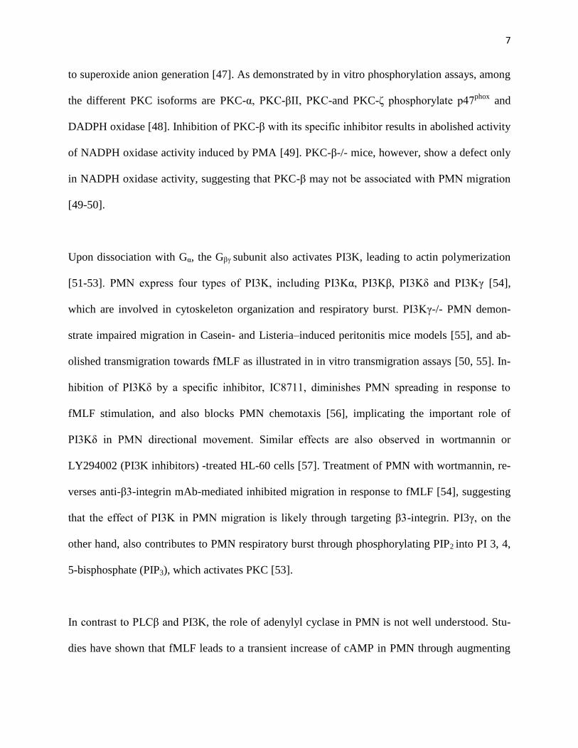
to superoxide anion generation [47]. As demonstrated by in vitro phosphorylation assays, among
the different PKC isoforms are PKC-α, PKC-βII, PKC-and PKC-δ phosphorylate p47phox
and
DADPH oxidase [48]. Inhibition of PKC-β with its specific inhibitor results in abolished activity
of NADPH oxidase activity induced by PMA [49]. PKC-β-/- mice, however, show a defect only
in NADPH oxidase activity, suggesting that PKC-β may not be associated with PMN migration
[49-50].
Upon dissociation with Gα, the Gβγ subunit also activates PI3K, leading to actin polymerization
[51-53]. PMN express four types of PI3K, including PI3Kα, PI3Kβ, PI3Kδ and PI3Kγ [54],
which are involved in cytoskeleton organization and respiratory burst. PI3Kγ-/- PMN demon-
strate impaired migration in Casein- and Listeria–induced peritonitis mice models [55], and ab-
olished transmigration towards fMLF as illustrated in in vitro transmigration assays [50, 55]. In-
hibition of PI3Kδ by a specific inhibitor, IC8711, diminishes PMN spreading in response to
fMLF stimulation, and also blocks PMN chemotaxis [56], implicating the important role of
PI3Kδ in PMN directional movement. Similar effects are also observed in wortmannin or
LY294002 (PI3K inhibitors) -treated HL-60 cells [57]. Treatment of PMN with wortmannin, re-
verses anti-β3-integrin mAb-mediated inhibited migration in response to fMLF [54], suggesting
that the effect of PI3K in PMN migration is likely through targeting β3-integrin. PI3γ, on the
other hand, also contributes to PMN respiratory burst through phosphorylating PIP2 into PI 3, 4,
5-bisphosphate (PIP3), which activates PKC [53].
In contrast to PLCβ and PI3K, the role of adenylyl cyclase in PMN is not well understood. Stu-
dies have shown that fMLF leads to a transient increase of cAMP in PMN through augmenting
8
the adenylyl cyclase response [58]. Elevated cAMP has been shown to inhibit PMN chemotaxis
to fMLF [59]. However, limited studies exploring the role of cAMP in PMN utilizing inhibitors
of phosphodiesterase have been performed. Blocking the breakdown pathway of cAMP using
inhibitors of phosphodiesterase results in markedly impaired adhesion of PMN to the endothe-
lium and decreased cell surface expression of CD11b/CD18 [60], suggesting adenylyl cyclase is
involving in the PMN adhesion step.
Moreover, fMLF activates Rho small GTPase, including Rho, Rac and Cdc42, all of which regu-
late PMN migration through affecting actin filament formation [39, 52]. The MAPK cascade is
also activated after fMLF stimulation, thus leading to chemotaxis activation and superoxide pro-
duction [38-39]. Considering the complex signaling cascades in stimulated PMN, the fMLF-
mediated signaling pathway is very critical to PMN chemotactic migration and superoxide pro-
duction.

9
Figure 1-2. fMLF-mediated activation pathway in PMN.
α βγ
fMLF
αβ
γ
GTP
Degranulation
CD47
Integrin
lactoferrin
SIRPα
Cell polarization
PI-PLC
DAG PKC
PI3-kinase
Tyrosine kinase
/ phosphatase
Ca2+
IP3
Cytoskeleton reorganizationActin filament polymerization
Cell migration
Rho GTPase
Small GTPase
10
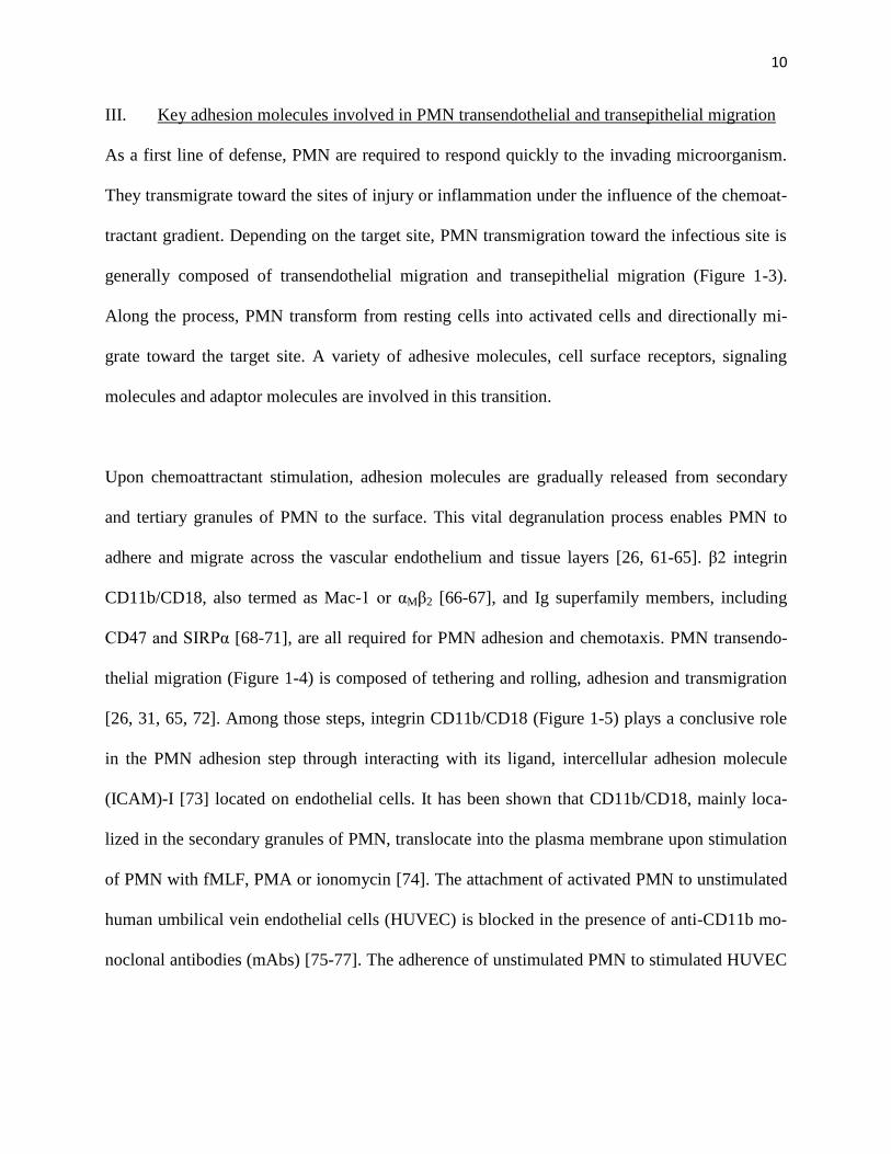
III. Key adhesion molecules involved in PMN transendothelial and transepithelial migration
As a first line of defense, PMN are required to respond quickly to the invading microorganism.
They transmigrate toward the sites of injury or inflammation under the influence of the chemoat-
tractant gradient. Depending on the target site, PMN transmigration toward the infectious site is
generally composed of transendothelial migration and transepithelial migration (Figure 1-3).
Along the process, PMN transform from resting cells into activated cells and directionally mi-
grate toward the target site. A variety of adhesive molecules, cell surface receptors, signaling
molecules and adaptor molecules are involved in this transition.
Upon chemoattractant stimulation, adhesion molecules are gradually released from secondary
and tertiary granules of PMN to the surface. This vital degranulation process enables PMN to
adhere and migrate across the vascular endothelium and tissue layers [26, 61-65]. β2 integrin
CD11b/CD18, also termed as Mac-1 or αMβ2 [66-67], and Ig superfamily members, including
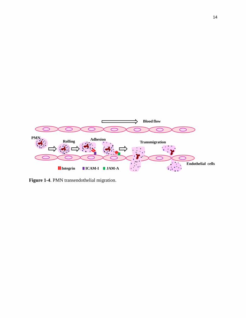
CD47 and SIRPα [68-71], are all required for PMN adhesion and chemotaxis. PMN transendo-
thelial migration (Figure 1-4) is composed of tethering and rolling, adhesion and transmigration
[26, 31, 65, 72]. Among those steps, integrin CD11b/CD18 (Figure 1-5) plays a conclusive role
in the PMN adhesion step through interacting with its ligand, intercellular adhesion molecule
(ICAM)-I [73] located on endothelial cells. It has been shown that CD11b/CD18, mainly loca-
lized in the secondary granules of PMN, translocate into the plasma membrane upon stimulation
of PMN with fMLF, PMA or ionomycin [74]. The attachment of activated PMN to unstimulated
human umbilical vein endothelial cells (HUVEC) is blocked in the presence of anti-CD11b mo-
noclonal antibodies (mAbs) [75-77]. The adherence of unstimulated PMN to stimulated HUVEC

11
is also inhibited by anti-CD11bAb, LM2/1 [78]. In CD11b/CD18 deficient mice, the attachment
of PMN to ICAM-1 is decreased compared with wild-type mice [79].
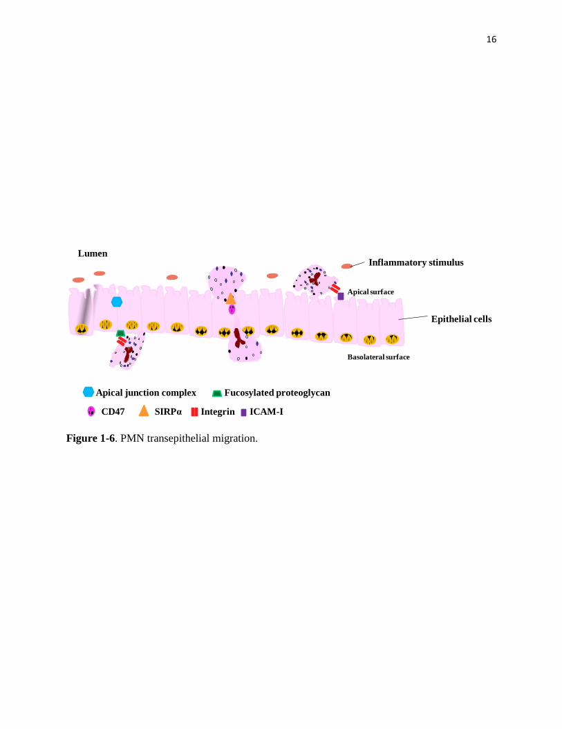
CD11b/CD18-mediated adhesion not only extensively contributes to transendothelial migration
but also transepithelial migration (Figure 1-6). PMN transepithelial migration consists of adhe-
sion, migration and post-migration [67, 80-81]. Adhesion of PMN to epithelial cells is mediated
by the interaction of CD11b/CD18 on PMN with its ligands, fucosylated proteoglycan(s), located
on epithelial cells. This interaction triggers the opening of apical junction complex mainly
through regulation of myosin phosphorylation. As a result, following phosphorylation, junction
complexes open and facilitate PMN transmigration. Upon reaching the lumen, CD11b/CD18 in-
teracts with ICAM-1 on the apical epithelial site and further assists PMN transepithelial migra-
tion toward the damaged site [65].
In addition to CD11b/CD18, CD47, SIRPα and JAM-A are also actively involved the process of
PMN transmigration. CD47 (Figure 1-7) is an important regulator in facilitating PMN transmi-
gration. In PMN, CD47 is kept in the secondary granules and is re-distributed to the cell surface
after chemoattractant stimulation, leading to the promotion of PMN migration [69]. It was first
discovered that PMN migration was inhibited in the presence of anti-CD47 antibodies [71, 82-
83]. Later research further demonstrated PMN transmigration across T84 monolayers and cell-
free collagen-coated filters is delayed when cell are treated with inhibitory anti-CD47 antibodies
C5D5 and B6H12 [69]. Indeed, functionally inhibitory anti-CD47 mAbs delay, but do not block,
the process of PMN transepithelial migration and migration across a collagen-coated transwell
toward fMLF [69]. When challenged with Escherichia coli, CD47-/- mice demonstrated a defect

12
in the early stages of PMN migration [84]. In addition to its effect in PMN, CD47 has been im-
plicated in the migration in other cell types. Likewise, CD47-/-epidermal dendritic cells (DCs)
demonstrate decreased migration toward draining lymph nodes in vivo [29]. Transduction of
neuroblastoma cells with CD47 results in neurite and filopodium formation [85]. Additionally,
through in vitro experimentation, it is uncovered that CD47 is important for the migratory activi-
ty of B cells [86].
SIRPα (Figure 1-7), a counter ligand for CD47 [87], also contributes greatly in PMN migration.
Similar to the distribution of CD47 in PMN, SIRPα translocates from secondary granules to the
cell surface after fMLF stimulation [68, 70]. PMN transepithelial migration is inhibited using
anti-SIRPα mAbs. The SIRPα-CD47 interaction leads to significantly decreased PMN migration
[68], suggesting that SIRPα delivers a negative signal to PMN migration through its immunore-
ceptor tyrosine-based inhibition motif (ITIM).
Junctional adhesion molecule-A (JAM-A) is also engaged in regulating PMN migration, particu-
larly in PMN transendothelial migration [81, 88]. JAM-A redistributes to the cell surface under
inflammatory condition [89], thus facilitating PMN migration. Treatment of HUVECs with a
blocking antibody against JAM-A [90] impairs PMN transmigration. The effect of JAM-A is
likely through the interaction of JAM-A with its ligand, β2 integrin LFA-1, on leukocytes [90].
13
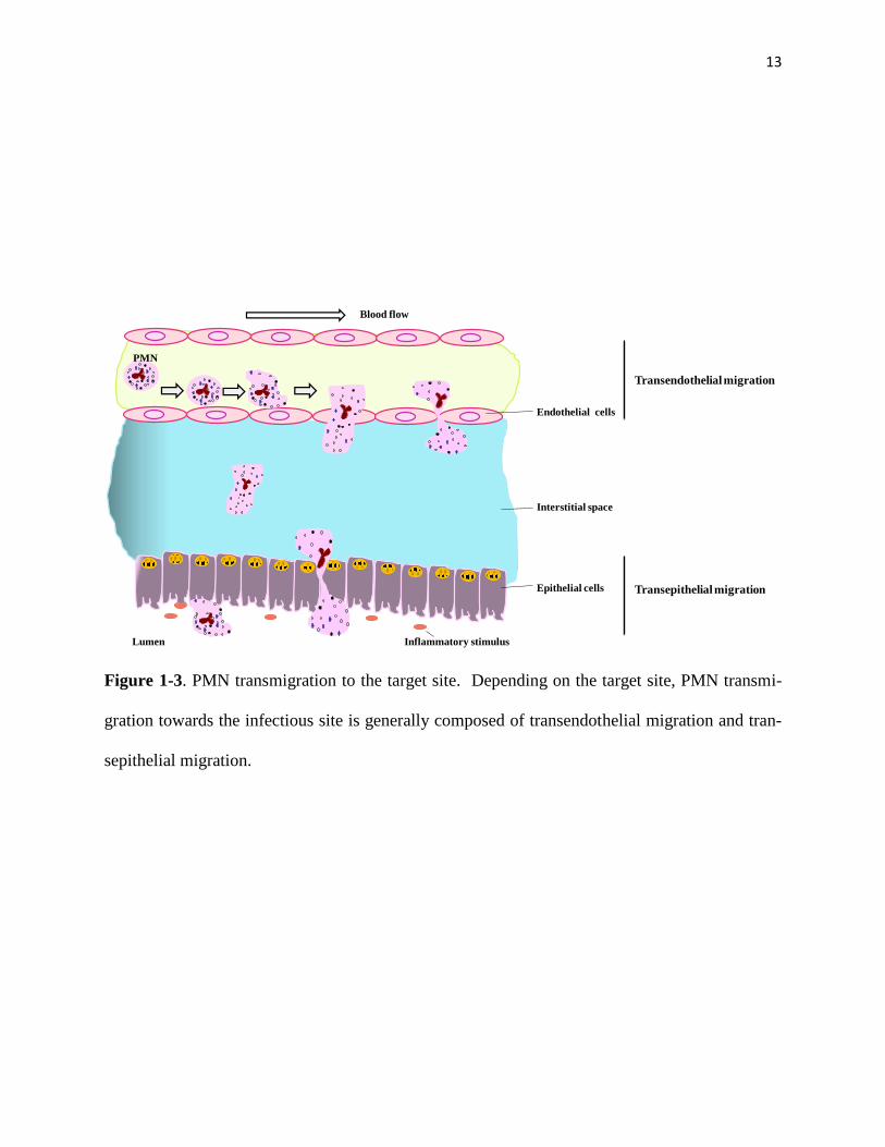
Figure 1-3. PMN transmigration to the target site. Depending on the target site, PMN transmi-
gration towards the infectious site is generally composed of transendothelial migration and tran-
sepithelial migration.
Interstitial space
Endothelial cells
Blood flow
Epithelial cells
Inflammatory stimulus
Transendothelial migration
Transepithelial migration
Lumen
PMN
14
Figure 1-4. PMN transendothelial migration.
Integrin ICAM-I
Transmigration RollingAdhesion
Endothelial cells
PMN
Blood flow
JAM-A
15
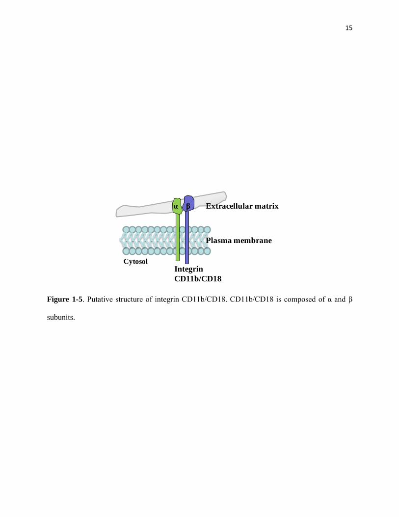
Figure 1-5. Putative structure of integrin CD11b/CD18. CD11b/CD18 is composed of α and β
subunits.
Extracellular matrix
Plasma membrane
α β
CytosolIntegrin
CD11b/CD18
16
Figure 1-6. PMN transepithelial migration.
Epithelial cells
Inflammatory stimulusLumen
Integrin
Fucosylated proteoglycan
ICAM-I
Apical junction complex
CD47 SIRPα
Apical surface
Basolateral surface

17
Figure 1-7. Putative structure of SIRPα (left) and CD47 (right).
ITIM
ITIM
YP
YP
YP
YP
Plasma membrane

18
2. Inflammation
Throughout evolutionary history, human beings have continued to defend against invasion by
microorganisms. We successfully resist to infections most of time because of our complex im-
mune system. The immune system includes both the innate immunity and the adaptive immunity.
Innate immune immunity takes place first and occurs within the first 96 hours. PMN and macro-
phages are the key players for innate immunity. Through phagocytosis, they eliminate invading
microorganisms. Incomplete clearance results in the response of the adaptive immune system
mainly involving in lymphocytes. Coordination of the innate immunity with the adaptive immun-
ity is crucial to fight efficiently against infection.
Inflammation is an innate immune response to local injury or infection [28] (Figure 1-8). The
occurrence of inflammation is due to insufficient removal of invading microorganism. In in-
flammation, innate immune response takes place first [91]. In the local environment, resident tis-
sue cells sense the presence of pathogens, secret cytokines acting on other cells and trigger the
innate immune response. Under the influence of cytokine networks, vascular dilation occurs and
edema takes place, characterized by the leaking of plasma and fluid into connective tissues due
to the permeability change of endothelial cells. At the same time, edema leads to a change in the
nervous system, thus resulting in a feeling of pain [28]. In addition to vascular events, cellular
events also play a primary role in inflammation. In the local environment, high concentrations of
chemoattractants secreted by activated stromal cells, including activated endothelia cells and ma-
crophages, etc. [31], and increased surface expression of adhesion molecules on the endothelium
lead PMN to undergo rolling, activation, adhesion and transmigration across the endothelial mo-

19
nolayer to the targeted tissue to release anti-microbial substances and phagocytize pathogens
[92].
PMN are vital for the beginning stage of inflammation [25]. PMN infiltration is the hallmark of
the early-stage of inflammatory response [28]. After they arrive at the target site, PMN execute
their function by releasing reactive oxygen species, antimicrobial proteins and proteolytic en-
zymes, as well as by producing cytokines and performing phagocytosis [27]. Thus, sufficient re-
cruitment of PMN is very important for eliminating invading microorganisms, recovering tissue
damage and maintaining homeostasis [26]. In particular, during acute infection and inflamma-
tion, PMN sense chemoattractant signals and swiftly migrate across the microvasculature and
underlying tissues to accumulate at inflammatory foci. Here, these cells execute forceful antimi-
crobial and tissue damaging functions. Therefore, the timely and precise response of PMN is crit-
ical for innate immune response.
In spite of the critical role of PMN in acute inflammation, efficient removal of PMN in the late
stage of inflammation is substantial [93-94]. PMN apoptosis [95] and removal by macrophages
control the extent of inflammation. The prolonged existence of PMN in the targeted tissue also
frequently results in host-tissue damage. PMN-mediated inflammation is one of the central com-
ponents of many pathophysiological processes; thus, diseases in which dysregulated PMN
transmigration and activation occur often result in severe tissue injuries and organ dysfunction
[25, 96]. Many active stage of human diseases are characterized by excessive PMN infiltration,
including bacterial infection, inflammatory bowel disease, ischemia-reperfusion injury, rheuma-
toid arthritis, etc. [31].

20
Furthermore, resident tissue cells such as macrophages and dendritic cells contribute largely to
inflammation [93-94, 97-98]. After engulfing pathogens, macrophages secrete cytokines to fur-
ther recruit other cells for assistance. After engulfing pathogens, dendritic cells migrate toward
secondary lymphoid organs to induce adaptive immune responses. Upon dendritic cell migration
into the secondary lymphoid organs, degraded pathogens are presented by major histocompatibil-
ity complex (MHC) on the surface and recognized by naive T lymphocytes. Activation through
recognition of the MHC-antigen complex with T cell receptors and co-stimulatory signals causes
pathogen-specific T lymphocytes to differentiate to T helper cells or cytotoxic T cells. T helper
cells are able to activate B cells leading to the generation of plasma cells and secrete antibodies.
Some plasma cells and antibodies leave lymphoid organs and enter infectious tissues for elimi-
nating pathogens. Through releasing cytokines, T helper cells also further activate macrophages
in the tissue to increase their abilities to phagocytize pathogens. Compared with T helper cells,
cytotoxic T cells migrate to the target tissue and directly remove pathogens through releasing
cytotoxins or inducing apoptosis through a Fas ligand-Fas interaction. In conclusion, inflamma-
tion is a complex process whereby innate immunity coordinates with adaptive immunity to fight
against pathogens.

21
Figure 1-8. PMN play a pivotal role in inflammation. PMN infiltration is the hallmark of the ear-
ly stage of the inflammatory response.
PMN
SkinWound Blood clot
Effector cell
Blood vessel
Cytokines
Bacteria
22
3. PMN granules
Different from other cells types, circulating PMN only express a few cell surface proteins [62].
Instead, they keep most of those proteins in special membrane bound storages called granules
[61] (Figure 1-9). According to size and morphology, PMN granules are composed of four cate-
gories, primary granules (azurophil granules), secondary granules (specific granules), tertiary
granules (gelatinase granules) and secretory vesicles [63]. Each granule contains different protein
components and possesses distinct functions.
Primary granules, regarded as lysosomes, undergo limited degranulation in response to outside
stimuli [99]. Primary granules are important storage for antimicrobial proteins, serine proteases
[100-101] including elastase and cathepsin G [102], and proteinase 3 [63]. Myeloperoxidase
(MPO) is a marker for primary granules [103]. It plays a key role in PMN phagocytic function,
due to its strong antimicrobial activity [104], through interaction with H2O2 formed by superox-
ide anions in the respiratory burst system. During the process of phagocytosis, serine proteases,
localized inside phagolysosome, work together with hypochloric acid, a final product generated
in a series of reactions catalyzed by MPO, to execute antimicrobial activities. In addition to kill-
ing microorganisms in phagolysosomes, elastase are also released during PMN activation in re-
sponse to various chemoattractants stimulation [105]. Upon release, serine proteases play a very
important role in chemokine modification that leads to an increased binding affinity towards the
chemokine receptor. Chemokines modified by serine proteases gain increased chemotactic ac-
tivity for PMN ,and thus, are essential for PMN migration [100]. Serine proteases are also capa-
ble of cleaving intercellular adhesion molecules, ICAM-1 [106], vascular cell-adhesion molecule
23
1 (VCAM-1) [107] and epithelial (E)-cadherin [108], thus facilitating PMN transmigration
across the endothelium and epithelial cells. As a result, certain primary granules not only func-
tion as lysosomes during PMN phagocytosis, but also contribute to the interaction between PMN
and the extracellular matrix during PMN migration.
Secondary granules are important stores of adhesion molecules, membrane receptors and other
critical proteins, such as lactoferrin, fMLF receptors, CD47, SIRPα, and CD11b/CD18, facilitat-
ing PMN migration [63, 68-69]. Lactoferrin is a marker of secondary granules [103], and its an-
timicrobial activity comes from competing with microorganisms for iron [109]. PMN from pa-
tients with neutrophil-specific granule deficiency [110] are defect in chemotaxis in response to
stimuli, decreased binding capability to fMLF, decreased antibacterial activity and less activity in
response to fMLF stimulation. Other cases of PMN secondary granule deficiency also illustrated
decreased chemotaxis and impaired killing of microorganisms [111]. These characteristics fur-
ther suggest that secondary granules, enriched in receptors and adhesion molecules, are indis-
pensable for PMN to execute anti-infection functions.
Tertiary granules serve as reservoirs of matrix-degrading enzymes [63]. The key component in
tertiary granules is gelatinase. Gelatinase, also termed matrix metalloproteinases (MMPs), enzy-
matically digest extracellular matrix proteins during PMN transmigration. In human PMN, two
MMPs have been identified. They are MMP-9 (92KDa) and leukolysin (MT6-MMP/MMP-25)
(56KDa) [63, 112]. Compared with secondary granules, tertiary granules are more mobilized and
easily exocytosed upon stimuli activation [99].

24
Figure 1-9. PMN granules types (A) and degranulation (B) in response to stimulation (e.g.
fMLF).
BAC
D
E. R.
Golgi
TGN
Plasma Membrane
Storage Granules
D
a bg
fMLF
abg
GTP
Degranulation
CD47
integrin
lactoferrin
SIRPα
PM
N G
ran
ule
s
A. Primary granules (azurophil granules)
B. Secondary granules (specific granules): lactoferrin, CD47, integrin, SIRPα
C. Tertiary granules (gelatinase granules)
D. Secretory vesicles
degranulation
A.
B.
Ral
25
Secretory vesicles are highly mobilized [99], and they contain membrane-associated receptors
and are able to furnish PMN with the first batch of proteins for sensing and triggering activation
during PMN-mediated immune responses [61]. Secretory vesicles contain the marker protein al-
bumin [113]. It has been demonstrated that albumin is originally from PMN plasma membranes,
not from new synthesis, suggesting that albumin comes from endocytosis pathway. The quick
interchange of albumin between the plasma membrane and secretory vesicles further implies that
secretory vesicles are very sensitive to exocytosis. Furthermore, PMN are able to mobilize secre-
tory vesicles even in the absence of stimulation [114].
The classification of granules does not only rely on the size and morphology of granules. The
components in each granule depends on the time that those proteins are synthesized (targeting-
by-timing hypothesis) and the characteristics of each protein, i.e. whether it is able to co-exist to
avoid degradation [99]. In addition to the presence or absence of MPO, the major difference be-
tween the secondary and primary granules lies in the fact that proteases stored in primary gra-
nules are in activated forms; proteases in secondary granules exist as latent forms and need to be
activated after release from PMN. However, the detailed mechanisms underlying processing and
targeting of granule proteins in PMN are unclear. It has been implied that the Golgi network is
likely to function as a sorting apparatus in PMN [61]. Studies of serine proteases stored in prima-
ry granules have shown that glycosylation, a major function of the Golgi network, is not required
for granule protein targeting [99]. Specifically, Cathepsin G does not require glycosylation for
granule targeting [115]. Through site-directed mutagenesis, Cathepsin G lacking an N-linked
glycosylation site was still shown to be transported to lysosomes. In addition to protein confor-

26
mation and modification, such as glycosylation, protein-protein interaction is also crucial for cor-
rect granule targeting, particularly for lactoferrin, which is a key component for secondary gra-
nules [103]. A study on transport of newly synthesized lactoferrin demonstrated that the transport
of lactoferrin was blocked by interfering ionic interaction at the plasma membrane and acidifica-
tion environment inside the cell [103].
To date, the most sufficient experimental approach to investigate the localization of proteins of
interest in PMN is subcellular fractionation, which has been previously demonstrated to success-
fully separate neutrophil plasma membranes from intracellular granules [70, 116]. Thus, in this
study, a series of these experiments were performed to identify the localization of Ral in PMN.
4. Mechanisms underlying granule release
Mobilization of intracellular granules and vesicles also contributes, to an extraordinary degree, to
PMN migration. As we know, cytoplasmic granules and vesicles of PMN are composed of not
only antimicrobial proteins and proteolytic enzymes for eliminating foreign microorganisms but
also a variety of cell surface receptors, functional proteins and granule matrix proteins involved
in PMN extravasation. During degranulation, the PMN cell surface is incorporated with granule
proteins and receptors. Subsequently, those fused proteins facilitate the activation of resting
PMN to engage in cell-cell interactions and cell-extracellular matrix interactions in the process
of transmigration. CD47, as an example, has been illustrated to facilitate PMN migration only
after re-distribution from secondary granules to the cell surface upon chemoattractant stimulation
[69]. As a result, controlled and effective degranulation of PMN is very important in inflamma-
tion and preventing overwhelming PMN-mediated tissue damage, thus maintaining homeostasis.
27
However, the molecular mechanism(s) by which PMN regulate individual intracellular granule
release is still unclear. The signaling events elicited by chemoattractant stimulation (e.g. fMLF)
leading to the release of each type of granule are still unclear. In other cell types, Ral, a Ras
family small GTPase, involves in intracellular vesicle transportation, including both endocytosis
and exocytosis [117-121]. Small GTPases are monomeric G proteins that are actively involved in
many cellular functions [122]. They are further divided into five families based on sequence:
Ras, Rho, Rab, Ran and Sar1/Arf [123]. According to structure, there are two interconvertible
forms of small GTPases: GTP-bound active form and GDP-bound inactive form (Figure 1-10).
Upon upstream activator binding, GDP is dissociated from the small GDP-bound form because
of the conformation change, and is replaced with GTP. The GTP-bound form relays the signal
from upstream to downstream through binding downstream effectors [122]. As a result, small
GTPases serve as a molecular switch in signaling cascades. The interexchange of the GTP-bound
form with the GDP-bound form is controlled by guanine nucleotide exchange factor (GEF) and
GTPase activating protein (GAP). GEF triggers the formation of GTP-bound active form. GAP,
increases the conversion of GTP to GDP, thus resulting in the formation of GDP-bound inactive
form [122].

28
Figure 1-10. Interchange of the GTP-bound form with the GDP-bound form of small GTPase.
GEF: guanine nucleotide exchange factor; GAP: GTPase activating protein.
Upstream activators
Small G-protein
Small G-protein
GDP
GTP
GEFGAP
GTP
GDP
p
Downstream effectors

29
Given the importance of Ral in other cell types, in this study, the potential role of Ral in regulat-
ing PMN granule mobilization and chemotactic function was investigated.
To date, the dynamics of actin cytoskeleton, Ca2+
signaling, src family kinases, β-arrestin, small
GTPases and SNARE molecules have been reported to be involved in PMN degranulation.
Small GTPases play important roles in regulating PMN granule release. Rac2, a Rho family
GTPases family member, is pivotal for neutrophil primary granule release [124-127]. Rac2-/-
mice have shown defects in PMN primary granule release but not secondary or tertiary granule
release. Rac2-/- mice also demonstrated defects in the translocation of primary granules [124].
Bone marrow PMN isolated from Rac2-/- mice fail to release primary and tertiary granules upon
fMLP, LTB4, and CB-LTB4 [125] stimulation. In addition, Rac2 is also associated with supe-
roxide anion (O2–·) production [125]. In animal models and patients, the Rac mutation D57N,
characterized by the loss of binding capability to GTP, has been [110, 127] found to prevent the
MPO release and superoxide anions production in response to fMLF and phorbol myristate ace-
tate (PMA), but not the release of secondary granules [126].
Rab, a family protein of small GTP, have been widely studied in targeting vesicles to particular
compartments through tethering. In PMN, Rab27a has been implicated in regulating primary
granule release through interacting with its effector protein JFC1/Slp1 [128-129]. Rab27-/- mice
fail to release MPO upon LPS challenge. Subcellular distribution of Rab27a by fractionation and
immunofluorescence staining further supports the fact that Rab27 is a constituent of exocytotic

30
machinery of primary granules in PMN. Rab27b has also been demonstrated to be the key com-
ponent in regulating PMN primary granule release [130].
Rap1 and Rap2 are associated with PMN secondary granules but not primary granules or cytosol,
as demonstrated by PMN subcellular fractionation and immunostaining. Accompanied with de-
granulation, Raps translocate to the plasma membrane from secondary granules, further suggest-
ing their roles in controlling PMN secondary granule release [131].
Studies have also found that Rho family small GTPases regulate granule release. Pretreatment of
PMN with MAPK phosphorylation inhibitors diminishes phagocytosis of C albicans and granule
mobilization. Similarly, overexpression of the dominant-negative Rac and Cdc42 in PMN,
through viral delivery, leads to abolished phagocytosis and granule release, implicating Rac and
Cdc42 in regulating PMN granule release through the MAPK signaling pathway [132].
As a key component of the cytoskeleton, actin filaments not only contribute to cell motility, but
also intracellular trafficking [133]. They are also involved in endocytosis and exocytosis [134].
Dynamic actin assembly and disassembly are associated with PMN primary granule release. In
particular, actin polymerization is likely to initiate the release of primary granules. Primary gra-
nules are released from PMN treated with fMLF and cytochalasin B (CB) [135]. However, stabi-
lization of actin results in inhibition of primary granule release. Inhibition of Rac also leads to
reduced primary granule release through jeopardizing F-actin formation. Another study has sug-
gested that the actin cytoskeleton might regulate the release of all types of granules in the PMN
in response to stimulation [136]. With the exception of secretory vesicles, CB significantly in-

31
creases fMLF-simulated secondary and tertiary granule release. Additionally, CB triggers prima-
ry granule release that is failed to be released by fMLF alone. All of the above suggest that the
actin cytoskeleton restricts the release of granules and maintains PMN in the resting condition. In
addition to actin filaments, microtubules have been demonstrated to be associated with PMN
granules [137]. Granule-microtubules, including primary granule-microtubule complexes and
secondary granule-microtubule complexes, have been isolated from PMN, suggesting the critical
role of microtubules in regulating PMN granule release.
During granule release, tethering and fusion of vesicles toward the target compartment involves a
complex process that is not well understood. SNAREs, (soluble NSF (N-ethylmaleimide-
sensitive factor), are key proteins involving in the docking and fusing of vesicles to the target
compartment [138-140]. In human PMN, (t-) SNARE isoform syntaxin 6 has been cloned and
shown to be localized only in the plasma membranes of resting and activated PMN [141]. In con-
trast to syntaxin 6, SNAP-23, another isoform of (t-) SNARE, localizes in tertiary and secondary
granules in PMN and translocates to the plasma membrane upon stimulation. Antibodies against
SNAP-23 block secondary granule release, whereas, anti-syntaxin 6 antibodies, block primary
and secondary granule release from PMN. The above suggest that SNAP-23 and syntaxin 6 par-
ticularly regulate secondary and primary granule release. Vesicle-associated membrane protein
(VAMP)-7, a (v-) SNARE in PMN [142], has been detected in primary granules in PMN. Treat-
ment of PMN with antibodies against VAMP-7 dose-dependently abrogates primary granule,
secondary granule and tertiary granule release. VAMP-2 and syntaxin 4 are associated with se-
cretion of secondary and tertiary granules from PMN [143]. VAMP-2 is identified in PMN. Ap-
plication of specific antibodies against 2-17aa in the N terminal of VAMP-2 to electropermeabi-

32
lized PMN leads to the inhibition of secondary and tertiary granule release, but not primary gra-
nule release from PMN. This suggests an important role for VAMP-2 in release of those two
granules PMN. Furthermore, VAMP-2 has also been suggested to control granule release in
PMN after stimulation through interaction with syntaxin 4, which was demonstrated by the co-
immunoprecipitation assay. Regarding the different types of granules in PMN, distinct SNAREs
are likely to associate with each individual granule, and thus regulate PMN granule release.
Although signaling pathways regulating PMN granule release are not well understood, GPCR-
mediated signaling cascades in PMN activation have been well established. As a scaffold pro-
tein, β-arrestin has been shown to be involved in PMN granule release. Upon IL-8 binding to
CXCR1 on PMN [144], β-arrestin binds to phosphorylated receptors and forms more β-arrestin-
Hck complexes. It has been demonstrated that β-arrestin translocates from the plasma membrane
to secondary granules in PMN upon stimulation, suggesting its potential role in regulating PMN
secondary granule release.
The effect of Src family kinases has also been demonstrated. P59Hck
was first found to be mainly
localized on granules, in contrast to plasma membranes and cytosol, in PMN [145]. Subsequent-
ly, it is found to be localized only on primary granules in PMN and is translocated to phagosomal
membranes in HL60 cells during phagocytosis, indicating its role in regulating fusion of primary
granules with lysosome. P55c-fgr
has also been detected in human PMN and it is present in PMN
membrane portions mainly on the plasma membrane of secondary and tertiary granules. P55c-fgr
partially translocates to the plasma membrane in PMN stimulated with fMLF together with
CB[146], suggesting its role in granule release. hck-/-
fgr-/-
PMN demonstrate the diminished re-

33
lease of lactoferrin in response to TNF stimulation when cells are placed in tissue culture plates
or on collagen coated surfaces, suggesting that hck and fgr are involved in PMN adhesion-
dependent granule release [147].
Calcium signaling also plays an essential role in PMN degranulation. It has been shown that in-
creased intracellular calcium levels leads to release each type of granules in a hierarchical order
[148]. Raised intracellular calcium is not only led by extracellular calcium influx, but also by
other factors. It has been demonstrated that intracellular calcium in PMN is increased by ligation
of L-selectin with its ligand, sulfatides [149], fMLF stimulation [149] and cross-linking of
CD11b/CD18 on the PMN surface using its mAb [150]. Particularly, ligation of CD11b/CD18
induces the activation of cytoplasmic protein tyrosine kinases, such as p58fgr
, resulting in
CD11b/ CD18 dependent tyrosine phosphorylation. Patients with loss of β2 integrins fail to re-
sponse to TNF-mediated tyrosine phosphorylation in PMN [151-152]. Furthermore, activation of
CD11b/CD18 phosphorylates and activates a series of proteins. The effect of CD11b/CD18
might be Ca2+
dependent [153-154].
In PMN, several annexins have been identified [155]. Among them, annexin I is associated with
secondary granules, the plasma membrane and secretory vesicles, but not primary granules. The
effect of Annexin I likely depends on high concentrations of Ca2+
[156]. Annexin II, IV and VI
are associated with all of the granule populations. The above suggests that different isoforms of
annexin convey distinct effects on the release of different categories of granules from PMN.
5. Ral
34
The molecular mechanisms underlying PMN degranulation are still poorly understood. Consider-
ing the complex signal cascades involved in each type of granule release, it still remains unclear
how PMN control the orderly release of individual granule subsets in response to different stimu-
li.
Ral is a member of Ras small GTPases. Like other small GTPases, Ral activity is regulated by
GAP and GEF. To date, six GEFs have been identified. Based on structural similarity, they are
further divided into two subfamilies. The first family of Ral GEFs is composed of RalGDS,
ralGDS-like (Rgl), RalGDS-like factor (Rlf), and Ral GEF-like 3 (Rgl3). They contain Ras-GTP-
binding domains, suggesting that Ral is activated through a Ras-dependent pathway. The other
family consists of RalGPS2/RalGEF2 and RalGPS2, without a Ras-GTP-binding domain, sug-
gesting the Ras-independent pathway for Ral activation [157]. Compared to GEFs, only two
GAPs, GAP1 and GAP2 [157], have been identified by in vitro experiments. Ral activation in-
volves complex signal cascades including both Ras-dependent [158-161] and Ras-independent
pathways [162-163]. Studies by Rob M.F. Wolthuis demonstrate that Ral activation requires Ras
activation [158]. Most importantly, Ral activation correlates with Ras activation. In contrast, oth-
er studies have also illustrated that the Ras-independent activation mechanism also extensively
contributed to Ral activation [162-163]. In Rat-2 cells [162], not only does Ral activation require
Ras activation, but is also triggered by increasing intracellular Ca2+
using ionomycin. Mean-
while, PLC is also required for Ral activation induced by LPA or EGF. Ca2+
-mediated Ral acti-
vation is observed in platelets as well. Increased intracellular calcium levels and/or increased
calcium influx lead to Ral activation in platelets [163]. Bhattacharya M et al., using human em-
bryonic kidney 293 cells (HEK293), discovers that RalGDS dissociation from beta-arrestin and

35
translocation to the plasma membrane leads to Ral activation [164]. This result implies an essen-
tial function for Ral in cytoskeletal reorganization and granule release.
The detailed mechanism that inactivates Ral, however, is still unknown. Thus, it is critical that
more studies exploring this mechanism be performed. In particular, Ral deactivation is critical
for PMN. Results from my study suggest that the loss of Ral activity results in secondary granule
release; through secondary granule release, adhesion molecules incorporate into the PMN sur-
face, and thus facilitate PMN migration, interaction with extracellular matrix and communication
with other cells in inflammation.
There are two Ral genes in mammalian cells. The Genebank IDs for RalA and RalB of Homo
sapiens are AF493910.1 and X15015.1, respectively. RalA and RalB are highly homologous,
with about 85% identical amino acids [165] (Figure 1-12). The distinct sequences are mainly lo-
cated at the carboxyl-terminus, in amino acids 192-204 [166]. RalA and RalB contain the same
amino acid sequences within two effector binding regions, shown in Figure 1-12, suggesting that
they are likely to harbor overlaid functions. As a typical family member of Ras small GTPase, in
the C-terminus, Ral contains an atypical CAAX motif (where C stands for cystein, A stands for
aliphatic amino acid, X stands for methionine or serine); CAAX is a lipid modification motif
[167] and suggests that Ral is likely to be associated with the membrane.
As shown in Figure 1-11, Ral contains two effector binding regions that are also termed “switch
regions”, which mediate the recognition of effectors. Ral performs distinct and important bio-
logical functions through interacting with different downstream binding partner proteins. Five

36
binding partners have been identified to date. They include RalBP1, Sec5/Exo84 (subunit of ex-
ocyst), PLC-δ1, and ZONAB, and phospholipase D 1 (PLD1) [168]. Except for PLD1 binding to
the first 11 amino acids of Ral, others all bind to the switch regions. Through interaction with
diverse effectors, Ral executes different biological functions. Particularly, Ral employs primary
functions in vesicle trafficking through downstream effectors, RalBP1, Sec5/Exo84 and PLD1.
Although Ral engages in many aspects of cellular processes, it executes its major function in ex-
ocytosis and endocytosis, mainly by regulating exocyst, which is seen in a variety cell types
[169]. Exocyst is a protein complex composed of Sec3, Sec5, Sec6, Sec8, Sec10, Sec15, Exo70
and Exo84 subunits, and is implicated in the fusion of vesicles to the plasma membrane [170].
The regulation of the exocyst is largely dependent on small GTPases. Ral small GTPases are key
players in this process. A number of publications have demonstrated that Ral promotes exocyst
assembly [171-172]. Using the double-mutant protein Ral23V49N as a probe in the yeast two-
hybrid system, active Ral was found to directly associate with Sec5, a subunit of exocyst and the
association was confirmed by the decreased formation of exocyst in the absence of Ral [173].
The functions of RalA and RalB are not completely understood. They play a collaborative role in
regulating cytokinesis through exocyst [174], suggesting that RalA and RalB have compensatory
roles in exocytosis. It has been postulated that dense granule releases from permeabilized plate-
lets stimulated with GppNHp, a non-hydrolyzable analog of GTP are triggered by active Ra-
lA/RalB [175]. The release of dense granules is abrogated by overexpression of Sec5- GTP-Ral-
binding domain (RBD), which suggests the secretion of dense granules from platelets is regu-
lated by the Ral-Exocyst pathway.

37
The influence of Ral on vesicle release has also been demonstrated in the neuron signaling sys-
tem. The expression of a dominant-inhibitory mutant of Ral results in dramatically inhibited re-
lease of vesicles in the presynaptic nerve terminal in response to Ca2+
. The regulation is likely
through active Ral-exocyst interaction [118]. In addition, active RalA interacts with the mamma-
lian brain exocyst complex [119]. In neuronal cells, chromaffin and PC12 cells, Ral associates
with the plasma membrane in resting cells; however, after stimulation, it is thought to interact
with effector protein(s) and form a complex. Activation of Ral in stimulated PC12 cells by K+
triggers the release of large, dense core secretory granules. Furthermore, overexpression of a
constitutively active form of Ral increases the exocytosis of those granules from PC12 cells
[117].
In polarized epithelial cells, Ral directs vesicle transportation. Protein transportation that was
originally directed only towards the basolateral membrane was changed to the basolateral and
apical membranes in the presence of active Ral [173]. Overexpression of the constitutively active
form of Ral, Ral23V, or the inhibitory construct containing the Ral-binding domain leads to the
inhibition of granule exocytosis in PC12 cells [173]. RalB is required for exocyst complex for-
mation in rat kidney epithelial cells [176]. Active RalA binds exocyst and increases the translo-
cation of E-cadherin in epithelial cells [177]. Adhesion-mediated RalA activation is also required
for integrin-mediated membrane raft endocytosis and recycling during regulation of growth sig-
naling [120].

38
In addition to exocytosis, it is reasonable to predict that Ral is also associated with endocytosis,
based on studies demonstrating the function of RalBP1. RalBP1/RLIP76, another important
downstream effector of Ral, is a member of the machinery regulating receptor-mediated endocy-
tosis [121, 178-179]. Using a two-hybrid assay, RalBP1 is found to associate with µ2, the me-
dium chain of AP2 adaptor protein complex [121] that is requisite for clathrin-mediated endocy-
tosis by recruiting clathrin to plasma membrane [180]. Overexpression of constituently active
Ral also blocked endocytosis of transferrin in HeLa cells [121], A431 cells [178], and CHO-IR
cells [178].
Not only does Ral play a crucial role in regulating vesicle trafficking, its functions have also
been explored in tumorigenicity [181]. In human bladder cancer [181], RalA mRNA is signifi-
cantly upregulated and is associated with invasive tumors. The RalA mRNA level is also highly
associated with cancer protein expression [181]. RalA is required for the growth of human pan-
creatic carcinoma cells [182]. Also, inhibition of RalB by siRNA resulted in remarkably de-
creased cell migration in bladder carcinoma and prostate carcinoma cells [183]. RalB is involved
in host defense signaling through suppressing tumor apoptosis, thus leading to cancer cell sur-
vival [184-185]. It has been shown that RalB activation promotes the complex formed between
Sec5 and the exocyst subunit, along with TBK1 (Tank binding kinase 1), an atypical IκB kinase
member, resulting in cancer survival.
The effect of Ral in vesicle trafficking has also been elucidated through investigating the func-
tion of PLD. PLD1 and its product PA are actively involved in vesicle trafficking, exocytosis and
endocytosis [186-187]. The role of PLD1 is likely to be regulated by ARF6, a small GTPase, due
39
to the fact that 1) the activity of PLD1 depends on ARF6, and 2) the inhibition of PLD1 activity
by myr-Arf6 (N-terminal-myristoylated Arf6) [188] peptide leads to impaired exocytosis induced
by calcium in chromaffin cells [189]. PLD1 functions coupled with active Ral are required for
exocytosis. Furthermore, Ral, PLD1 and ARF6 physically associate with each other and form a
complex, mediating exocytosis upon stimulation. The expression of a vector bearing the coding
sequence for active Ral, but lacking in binding sequence for PLD1, fails to elicit granule release,
indicating that Ral functions in concert with PLD1 to modulate exocytosis [117].
The effect of RalA and RalB in PMN is still inconclusive. Using RT-PCR (Figure 3-19), in this
study it was demonstrated that RalA and RalB are both present in human PMN, and RalB is
more dominant than RalA. In my experimental approaches, I utilized a Ral pull-down assay to
investigate the activity switch of Ral. Pull-down assays are widely accepted and used to address
the activity of a variety of small GTPases including Ras, Rac1, Cdc42, Rho, Rap, Arf and Ral.
The assay takes advantage of the specific interaction between RalBP1 and active Ral. It has been
illustrated that amino acids 375-626 of RalBP1 comprise the putative binding domain that me-
diates the interaction with amino acids 37-56 of RalA [190]. As shown in Figure 1-11 and Figure
1-12, RalA and RalB bear identical amino acid sequences in the binding domain of RalBP1. Al-
though, the Ral pull-down assay was designed originally for the detection of RalA activity, it is
expected that RalBP1 will be able to pull-down the active form of RalB, which has been illu-
strated in another study [183].
Taken together, the current research is not focused on differentiating the role of RalA and RalB
in regulating secondary granule release in PMN. As shown in Figure 1-11 and Figure 1-12, RalA
40
and RalB are highly homologous proteins and share most (if not all) of the functional domains
identified to date. The possible Ral downstream effector proteins relevant to our study involving
in receptor endocytosis, such as RalBP1/RLIP76 [121, 178-179], were found to bind to RalA and
RalB at sites with an identical amino acid sequence (aa 37-56). Given the overlapping functions
of RalA and RalB, and the fact that both of these forms are expressed in PMN, we decided to use
Ral in my study.
In this dissertation, Ral, another family member of small GTPases, is demonstrated to play a vital
role for PMN secondary granule release.

41
Figure 1-11. Ral domain structure. The Ral domain structure was adopted from VanDam and
Robinson [168] with modifications using NCBI’s conserved domains software.
PLD orPLC-gbinding
1 11 23 29 39 40 52 70 78
71
127128
130
157 158 203 206
206
GTP binding And hydrolysis
Effector binding I
Effector
binding II
Calmodulin binding domain
46
CAAX motif
Amino Acids

42
82.1% identity in 206 residues overlap; Score: 855.0; Gap frequency: 1.0%
RalA, 1 MAANKPKGQNSLALHKVIMVGSGGVGKSALTLQFMYDEFVEDYEPTKADSYRKKVVLDGE
RalB, 1 MAANKSKGQSSLALHKVIMVGSGGVGKSALTLQFMYDEFVEDYEPTKADSYRKKVVLDGE
***** *** **************************************************
RalA, 61 EVQIDILDTAGQEDYAAIRDNYFRSGEGFLCVFSITEMESFAATADFREQILRVK-EDEN
RalB, 61 EVQIDILDTAGQEDYAAIRDNYFRSGEGFLLVFSITEHESFTATAEFREQILRVKAEEDK
****************************** ****** *** ***+ ********* *++
RalA, 120 VPFLLVGNKSDLEDKRQVSVEEAKNRAEQWNVNYVETSAKTRANVDKVFFDLMREIRARK
RalB, 121 IPLLVVGNKSDLEERRQVPVEEARSKAEEWGVQYVETSAKTRANVDKVFFDLMREIRTKK
+ *+*+ ********+ +***+ ****++ +** * *+ ************************ +*
RalA, 180 MEDSKEKNGKKKRKSLAKRIRERCCIL
RalB, 181 MSENKDKNGKKSSKN-KKSFKERCCLL
* + +*+ ***** *+ * + + ****+*
Figure 1-12. Sequence alignment of RalA and RalB. Protein sequence alignment and domain
structures of human RalA and RalB. Alignment of the primary structures of RalA and RalB re-
veals a high similarity between these proteins. The identical amino acids are indicated by “*” and
the homologous amino acids are indicated by “+”. The different amino acids are highlighted in
purple.

43
Summary
This dissertation describes the regulatory function of Ral, a small GTPase, in vesicle trafficking
in the aspect of exocytosis. In particular, my dissertation work investigates the Ral-mediated
complex signaling pathway in the regulation of granule release from a very unique cell type,
PMN. My work, for the first time, reports the novel finding that Ral controls secondary granule
release from PMN (Figure 1-13). Active Ral prevents degranulation of secondary granules from
PMN. The function of Ral as a gate-keeper for secondary granules is further illustrated by over-
expression of a constitutively active Ral during PMN chemoattractant stimulation. Inhibition of
Ral activity is intimately related with PMN dysfunction. Thus, my work suggests that Ral is vital
for PMN secondary granule mobilization and is intimately linked to chemotactic function. Re-
garding the pivotal role of PMN in innate immune response and inflammation, my work will
shed new light on drug design through targeting Ral in PMN in order to defend against a variety
of inflammatory conditions.

44
Figure 1-13. Control of secondary granule release in neutrophils by Ral
BAC
D
E. R.
Golgi
TGN
Plasma Membrane
Storage Granules
D a bg
fMLF
a b
g
GTP
Degranulation
CD47
Integrin
lactoferrin
SIRPα
Ral
Cell polarization
PI-PLC
DAG PKC
PI3-kinase
Tyrosine kinase
/ phosphatase
Ca2+
IP3
Cytoskeleton reorganizationActin filament polymerization
Cell migration
Rho GTPase
Small GTPase
A: primary granuels
B: Secondary granules
C: Tertiaty granules
D: Secretory vesicles
45
CHAPTER II:
MATERIAL AND METHODS
2.1 PMN isolation
2.1.1 PMN isolation by dextran-ficoll (DEAE-Ficoll)
Human whole blood was drawn from volunteers followed by centrifugation at 200×g for 10 min
and stopped, without breaking, to separate the platelet-rich plasma and blood cells. After decant-
ing plasma, 2% dissolved in 0.9% NaCl was utilized to separate red blood cells (RBC) from leu-
kocytes (35min, 4 °C). The upper layer containing leukocytes were collected into a new tube fol-
lowed by centrifugation (10min, 4 °C). The 10ml respuspended pellet in 0.9% NaCl was placed
onto 3ml ficoll followed by centrifugation (40min, 4 °C). Pellet on the bottom of the tube was
composed of PMN and RBCs, while peripheral blood mononuclear cells (PBMCs) including
lymphocytes, monocytes and macrophages formed a layer approximately 1ml above the pellet.
After discarding the top solution and the PBMC layer, the pellet containing PMN and RBCs was
resuspended in cold RBC lysis buffer composed of 155 mM NH4Cl, 2 mM NaHCO3 and 0.1 mM
EDTA. After centrifugation, PMN were resuspended in cold HBSS (-) after washing once. PMN
were used within 4 hours of isolation. The purity of PMN isolated using DEAE-Ficoll method
was greater than 95%.
2.1.2 PMN isolation from “buffy coat”
Human whole blood was centrifuged at 1300 rpm for 10 min at 4 °C. After removing the plasma
and top “buffy coat”, a thin layer containing PMN was collected and kept in a new conical tube.
In order to maximally retract PMN from the buffy coat, a thin layer of RBCs underneath the
PMN were also collected and transferred to the same tube. To remove RBCs, two rounds of lysis

46
using hypotonic buffer containing 155 mM NH4Cl, 2 mM NaHCO3, 0.1 mM EDTA were ap-
plied. After centrifugation, PMN were resuspended in cold HBSS (-) after washing once. The
purity of PMN isolated using this method was about 70% to 80%.
2.1.3. PMN isolation using lympholyte-poly
Human whole blood drawn from healthy donors was added on top of an equal volume of lym-
pholyte-poly (Cedarlane Laboratories Limited) in a 15ml centrifuge tube followed by centrifuga-
tion at 500g for 40 min at 25°C. After centrifugation, the RBCs were in the pellet on the bottom
of the tube, while two visible leukocyte bands were formed at 1/3 the length of the tube. The
PBMCs remained at the sample/medium interface (upper band) and the PMN formed a band
(lower band). After removing the upper band containing PBMCs, PMN in the lower band were
harvested into a new conical tube. PMN were washed twice with HBSS (-) without Ca2+
and
Mg2+
followed by centrifugation at 400g for 10 min at 25°C. The PMN pellet was then lysed
once with lysis buffer (Roche, Inc) to remove excessive RBCs followed by centrifugation at
400g for 10 min at 25°C. After centrifugation, PMN were resuspended in cold HBSS (-) after
washing once
2.2 Platelet isolation
Human whole blood was centrifuged at 1000 rpm for 10 min at 4 °C, without breaking, to sepa-
rate the platelet-rich plasma and blood cells. The top layer containing the platelet-rich plasma
was placed into a new tube. To isolate platelets and prevent activation during the isolation
process, the collected plasma was supplemented with 0.1 volume ACD (2.5% trisodium citrate,
1.5% citric acid, 2% D-glucose) followed by centrifugation at 3000rpm for 15 min. The pelleted

47
platelets were washed with a citrate buffer (120mM NaCl, 4.26mM NaH2PO4, 5.5mM glucose,
4.77mM sodium citrate and 2.35mM citritic acid, pH 6.5), and finally resuspended in HBSS (-)
at 4 °C.
2.3 Treatment of PMN with inhibitors and agents
For treating PMN, damnacanthal (Calbiochem) was freshly dissolved in DMSO, followed by fur-
ther dilution in HBSS(-) at concentrations ranging from 0.1µM to 18µM before incubation with
PMN (5×10⁶ cells) at 25ºC for 15 min. Other inhibitors, such as Bruton tyrosine kinase inhibitor
LMF-A13, Src family inhibitors PP1 and genistein, Syk inhibitor piceatannol, mitogen-activated
protein kinase (MAPK) inhibitor SB203580 and MEK inhibitor PD98059 (all from Biomol), PI3
kinases inhibitor LY294002 (both from Calbiochem) and others, were used as described pre-
viously [191-192]. To block the clathrin-mediated endocytosis, dynamin inhibitor dynasore [193]
and an AP-2 binding peptide fragment of the EGFR protein tyrosine kinase substrate #15
(Eps15-DIII) [194-195] were used as previously described. Eps15-DIII was delivered into PMN
by tagging with HIV Tat. PMN were pre-treated with these reagents for 5-15 minutes before sti-
mulation with 1µM fMLF at 37°C.
The endocytosis inhibitors phenylarsine oxide (POA) and chlorpromazine hydrochloride (CPZ)
[196] were purchased from Sigma and freshly prepared prior to the study. POA was dissolved in
DMSO followed by dissolving in HBSS (+) at the final concentration of 20µM, followed by in-
cubation with PMN (1.5x 10⁷) at 37ºC for 5 min before fMLP stimulation. DMSO, at a final
concentration of 0.06%, was used as the vehicle control for POA. CPZ, sucrose (Thermo Fisher
Scientific, Inc), NaCl (Thermo Fisher Scientific, Inc), and succinic acid pH 5.5 (Thermo Fisher

48
Scientific, Inc) [197-198] were dissolved in HBSS (+) at a final concentration of 50µM,
500mM, 760mM and 20mM, respectively, followed by incubation with PMN (1.5x 10⁷) at 37ºC
for 5 min.
2.4 PMN degranulation assays
To test the effect of inhibitors on resting PMN, PMN (5×10⁶) were incubated in 400µl of HBSS
(-) with different inhibitors or the same dilution of DMSO at 25ºC for 15 min followed by centri-
fugation. Clear supernatants without cell debris were collected and assayed for specific granular
markers including myeloperoxidase (MPO), lactoferrin and gelatinase correlating to the release
of primary, secondary and tertiary granules, respectively [69, 191]. MPO activity was assayed
using 2, 2’-azino-bis (3-ethylbenzthiazoline-6-sulfonic acid (ABTS). Lactoferrin was assayed by
ELISA using anti-lactoferrin antibody (Jackson ImmunoResearch Laboratories). Gelatinase was
assayed by zymogram using gelatin-embedded gels (BioRad).
To assay degranulation after chemoattractant stimulation, PMN, untreated or treated with Tat-
tagged peptides, PMN (2.5 x 10⁶) were incubated with purified Tat-tagged wild-type Ral (Ral),
the constitutive active form (RalG23V) and dominant negative form (RalS28N) of RalA at con-
centrations of 10nM and 40nM for 15 min at 15°C followed by stimulation in 250µl HBSS (+)
with fMLP (1µM) for 20 min at 37ºC. Following a similar procedure, supernatants without cell
debris were collected and assayed for the release of granular markers. To assay PMN degranula-
tion after treatment with endocytosis inhibitors, PMN (1.5 x10⁷) were incubated in 1.2ml HBSS
(+) with different inhibitors at 37ºC for 5 min followed by fMLP (1µM) stimulation at 37ºC for

49
10 min followed by assaying for the release of granular markers. Positive degranulation controls
were performed by stimulating the same amount of PMN with fMLF plus 10µM CB [135].
2.5 PMN transmigration assay
PMN migration across collagen-coated transfilters was performed as described previously [69,
191]. Briefly, freshly isolated PMN (1 × 10⁶), non-treated or treated with inhibitors (15 min,
25oC) or Tat-tagged Ral peptides (30 min, 4
oC) at final concentration of 10nM and 40nM were
loaded into the upper chambers of the transmigration setups in 150μl HBSS (+). Transmigration
was initiated by adding 0.1μM fMLF in the lower chambers containing 500μl HBSS (+) and in-
cubation at 37°C. PMN that migrated into the lower chambers were quantified by measuring the
activity of myeloperoxidase (MPO), a marker for primary granules. MPO was released by lysing
plasma membrane using Triton X-100. 25μl 10% Triton X-100 was added to the lower chamber
containing migrated PMN at a final concentration of 5%. 25μl 1M citrate buffer pH 4.2 was also
added to the lower chamber to adjust the pH to 4.2. Equal amounts of sample were mixed with
the solution containing 1 mM ABTS, 10 mM H2O2, and 100 mM citrate buffer pH 4.2. Color
matrix was developed at 405nM on a plate reader. In order to quantify the number of the mi-
grated cells in the lower chamber, a standard curve was also generated using known concentra-
tions of PMN.
2.6 Ras family GTPase activation assay
Ras GTPases activation assay in PMN
The GTPase activities of Ras, Rap and Ral in PMN were assayed using Ras, Rap and Ral activa-
tion assay kits (Millipore) with modifications. Briefly, PMN (2×10⁷ cells/per condition) or plate-
50
lets (3×10⁸ cells/per condition), with or without different treatment, were lysed with a buffer con-
taining 25mM Hepes, pH7.5, 150mM NaCl, 2mM MgCl2, 1% NP-40, 0.5% Na deoxycholate,
1% Triton X-100, 2% glycerol and protease inhibitors (cocktail (Sigma) and 10mM PMSF) fol-
lowed by incubation with Raf-1 RBD, RalGDS-RBD or RalBP1 (residues 397-519 of rat
RalBP1)-conjugated agarose for 45 min at 4°C. After washing the agarose, the pulled-down ac-
tive Ras, Rap and Ral were detected by Western blot. Positive and negative controls were per-
formed by addition of GTPγS or GDPγS to the cell lysates before pull-down. In addition, whole
lysates from both control and treated cells were saved and run in parallel, serving as an internal
loading control to determine the total amount of Ras, Rap and Ral proteins.
Alternatively, active Ral in PMN was also pulled down using a RalBP1 of human origin (amino
acids 397-518 of human RalBP1) and GST fusion protein, a gift from Dr. Johannes Bos (Utrecht
University, Netherlands) [163]. Our results indicated that RalBP1 of either human or rat origin
worked equally well to pull down active human Ral.
Ral GTPases activation assay in platelets
Fresh isolated human platelets (3 × 10⁸ cells/per condition) with or without thrombin stimulation
were subjected to the Ral activation assay. Isolated platelets were stimulated with human recom-
binant thrombin (Sigma, Saint Louis) at a final concentration of 0.1UN/ml in tissue culture-
treated 6-well plates (Costar, Corning Incorporated) at 37°C for 20 min. Cells were lysed in the
buffer containing 25mM Hepes, pH 7.5, 150mM NaCl, 1% NP-40, 1% Triton, 2% glycerol,
0.5% Na-deoxycholate, 10 µg/ml leupeptin, 10 µg/ml aprotinin, and 1 mM PMSF. After centri-
fugation, the cleared supernatant was incubated with 30 µg GST-RalBP1 (human sequence) fu-

51
sion protein-coupled glutathione-sepharose with gentle agitation for 1 hour at 4°C to pull down
the active form of Ral. The sepharose mixture was pelleted down, washed with lysis buffer. Ac-
tive Ral was detected by western blot using an anti-Ral antibody. In addition, whole cell lysates
from unstimulated and stimulated platelets were saved and applied to western blots to assay the
total Ral proteins.
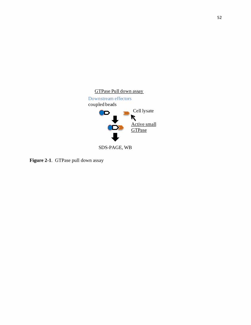
52
Figure 2-1. GTPase pull down assay
Cell lysate
Active small
GTPase
SDS-PAGE, WB
Downstream effectors
coupled beads
GTPase Pull down assay
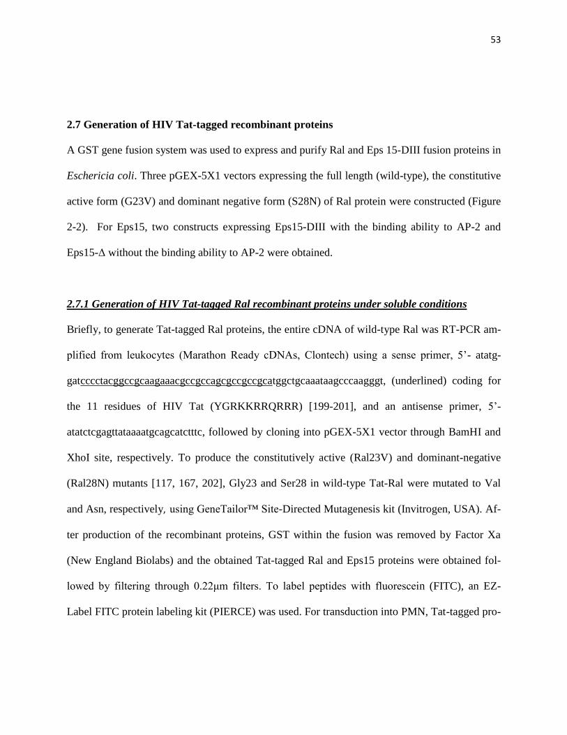
53
2.7 Generation of HIV Tat-tagged recombinant proteins
A GST gene fusion system was used to express and purify Ral and Eps 15-DIII fusion proteins in
Eschericia coli. Three pGEX-5X1 vectors expressing the full length (wild-type), the constitutive
active form (G23V) and dominant negative form (S28N) of Ral protein were constructed (Figure
2-2). For Eps15, two constructs expressing Eps15-DIII with the binding ability to AP-2 and
Eps15-Δ without the binding ability to AP-2 were obtained.
2.7.1 Generation of HIV Tat-tagged Ral recombinant proteins under soluble conditions
Briefly, to generate Tat-tagged Ral proteins, the entire cDNA of wild-type Ral was RT-PCR am-
plified from leukocytes (Marathon Ready cDNAs, Clontech) using a sense primer, 5’- atatg-
gatcccctacggccgcaagaaacgccgccagcgccgccgcatggctgcaaataagcccaagggt, (underlined) coding for
the 11 residues of HIV Tat (YGRKKRRQRRR) [199-201], and an antisense primer, 5’-
atatctcgagttataaaatgcagcatctttc, followed by cloning into pGEX-5X1 vector through BamHI and
XhoI site, respectively. To produce the constitutively active (Ral23V) and dominant-negative
(Ral28N) mutants [117, 167, 202], Gly23 and Ser28 in wild-type Tat-Ral were mutated to Val
and Asn, respectively, using GeneTailor™ Site-Directed Mutagenesis kit (Invitrogen, USA). Af-
ter production of the recombinant proteins, GST within the fusion was removed by Factor Xa
(New England Biolabs) and the obtained Tat-tagged Ral and Eps15 proteins were obtained fol-
lowed by filtering through 0.22μm filters. To label peptides with fluorescein (FITC), an EZ-
Label FITC protein labeling kit (PIERCE) was used. For transduction into PMN, Tat-tagged pro-
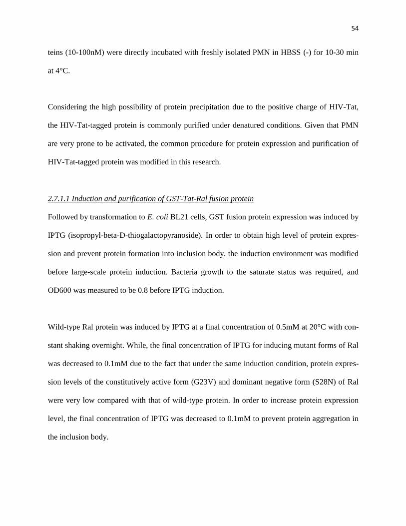
54
teins (10-100nM) were directly incubated with freshly isolated PMN in HBSS (-) for 10-30 min
at 4°C.
Considering the high possibility of protein precipitation due to the positive charge of HIV-Tat,
the HIV-Tat-tagged protein is commonly purified under denatured conditions. Given that PMN
are very prone to be activated, the common procedure for protein expression and purification of
HIV-Tat-tagged protein was modified in this research.
2.7.1.1 Induction and purification of GST-Tat-Ral fusion protein
Followed by transformation to E. coli BL21 cells, GST fusion protein expression was induced by
IPTG (isopropyl-beta-D-thiogalactopyranoside). In order to obtain high level of protein expres-
sion and prevent protein formation into inclusion body, the induction environment was modified
before large-scale protein induction. Bacteria growth to the saturate status was required, and
OD600 was measured to be 0.8 before IPTG induction.
Wild-type Ral protein was induced by IPTG at a final concentration of 0.5mM at 20°C with con-
stant shaking overnight. While, the final concentration of IPTG for inducing mutant forms of Ral
was decreased to 0.1mM due to the fact that under the same induction condition, protein expres-
sion levels of the constitutively active form (G23V) and dominant negative form (S28N) of Ral
were very low compared with that of wild-type protein. In order to increase protein expression
level, the final concentration of IPTG was decreased to 0.1mM to prevent protein aggregation in
the inclusion body.

55
After IPTG induction and centrifugation, cell pellet was resuspended in 1× ice cold PBS, fol-
lowed by sonication. A clear cell lysate was obtained after centrifugation at 15000rpm for 10 min
at 4°C. GST fusion proteins were purified by affinity purification using glutathione-sepharose 4B
(Amersham Biosciences). Glutathione-sepharose was incubated with the clear lysate with gentle
agitation for 2 hours at 4°C. According to the binding capacity that 1 ml drained glutathione-
sepharose is able to bind 8 mg fusion protein, a sufficient amount of glutathione-sepharose was
added to the lysate. Medium mixed with cell lysate and sepharose was then packed into column
(Pierce) followed by washing with 500 ml 1× ice cold PBS and eluted with10mM reduced gluta-
thione in 50mM Tris pH 8. The eluted protein was then concentrated using Millipore Amicon
Ultra-4 10,000 mwco to the final concentration of approximately 0.6 mg/ml, followed by dialysis
against 1×PBS by using dialysis tubing (Spectrum Spectra, 12-14,000 mwco). In order to prevent
activation of the protein precipitation cascade, protein concentration was modified below 1
mg/ml, and several rounds of high speed centrifugation were required to remove any small pre-
cipitants before dialysis. After dialysis and sterilization through 0.2µm filter, protein purity was
analyzed by coomassie blue staining and western blot.
2.7.1.2 Cleavage of GST-Tat-Ral fusion protein
In order to obtain Tat-Ral proteins, GST-tag was cleaved from GST fusion protein by Factor Xa
(NEB). Briefly, GST fusion proteins were incubated with Factor Xa at 0.3µg Factor Xa to 25 µg
GST fusion protein at 25°C for 2.5 hours followed by incubation with glutathione-agarose with
agitation at 4°C for 4 hours. The Tat-Ral protein in the run-through was obtained after passing
protein mixture through the column (Pierce). Purity was then analyzed by coomassie blue stain-
ing. In particular, before cleavage, GST fusion protein concentration was adjusted below 1mg/ml

56
to prevent Tat-Ral protein precipitation during cleavage, likely due to the exposure of highly
charged amino acids.
2.7.2 Generation of HIV Tat-tagged Eps15 recombinant proteins under soluble conditions
The same approach of generation and purification of HIV Tat-tagged Eps 15 protein as HIV Tat-
tagged Ral protein was applied. Two Tat-tagged Eps15 C-terminal peptides were produced, the
AP-2 -binding peptide (amino acids 563-885, designated as Eps15-DIII) and non-binding peptide
(amino acids 740-885, designated as Eps15-Δ) [194-195]. To construct these proteins, RT-PCR
were performed using an antisense primer, 5’-tagccagttctaagtcttc, and Tat sequence-containing
sense primers, 5’-atatggatcccctacggccgcaagaaacgccgccagcgccgccgccctgaactactgccttctggtgtga
and 5’-atatggatcccctacggccgcaagaaacgccgccagcgccgccgctcagccacatcgagctctgtcagcaac, respec-
tively. HIV Tat-tagged Eps15 protein was expressed and purified in the same way describe in
2.7.1.
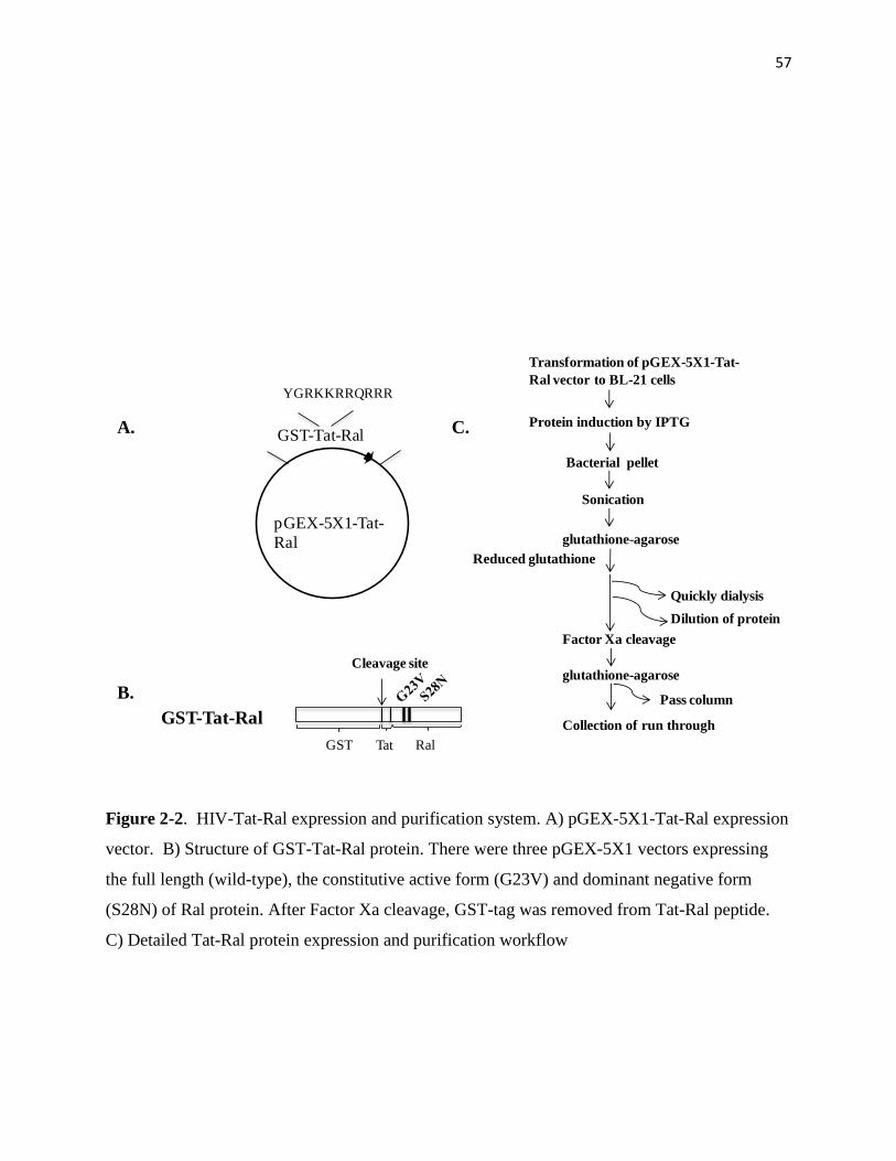
57
Figure 2-2. HIV-Tat-Ral expression and purification system. A) pGEX-5X1-Tat-Ral expression
vector. B) Structure of GST-Tat-Ral protein. There were three pGEX-5X1 vectors expressing
the full length (wild-type), the constitutive active form (G23V) and dominant negative form
(S28N) of Ral protein. After Factor Xa cleavage, GST-tag was removed from Tat-Ral peptide.
C) Detailed Tat-Ral protein expression and purification workflow
B.
GST-Tat-Ral
Cleavage site
GST Tat Ral
A. GST-Tat-Ral
pGEX-5X1-Tat-
Ral
YGRKKRRQRRR
C.
Transformation of pGEX-5X1-Tat-
Ral vector to BL-21 cells
Protein induction by IPTG
Bacterial pellet
Sonication
glutathione-agarose
Reduced glutathione
Quickly dialysis
Factor Xa cleavage
glutathione-agarose
Pass column
Collection of run through
Dilution of protein

58
2.8 Zymogram assay
Gelatinase activity was assayed by gelatin zymography (Bio-Rad, Inc). Briefly, the clear super-
natant was mixed with non-reducing SDS gel sample buffer and subjected to a gelatin gel with
10 μg protein per lane. The gel was then washed twice in 2.5% TritonX-100 in 50 mM Tris pH
7.4, 5 mM CaCl2, 1 microM ZnCl2 at 25°C followed by incubation in the same buffer without
Triton X-100 at 37°C for 20 h. The polyacrylamide gel was then stained with coomassie blue R-
250 for 1 hour at 25°C followed by destaining for 30 min at 25°C . Gelatinase activity was
shown by the white areas of loss of coomassie blue staining.
2.9 Subcellular fractionation of PMN
Freshly isolated PMN (108/per condition), with or without fMLF stimulation or inhibitor treat-
ment, were lysed in a buffer containing 25mM Hepes pH7.5, 150mM NaCl, 10% sucrose, 2mM
MgCl2 and protease inhibitors using a dounce homogenizer followed by centrifugation at 200×g
for 5min. Meanwhile, discontinuous sucrose density gradients were prepared by layering 5 layers
of successive decreasing sucrose density solutions (5ml/layer) upon one another (60%, 50%,
40%, 30% and 20%). PMN lysates (10ml) were then layered on top of the gradients. After centri-
fugation with a Beckman SW28 swinging bucket ultracentrifuge rotor for 3h at 28, 000rpm
(141,000×g) (4°C), the gradients were fractionated (1ml/fraction) followed by analysis for plas-
ma membrane (alkaline phosphatase), primary granule (myeloperoxidase), secondary granule
(lactoferrin) and tertiary granule (gelatinase) [69-70].
59
ml protein protein concentration
mmol
Xmg
24mmol
Xmg
389
mmol
100µl
1mg
X=Molecular Weight of the protein
microliters of FITC solution to add to labeling reaction=
2.10 FITC Protein Labeling
Tat-tagged Ral protein was fluorescently labeled using EZ-Label™ FITC Protein Labeling Kit
(PIERCE). Briefly, Tat-tagged Ral proteins were dialyzed against BupH Borate buffer contain-
ing 50 mM borate, pH 8, using Slide-A-Lyzer MINI Dialysis Unite with 3500 MWCO (PIERCE)
for 1 hour at room temperature to exchange protein buffer for optimizing protein labeling. After
buffer exchange, FITC was added into protein according to the formula* and incubated at room
temperature for 1 hour. FITC-protein mixture was then dialyzed against 1×PBS at 25°C for 1
hour in the same dialysis unit to remove excess fluorescent dye followed by transduction to
PMN.
Simplified formula*:
2.11. Immunofluorescence labeling and flow cytometry
Fresh isolated PMN (5 × 10⁶) were pretreated with tyrosine kianses and other pharmacological
inhibitors in HBSS (-) for 10 min (20°C). After centrifugation at 1300rpm for 5 min at 4°C, the

60
cell pellet was resuspended with blocking buffer containing 0.5% BSA. Cell surface expression
of CD11b, CD47 and SIRPα was assessed by labeling non-permeabilized PMN with anti-CD11b
mAb LM2/1, anti-CD47 mAb B2H6 and anti-SIRP antibodies (10µg/ml each, 1h, 4°C), respec-
tively. After washing, PMN were briefly fixed with 3% paraformaldehyde (PF) for 5 min at 25°C
followed by incubation with fluorescence conjugated secondary antibodies (1:1000 dilution in
blocking solution) for 30 min. Cells were observed under confocal microscope (Zeiss) or ana-
lyzed by flow cytometry (BD). Cell surface labeling by various antibodies was compared with
that obtained with isotype-matched control IgG. For intracellular localization staining, cells were
fixed with 3% PF and then permeabilized by 0.03% Triton X-100 for 10 min at 4oC before anti-
body labeling. For plasma membrane labeling, cells were incubated with wheat germ agglutinin
(WGA) at the final concentration of 5 μg/mL for 1 min at 25 oC followed by washing with HBSS
(-).
2.12. Other reagents
Hybridoma cell line that produces anti-CD11b antibody LM2/1 was obtained from the American
Type Culture Collection (ATCC). Monoclonal antibody against the extracellular domain of
CD47 (PF3.1) was generated previously [69]. Rabbit polyclonal antibodies against the extracel-
lular domains of SIRPα (anti-SIRPα.ex) and JAM-A (anti-JAM-A.ex) were raised by immuniz-
ing rabbits with purified fusion proteins comprised of rabbit Fc and the entire extracellular do-
mains of SIRPα or JAM-A[87, 203]. Antibodies against Ral, Ras and Rap1 were purchased from
Millipore. Anti-β actin mAb was purchased from Sigma. Rabbit anti-α-adaptin was purchased
from Santa Cruz Biotechnology. Rabbit anti-RalGDS was purchased from EMD Millipore.
Alexa Fluor 594 conjugated WGA was purchased from Invitrogen.

61
2.13 Statistical analyses
All the data was analyzed using Prism software. P value for statistical significance was below
0.05.
62
CHAPTER III:
CONTROL OF SECONDARY GRANULE RELEASE IN
NEUTROPHILS BY RAL GTPASE
PUBLICATION: CHEN CX, SOTO I, GUO YL, LIU Y. CONTROL OF SECONDARY
GRANULE RELEASE IN NEUTROPHILS BY RAL GTPASE. J BIOL CHEM, 2011 APR
1; 286(13): 11724-33.
NOTE:
I PERFORMED ALL OF THE EXPERIMENTS IN THIS CHAPTER, EXCEPT FOR TABLE
1.
DR. YUAN LIU AND I PERFORMED THE EXPERIMENTS SHOWN IN FIGURE 3-1.
DR. YAUN LIU AND I PERFORMED THE EXPERIMENTS SHOWN IN FIGURE 3-2.
YAN-LAN GUO AND I PERFORMED THE EXPERIMENT SHOWN IN FIGURE 3-3 and 3-
4.
DR. YUAN LIU PERFORMED THE EXPERIMENTS FOR FIGURE 4-2A.
63
Introduction
Neutrophils, or polymorphonuclear leukocytes (PMN), occupy the largest population in
peripheral leukocytes and play a crucial role in innate immunity. During acute infection and
inflammation, PMN sense chemoattractant signals and swiftly migrate across the
microvasculature and underlying tissues to accumulate at inflammatory foci, where these cells
execute forceful antimicrobial and tissue damaging functions. Therefore, the timely and precise
response of PMN constitutes an essential first-line defense against invading microbial. On the
other hand, PMN-mediated inflammation is also a central component of many
pathophysiological processes and diseases in which dysregulated PMN transmigration and
activation often result in severe tissue injuries and organ dysfunction [25, 96]
Different from most of other cell types, PMN contain a highly granular cytoplasm. At least four
types of granules and vesicles have been classified in PMN including peroxidase-positive
primary granules (also termed azurophil granules), peroxidase-negative secondary and tertiary
granules (also termed as specific and gelatinase granules, respectively), and a group of highly
mobilizable secretory vesicles [61-62]. These various granules and vesicles in PMN constitute an
important reservoir of antimicrobial peptides, proteases, respiratory burst oxidases, as well as
many membrane-bound receptors and adhesion molecules. PMN functions in response to
inflammatory stimuli are largely dependent on the mobilization and release (degranulation) of
these cytoplasmic granules and vesicles. In general, the secretory vesicles are highly mobilized
for extracellular release. These vesicles provide the PMN cell surface with essential, but low
levels of receptors and molecules that are important in PMN sensing and triggering activation
[61-63]. Upon chemoattractant stimulation, secondary and tertiary granules are released, and this

64
vital degranulation process enables PMN to adhere to and migrate across vascular endothelium
and tissue layers [26, 61-64]. PMN secondary granules contain a large number of key cell
adhesion molecules, such as the β2 integrin CD11b/CD18 [66-67] and the Ig superfamily
members CD47 and SIRPα [68-70], all of which are required for PMN adhesion and chemotaxis.
Degranulation of secondary granules also releases the anti-pathogen protein lactoferrin. Tertiary
granules have a high content of gelatinases; release of these collagenolytic metalloproteases
plays a critical role in degradation of the extracellular matrix during PMN extravasation. In
addition to these granules / vesicles, studies have also found that certain primary granules are
also released during PMN activation and that some of the primary granule components, such as
elastase, are indispensable for PMN migration in the tissues.
The molecular mechanisms that control PMN degranulation are, however, still unclear. The sig-
naling events elicited by chemoattractant stimulation leading to PMN degranulation of each type
of granule are complex, and only inadequately understood. In other cell types, Ras family small
GTPase Ral has been shown to be required in intracellular vesicle transportation, including both
endocytosis and exocytosis [117-121]. Here, we report our experimental findings which reveal
for the first time that Ral controls PMN release of secondary granules. In particular, our studies
suggest that active Ral serves as an essential gatekeeper which prevents secondary granule re-
lease. Disruption of Ral regulation, either by inhibition of Ral activity in resting PMN or over-
expression of a constitutively active Ral during chemoattractant stimulation, results in dysregula-
tion of secondary granule mobilization and impairment of PMN chemotactic function.
Results

65
Ras family inhibitor damnacanthal triggeres release of PMN secondary granules.
PMN surface adhesive proteins, such as CD47 [69, 71], CD11b/CD18 [66-67] and SIRPα [68,
204], play important roles in PMN adhesion, transmigration and phagocytosis. Instead of being
constitutively expressed on the cell surface, these essential proteins are mainly stored in intracel-
lular granules under resting conditions, but are rapidly upregulated onto the cell surface in re-
sponse to inflammatory chemoattractants following the process of PMN degranulation [69-70].
To reveal the mechanisms involved in control of PMN storage of these important cell surface
proteins and PMN degranulation, a panel of pharmacological probes / inhibitors targeted differ-
ent signaling molecules and pathways were tested for their effects on the cell surface protein ex-
pression. Treatment of PMN with damnacanthal, a membrane-permeable inhibitor of the Ras
family GTPases and the Src family tyrosine kinase Lck [205-207], was found to result in a dra-
matic increase of CD11b/CD18, CD47 and SIRPα on the PMN cell surface despite the lack of
chemoattractant stimulation. As shown in Table 1, after damnacanthal treatment, FACS analysis
showed increases of the mean fluorescence intensities (MFI) of cell surface CD11b/CD18, CD47
and SIRPα labeling from the basal level of 90.7, 35.2 and 14.2 up to 203.8, 112.8 and 67.8, re-
spectively. In contrast, treating PMN with the vehicle control or other inhibitors, such as geniste-
in, piceatannol, LFM-A13, PP1, PD98059, LY294002 and others, did not change the levels of
these proteins on PMN cell surface. Analysis of the cell shape and/or sizes by FSC and SSC pa-
rameters also indicated a visible shift of the PMN population towards lower FSC/SSC after dam-
nacanthal treatment (data not shown), suggesting a potential of PMN degranulation.
Since PMN contain multiple types of intracellular granules, and CD47, CD11b/CD18 and SIRPα
are mainly stored in secondary granules [69-70], it was thus highly possible that increase of these
66
proteins on the cell surface by damnacanthal treatment was through PMN degranulation. There-
fore, the release of primary, secondary and tertiary granules after damnacanthal treatment was
analyzed. As shown in Figure 3-1, compared to other inhibitors that did not cause PMN degranu-
lation, damnacanthal dose-dependently induced PMN release of secondary granules. In contrast,
no apparent release of primary or tertiary granules was observed by treating PMN with damna-
canthal. In the experiments, PMN were also treated with chemoattractant fMLF and a combina-
tion of fMLF and actin filament disruption reagent cytochalasin B (CB) to serve as positive de-
granulation controls [135]. As shown in Figure 3-1, as predicted, treating PMN with only fMLF
resulted in significant degranulation of secondary and tertiary granules, but not primary granules,
while treatment with fMLF-CB combination induced a robust and complete degranulation of all
the granules. At a concentration of 18μM, damnacanthal treatment could generate amounts of
lactoferrin release comparable to the combined fMLF and CB treatment, indicating a nearly
complete degranulation of secondary granules (Figure 3-1). In conclusion, these studies found
that treatment of PMN with damnacanthal selectively induced the degranulation of secondary
granules.
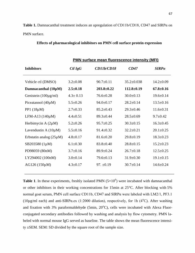
67
Table 1. Damnacanthal treatment induces an upregulation of CD11b/CD18, CD47 and SIRPα on
PMN surface.
Effects of pharmacological inhibitors on PMN cell surface protein expression
PMN surface mean fluorescence intensity (MFI)
Inhibitors Ctl IgG CD11b/CD18 CD47 SIRPα
Vehicle ctl (DMSO) 3.2±0.08 90.7±0.11 35.2±0.038 14.2±0.09
Damnacanthal (10µM) 2.5±0.18 203.8±0.22 112.8±0.19 67.8±0.16
Genistein (100µg/ml) 4.3± 0.13 76.6±0.28 30.0±0.13 19.6±0.14
Piceatannol (40µM) 5.5±0.26 94.0±0.17 28.2±0.14 13.5±0.16
PP1 (18µM) 2.7±0.33 85.2±0.43 29.3±0.46 11.6±0.31
LFM-A13 (140µM) 4.4±0.51 89.3±0.44 28.5±0.69 9.7±0.42
Herbimycin A (2µM) 5.2±0.26 95.7±0.25 30.3±0.15 16.3±0.45
Lavendustin A (10µM) 5.5±0.16 91.4±0.32 32.2±0.21 20.1±0.25
Erbstatin analog (25µM) 4.8±0.17 81.6±0.20 29.8±0.19 18.3±0.23
SB203580 (1µM) 6.1±0.30 83.8±0.40 28.8±0.15 15.2±0.23
PD98059 (80nM) 3.7±0.16 89.9±0.24 26.7±0.18 12.5±0.25
LY294002 (100nM) 3.0±0.14 79.6±0.13 31.9±0.30 19.1±0.15
AG126 (150µM) 4.3±0.17 97. ±0.19 30.7±0.14 14.6±0.24
Table 1. In these experiments, freshly isolated PMN (5×10⁶) were incubated with damnacanthal
or other inhibitors in their working concentrations for 15min at 25°C. After blocking with 5%
normal goat serum, PMN cell surface CD11b, CD47 and SIRPα were labeled with LM2/1, PF3.1
(10µg/ml each) and anti-SIRPα.ex (1:2000 dilution), respectively, for 1h (4oC). After washing
and fixation with 3% paraformaldehyde (5min, 20oC), cells were incubated with Alexa Fluor-
conjugated secondary antibodies followed by washing and analysis by flow cytometry. PMN la-
beled with normal mouse IgG served as baseline. The table shows the mean fluorescence intensi-
ty ±SEM. SEM: SD divided by the square root of the sample size.

68
Figure 3-1. Damnacanthal treatment induces specific release of PMN secondary granules. Fresh-
ly isolated PMN were treated with damnacanthal (Damn) of varied concentrations and other in-
hibitors for 15min at 25°C. A control treatment was performed using vehicle (DMSO) of the
same dilution (0.1%). After centrifugation to pellet cells, PMN degranulation was assessed by
assaying different granular markers in the cell-free supernatants. Positive degranulation controls
were performed by stimulation of PMN with 1µM fMLF with and without an addition of 10µM
cytochalasin B (CB). Concentrations of LY94002, Piceatannol, SB203580, genistein and
PD98059 used in the treatment were the same as those in Table 1. A) Assaying secondary gra-
nule release by measuring lactoferrin in the supernatants. B) Assaying primary granule release by
measuring MPO activity in the supernatants. C) Assaying tertiary granule release by zymogram
detecting gelatinase in the supernatants. The arrows indicate the two major forms of gelatinase
(92kD and 56kD, respectively) in PMN. As shown, damnacanthal (Damn.) specifically and dose-
dependently induced secondary granule degranulation. Data (mean ± SD) represent more than
five independent experiments. Significant degranulation was marked by “*”.
A)L
ac
tofe
rrin
(O
D4
05
)
0
1
2
3
4 **
*
*
*
Secondary granule releaseB)
MP
O (
OD
40
5)
0
1
2
3
4 *
Primary granule release
C)(Gelatinase zymogram)
* *
Tertiary granule release

69
Damnacanthal treatment results in defect of PMN function.
In general, PMN degranulation of secondary granules is triggered by chemoattractants binding to
the cell surface receptors followed by signaling relays involving G protein and a series of down-
stream signaling molecules [208]. Since damnacanthal caused release of secondary granules in
the absence of chemoattractant stimulation, I questioned whether this kind of “dysregulated” de-
granulation would affect PMN function and thus assayed chemoattractant-induced PMN trans-
migration and adhesion/spreading. As shown in Figure 3-2, in contrast to that PMN vigorously
transmigrated towards fMLF in the control condition, damnacanthal dose-dependently inhibited
PMN transmigration and completely abolished PMN migration at a concentration of 18μM. Also
as shown in Figure 3-3A, compared to control PMN which responded to fMLF stimulation and
quickly spread on collagen-coated surfaces displaying a polarized F-actin labeling pattern, dam-
nacanthal treatment impaired PMN spreading. Indeed, PMN adhesion to collagen induced by
fMLF was significantly weakened in the presence of damnacanthal (Figure 3-3B). Next I ex-
amined whether damnacanthal treatment affected cell surface protein function. Since CD47 and
SIRPα are extracellular counter-ligands [68, 87], we thus tested cell surface CD47 direct binding
to a SIRPα extracellular domain fusion protein, SIRPα1.ex-Fc [70, 87]. As shown in Figure 3-4,
although damnacanthal induced a significant increase of CD47 on PMN cell surface, these upre-
gulated CD47 were not binding to its ligand SIRPα. In contrast, CD47 was also increased on
PMN surface after fMLF stimulation, and these CD47 directly bound to SIRPα1.ex-Fc (Figure 3-
4).

70
Figure 3-2. Damnacanthal inhibits chemoattractant-triggered PMN function. A) Damnacanthal
inhibits PMN chemotaxis. PMN (106) were treated with damnacanthal of varying concentrations
for 15min at 25°C followed by assaying transmigration across collagen-coated filters towards
fMLF (0.1µM) in 1h. Data (mean ± SD) represent three independent experiments with triplicates
in each condition (*, p<0.05, **, p<0.01).
Damnacanthal
Tra
ns
mig
rate
d P
MN
(x1
04)
0.9
mM
3.6
mM
9.0
mM
18
mM
1.8
mM
*
**
Ve
hic
le
0
10
20
30
40
50
-fM
LF
Ve
hic
le
+ fMLF

71
Figure 3-3. Damnacanthal inhibits chemoattractant-triggered PMN function. Damnacanthal in-
hibits PMN F-actin polarization (A) and spreading (B). PMN (106), untreated or treated with
damnacanthal (10 µM), were stimulated with fMLF (1µM, 37°C) in collagen-coated wells fol-
lowed by observation of PMN adhesion and spreading (phase-contrast images). After stimulation
for 20min, the cells were then fixed and permeabilized followed by labeling F-actin with rhoda-
mine-conjugated phalloidin.
Vehicle Damnacanthal
+ fMLF
F-a
cti
n
20µm
Vehicle
- fMLF
Vehicle Damnacanthal
+ fMLF
20µm
A)
B)
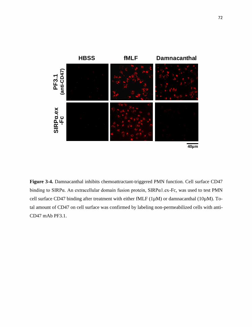
72
Figure 3-4. Damnacanthal inhibits chemoattractant-triggered PMN function. Cell surface CD47
binding to SIRPα. An extracellular domain fusion protein, SIRPα1.ex-Fc, was used to test PMN
cell surface CD47 binding after treatment with either fMLF (1µM) or damnacanthal (10µM). To-
tal amount of CD47 on cell surface was confirmed by labeling non-permeabilized cells with anti-
CD47 mAb PF3.1.
PF
3.1
(an
ti-C
D4
7)
SIR
Pα
.ex
-Fc
fMLF DamnacanthalHBSS
40µm
73
Identification of the specific GTPase associated with secondary granule release.
Efforts have been made to identify the target molecule of damnacanthal that plays an important
role in preventing secondary granule release in PMN prior to chemoattractant stimulation. We
have found that, although damnacanthal is a potent inhibitor of the tyrosine kinase Lck, its effect
on PMN degranulation was not through targeting Lck. In agreement with many studies by others,
I observed that Lck is mainly expressed in T cells but not in PMN (Figure 3-5). In addition, Dr.
Liu obtained Lck-knockout mice [209] from Dr. Tak Mak (Ontario Cancer Institute, Canada).
Comparing PMN isolated from Lck knockout mice with the cells from their wild-type littermates
yielded no difference in the levels of cell surface protein expression, PMN response to chemoat-
tractant stimulation, degranulation and chemotactic transmigration (data not shown), suggesting
that Lck is dispensable in PMN function. A previous study by Brumell et al. also indicated no
Lck activity in PMN[210]. In addition to Lck, damnacanthal is also an established inhibitor of
Ras family small GTPases [206-207]. Since some of these GTPases, such Ras family member
Ral, have been demonstrated to play important roles in vesicle transportation, thus it was likely
that the effect of damnacanthal on PMN through inhibition of Ras family members.
The Ras family small GTPases are traditionally classified into three major subfamilies, Ras (H,
R, etc.), Rap (1/2) and Ral (A/B) [211-213]. In addition, three new members of Ras family small
GTPases have been identified recently including Rheb, Rin, and Rit [214]. To identify which is
potentially involved in the degranulation of secondary granules, I screened the Ras members for
activity changes before and after chemoattractant stimulation. Given that damnacanthal treatment
caused secondary granule release in the absence of stimulation, I speculated that the candidate
Ras member must be in its active form in resting PMN. Also, since chemoattractant stimulation

74
triggers secondary granule release, we reasoned that this specific Ras should be deactivated in
order to coordinate secondary granule release. Based on this rationale, I assayed the activities of
Ras, Rap and Ral in PMN before and after fMLF stimulation, and with or without damnacanthal
treatment. As shown in Figure 3-6, Ras, Rap and Ral were all expressed in PMN. Among the
three family members, Ras was found mainly in its inactive form in unstimulated PMN and was
quickly activated after fMLF stimulation. Rap, on the other hand, showed minor activity changes
before or after fMLF stimulation and, to our surprise, was insensitive to damnacanthal treatment
(Figure 3-6). Therefore, neither Ras nor Rap was likely the candidate that regulates secondary
granule release.
In contrast to Ras and Rap, Ral demonstrated a dynamic activity change that correlated with
chemoattractant stimulation and PMN degranulation. As shown in Figure 3-6, Ral appeared to be
in its active form in unstimulated PMN, and quickly became inactive following fMLF stimula-
tion. Pull-down experiments using RalBP1, a downstream effector that specifically binds to ac-
tive Ral, recovered significant amount of active Ral from resting PMN. On the other hand, stimu-
lation with fMLF resulted in a quick diminishing of RalBP1 pull-down within 2-5min, a time
course paralleled with secondary granule release that was assayed simultaneously (Figure 3-6,
right). Same results of were obtained when PMN were stimulated by another chemoattractant IL-
8 (Figure 3-7). Deactivation of Ral was also observed in PMN that had chemotactically transmi-
grated across collaged-coated filters towards fMLF (Figure 3-6, migrated). In addition, activity
of Ral in resting PMN was inhibited after damnacanthal treatment (Figure 3-6), which correlated
with damnacanthal-induced secondary granule release. Thus, these results suggested Ral to be
the regulator that controls secondary granule release in PMN.

75
Figure 3-5. Lck is not expressed in neutrophils (PMN). A) Western blot detecting Lck in PMN
and PBMC. B) Immunofluorescence labeling demonstrating that Lck is expressed in SIRPα-
negative cells in PBMC, mainly lymphocytes, but not in SIRPα-positive monocytes or PMN.
(Antibody against Lck was purchased from Cell Signaling Technology.) Note: SIRPα is re-
stricted in myeloid lineages, including PMN, monocytes and dendritic cells, but not expressed in
lymphocytes. Few Lck-positive cells detected in isolated PMN were likely due to an unavoidable
contamination of PBMC. In general, PMN are separated from PBMC by Ficoll-Paque density
gradient (used for this study) or gelatin sedimentation. The former method normally produces
PMN of 90-95% purity while the latter method produces PMN of varied purity, between 70-
90%. Expression of Lck in eosinophils was also reported (Stafford et al. The Journal of Immu-
nology, 2002; Lynch et al. Blood, 2000) and may account for few Lck-positive cells in the prepa-
ration of PMN.
Anti-Lck
PB
MC
PM
N
Anti-β actin
55kD
42kD
A)
WB by:
B)
Phase MergeAnti-LckAnti-SIRPα.ex
PBMC
PMN
50µm
76
To further explore the mechanism that regulates Ral activity in PMN, the localization of RalGDS
in PMN was investigated (Figure 4-1). The effect of Ca2+
and Mg2+
on Ral activity was ex-
amined (Figure 4-2, 4-3, 4-4). Results showed that Mg2+
was the potential regulator in control-
ling Ral activity.
To confirm my studies in PMN and verify the pull-down assays, in parallel experiments, I simul-
taneously assayed Ral activity in platelets. As shown in Figure 3-7B, RalBP1 pull-down results
agreed with the previous report by others showing that Ral is inactive in resting platelets and
activated after thrombin stimulation [163]. The different Ral activity profiles in PMN and plate-
lets thus suggest that these cells employ distinctive signaling mechanisms to regulate Ral activity
and that Ral may have different functional aspects in PMN and platelets.
In addition, Ral activity was also assayed in PMN isolated directly from the “buffy coat” (Figure
3-8) or isolated using lympholyte-poly (Figure 3-9, Figure 3-10) to further investigate the effects
of Ral activity on PMN secondary granule release.
Comparing PMN isolated using DEAE-DEXTRAN with the cells isolated from the “buffy coat”,
Ral demonstrated the same activity profile. As shown in Figure 3-8, Ral was in its active form in
unstimulated PMN, and became inactive following fMLF stimulation.
Ral in PMN isolated using commercial separation chemical, lympholyte-poly, however, retained
minimal activity in unstimulated cells (Figure 3-9 and Figure 3-10). fMLF stimulation (Figure 3-
9) rendered a quick activation of Ral within 1 min, and a prompt diminishment of Ral activity
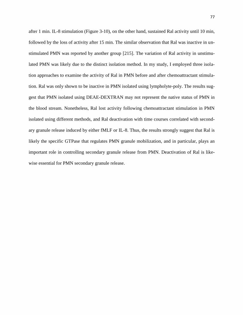
77
after 1 min. IL-8 stimulation (Figure 3-10), on the other hand, sustained Ral activity until 10 min,
followed by the loss of activity after 15 min. The similar observation that Ral was inactive in un-
stimulated PMN was reported by another group [215]. The variation of Ral activity in unstimu-
lated PMN was likely due to the distinct isolation method. In my study, I employed three isola-
tion approaches to examine the activity of Ral in PMN before and after chemoattractant stimula-
tion. Ral was only shown to be inactive in PMN isolated using lympholyte-poly. The results sug-
gest that PMN isolated using DEAE-DEXTRAN may not represent the native status of PMN in
the blood stream. Nonetheless, Ral lost activity following chemoattractant stimulation in PMN
isolated using different methods, and Ral deactivation with time courses correlated with second-
ary granule release induced by either fMLF or IL-8. Thus, the results strongly suggest that Ral is
likely the specific GTPase that regulates PMN granule mobilization, and in particular, plays an
important role in controlling secondary granule release from PMN. Deactivation of Ral is like-
wise essential for PMN secondary granule release.
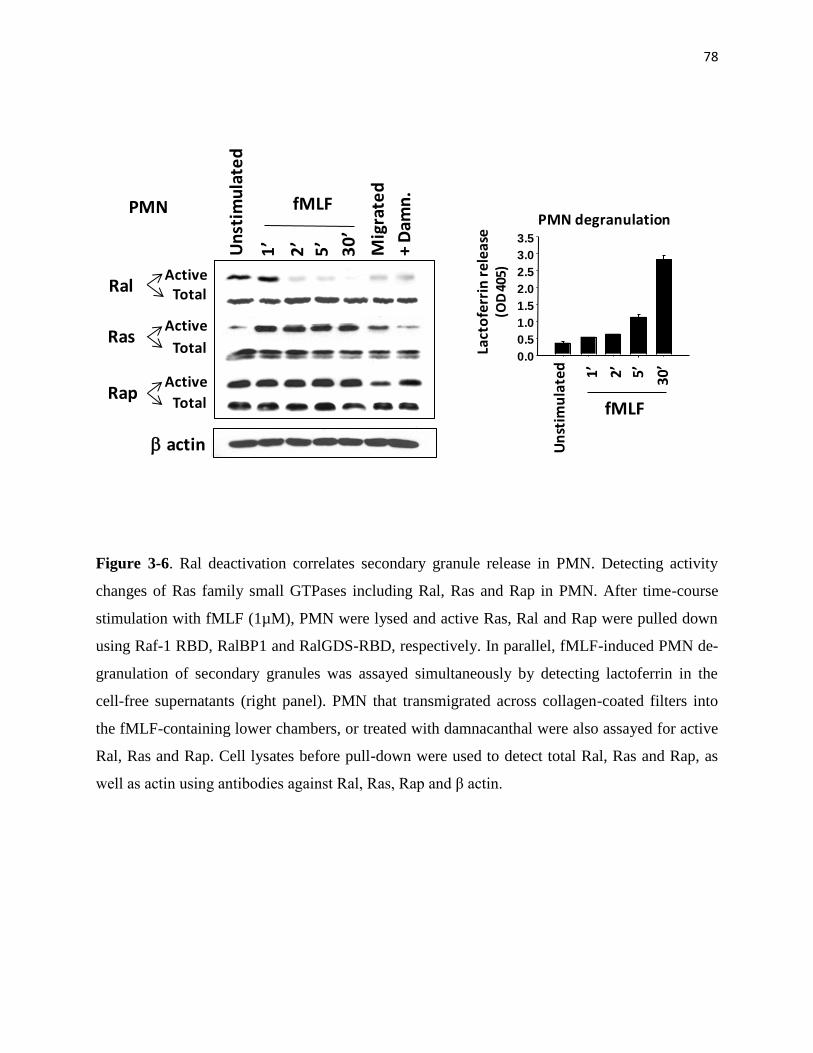
78
Figure 3-6. Ral deactivation correlates secondary granule release in PMN. Detecting activity
changes of Ras family small GTPases including Ral, Ras and Rap in PMN. After time-course
stimulation with fMLF (1µM), PMN were lysed and active Ras, Ral and Rap were pulled down
using Raf-1 RBD, RalBP1 and RalGDS-RBD, respectively. In parallel, fMLF-induced PMN de-
granulation of secondary granules was assayed simultaneously by detecting lactoferrin in the
cell-free supernatants (right panel). PMN that transmigrated across collagen-coated filters into
the fMLF-containing lower chambers, or treated with damnacanthal were also assayed for active
Ral, Ras and Rap. Cell lysates before pull-down were used to detect total Ral, Ras and Rap, as
well as actin using antibodies against Ral, Ras, Rap and β actin.
PMN degranulation
Un
stim
ula
ted
2’ 5’ 30’
1’
Lact
ofe
rrin
re
leas
e(O
D 4
05)
fMLF
Un
stim
ula
ted
fMLF
2’
5’
30’
Mig
rate
d
+ D
amn
.
PMN
b actin
1’
ActiveTotal
Active
Total
Active
Total
Ral
Ras
Rap
0.0
0.5
1.0
1.5
2.0
2.5
3.0
3.5
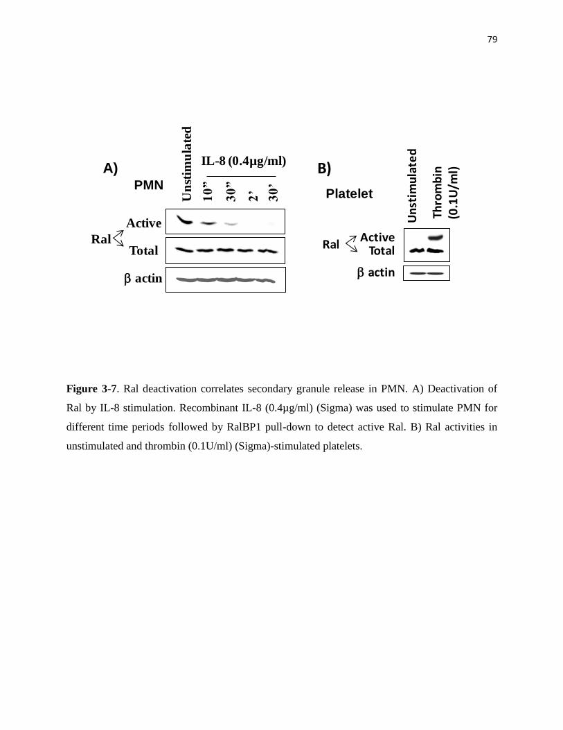
79
Figure 3-7. Ral deactivation correlates secondary granule release in PMN. A) Deactivation of
Ral by IL-8 stimulation. Recombinant IL-8 (0.4µg/ml) (Sigma) was used to stimulate PMN for
different time periods followed by RalBP1 pull-down to detect active Ral. B) Ral activities in
unstimulated and thrombin (0.1U/ml) (Sigma)-stimulated platelets.
B)
Thro
mb
in(0
.1U
/ml)
ActiveTotal
b actin
Platelet
Un
stim
ula
ted
Ral
Un
stim
ula
ted
IL-8 (0.4µg/ml)
10”
30”
2’
30’
A)PMN
Active
Total
b actin
Ral
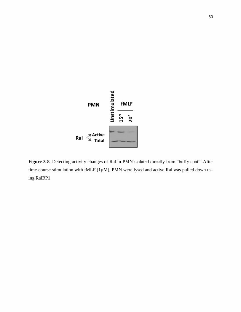
80
Figure 3-8. Detecting activity changes of Ral in PMN isolated directly from “buffy coat”. After
time-course stimulation with fMLF (1µM), PMN were lysed and active Ral was pulled down us-
ing RalBP1.
ActiveTotalRal
Un
stim
ula
ted
fMLF
20’
PMN
15’’
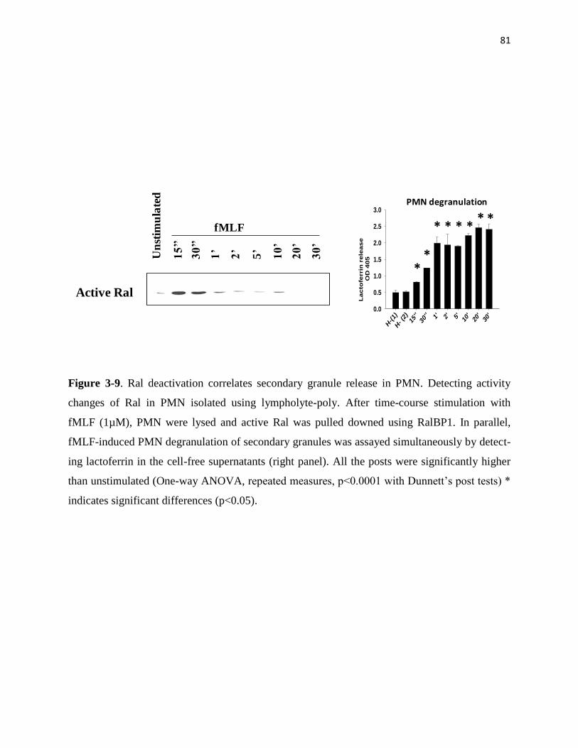
81
Figure 3-9. Ral deactivation correlates secondary granule release in PMN. Detecting activity
changes of Ral in PMN isolated using lympholyte-poly. After time-course stimulation with
fMLF (1µM), PMN were lysed and active Ral was pulled downed using RalBP1. In parallel,
fMLF-induced PMN degranulation of secondary granules was assayed simultaneously by detect-
ing lactoferrin in the cell-free supernatants (right panel). All the posts were significantly higher
than unstimulated (One-way ANOVA, repeated measures, p<0.0001 with Dunnett’s post tests) *
indicates significant differences (p<0.05).
fMLF
15’’
30’’
20’
1’
2’
5’
10’
Un
stim
ula
ted
Active Ral
30’
H-(1)
H- (2
)15
''30
'' 1' 2' 5' 10'
20'
30'
Lac
tofe
rrin
rele
ase
OD
405
0.0
0.5
1.0
1.5
2.0
2.5
3.0PMN degranulation
**
* * * ** *

82
Figure 3-10. Ral deactivation correlates secondary granule release in PMN. Detecting activity
changes of Ral in PMN isolated using lympholyte-poly. After time-course stimulation with re-
combinant IL-8 (0.4µg/ml) (Sigma), PMN were lysed and active Ral was pulled downed using
RalBP1. In parallel, fMLF-induced PMN degranulation of secondary granules was assayed si-
multaneously by detecting lactoferrin in the cell-free supernatants (right panel). All the posts
were significantly higher than unstimulated (One-way ANOVA, repeated measures, p<0.0001
with Dunnett’s post tests) * indicates significant differences (p<0.05).
Un
stim
ula
ted
IL-8 (4µg/ml)
30’’
15’
Active
TotalRal
PMN degranulation
Un
stim
ula
ted
15’
30’’
Lact
ofe
rrin
rel
ease
(OD
405
)
IL-8
0.0
0.5
1.0
1.5
2.0
2.5
3.0
H-(1
)
H- (
2)15
''30
'' 1' 2' 5' 10'
30'
0.0
0.5
1.0
1.5
2.0
2.5
IL-8 (4µg/ml)
A)
B)
Un
stim
ula
ted
(2)
IL-8 (4µg/ml)
15’’
30’’
30’
1’ 2’ 5’ 10’
Un
stim
ula
ted
(1)
Active Ral Lact
ofe
rrin
re
leas
e(O
D 4
05)
*
*
* * **
**
*

83
Active Ral blocks secondary granule release in PMN.
To test that active Ral in resting PMN serves to prevent secondary granule release and that Ral
deactivation in response to chemoattractant stimulation is required for degranulation, I designed
experiments to over-express a constitutively active Ral, Ral23V, in PMN and tested its effect on
fMLF-induced degranulation. Since freshly isolated PMN are short-lived and not suitable for
protein expression by conventional gene transfection methods, I employed protein transduction
technology and directly delivered purified recombinant Ral into PMN by tagging the proteins
with an HIV Tat peptide (YGRKKRRQRRR) [54, 200, 216]. As demonstrated by previous stu-
dies, HIV Tat and other poly-arginine peptides have been widely used to transduce proteins of up
to 120kD into live cells [54, 200, 216] . Although PMN are highly sensitive and difficult to han-
dle, using HIV Tat to study important intracellular molecules has been very successful [54, 217-
218]. In this study, three Tat-tagged Ral proteins were generated, including a wild-type Ral
(WT), a constitutively active mutant Ral (Ral23V), and a dominant negative mutant Ral
(Ral28N) [117, 167, 202]. As depicted in Figure 3-11, these proteins were first produced as re-
combinant GST fusions, followed by removal of GST and yielding Tat-tagged Ral. Figure 3-
11B&C shows the stepwise production of Tat-tagged WT, Ral23V and Ral28N, and RalBP1
pull-down confirming that Ral23V was a constitutively active form.
To determine that Tat-tagged Ral proteins were capable of directly delivery into PMN, I chemi-
cally labeled the purified proteins with fluorescein (FITC), followed by incubation with PMN.
As shown in Figure 3-12A, fluorescence microscopy revealed that the three Tat-tagged Ral pro-
teins (Tat-WT, Tat-Ral23V and Tat-Ral28N) were all rapidly delivered into PMN after only a
short incubation (10min) at a low temperature (4°C). Subsequent Western blot detected an over

84
two-folds increase of Ral in PMN after incubating with each Tat-tagged Ral protein (Figure 3-
12B). I also performed RalBP1 pull-down assays to test Ral activity after delivery of each Tat-
tagged Ral protein into PMN. As shown in Figure 3-13, transduction of exogenous Ral23V into
PMN led to a sustained Ral activity in PMN even in the presence of fMLF stimulation, a condi-
tion that the endogenous Ral activity was completely diminished as that observed in the control,
non-peptide transduction PMN. Transduction of WT Ral or Ral28N, on the other hand, only
slightly (WT) or did not (Ral28N) increase Ral activity in PMN (Figure 3-13).
I next investigated whether Ral activity blocks fMLF-induced PMN degranulation. In these ex-
periments, PMN were first loaded with Tat-tagged WT Ral, Ral23V or Ral28N, and then stimu-
lated with fMLF. As shown in Figure 3-14A, no spontaneous PMN degranulation was detected
by just loading Tat-tagged Ral proteins (unstimulated). Prominent release of secondary granules
was induced by fMLF in PMN loaded with WT Ral or Ral28N, or without peptide delivery. On
the contrary, transduction of the constitutively active Ral, Ral23V, into PMN strikingly blocked
secondary granule release in the presence of fMLF. As shown in Figure 3-14, both lactoferrin
release (Figure 3-14A) and CD47 translocation to the cell surface plasma membrane (Figure 3-
14B) were inhibited by Ral23V in PMN in spite of fMLF stimulation. However, Ral23V had no
effect on fMLF-induced tertiary granule release or the retention of primary granules (Figure 3-
15A), further suggesting that Ral selectively regulates secondary granule mobilization.
Given that PMN degranulation of secondary granules is crucial for chemotaxis, I thus performed
PMN transmigration assays and examined the effect of Ral23V. As shown in Figure 3-15B, as
expected, transduction of Ral23V into PMN significantly reduced PMN transmigration towards
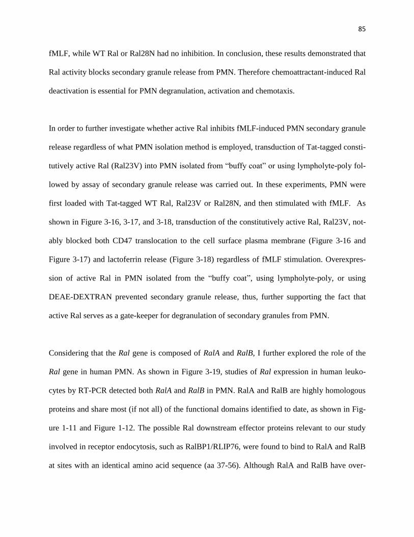
85
fMLF, while WT Ral or Ral28N had no inhibition. In conclusion, these results demonstrated that
Ral activity blocks secondary granule release from PMN. Therefore chemoattractant-induced Ral
deactivation is essential for PMN degranulation, activation and chemotaxis.
In order to further investigate whether active Ral inhibits fMLF-induced PMN secondary granule
release regardless of what PMN isolation method is employed, transduction of Tat-tagged consti-
tutively active Ral (Ral23V) into PMN isolated from “buffy coat” or using lympholyte-poly fol-
lowed by assay of secondary granule release was carried out. In these experiments, PMN were
first loaded with Tat-tagged WT Ral, Ral23V or Ral28N, and then stimulated with fMLF. As
shown in Figure 3-16, 3-17, and 3-18, transduction of the constitutively active Ral, Ral23V, not-
ably blocked both CD47 translocation to the cell surface plasma membrane (Figure 3-16 and
Figure 3-17) and lactoferrin release (Figure 3-18) regardless of fMLF stimulation. Overexpres-
sion of active Ral in PMN isolated from the “buffy coat”, using lympholyte-poly, or using
DEAE-DEXTRAN prevented secondary granule release, thus, further supporting the fact that
active Ral serves as a gate-keeper for degranulation of secondary granules from PMN.
Considering that the Ral gene is composed of RalA and RalB, I further explored the role of the
Ral gene in human PMN. As shown in Figure 3-19, studies of Ral expression in human leuko-
cytes by RT-PCR detected both RalA and RalB in PMN. RalA and RalB are highly homologous
proteins and share most (if not all) of the functional domains identified to date, as shown in Fig-
ure 1-11 and Figure 1-12. The possible Ral downstream effector proteins relevant to our study
involved in receptor endocytosis, such as RalBP1/RLIP76, were found to bind to RalA and RalB
at sites with an identical amino acid sequence (aa 37-56). Although RalA and RalB have over-

86
lapping functions, which have been reported by various studies, I further investigated the specific
role of RalB in PMN secondary granule release. In these experiments, I generated a Tat-tagged
constitutively active RalB (RalB23V) protein, followed by transduction of the protein into PMN,
and assayed its effect on secondary granule release. Transduction of RalB23V into PMN also
inhibited PMN mobilization of secondary granules shown by impaired lactoferrin release and
blockage of CD47 translocation to plasma membrane (Figure 3-20).

87
Figure 3-11. Generation of Tat-tagged wild-type Ral (WT), constitutively active Ral (Ral23V),
and dominant negative Ral (28N) protein. A) A schematic depiction of Tat-tagged Ral produc-
tion. The recombinant fusion proteins containing GST and Tat-Ral were produced, followed by
cleavage of GST with Factor Xa to yield Tat-Ral peptides. The figure also shows that two point
mutations, G23V and S28N, were created in Ral to produce constitutively active Ral (23V) and
dominant negative Ral (28N). B) A stepwise analysis of Tat-tagged Ral protein production by
SDS-PAGE and Coomassie blue staining. C) RalBP1 pull-down confirming that Tat-Ral23V was
a constitutively active GTPase. Purified Tat-tagged wild-type Ral, Ral23V and Ral28N (0.2µg
each) were incubated with GST-RalBP1 (1µg) and glutathione Sepharose in HBSS containing
5% non-fat dry milk, 2% BSA and 0.01% Tween 20 for 1h. After washing the precipitates,
Western blot was performed to detect active Ral pulled down by RalBP1.
A)
S28N (dominant negative)
GST RalN
+ Factor Xa
GSTTat-Ral
Ta
t
G23V (constitutively active)
Ra
l 2
8N
Ra
l 2
3V
WT
46
30
25
58
KDa
B)
GST
GST-Tat-Ral
Tat-Ral
WT
Ra
l23
V
Ra
l28
N
Cle
av
ag
e
Elu
tio
n
Cle
av
ag
e
Elu
tio
n
Cle
av
ag
e
Elu
tio
n
Ta
t-W
T
Ta
t-R
al2
3V
Ta
t-R
al2
8N
C)
Pre-bound
RalBP1 pull-
down
WB: Anti-Ral

88
Figure 3-12. Transduction of Tat-tagged Ral proteins including wild-type Ral (WT), constitu-
tively active Ral (Ral23V), and dominant negative Ral (28N) to PMN. A) Assaying Tat-tagged
Ral transduction into PMN. Tat-tagged wild-type Ral (WT), Ral23V and Ral28N (0.1µM of
each) were labeled with FITC followed by incubation with PMN for 10min at 4°C. B) Effective
transduction into PMN was then analyzed by Western blot.
Ta
t-R
al2
3V
Incubating
PMN with: -
Ta
t-W
T
Ta
t-R
al2
8N
Total Ral
β actin
A)
Tat-WT
Tat-Ral23V
Tat-Ral28N
No peptide
FITC Phase
100µm
B)
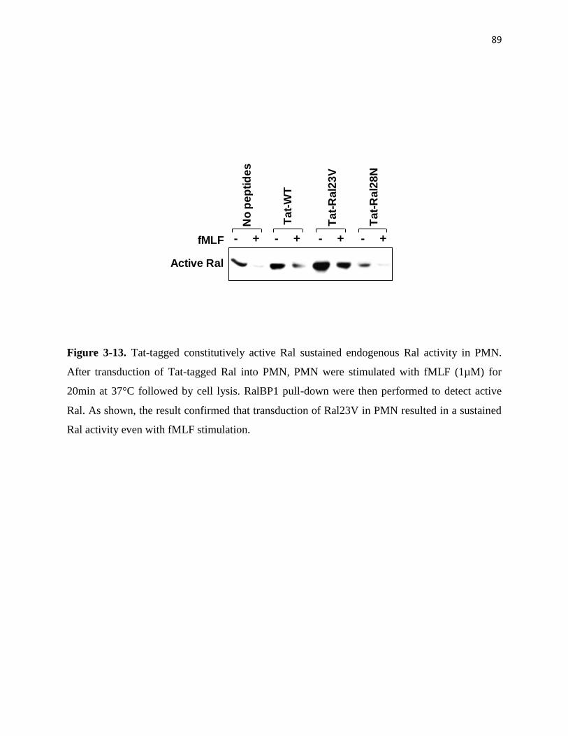
89
Figure 3-13. Tat-tagged constitutively active Ral sustained endogenous Ral activity in PMN.
After transduction of Tat-tagged Ral into PMN, PMN were stimulated with fMLF (1µM) for
20min at 37°C followed by cell lysis. RalBP1 pull-down were then performed to detect active
Ral. As shown, the result confirmed that transduction of Ral23V in PMN resulted in a sustained
Ral activity even with fMLF stimulation.
Ta
t-R
al2
3V
Ta
t-R
al2
8N
Active Ral
fMLFN
o p
ep
tid
es
Ta
t-W
T
+- +- +- +-
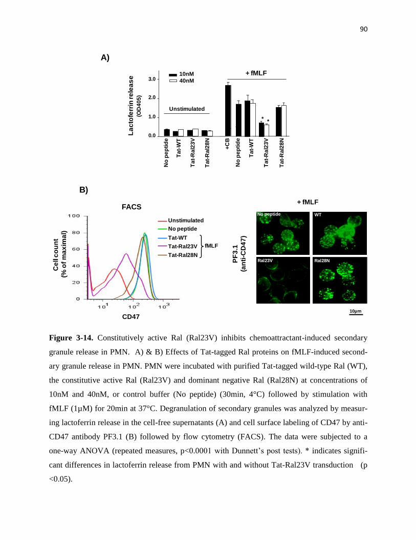
90
Figure 3-14. Constitutively active Ral (Ral23V) inhibits chemoattractant-induced secondary
granule release in PMN. A) & B) Effects of Tat-tagged Ral proteins on fMLF-induced second-
ary granule release in PMN. PMN were incubated with purified Tat-tagged wild-type Ral (WT),
the constitutive active Ral (Ral23V) and dominant negative Ral (Ral28N) at concentrations of
10nM and 40nM, or control buffer (No peptide) (30min, 4°C) followed by stimulation with
fMLF (1µM) for 20min at 37°C. Degranulation of secondary granules was analyzed by measur-
ing lactoferrin release in the cell-free supernatants (A) and cell surface labeling of CD47 by anti-
CD47 antibody PF3.1 (B) followed by flow cytometry (FACS). The data were subjected to a
one-way ANOVA (repeated measures, p<0.0001 with Dunnett’s post tests). * indicates signifi-
cant differences in lactoferrin release from PMN with and without Tat-Ral23V transduction (p
<0.05).
A)
No
pe
pti
de
La
cto
ferr
in re
lea
se
(OD
40
5)
Ta
t-W
T
Ta
t-R
al2
3V
Ta
t-R
al2
8N
+ fMLF
0.0
1.0
2.0
3.0
* *
+C
B
Unstimulated
10nM
40nM
No
pe
pti
de
Ta
t-W
T
Ta
t-R
al2
3V
Ta
t-R
al2
8N
B)
Ral23V
WT
Ral28N
No peptide
+ fMLF
PF
3.1
(an
ti-C
D4
7)
Unstimulated
No peptide
Tat-WT
Tat-Ral23V
Tat-Ral28N
fMLF
CD47
Ce
ll c
ou
nt
(% o
f m
ax
ima
l)
FACS
10µm
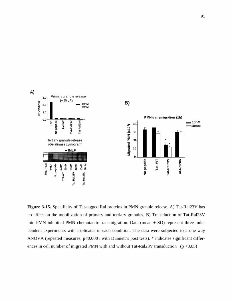
91
Figure 3-15. Specificity of Tat-tagged Ral proteins in PMN granule release. A) Tat-Ral23V has
no effect on the mobilization of primary and tertiary granules. B) Transduction of Tat-Ral23V
into PMN inhibited PMN chemotactic transmigration. Data (mean ± SD) represent three inde-
pendent experiments with triplicates in each condition. The data were subjected to a one-way
ANOVA (repeated measures, p<0.0001 with Dunnett’s post tests). * indicates significant differ-
ences in cell number of migrated PMN with and without Tat-Ral23V transduction (p <0.05)
A)
(+ fMLF)
No
pep
tid
e
10nM40nM
MP
O (O
D405)
0.0
1.0
2.0
3.0
+C
B
Tat-
WT
Tat-
Ral2
3V
Tat-
Ral2
8N
Primary granule release
No
pep
tid
e
40n
M
10n
M
40n
M
10n
M
40n
M
10n
M
Tat-
WT
Tat-
Ral2
3V
Tat-
Ral2
8N
-fM
LF
fML
F+
CB
+ fMLF
(Gelatinase zymogram)
Tertiary granule release
40nM10nM
PMN transmigration (1h)
No
pe
pti
de
Ta
t-R
al2
3V
Ta
t-R
al2
8N
Ta
t-W
T
Mig
rate
d P
MN
(x
10
4)
0
10
20
30
40
*
*
B)
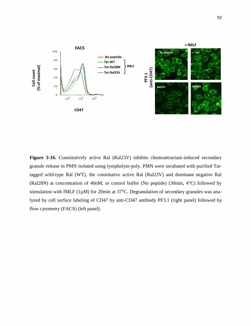
92
Figure 3-16. Constitutively active Ral (Ral23V) inhibits chemoattractant-induced secondary
granule release in PMN isolated using lympholyte-poly. PMN were incubated with purified Tat-
tagged wild-type Ral (WT), the constitutive active Ral (Ral23V) and dominant negative Ral
(Ral28N) at concentration of 40nM, or control buffer (No peptide) (30min, 4°C) followed by
stimulation with fMLF (1µM) for 20min at 37°C. Degranulation of secondary granules was ana-
lyzed by cell surface labeling of CD47 by anti-CD47 antibody PF3.1 (right panel) followed by
flow cytometry (FACS) (left panel).
Ral23V
WT
Ral28N
No peptide
+ fMLF
PF3
.1(a
nti
-CD
47
)
No peptide
Tat-WT
Tat-Ral23V
Tat-Ral28N fMLF
CD47
Ce
ll c
ou
nt
(% o
f m
axim
al)
FACS
Ral23V

93
Figure 3-17. Constitutively active Ral (Ral23V) inhibits chemoattractant-induced secondary
granule release in PMN isolated directly from “buffy coat”. PMN were incubated with purified
Tat-tagged wild-type Ral (WT), the constitutive active Ral (Ral23V) and dominant negative Ral
(Ral28N) at concentration of 40nM, or control buffer (No peptide) (30min, 4°C) followed by
stimulation with fMLF (1µM) for 20min at 37°C. Degranulation of secondary granules was ana-
lyzed by cell surface labeling of CD47 by anti-CD47 antibody PF3.1 (right panel) followed by
flow cytometry (FACS) (left panel).
Ral23V
WT
Ral28N
No peptide
+ fMLF
PF3
.1(a
nti
-CD
47
)
No peptide
Tat-WT
Tat-Ral23V
Tat-Ral28N fMLF
CD47
Ce
ll c
ou
nt
(% o
f m
axim
al)
FACS

94
Figure 3-18. Constitutively active Ral (Ral23V) inhibits chemoattractant-induced secondary
granule release in PMN isolated from “buffy coat” (A) and using lympholyte-poly (B). A) & B)
Effects of Tat-tagged Ral proteins on fMLF-induced secondary granule release in PMN. PMN
were incubated with purified Tat-tagged wild-type Ral (WT), the constitutive active Ral
(Ral23V) and dominant negative Ral (Ral28N) at concentration of 40nM, or control buffer (No
peptide) (30min, 4°C) followed by stimulation with fMLF (1µM) for 20min at 37°C. Degranula-
tion of secondary granules was analyzed by measuring lactoferrin release in the cell-free super-
natants. A) All the posts had significant difference compared with than no peptides (One-way
ANOVA, repeated measures, p<0.0002 with Dunnett’s post tests) B) All the posts had signifi-
cant difference compared with no peptide treatment (One-way ANOVA, repeated measures,
p<0.0001 with Dunnett’s post tests) * indicates significant differences in lactoferrin release from
PMN with and without Tat-Ral23V transduction (p<0.05).
0.0
0.5
1.0
1.5
2.0
2.5
Lact
ofe
rrin
re
leas
e(O
D 4
05)
+ fMLF
Un
sti
mu
late
d
No
pe
pti
de
Ta
t-W
T
Ta
t-R
al2
3V
Ta
t-R
al2
8N
fML
F+
CB
A) B)
0.0
0.5
1.0
1.5
2.0
2.5
3.0
3.5+ fMLF
Lact
ofe
rrin
re
lea
se(O
D 4
05)
Un
sti
mu
late
d
No
pe
pti
de
Ta
t-W
T
Ta
t-R
al2
3V
Ta
t-R
al2
8N
fML
F+
CB
**

95
Figure 3-19. Detection of RalA and RalB in human leukocytes. The RalA-specific and the
RalB-specific cDNA fragments, 777bps and 648bps, respectively, were RT-PCR amplified from
freshly isolated PMN and PMBC. The same DNA fragments were also PCR amplified from a
human leukocyte cDNA library (Marathon Ready cDNAs, Clontech). Given that RalA and RlaB
are highly homologous, the PCR primers were specially chosen based on the most dissimilar re-
gions of their cDNAs. The primers for amplifying RalA were 5-ttccaggcgacaaggaccg agta and 5-
aagataagaaaggagtttggg. The primers for amplifying RalB were 5-atggctgccaacaagagtaag and 5-
agctggcttcatccgtcacctt. The amplified PCR fragments were DNA sequenced to confirm their
identities.
RalA RalB
400500600700
bPP
MN
PB
MC
PM
N
PB
MC
Le
uk
ocyte
cD
NA
s
Le
uk
ocyte
cD
NA
s
800
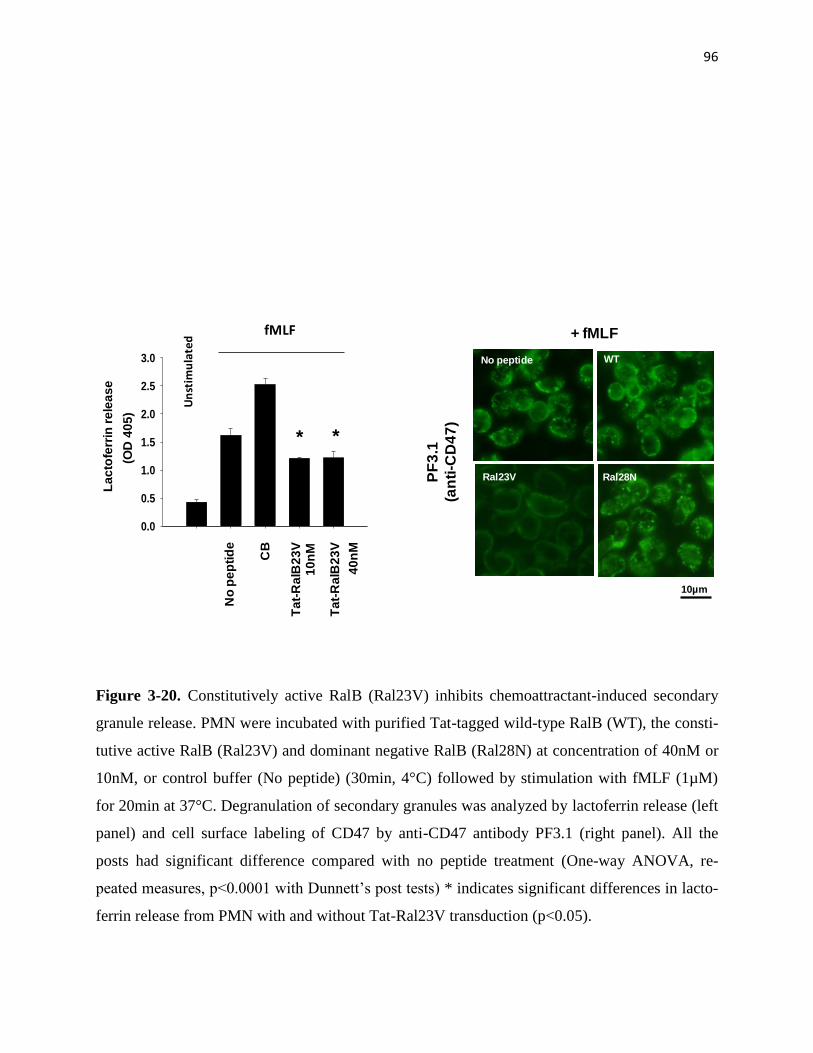
96
Figure 3-20. Constitutively active RalB (Ral23V) inhibits chemoattractant-induced secondary
granule release. PMN were incubated with purified Tat-tagged wild-type RalB (WT), the consti-
tutive active RalB (Ral23V) and dominant negative RalB (Ral28N) at concentration of 40nM or
10nM, or control buffer (No peptide) (30min, 4°C) followed by stimulation with fMLF (1µM)
for 20min at 37°C. Degranulation of secondary granules was analyzed by lactoferrin release (left
panel) and cell surface labeling of CD47 by anti-CD47 antibody PF3.1 (right panel). All the
posts had significant difference compared with no peptide treatment (One-way ANOVA, re-
peated measures, p<0.0001 with Dunnett’s post tests) * indicates significant differences in lacto-
ferrin release from PMN with and without Tat-Ral23V transduction (p<0.05).
No
pe
pti
de
Ta
t-R
alB
23
V
0.0
0.5
1.0
1.5
2.0
2.5
3.0
La
cto
ferr
in r
ele
as
e
(OD
40
5)
Un
stim
ula
ted
fMLF
CB
Ta
t-R
alB
23
V
40
nM
10
nM
Ral23V
WT
Ral28N
No peptide
+ fMLF
PF
3.1
(an
ti-C
D4
7)
10µm
* *

97
Ral translocation between plasma membranes and secondary granules
Like Ras family GTPases, Ral contains a lipid modification site at the carboxyl terminus, sug-
gesting that Ral may be associated with the cell membranes. To test this, I separated PMN mem-
branes from the cytosol by ultracentrifugation in the absence of detergent. As shown in Figure
10A, Western Blot indicated that Ral is exclusively associated with the cell membranes in PMN.
To further localize Ral and define whether Ral is dynamically coupled with secondary granules, I
performed subcellular fractionation using sucrose density gradients. As shown in Figure 3-21,
the fractionations successfully separated PMN plasma membranes (alkaline phosphatase) from
secondary granules (lactoferrin) and primary granules (MPO). To my surprise, detection of Ral
in the fractions by western blots revealed that Ral is entirely associated with the plasma mem-
brane in unstimulated PMN that is co-localized with another plasma membrane protein JAM-A.
Stimulation with fMLF, on the other hand, resulted in translocation of a considerable amount of
Ral to secondary granules (Figure 3-21B). In agreement with previous observation [68, 70], pro-
teins initially stored in secondary granules, such as SIRPα, were redistributed to the plasma
membrane after fMLF stimulation following degranulation. As predicted, RalBP1 pull-down re-
covered active Ral from the plasma membrane fractions of unstimulated PMN, but not from sec-
ondary granule-rich fractions (Figure 3-22). I also performed immunofluorescence labeling and
observed that, as shown in Figure 3-23, Ral in unstimulated PMN was co-localized with the
plasma membrane marker wheat germ agglutinin (WGA) [219]. Stimulation with fMLF induced
a significant dissociation of Ral from WGA, suggesting Ral redistribution in PMN.
One major mechanism of plasma membrane internalization in PMN after chemoattractant stimu-
lation is through clathrin-mediated cell surface receptor endocytosis [208, 220], and a previous

98
study demonstrated that a Ral effector is complexed with a component of clathrin pits [121].
Therefore, I examined whether fMLF-induced Ral translocation from the plasma membrane to
intracellular granules followed this pathway.
Two inhibitors, dynasore [193] and a AP-2 binding Eps15 C-terminal peptide (Eps15-DIII)[194-
195], which specifically act on the clathrin-dependent endocytosis were examined. As shown in
Figure 3-24A, pre-treatment of PMN with dynasore (80 µM) and recombinant Eps15-DIII (40
nM), or their combination, appeared to have no inhibition on Ral translocation to secondary gra-
nules after fMLF stimulation. Dynasore inhibits dynamin, an essential GTPase regulating clath-
rin-coated pit fission, while Eps15-DIII inhibits clathrin-mediated endocytosis through sequestra-
tion of AP-2 thus interferes formation of the clathrin coat. To effectively use Eps15-DIII in the
study, this peptide was tagged with HIV-Tat, which significantly facilitated protein transduction
into PMN (Figure 3-24B). I also confirmed the function of the Eps15-DIII by protein pull-down
assays, which demonstrated that Eps15-DIII successfully co-precipitated AP-2 from PMN ly-
sates (Figure 3-24C).
In addition to dynasore and a AP-2 binding Eps15 C-terminal peptide (Eps15-DIII), I also in-
cluded several other traditional endocytosis inhibitors that act on the clathrin-dependent endocy-
tosis including hypertonic sucrose (0.5M)[221], salt (0.76M NaCl)[222], phenylarsine oxide
(POA)[223], chlorpromazine (CPZ)[196], and cytosolic acidification (succinic acid, pH5.5)
[197-198]. As shown in Figure 3-25A, blocking clathrin-mediated endocytosis in PMN with
classical inhibitors resulted in a partial or complete inhibition of fMLF-induced secondary gra-
nule release. Remarkably, as shown in Figure 3-25B, these reagents also abolished Ral deactiva-
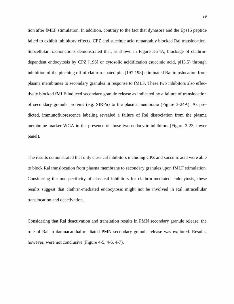
99
tion after fMLF stimulation. In addition, contrary to the fact that dynasore and the Eps15 peptide
failed to exhibit inhibitory effects, CPZ and succinic acid remarkably blocked Ral translocation.
Subcellular fractionations demonstrated that, as shown in Figure 3-24A, blockage of clathrin-
dependent endocytosis by CPZ [196] or cytosolic acidification (succinic acid, pH5.5) through
inhibition of the pinching off of clathrin-coated pits [197-198] eliminated Ral translocation from
plasma membranes to secondary granules in response to fMLF. These two inhibitors also effec-
tively blocked fMLF-induced secondary granule release as indicated by a failure of translocation
of secondary granule proteins (e.g. SIRPα) to the plasma membrane (Figure 3-24A). As pre-
dicted, immunofluorescence labeling revealed a failure of Ral dissociation from the plasma
membrane marker WGA in the presence of those two endocytic inhibitors (Figure 3-23, lower
panel).
The results demonstrated that only classical inhibitors including CPZ and succinic acid were able
to block Ral translocation from plasma membrane to secondary granules upon fMLF stimulation.
Considering the nonspecificity of classical inhibitors for clathrin-mediated endocytosis, these
results suggest that clathrin-mediated endocytosis might not be involved in Ral intracellular
translocation and deactivation.
Considering that Ral deactivation and translation results in PMN secondary granule release, the
role of Ral in damnacanthal-mediated PMN secondary granule release was explored. Results,
however, were not conclusive (Figure 4-5, 4-6, 4-7).

100
Figure 3-21. Localization of Ral in PMN. A) Ral is associated with membranes. PMN
(2.5×10⁷) were dounce-lysed followed by ultracentrifugation to separate cytosol from cell mem-
branes. SDS-PAGE and Western blot were performed to detect Ral in the total lysate before cen-
trifugation (total PMN) and in the separated cytosol and membranes. B) Subcellular localization
of Ral in PMN. PMN (108/per condition), unstimulated and fMLF-stimulated, were lysed and
subjected to sucrose density gradient centrifugation to separate the plasma membrane and intra-
cellular granules followed by fractionation. Western blots (lower panel) were performed to detect
Ral, SIRPα and JAM-A in the fractions using antibodies against Ral, anti-SIRPα.ex and anti-
JAM-A.ex. JAM-A is a transmembrane protein consistently expressed in the plasma membranes.
1-1011 12 13 14 15 16 17 18 19 20 21 22
0.0
0.5
1.0
1.5
2.0
2.5
3.0
SIRPα
# 1 11 12 13 14 15 16 17 18 19 20 21 22# 1 10 11 12 13 14 15 16 17 18 19 20 21 22
Ral
Mem
bra
nes
Cy
toso
l
To
tal P
MN
A)
Ral
B)
Fraction number (#)
1-9 10 11 12 13 14 15 16 17 18 19 20 21 22
0.0
0.5
1.0
1.5
2.0
2.5
3.0
B)Unstimulated PMN
Reco
vere
d a
ctiv
ity
(O
D 4
05)
Stimulated PMN
Reco
vere
d a
ctiv
ity
(O
D 4
05)Plasma membrane ( alkaline phosphatase)
Primary granules ( MPO)
Secondary granules ( lactoferrin)
Plasma membrane ( alkaline phosphatase)
Primary granules ( MPO)
Secondary granules ( lactoferrin)
Fraction number (#)
Fraction number
JAM-A
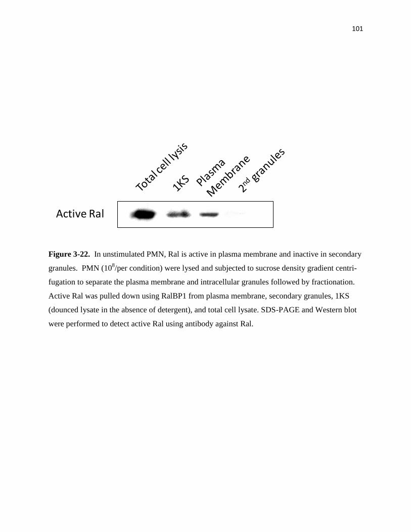
101
Figure 3-22. In unstimulated PMN, Ral is active in plasma membrane and inactive in secondary
granules. PMN (108/per condition) were lysed and subjected to sucrose density gradient centri-
fugation to separate the plasma membrane and intracellular granules followed by fractionation.
Active Ral was pulled down using RalBP1 from plasma membrane, secondary granules, 1KS
(dounced lysate in the absence of detergent), and total cell lysate. SDS-PAGE and Western blot
were performed to detect active Ral using antibody against Ral.
Active Ral
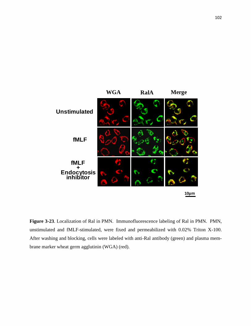
102
Figure 3-23. Localization of Ral in PMN. Immunofluorescence labeling of Ral in PMN. PMN,
unstimulated and fMLF-stimulated, were fixed and permeabilized with 0.02% Triton X-100.
After washing and blocking, cells were labeled with anti-Ral antibody (green) and plasma mem-
brane marker wheat germ agglutinin (WGA) (red).
Unstimulated
fMLF
fMLF +
Endocytosis inhibitor
WGA RalA Merge
10µm

103
Figure 3-24. Ral translocation from the plasma membrane to secondary granules may follow
fMLF-induced endocytic process. A) Effect of clathrin-dependent endocytic inhibitors on Ral
redistribution in PMN. In these experiments, PMN were pre-incubated with dynasore (80µM),
Tat-tagged Eps15 peptide (Eps15-DIII) (40nM), or a combination of the two reagents for 15min
(25°C) before fMLF stimulation (20min at 37°C). After cell lysis, subcellular fractionation was
performed followed by detection of Ral by Western blots. The effect of other endocytosis inhibi-
tors, CPZ (50µM) and succinic acid (20mM, pH5.5), on Ral distribution was also tested. B) Pro-
tein transduction of Tat-tagged Eps15-DIII into PMN. Purified Tat-tagged Eps15-DIII was fluo-
rescent labeled followed by incubating with PMN for 10min (4°C). The image shows that Eps15-
DIII was successfully delivered into the cells. C) Pull-down assay confirmed that Eps15-DIII di-
rectly binds to AP-2 complexes. PMN (1.5x10⁷) were lysed in a buffer containing 1% Trition X-
100 and protease inhibitors followed by incubation with 10µg purified Tat-tagged Eps15-DIII in
the form of GST fusion protein or its non-binding control peptide Tat-tagged Eps15-Δ for 3h
(4°C). After pull-down with glutathione Sepharose, the precipitated protein complexes were
washed and analyzed by western blot using antibody against α-adaptin, an important component
of AP-2 complexes.
A) B)
Eps15-DIII
No peptide
FITC Phase
20µm
pull-down
WB:Anti-α-adaptin
175kD
80kD
postbond
C)
Vehicle
CPZ
Plasma membrane
Ral
SIRPα
Ral
SIRPα
Ral
SIRPα
Dynasore
Ral
SIRPα
Dynasore+
Eps15-DIII
Eps15-DIII
Ral
SIRPα
Ral
SIRPα
+ fMLF
pH 5.5

104
Figure 3-25. Blockage of clathrin-dependent endocytosis inhibits fMLF-induced secondary gra-
nule release (A), Ral deactivation (B) and Ral translocation (C). In these experiments, PMN were
incubated with hypertonic solutions (0.5M sucrose and 0.76M NaCl), POA (20µM), CPZ
(50µM), succinic acid (20mM, pH5.5), or a mixture of later three reagents (combination) for
5min (25°C) before fMLF stimulation (20min at 37°C). Release of lactoferrin into the superna-
tants was measured (A). Ral activity was detected by RalBP1 pull down assays (B). All the posts
were significantly lower than HBSS and vehicle control (One-way ANOVA, repeated measures,
p<0.0001 with Dunnett’s post tests) * indicates significant differences (p<0.05). There was no
significant difference between HBSS and vehicle control (p>0.05).
A) B)
+ fMLF
pH
5.5
PO
A
Un
sti
mu
late
d
0.5
M s
ucro
se
CP
Z
Veh
icle
0.7
6M
NaC
l
Active Ral
Total Ral
β actin
Co
mb
inati
on
HB
SS
La
cto
ferr
in r
ele
as
e
(OD
40
5)
+ fMLF
Un
sti
mu
late
d
HB
SS
0.5
M s
ucro
se
0.7
6M
NaC
l
Veh
icle
pH
5.5
PO
A
CP
Z
Co
mb
inati
on
0.0
0.5
1.0
1.5
2.0
2.5
3.0
*
* * * * *

105
CHAPTER IV:
DISCUSSION
As a type of highly specialized and fast responsive cells, PMN store a large number of function-
ally important molecules in the intracellular granules and vesicles. Upon stimulation, PMN
promptly mobilize these granules/vesicles and rapidly release arrays of cell adhesion molecules,
receptors, and proteases to the cell surfaces to facilitate PMN adhesion, chemotaxis and microbi-
al killing. At least four types of granules and vesicles varied in size/shape and content have been
identified in PMN [25, 96], but the mechanisms that control their mobilization and release during
PMN response to inflammatory stimulation remain largely undefined. Among these granules and
vesicles, secondary granules are defined as being mostly rich in leukocyte integrins and adhesive
receptor-like proteins. Degranulation of secondary granules thus serves as a major means to fur-
nish the cell surface plasma membrane with the essential cell adhesion and migration molecules.
Here, we report our studies of PMN degranulation which reveal, for the first time, that the Ras
family member, Ral, is a critical regulator specifically in the degranulation of secondary gra-
nules.
As shown by my results, Ral exits as an active GTPase in freshly isolated PMN and its activity
plays a key role in keeping secondary granules in the cell. Inhibition of Ral in resting PMN by
damnacanthal, a Ras family GTPase inhibitor with unclear mechanism [206-207], causes “pre-
mature” liberation of secondary granules in the absence of chemoattractant stimulation. Howev-
er, this dysregulated degranulation has no benefit to, but instead impeded PMN function. As I
observed, although damnacanthal increased cell surface integrins and adhesive molecules, these
upregulated molecules failed to mediate effective PMN interactions, adhesion and migration. In

106
particular, my results (Figure 3-4) showed that, in contrast to fMLF-induced CD47 on the cell
surface that mediates binding interactions with its extracellular ligand, damnacanthal-induced
CD47 on PMN were incapable of binding interaction. Similarly, cell surface upregulated
CD11b/CD18 by damnacanthal did not appear to mediate strong extracellular interactions, which
count, in part, for the decrease of PMN adhesion and spreading in response to fMLF. Thus, these
results suggest that a critical mechanism, which is generally elicited by chemoattractant stimula-
tion, is required to control secondary granule degranulation and, along this process, also converts
essential cell surface proteins to be functional and ready to mediate cell surface interactions.
These studies thus further signify the important role of Ral in PMN. By preventing irregular de-
granulation in the absence of stimuli, the Ral-mediated mechanism preserves PMN functional
capability for potent and effective response during inflammation.
In this study, I also obtained the important result that chemoattractant induces a rapid deactiva-
tion of Ral in PMN, which explains a dynamic regulation required for secondary granule release.
In contrast, experimentally over-expression of a constitutively active Ral, Ral23V [117, 167,
202], into PMN to sustain Ral activity even in the presence of fMLF resulted in prohibition of
secondary granule release, and hence hampered PMN chemotactic transmigration. While its role
in secondary granule mobilization is evident, Ral has no control of the mobilization of primary
and tertiary granules, and both damnacanthal and Ral23V only had effect on the degranulation of
secondary granules.
The mechanism by which Ral regulates secondary granule storage and release is unclear. By
subcellular fractionation, I found that Ral is associated with the plasma membrane under resting

107
conditions, while chemoattractant stimulation triggers a translocation of Ral to secondary gra-
nules. Presumably, chemoattractant-induced cell surface endocytosis plays an important role in
Ral intracellular translocation, and thus PMN degranulation. Since PMN chemoattractant recep-
tors are G protein-coupled, and clathrin-coated pits are generally employed to internalize these
receptors after ligand binding, one possible hypothesis would be that Ral is in complex with the
chemoattractant receptor and assembled into the clathrin pits after chemoattractant stimulation.
However, testing this hypothesis using two inhibitors, dynasore and an AP-2 binding Eps15 pep-
tide, both specific blockers of clathrin-dependent endocytosis, failed to inhibit Ral translocation
to the pool of secondary granules. Interestingly, several other inhibitors that generally inhibit en-
docytosis, vesicle transportation and/or cytoskeleton structures blocked Ral intracellular translo-
cation in the experiments.
In different cells, Ral has also been implicated in other types of vesicle transportation, including
both endocytosis and exocytosis [117-121]. However, the detailed mechanism regulating Ral ac-
tivity is not clearly understood.
In platelets, it was found that Ral is inactive under resting conditions but becomes activated after
stimulation [163]. Contrary to our observation in this study, scientists in another study showed
that Ral is inactive in resting PMN [215], a variation that was likely caused by different methods
of cell preparation. In other studies, a guanine nucleotide dissociation stimulator, RalGDS, has
been suggested to be important in regulating Ral [164, 224], and such regulation involves Ras. In
particular, RalGDS was shown to directly bind to Ras, suggesting that Ral acts as a downstream
effector in the Ras network [167, 225]. However, evidence obtained from my studies fall short of
108
supporting an important role of RalGDS in Ral-mediated degranulation in PMN. First, I found
that the expression of RalGDS in PMN is trivial compared to many other cell types (Figure 4-1),
and is also in no match for the abundance of Ral (Figure 4-1A). Second, subcellular fractiona-
tions detected no co-localization of RalGDS and Ral in PMN under either resting or fMLF-
stimulated conditions. Contrary to Ral, which is translocated between plasma membrane and
granules, RalGDS is consistently localized in the cytosolic fractions (Figure 4-1B). Studies of
other cells have shown that RalGDS directly binds to Ras, suggesting that Ral acts as a down-
stream effector in the Ras network [201, 225]. However, I found that, in PMN, Ral has a com-
pletely different activity profile from that of Ras (Figure 3-6), suggesting a dissociation of Ral
from the Ras pathway and a dispensable role for RalGDS in this regard.
While the mechanism of activation of Ral is vital, especially during PMN maturation, in order to
restrain produced granules in PMN, my results suggest the later mechanism that deactivates Ral
is also undoubtedly crucial for converting naive PMN into potent, functional inflammatory leu-
kocytes. This mechanism, which is evidently important for PMN degranulation, remains un-
known. Specific molecules that promote Ral deactivation, such as GTPase activating proteins
(GAPs), are hardly detected in any cell types including PMN.
Previous studies have indicated a role of intracellular calcium in secondary granule release, the
mechanism of how calcium functions is unclear. During chemoattractant (e.g. fMLF or IL-8)
stimulation, the intracellular calcium concentration in neutrophils is generally increased to the
peak ~10sec post-stimulation, followed by a quick decrease to the basal level (in the next ~1min)
regardless of the continuous presence of the chemoattractant (Figure 4-2A). However, in the

109
same experimental setting, the robust release of secondary granules is observed approximately
starting at ~1min and continuously to a much later time point (>30min) (Figure 4-2B). Thus, the
time-course discrepancies of transient calcium increase in neutrophils versus the elongated re-
lease of secondary granule release after chemoattractant stimulation suggest that calcium likely
serves as an important secondary messenger or a modulator, which activates downstream signal-
ing molecules directly involved in the control of secondary granule release. Since inhibition of
Ral by damnacanthal triggers neutrophil degranulation in the buffer without calcium (in HBSS (-
)) (Table 1 & Figure. 3-1), I thus do not consider that damnacanthal-mediated downstream events
(deactivation of Ral and secondary granule release) involve calcium or calcium signaling. How-
ever, my results did not exclude the possibility that calcium may regulate a step upstream of Ral
deactivation. Indeed, Ral structurally contains a calmodulin binding site. Detailed studies of Ral
activity at the very early phase of fMLF stimulation (within 1min) also sometimes showed a
slight increase (10-20%) of Ral activity at 10-30sec (Figure 4-3A), a time-course correlating with
the increase of intracellular calcium concentration. However, in vitro binding assays using the
purified recombinant Ral and RalBP1 (one of the effectors of Ral) failed to demonstrate a direct
involvement of calcium in Ral binding to this effector (Figure 4-3B).
Surprisingly, during my work, I found that Mg2+
inhibited Ral activity in PMN, as demonstrated
by a pull-down assay (Figure 4-4). Ral activity was strikingly increased in the presence of 2 mM
EDTA, while EDTA did not change Ral activity in resting platelets (Figure 4-4A and Figure 3-
7B). Since EDTA is a chelator for both Ca2+
and Mg2+
, both cations are likely to be involved in
regulating Ral activity. In order to dissect further which cation was the possible upstream inhibi-
tor of Ral, I first stimulated PMN with fMLF in HBSS (-) buffer without Ca2+
and assayed Ral

110
activity by pull-down. As shown in Figure 4-4B, stimulation of PMN in HBSS (-) failed to inhi-
bit Ral activity compared with stimulation of cells in HBSS (+). In addition, stimulation of PMN
in the buffer supplemented with 5mM EGAT, a specific chelator for Ca2+
, did not have any ef-
fect on Ral activity change. These results agree with other results (Figure 4-3B) and suggest that
Ca2+
is not a regulator of Ral activity. Mg2+
, however, inhibited Ral activity, which is shown in
Figure 4-4C. Lysis of PMN in the presence of 30 mM Mg2+
abolished Ral activity. All of these
results illustrate that Mg2+
is the potential upstream inhibitor of Ral in PMN.
Treatment of PMN with damnacanthal in the absence of chemoattractant stimulation results in
secondary granule release. However, the detailed mechanisms underlying damnacanthal-
mediated secondary granule release from PMN are unclear.
It is obvious that the effect of damnacanthal on PMN is different from that of CB. As shown in
Figure 3-1, treatment of PMN with fMLF+CB triggered robust PMN degranulation of all the
granules including primary, secondary and tertiary granules. Treatment of PMN with damnacan-
thal, with or without fMLF, affected only the release of secondary granules. In experiments, I
observed that pre-treatment of PMN with low dose damnacanthal (< 10 µM) followed by fMLF
stimulation resulted in further degranulation in addition to the effect of damnacanthal alone (Fig-
ure 4-5). However, this manner of “synergenic” effect on secondary granule release was not ob-
served with pre-treatment of PMN with higher doses of damnacanthal (>10 µM), which alone
triggered a near complete degranulation or complete degranulation without fMLF stimulation
(Figure 3-1). Pretreatment of PMN with damnacanthal had no effect on fMLF-induced tertiary
granule release (Figure 4-5). I further questioned whether receptor-mediated endocytosis in-
111
volves in damnacanthal-mediated PMN secondary granule release. As shown in Figure 4-6 and
Figure 4-7, treatment of PMN with only the endocytosis inhibitors had no effect on Ral distribu-
tion. In addition, pretreatment with endocytosis inhibitors appeared to have no effect, or only
partial effect on damnacanthal-induced secondary granule release and Ral translocation from the
plasma membranes to the granules. These results, together with our other data (Figure 3-24),
suggest that Ral deactivation might not be downstream of endocytosis or along the process of
endocytic pathway.
Taking everything into consideration, our results demonstrate that it is important to elucidate Ral
activation and its importance in PMN; however, the mechanism that deactivates Ral is crucial for
PMN degranulation, thus converting to functional inflammatory leukocytes. Ral deactivation
correlates with clathrin-mediated endocytosis. Further studies of the signaling pathways and the
precise protein interactions involved in clathrin-dependent Ral translocation would provide clues
to resolve Ral conformational changes. In addition to Ral, other small GTPases, such as Ras fam-
ily members Ras and Rap, and Rho family members Rho, Rac and Cdc42 are also expressed in
PMN [226-227] and some of these are found to be associated with granules [124, 131, 228].
Thus, detailed investigation of these small GTPases in PMN, and correlation of their activity
changes with PMN activation and degranulation will shed new light on the understanding of
PMN inflammatory functions. Given the critical role of PMN in innate immunity and inflamma-
tion, our findings illustrate a novel mechanism that modulates the PMN inflammatory response,
which may provide a potential therapeutic target for treating various inflammatory conditions.
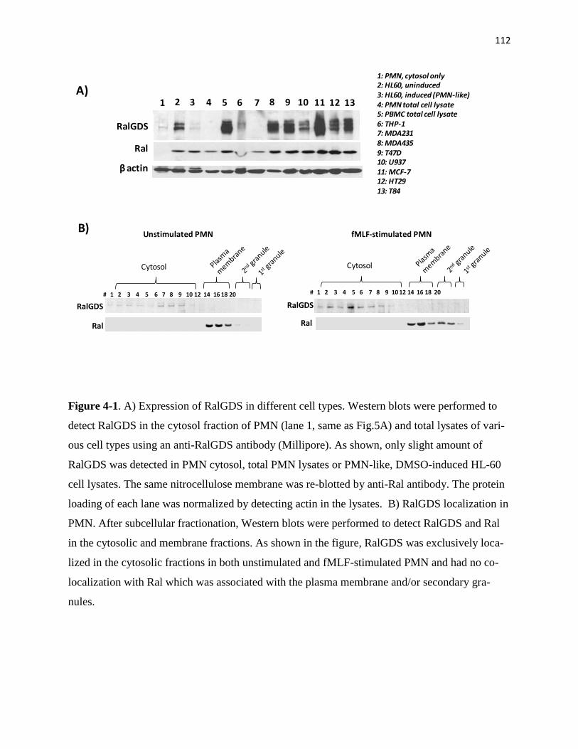
112
Figure 4-1. A) Expression of RalGDS in different cell types. Western blots were performed to
detect RalGDS in the cytosol fraction of PMN (lane 1, same as Fig.5A) and total lysates of vari-
ous cell types using an anti-RalGDS antibody (Millipore). As shown, only slight amount of
RalGDS was detected in PMN cytosol, total PMN lysates or PMN-like, DMSO-induced HL-60
cell lysates. The same nitrocellulose membrane was re-blotted by anti-Ral antibody. The protein
loading of each lane was normalized by detecting actin in the lysates. B) RalGDS localization in
PMN. After subcellular fractionation, Western blots were performed to detect RalGDS and Ral
in the cytosolic and membrane fractions. As shown in the figure, RalGDS was exclusively loca-
lized in the cytosolic fractions in both unstimulated and fMLF-stimulated PMN and had no co-
localization with Ral which was associated with the plasma membrane and/or secondary gra-
nules.
1 2 3 4 5 6 7 8 9 10 11 12 13
RalGDS
Ral
βactin
A)1: PMN, cytosol only2: HL60, uninduced3: HL60, induced (PMN-like)4: PMN total cell lysate5: PBMC total cell lysate6: THP-17: MDA2318: MDA4359: T47D10: U93711: MCF-712: HT2913: T84
# 1 2 3 4 5 6 7 8 9 10 12 14 16 18 20 # 1 2 3 4 5 6 7 8 9 10 12 14 16 18 20
B)
Cytosol Cytosol
RalGDS
Ral
RalGDS
Ral
Unstimulated PMN fMLF-stimulated PMN
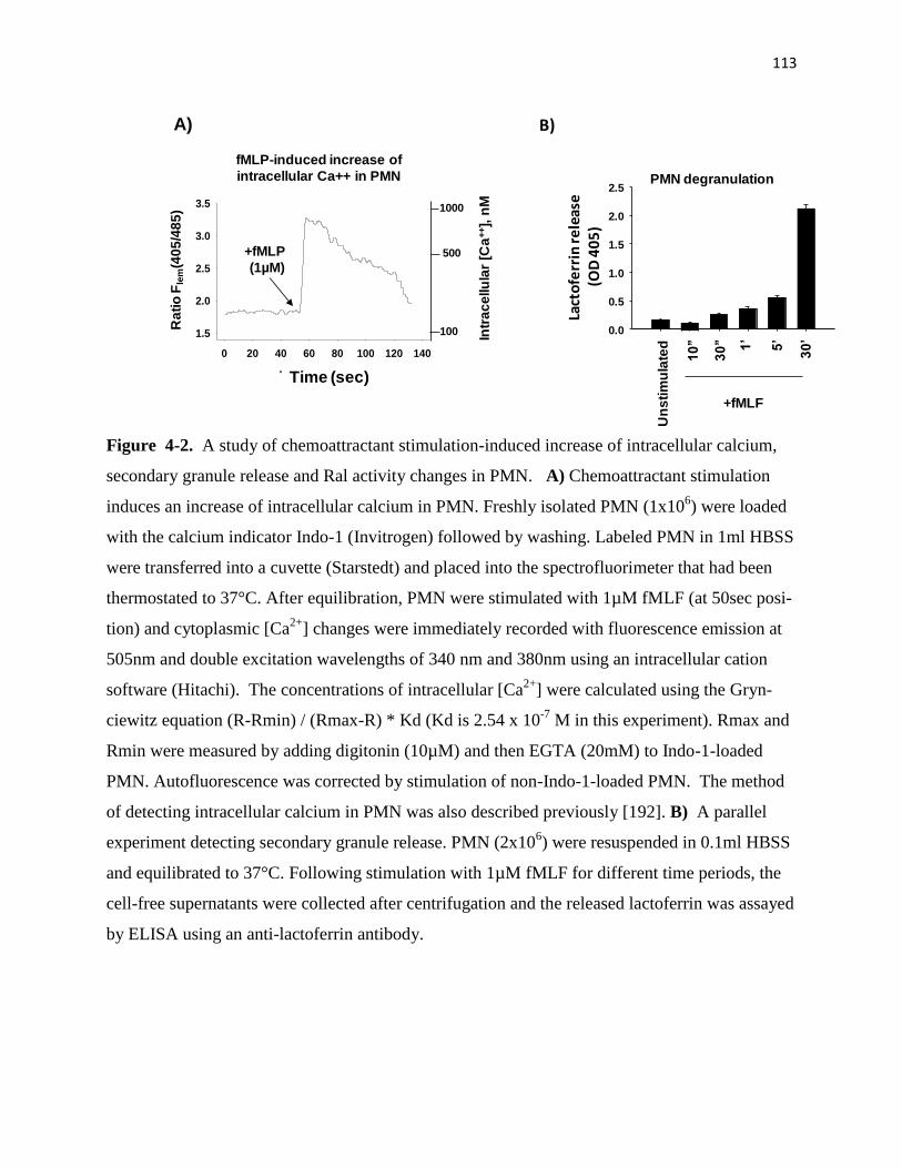
113
Figure 4-2. A study of chemoattractant stimulation-induced increase of intracellular calcium,
secondary granule release and Ral activity changes in PMN. A) Chemoattractant stimulation
induces an increase of intracellular calcium in PMN. Freshly isolated PMN (1x10⁶) were loaded
with the calcium indicator Indo-1 (Invitrogen) followed by washing. Labeled PMN in 1ml HBSS
were transferred into a cuvette (Starstedt) and placed into the spectrofluorimeter that had been
thermostated to 37°C. After equilibration, PMN were stimulated with 1µM fMLF (at 50sec posi-
tion) and cytoplasmic [Ca2+
] changes were immediately recorded with fluorescence emission at
505nm and double excitation wavelengths of 340 nm and 380nm using an intracellular cation
software (Hitachi). The concentrations of intracellular [Ca2+
] were calculated using the Gryn-
ciewitz equation (R-Rmin) / (Rmax-R) * Kd (Kd is 2.54 x 10⁻⁷
M in this experiment). Rmax and
Rmin were measured by adding digitonin (10µM) and then EGTA (20mM) to Indo-1-loaded
PMN. Autofluorescence was corrected by stimulation of non-Indo-1-loaded PMN. The method
of detecting intracellular calcium in PMN was also described previously [192]. B) A parallel
experiment detecting secondary granule release. PMN (2x10⁶) were resuspended in 0.1ml HBSS
and equilibrated to 37°C. Following stimulation with 1µM fMLF for different time periods, the
cell-free supernatants were collected after centrifugation and the released lactoferrin was assayed
by ELISA using an anti-lactoferrin antibody.
A)
0 20 40 60 80 100 120 140
1.5
2.0
2.5
3.0
3.5
Time (sec)
Ra
tio
o (
F1
/F2
)R
ati
o F
Iem
(40
5/4
85
)
+fMLP
(1µM)
100
500
1000
Intr
ac
ellu
lar
[Ca
++],
nM
Time (sec)
B)
Lact
ofe
rrin
re
leas
e(O
D 4
05
)
PMN degranulation
+fMLF
10’’
30’’ 1’
5’
30’
Un
sti
mu
late
d
0.0
0.5
1.0
1.5
2.0
2.5
fMLP-induced increase of
intracellular Ca++ in PMN

114
Figure 4-3. A study of chemoattractant stimulation-induced increase of intracellular calcium,
secondary granule release and Ral activity changes in PMN. A) Ral activity changes in PMN
after fMLF stimulation. In this experiment, PMN (1x10⁷) were stimulated with 1µM fMLF at
37°C in HBSS for different time periods. After cell lysis, active Ral in PMN was recovered by
RalBP1 pull-down followed by Western blot using an anti-Ral antibody. As can be seen in the
figure, Ral activity was slightly increased (10-30%) immediately after fMLF stimulation (at ~10-
30 second). B) Calcium concentrations have no effect on the direct binding of RalA to RalBP1.
In these experiments recombinant GST-RalBP1 and Tat-tagged wild-type and mutant RalA were
produced by expression in E.coli followed by protein purification. Pull-down assays were per-
formed by incubation (30min, 4°C) of GST-RalBP1(1µg) with Tat-tagged Ral (0.2µg each) in
HBSS containing 5% non-fat dry milk, 2% BSA, 0.01% Tween 20 and different concentrations
of CaCl2. Glutathione-conjugated Sepharose (20µl) were also added. After washing, proteins
pulled down by the Sepharose were analyzed by Western blot using an anti-Ral antibody. As can
be seen in the figure, only the constitutively active Ral (Ral23V) was directly pulled down by
RalBP1. Increasing calcium concentrations had no effect on the direct binding of Ral to RalBP1.
Active Ral
Total Ral
+fMLF
10’’
30’’
1’
2’
5’
Un
sti
mu
late
d
Ral activity in PMNA) B)
Tat-
Ral2
3V
Tat-
Ral2
8N
RalBP1 pull-down
WB: Anti-Ral
Tat-
WT
[Ca2+]nM 10
00
50
0
0 10
00
50
0
0 10
00
50
0
0
RalBP1 pull-down
WB: Anti-Ral

115
Figure 4-4. Mg2+
inhibits Ral activity in PMN. A) Ral activity in freshly isolated PMN was in-
creased in the presence of EDTA. PMN were lysed in the presence or absence of 2mM EDTA,
and active Ral was pulled down using RalBP1. In addition, resting platelets were lysed in the
presence of 2mM EDTA, and active Ral was pulled down by RalBP1. B) Detecting activity
changes of Ral in PMN. PMN were stimulated with fMLF (1µM) in HBSS (-), HBSS (+), or
HBSS (+) supplemented with 5mM EGTA. After time-course stimulation, PMN were lysed, and
active Ral was pulled down. Cell lysates before pull-down were used to detect total Ral as well
as actin using antibodies against Ral and β actin. C) Detecting activity changes of Ral in PMN.
PMN were lysed in the presence of 5mM EDTA or 30nM Mg2+
, and active Ral was detected by
pull down assay.
Active RalA
Total RalA
Lysis buffer
2mM EDTA- ++
Active RalA
Total RalA
H-5’ 20’
fMLF
5’ 20’ 5’ 20’
β actin
Active RalA
Total RalA
-
A) B)
C)

116
Figure 4-5. Effects of damnacanthal alone or in a combination with fMLF on PMN granule re-
lease. In these experiments, aliquots of freshly isolated PMN (1x10⁷) were treated with vehicle
(0.1% DMSO, 15min, 25°C), damnacanthal (3.6µM and 9µM, 15min, 25°C), fMLF (1µM,
15min, 37°C), or damnacanthal (3.6µM, 15min, 25°C) followed by fMLF (1µM, 15min, 37°C).
An aliquot of PMN resuspended in HBSS(-) and kept at 25°C (15min) was also used as a control
condition. After incubation, cell-free supernatants were collected after centrifugation and assayed
for granule release. Left: assay of PMN secondary granule release by detecting lactoferrin in the
supernatants. Right: Assay of PMN secondary granule release by zymogram detecting liberated
gelatinase in the supernatants.
HB
SS
(-)
Ve
hic
le c
tl
Da
mn
. (3
.6µ
M)
fML
F (
1µ
M)
Da
mn
.(3
.6µ
M)+
fM
LP
Da
mn
. (9
µM
)
Secondary granule release
La
cto
ferr
in r
ele
as
e
(OD
40
5)
HB
SS
(-)
Ve
hic
le c
tl
Da
mn
. (3
.6µ
M)
fML
F (
1µ
M)
Da
mn
.(3
.6µ
M)+
fM
LP
Da
mn
. (9
µM
)
0.0
0.5
1.0
1.5
2.0
2.5
Tertiary granule release
(Gelatinase zymogram)

117
Figure 4-6. Subcellular fractionations studying the effects of damnacanthal and endocytosis in-
hibitors on Ral distribution in PMN. Damnacanthal triggers Ral redistribution from the plasma
membranes into secondary granules. Freshly isolated PMN (1x10⁸/per condition) were treated
with vehicle (0.1%DMSO) or damnacanthal (9µM) for 15min (25°C) before lysis and subjected
to sucrose density gradients to separate subcellular organelles. After fractionation, the localiza-
tions of plasma membranes and intracellular granules in respective fractions were determined by
their specific marker proteins (upper panels). The lower panels show the Western blot results
detecting Ral, SIRPα and JAM-A (junction adhesion molecule A) in the fractions. In PMN,
SIRPα is mainly stored in secondary granules in the absence of stimulation, while JAM-A is
consistently associated with the plasma membrane (unpublished data). As can be seen, a portion
of Ral was redistributed from the plasma membranes into secondary granules after damnacanthal
treatment. Given that damnacanthal induces secondary granule release, it was predictable to ob-
serve that a portion of SIRPα was translocated to the plasma membranes. The indifferent JAM-A
localization demonstrated that plasma membrane integrity was not jeopardized by damnacanthal.
1-5 6 7 8 9 10 11 12 13 14 15 16 17 18
0.0
0.2
0.4
0.6
0.8
1.0
1.2
1.4
1.6
1.8
2.0
2.2
Plasma membrane ( Alkaline phosphatase)
Secondary granules ( Lactoferrin)
Primary granules (MPO)
Reco
vered
acti
vit
y (
OD
40
5)
1-5 6 7 8 9 10 11 12 13 14 15 16 17 18
0.0
0.5
1.0
1.5
2.0
2.5
3.0
Plasma membrane ( Alkaline phosphatase)
Secondary granules ( Lactoferrin)
Primary granules ( MPO)
Reco
vered
acti
vit
y (
OD
40
5)
Damnacanthal (-fMLF)
Fraction number (#) Fraction number (#)
SIRPα
Ral
JAM-A
Vehicle ctl (-fMLF)
# 1 6 7 8 9 10 11 12 13 14 15 16 17 18 # 1 6 7 8 9 10 11 12 13 14 15 16 17 18 Fraction number

118
Figure 4-7. Subcellular fractionations studying the effects of damnacanthal and endocytosis in-
hibitors on Ral distribution in PMN. Inhibitors acting on clathrin-mediated endocytosis had no
effect on damnacanthal-triggered Ral redistribution in PMN. In these experiments, PMN, pre-
treated with an individual endocytosis inhibitor (e.g. CPZ, 50µM) or a combination of several
inhibitors (50µM CPZ + 20µM POA + 20mM succinic acid (pH5.5)) (10min, 25°C), were either
subjected or not subjected to further treatment with damnacanthal (9µM) (15min, 25°C). After
treatment, the cells were lysed followed by to sucrose density gradient centrifugation to separate
subcellular organelles. After fractionation, Western blots were performed to detect Ral distribu-
tion in the fractions using an anti-Ral antibody. As shown in the figure, none of the endocytosis
inhibitors markedly blocked damnacanthal-triggered Ral redistribution from the plasma mem-
branes to secondary granules.
1-5 6 7 8 9 10 11 12 13 14 15 16 17 18
0.0
0.5
1.0
1.5
2.0
1-5 6 7 8 9 10 11 12 13 14 15 16 17 18
0.0
0.5
1.0
1.5
2.0
1-5 6 7 8 9 10 11 12 13 14 15 16 17 18
0.0
0.5
1.0
1.5
2.0
2.5
3.0
Endocytosis inhibitors
(CPZ + POA+ pH5.5)
Endocytosis inhibitors
(CPZ + POA+ pH5.5)
+ Damnacanthal
CPZ +
Damnacanthal
Fraction number (#)
Ral
# 1 6 7 8 9 10 11 12 13 14 15 16 17 18
Fraction number (#)
# 1 6 7 8 9 10 11 12 13 14 15 1617 18
Fraction number (#)
# 1 6 7 8 9 10 11 12 13 14 15 16 17 18
Reco
vere
d a
ctiv
ity
(O
D4
05
)
Reco
vere
d a
ctiv
ity
(O
D4
05
)
Reco
vere
d a
ctiv
ity
(O
D4
05
)
Fraction
number
Plasma membrane (alkaline phosphatase)Primary granules (MPO)
Secondary granules ( lactoferrin)
Plasma membrane (alkaline phosphatase)Primary granules (MPO)
Secondary granules ( lactoferrin)
Plasma membrane (alkaline phosphatase)Primary granules (MPO)
Secondary granules ( lactoferrin)
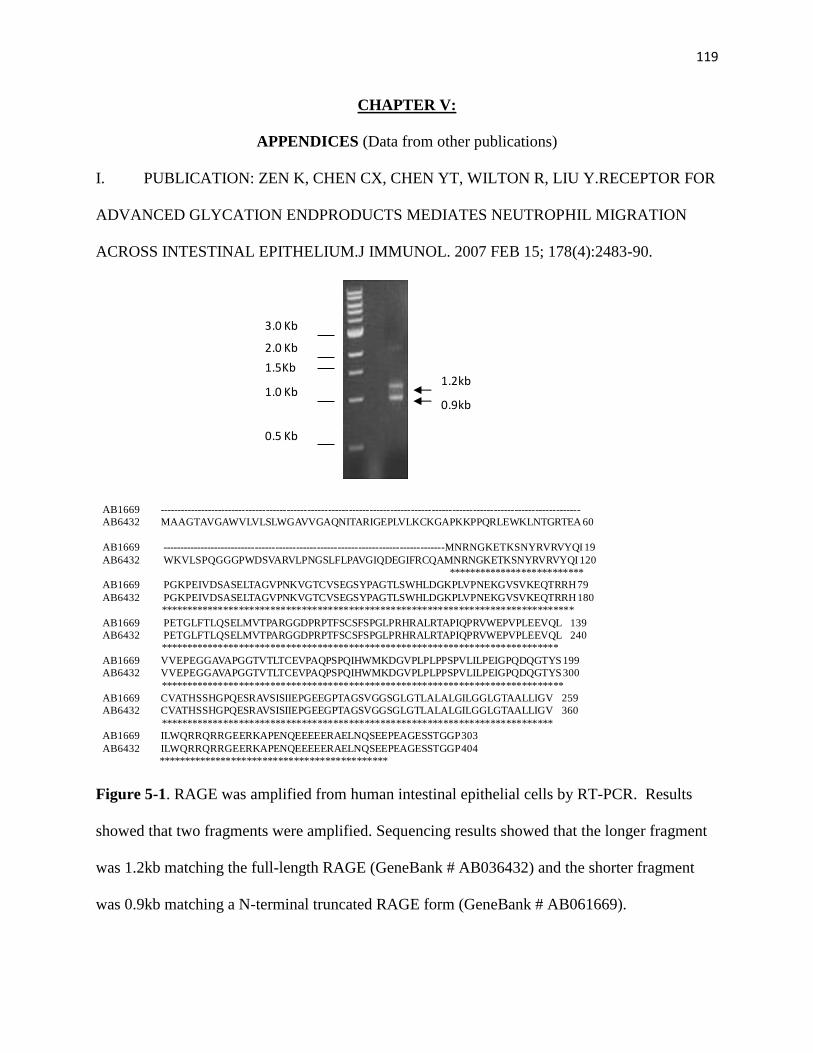
119
CHAPTER V:
APPENDICES (Data from other publications)
I. PUBLICATION: ZEN K, CHEN CX, CHEN YT, WILTON R, LIU Y.RECEPTOR FOR
ADVANCED GLYCATION ENDPRODUCTS MEDIATES NEUTROPHIL MIGRATION
ACROSS INTESTINAL EPITHELIUM.J IMMUNOL. 2007 FEB 15; 178(4):2483-90.
Figure 5-1. RAGE was amplified from human intestinal epithelial cells by RT-PCR. Results
showed that two fragments were amplified. Sequencing results showed that the longer fragment
was 1.2kb matching the full-length RAGE (GeneBank # AB036432) and the shorter fragment
was 0.9kb matching a N-terminal truncated RAGE form (GeneBank # AB061669).
3.0 Kb
2.0 Kb
1.5Kb
1.0 Kb
0.5 Kb
1.2kb
0.9kb
AB1669 --------------------------------------------------------------------------------------------------------------------------
AB6432 MAAGTAVGAWVLVLSLWGAVVGAQNITARIGEPLVLKCKGAPKKPPQRLEWKLNTGRTEA 60
AB1669 ----------------------------------------------------------------------------------MNRNGKETKSNYRVRVYQI 19
AB6432 WKVLSPQGGGPWDSVARVLPNGSLFLPAVGIQDEGIFRCQAMNRNGKETKSNYRVRVYQI 120
**************************
AB1669 PGKPEIVDSASELTAGVPNKVGTCVSEGSYPAGTLSWHLDGKPLVPNEKGVSVKEQTRRH 79
AB6432 PGKPEIVDSASELTAGVPNKVGTCVSEGSYPAGTLSWHLDGKPLVPNEKGVSVKEQTRRH 180
*******************************************************************************
AB1669 PETGLFTLQSELMVTPARGGDPRPTFSCSFSPGLPRHRALRTAPIQPRVWEPVPLEEVQL 139
AB6432 PETGLFTLQSELMVTPARGGDPRPTFSCSFSPGLPRHRALRTAPIQPRVWEPVPLEEVQL 240
****************************************************************************
AB1669 VVEPEGGAVAPGGTVTLTCEVPAQPSPQIHWMKDGVPLPLPPSPVLILPEIGPQDQGTYS 199
AB6432 VVEPEGGAVAPGGTVTLTCEVPAQPSPQIHWMKDGVPLPLPPSPVLILPEIGPQDQGTYS 300
*****************************************************************************
AB1669 CVATHSSHGPQESRAVSISIIEPGEEGPTAGSVGGSGLGTLALALGILGGLGTAALLIGV 259
AB6432 CVATHSSHGPQESRAVSISIIEPGEEGPTAGSVGGSGLGTLALALGILGGLGTAALLIGV 360
***************************************************************************
AB1669 ILWQRRQRRGEERKAPENQEEEEERAELNQSEEPEAGESSTGGP 303
AB6432 ILWQRRQRRGEERKAPENQEEEEERAELNQSEEPEAGESSTGGP 404
********************************************

120
Figure 5-2. RAGE was detected from epithelial cell lines by western blot. RAGE was detected
from human colonic epithelial cells lines including T84 and HT29. RAGE was also detected
from epithelial cell A549.
170KD
62KD
48KD
32KD
25KD
83KD
35KD
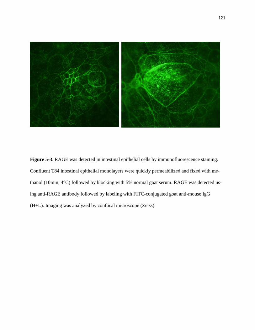
121
Figure 5-3. RAGE was detected in intestinal epithelial cells by immunofluorescence staining.
Confluent T84 intestinal epithelial monolayers were quickly permeabilized and fixed with me-
thanol (10min, 4°C) followed by blocking with 5% normal goat serum. RAGE was detected us-
ing anti-RAGE antibody followed by labeling with FITC-conjugated goat anti-mouse IgG
(H+L). Imaging was analyzed by confocal microscope (Zeiss).
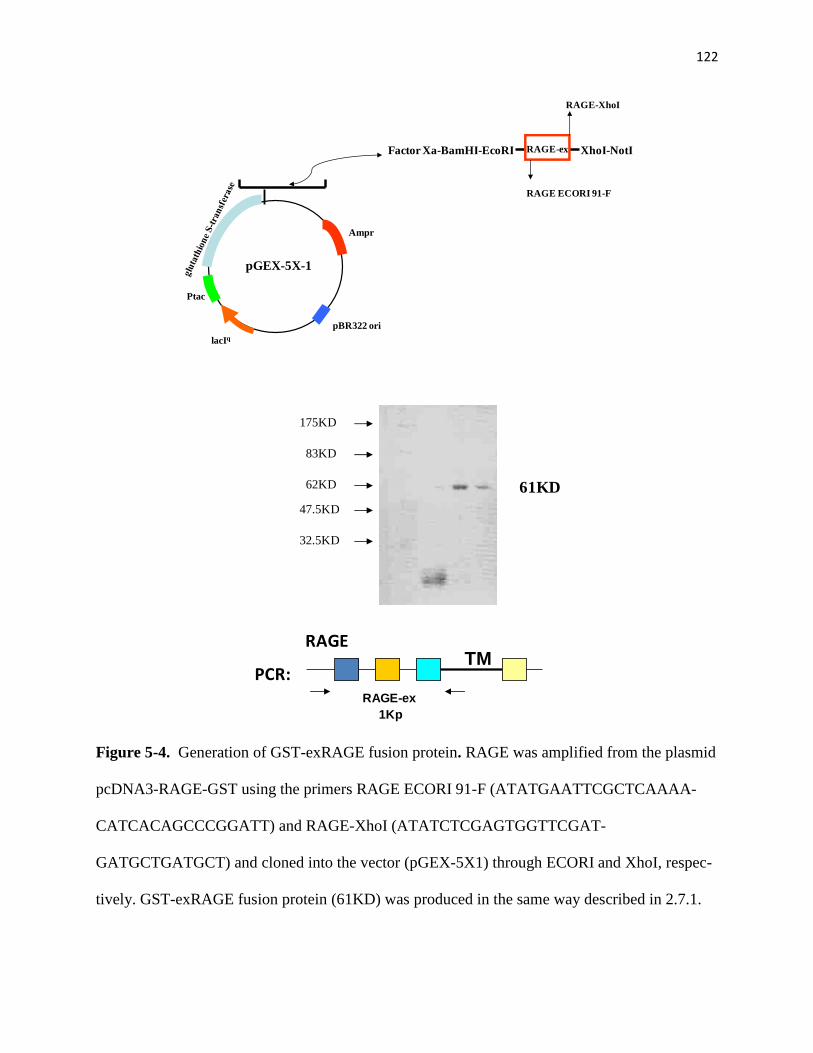
122
Figure 5-4. Generation of GST-exRAGE fusion protein. RAGE was amplified from the plasmid
pcDNA3-RAGE-GST using the primers RAGE ECORI 91-F (ATATGAATTCGCTCAAAA-
CATCACAGCCCGGATT) and RAGE-XhoI (ATATCTCGAGTGGTTCGAT-
GATGCTGATGCT) and cloned into the vector (pGEX-5X1) through ECORI and XhoI, respec-
tively. GST-exRAGE fusion protein (61KD) was produced in the same way described in 2.7.1.
pGEX-5X-1
Ampr
pBR322 ori
lacIq
Ptac
RAGE-ex
RAGE ECORI 91-F
RAGE-XhoI
XhoΙ-NotΙFactor Xa-BamHΙ-EcoRΙ
175KD
83KD
62KD
47.5KD
32.5KD
RAGETM
RAGE-ex
1Kp
61KD
PCR:

123
Figure 5-5. RAGE antibody titer test by ELISA. 96-well microtiter plate wells were coated with
RAGE-ex/GST fusion proteins (5μg/ml each) overnight at 4ºC.After blocking with 1% BSA
(30min, 25ºC), the wells were incubated with serum (1:200; 1:500; 1:1000; 1:2000; 1:5000;
1:100000) for 30min. After washing, fusion protein binding to the antibody (serum) was detected
by peroxidase conjugated goat anti-mouse Fc antibody.
serum dilution
1-200 1-500 1-1000 1-2000 1-5000 1-10000 1%BSA
OD
405
0.0
0.5
1.0
1.5
2.0
2.5
3.0
RAGE
RAGE(RL)

124
Figure 5-6. RAGE antibody titer test by western blot. The serum from the mouse RAGE-ex(RL)
and RAGE-ex(R) recognized the RAGE-Fc fusion protein, including the nonreducing form (A)
and reducing form. (B). Antibody also picked the band form the HT29, PMN and monocyte. Af-
ter immunization for 5 times, the serum was used for WB at 1:1000 dilution.
Serum: A: RAGE(RL) B: RAGE (R)
1: RAGE-Fc fusion protein 0.2ug (R & D)
2: HT29
3: Monocyte
4: PMN
A B A B
1758362
32.5
47.5
A B A B
1 2 3 4 1 2 3 4 1 2 3 4 1 2 3 4
Serum dilution: 1:500 1:1000
KD
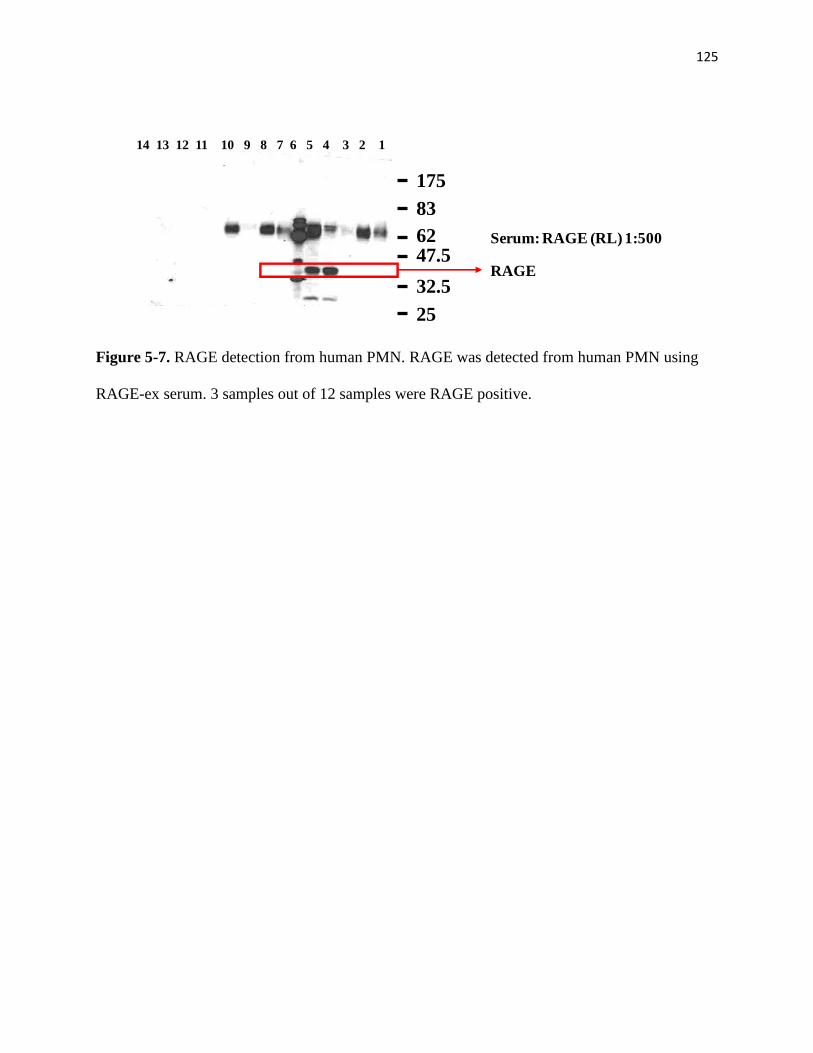
125
Figure 5-7. RAGE detection from human PMN. RAGE was detected from human PMN using
RAGE-ex serum. 3 samples out of 12 samples were RAGE positive.
175
83
62
32.5
47.5
25
14 13 12 11 10 9 8 7 6 5 4 3 2 1
Serum: RAGE (RL) 1:500
RAGE
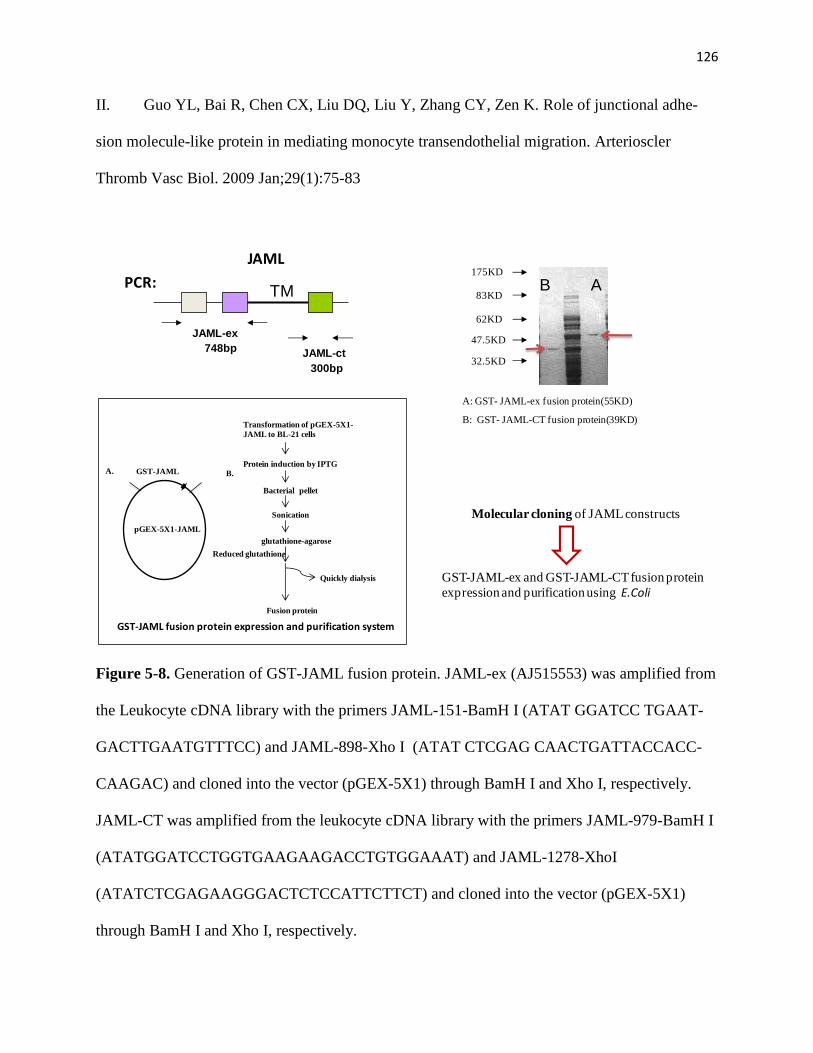
126
II. Guo YL, Bai R, Chen CX, Liu DQ, Liu Y, Zhang CY, Zen K. Role of junctional adhe-
sion molecule-like protein in mediating monocyte transendothelial migration. Arterioscler
Thromb Vasc Biol. 2009 Jan;29(1):75-83
Figure 5-8. Generation of GST-JAML fusion protein. JAML-ex (AJ515553) was amplified from
the Leukocyte cDNA library with the primers JAML-151-BamH I (ATAT GGATCC TGAAT-
GACTTGAATGTTTCC) and JAML-898-Xho I (ATAT CTCGAG CAACTGATTACCACC-
CAAGAC) and cloned into the vector (pGEX-5X1) through BamH I and Xho I, respectively.
JAML-CT was amplified from the leukocyte cDNA library with the primers JAML-979-BamH I
(ATATGGATCCTGGTGAAGAAGACCTGTGGAAAT) and JAML-1278-XhoI
(ATATCTCGAGAAGGGACTCTCCATTCTTCT) and cloned into the vector (pGEX-5X1)
through BamH I and Xho I, respectively.
GST-JAML-ex and GST-JAML-CT fusion protein
expression and purification using E.Coli
Molecular cloning of JAML constructs
JAML
TM
JAML-ex
JAML-ct748bp
300bp
A. GST-JAML
pGEX-5X1-JAML
B.
Transformation of pGEX-5X1-
JAML to BL-21 cells
Protein induction by IPTG
Bacterial pellet
Sonication
glutathione-agarose
Reduced glutathione
Quickly dialysis
Fusion protein
GST-JAML fusion protein expression and purification system
PCR:
A: GST- JAML-ex fusion protein(55KD)
B: GST- JAML-CT fusion protein(39KD)
175KD
83KD
62KD
47.5KD
32.5KD
AB
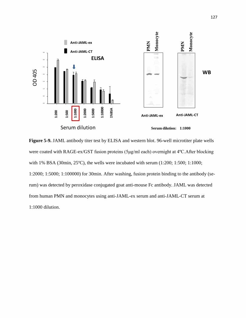
127
Figure 5-9. JAML antibody titer test by ELISA and western blot. 96-well microtiter plate wells
were coated with RAGE-ex/GST fusion proteins (5μg/ml each) overnight at 4ºC.After blocking
with 1% BSA (30min, 25ºC), the wells were incubated with serum (1:200; 1:500; 1:1000;
1:2000; 1:5000; 1:100000) for 30min. After washing, fusion protein binding to the antibody (se-
rum) was detected by peroxidase conjugated goat anti-mouse Fc antibody. JAML was detected
from human PMN and monocytes using anti-JAML-ex serum and anti-JAML-CT serum at
1:1000 dilution.
Anti-JAML-CT serum
serum dilution
1-200 1-500 1-1000 1-2000 1-5000 1-10000 1%BSA
0.0
0.5
1.0
1.5
2.0
2.5
3.0
3.5
0.0
0.5
1.0
1.5
2.0
2.5
3.0
3.5
OD
405
Serum dilution
1:20
00
1:50
00
1:10
000
1%B
SA
1:20
0
1:50
0
1:10
00
ELISA
Anti-JAML-ex
Anti-JAML-CT
WB
PM
N
Mo
no
cyte
Serum dilution: 1:1000
Anti-JAML-ex
PM
N
Mo
no
cyte
Anti-JAML-CT
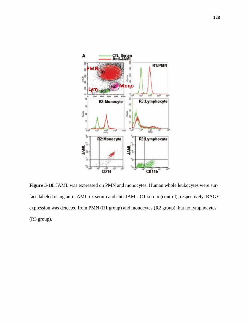
128
Figure 5-10. JAML was expressed on PMN and monocytes. Human whole leukocytes were sur-
face labeled using anti-JAML-ex serum and anti-JAML-CT serum (control), respectively. RAGE
expression was detected from PMN (R1 group) and monocytes (R2 group), but no lymphocytes
(R3 group).
PMN
MonoLym

129
III. PUBLICATION: Liu Y, Tong Q, Zhou Y, Lee HW, Yang JJ, Bühring HJ, Chen YT, Ha
B, Chen CX, Yang Y, Zen K. Functional elements on SIRPalpha IgV domain mediate cell sur-
face binding to CD47. J Mol Biol. 2007 Jan 19;365(3):680-93
Figure 5-11. Ala57, Gln67, and Val57 were key amino acid residues mediating binding interac-
tion of SIRPα to CD47.
Generation of SIRPα -IgV DNA constructs
Site-mutagenesis of the residues in Bit IgV loop to corresponding counter amino acids in SIRPβ
Transient transfection to COS cells
Purification of SIPRα fusion protein
Assaying the binding capacity of SIRPα to its ligand, CD47

130
REFERENCES
1. Zychlinsky, A., Y. Weinrauch, and J. Weiss, Introduction: Forum in immunology on
neutrophils. Microbes Infect, 2003. 5(14): p. 1289-91.
2. Parham, P., The immune system. 2009(3rd edition): Garland Science.
3. Borregaard, N., Neutrophils, from marrow to microbes. Immunity, 2010. 33(5): p. 657-
70.
4. Gordy, C., et al., Regulation of steady-state neutrophil homeostasis by macrophages.
Blood, 2011. 117(2): p. 618-29.
5. Christopher, M.J. and D.C. Link, Regulation of neutrophil homeostasis. Curr Opin
Hematol, 2007. 14(1): p. 3-8.
6. Weinmann, P., et al., A role for apoptosis in the control of neutrophil homeostasis in the
circulation: insights from CD18-deficient mice. Blood, 2003. 101(2): p. 739-46.
7. Lieschke, G.J., et al., Mice lacking granulocyte colony-stimulating factor have chronic
neutropenia, granulocyte and macrophage progenitor cell deficiency, and impaired
neutrophil mobilization. Blood, 1994. 84(6): p. 1737-46.
8. Schwarzenberger, P., et al., Requirement of endogenous stem cell factor and granulocyte-
colony-stimulating factor for IL-17-mediated granulopoiesis. J Immunol, 2000. 164(9): p.
4783-9.
9. Hernandez, P.A., et al., Mutations in the chemokine receptor gene CXCR4 are associated
with WHIM syndrome, a combined immunodeficiency disease. Nat Genet, 2003. 34(1): p.
70-4.
131
10. Broxmeyer, H.E., et al., Rapid mobilization of murine and human hematopoietic stem and
progenitor cells with AMD3100, a CXCR4 antagonist. J Exp Med, 2005. 201(8): p. 1307-
18.
11. Nagasawa, T., et al., Defects of B-cell lymphopoiesis and bone-marrow myelopoiesis in
mice lacking the CXC chemokine PBSF/SDF-1. Nature, 1996. 382(6592): p. 635-8.
12. Zou, Y.R., et al., Function of the chemokine receptor CXCR4 in haematopoiesis and in
cerebellar development. Nature, 1998. 393(6685): p. 595-9.
13. Kawabata, K., et al., A cell-autonomous requirement for CXCR4 in long-term lymphoid
and myeloid reconstitution. Proc Natl Acad Sci U S A, 1999. 96(10): p. 5663-7.
14. Liles, W.C., et al., Mobilization of hematopoietic progenitor cells in healthy volunteers
by AMD3100, a CXCR4 antagonist. Blood, 2003. 102(8): p. 2728-30.
15. Suratt, B.T., et al., Role of the CXCR4/SDF-1 chemokine axis in circulating neutrophil
homeostasis. Blood, 2004. 104(2): p. 565-71.
16. Ma, Q., et al., Impaired B-lymphopoiesis, myelopoiesis, and derailed cerebellar neuron
migration in CXCR4- and SDF-1-deficient mice. Proc Natl Acad Sci U S A, 1998.
95(16): p. 9448-53.
17. Ma, Q., D. Jones, and T.A. Springer, The chemokine receptor CXCR4 is required for the
retention of B lineage and granulocytic precursors within the bone marrow
microenvironment. Immunity, 1999. 10(4): p. 463-71.
18. Link, D.C., Neutrophil homeostasis: a new role for stromal cell-derived factor-1.
Immunol Res, 2005. 32(1-3): p. 169-78.

132
19. Lee, A., M.K. Whyte, and C. Haslett, Inhibition of apoptosis and prolongation of
neutrophil functional longevity by inflammatory mediators. J Leukoc Biol, 1993. 54(4):
p. 283-8.
20. Liles, W.C. and S.J. Klebanoff, Regulation of apoptosis in neutrophils--Fas track to
death? J Immunol, 1995. 155(7): p. 3289-91.
21. Luo, H.R. and F. Loison, Constitutive neutrophil apoptosis: mechanisms and regulation.
Am J Hematol, 2008. 83(4): p. 288-95.
22. Doerschuk, C.M., et al., Marginated pool of neutrophils in rabbit lungs. J Appl Physiol,
1987. 63(5): p. 1806-15.
23. Saverymuttu, S.H., et al., The kinetics of 111indium distribution following injection of
111indium labelled autologous granulocytes in man. Br J Haematol, 1985. 61(4): p. 675-
85.
24. Suratt, B.T., et al., Neutrophil maturation and activation determine anatomic site of
clearance from circulation. Am J Physiol Lung Cell Mol Physiol, 2001. 281(4): p. L913-
21.
25. Kobayashi, S.D., J.M. Voyich, and F.R. DeLeo, Regulation of the neutrophil-mediated
inflammatory response to infection. Microbes Infect, 2003. 5(14): p. 1337-44.
26. Kelly, M., J.M. Hwang, and P. Kubes, Modulating leukocyte recruitment in
inflammation. J Allergy Clin Immunol, 2007. 120(1): p. 3-10.
27. Nathan, C., Neutrophils and immunity: challenges and opportunities. Nat Rev Immunol,
2006. 6(3): p. 173-82.
28. Ryan, G.B. and G. Majno, Acute inflammation. A review. Am J Pathol, 1977. 86(1): p.
183-276.
133
29. Murphy, P.M., The molecular biology of leukocyte chemoattractant receptors. Annu Rev
Immunol, 1994. 12: p. 593-633.
30. Zlotnik, A. and O. Yoshie, Chemokines: a new classification system and their role in
immunity. Immunity, 2000. 12(2): p. 121-7.
31. Witko-Sarsat, V., et al., Neutrophils: molecules, functions and pathophysiological
aspects. Lab Invest, 2000. 80(5): p. 617-53.
32. Ali, H., et al., Chemoattractant receptor cross-desensitization. J Biol Chem, 1999.
274(10): p. 6027-30.
33. Sogawa, Y., et al., Formyl peptide receptor 1 and 2 dual agonist inhibits human
neutrophil chemotaxis by the induction of chemoattractant receptor cross-desensitization.
J Pharmacol Sci. 115(1): p. 63-8.
34. Sabroe, I., et al., Chemoattractant cross-desensitization of the human neutrophil IL-8
receptor involves receptor internalization and differential receptor subtype regulation. J
Immunol, 1997. 158(3): p. 1361-9.
35. Foxman, E.F., J.J. Campbell, and E.C. Butcher, Multistep navigation and the
combinatorial control of leukocyte chemotaxis. J Cell Biol, 1997. 139(5): p. 1349-60.
36. Kitayama, J., et al., Contrasting responses to multiple chemotactic stimuli in
transendothelial migration: heterologous desensitization in neutrophils and
augmentation of migration in eosinophils. J Immunol, 1997. 158(5): p. 2340-9.
37. Foxman, E.F., E.J. Kunkel, and E.C. Butcher, Integrating conflicting chemotactic signals.
The role of memory in leukocyte navigation. J Cell Biol, 1999. 147(3): p. 577-88.
38. Selvatici, R., et al., Signal transduction pathways triggered by selective formylpeptide
analogues in human neutrophils. Eur J Pharmacol, 2006. 534(1-3): p. 1-11.
134
39. Rabiet, M.J., E. Huet, and F. Boulay, The N-formyl peptide receptors and the
anaphylatoxin C5a receptors: an overview. Biochimie, 2007. 89(9): p. 1089-106.
40. Putney, J.W., Jr. and C.M. Ribeiro, Signaling pathways between the plasma membrane
and endoplasmic reticulum calcium stores. Cell Mol Life Sci, 2000. 57(8-9): p. 1272-86.
41. Hofer, A.M. and E.M. Brown, Extracellular calcium sensing and signalling. Nat Rev
Mol Cell Biol, 2003. 4(7): p. 530-8.
42. Nowycky, M.C. and A.P. Thomas, Intracellular calcium signaling. J Cell Sci, 2002.
115(Pt 19): p. 3715-6.
43. Gerthoffer, W.T., Mechanisms of vascular smooth muscle cell migration. Circ Res, 2007.
100(5): p. 607-21.
44. Ginis, I. and A.I. Tauber, Activation mechanisms of adherent human neutrophils. Blood,
1990. 76(6): p. 1233-9.
45. Merrill, J.T., et al., Two pathways of CD11b/CD18-mediated neutrophil aggregation with
different involvement of protein kinase C-dependent phosphorylation. J Immunol, 1990.
145(8): p. 2608-15.
46. Panaro, M.A. and V. Mitolo, Cellular responses to FMLP challenging: a mini-review.
Immunopharmacol Immunotoxicol, 1999. 21(3): p. 397-419.
47. Pongracz, J. and J.M. Lord, Superoxide production in human neutrophils: evidence for
signal redundancy and the involvement of more than one PKC isoenzyme class. Biochem
Biophys Res Commun, 1998. 247(3): p. 624-9.
48. Fontayne, A., et al., Phosphorylation of p47phox sites by PKC alpha, beta II, delta, and
zeta: effect on binding to p22phox and on NADPH oxidase activation. Biochemistry,
2002. 41(24): p. 7743-50.

135
49. Dekker, L.V., et al., Protein kinase C-beta contributes to NADPH oxidase activation in
neutrophils. Biochem J, 2000. 347 Pt 1: p. 285-9.
50. Li, Z., et al., Roles of PLC-beta2 and -beta3 and PI3Kgamma in chemoattractant-
mediated signal transduction. Science, 2000. 287(5455): p. 1046-9.
51. Merlot, S. and R.A. Firtel, Leading the way: Directional sensing through
phosphatidylinositol 3-kinase and other signaling pathways. J Cell Sci, 2003. 116(Pt 17):
p. 3471-8.
52. Rickert, P., et al., Leukocytes navigate by compass: roles of PI3Kgamma and its lipid
products. Trends Cell Biol, 2000. 10(11): p. 466-73.
53. Wymann, M.P., et al., Lipids on the move: phosphoinositide 3-kinases in leukocyte
function. Immunol Today, 2000. 21(6): p. 260-4.
54. Bruyninckx, W.J., et al., Phosphoinositide 3-kinase modulation of beta(3)-integrin
represents an endogenous "braking" mechanism during neutrophil transmatrix
migration. Blood, 2001. 97(10): p. 3251-8.
55. Sasaki, T., et al., Function of PI3Kgamma in thymocyte development, T cell activation,
and neutrophil migration. Science, 2000. 287(5455): p. 1040-6.
56. Sadhu, C., et al., Essential role of phosphoinositide 3-kinase delta in neutrophil
directional movement. J Immunol, 2003. 170(5): p. 2647-54.
57. Wang, F., et al., Lipid products of PI(3)Ks maintain persistent cell polarity and directed
motility in neutrophils. Nat Cell Biol, 2002. 4(7): p. 513-8.
58. Iannone, M.A., G. Wolberg, and T.P. Zimmerman, Chemotactic peptide induces cAMP
elevation in human neutrophils by amplification of the adenylate cyclase response to
endogenously produced adenosine. J Biol Chem, 1989. 264(34): p. 20177-80.

136
59. Harvath, L., et al., cAMP and human neutrophil chemotaxis. Elevation of cAMP
differentially affects chemotactic responsiveness. J Immunol, 1991. 146(1): p. 224-32.
60. Derian, C.K., et al., Inhibition of chemotactic peptide-induced neutrophil adhesion to
vascular endothelium by cAMP modulators. J Immunol, 1995. 154(1): p. 308-17.
61. Borregaard, N. and J.B. Cowland, Granules of the human neutrophilic
polymorphonuclear leukocyte. Blood, 1997. 89(10): p. 3503-21.
62. Borregaard, N., O.E. Sorensen, and K. Theilgaard-Monch, Neutrophil granules: a library
of innate immunity proteins. Trends Immunol, 2007. 28(8): p. 340-5.
63. Faurschou, M. and N. Borregaard, Neutrophil granules and secretory vesicles in
inflammation. Microbes Infect, 2003. 5(14): p. 1317-27.
64. Nourshargh, S. and F.M. Marelli-Berg, Transmigration through venular walls: a key
regulator of leukocyte phenotype and function. Trends Immunol, 2005. 26(3): p. 157-65.
65. Ley, K., et al., Getting to the site of inflammation: the leukocyte adhesion cascade
updated. Nat Rev Immunol, 2007. 7(9): p. 678-89.
66. Mayadas, T.N. and X. Cullere, Neutrophil beta2 integrins: moderators of life or death
decisions. Trends Immunol, 2005. 26(7): p. 388-95.
67. Parkos, C.A., Cell adhesion and migration. I. Neutrophil adhesive interactions with
intestinal epithelium. Am J Physiol, 1997. 273(4 Pt 1): p. G763-8.
68. Liu, Y., et al., Signal regulatory protein (SIRPalpha), a cellular ligand for CD47,
regulates neutrophil transmigration. J Biol Chem, 2002. 277(12): p. 10028-36.
69. Liu, Y., et al., The role of CD47 in neutrophil transmigration. Increased rate of
migration correlates with increased cell surface expression of CD47. J Biol Chem, 2001.
276(43): p. 40156-66.
137
70. Liu, Y., et al., SIRPbeta1 is expressed as a disulfide-linked homodimer in leukocytes and
positively regulates neutrophil transepithelial migration. J Biol Chem, 2005. 280(43): p.
36132-40.
71. Parkos, C.A., et al., CD47 mediates post-adhesive events required for neutrophil
migration across polarized intestinal epithelia. J Cell Biol, 1996. 132(3): p. 437-50.
72. Kubes, P. and S.M. Kerfoot, Leukocyte recruitment in the microcirculation: the rolling
paradigm revisited. News Physiol Sci, 2001. 16: p. 76-80.
73. Diamond, M.S., et al., ICAM-1 (CD54): a counter-receptor for Mac-1 (CD11b/CD18). J
Cell Biol, 1990. 111(6 Pt 2): p. 3129-39.
74. Sengelov, H., et al., Subcellular localization and dynamics of Mac-1 (alpha m beta 2) in
human neutrophils. J Clin Invest, 1993. 92(3): p. 1467-76.
75. Pohlman, T.H., et al., An endothelial cell surface factor(s) induced in vitro by
lipopolysaccharide, interleukin 1, and tumor necrosis factor-alpha increases neutrophil
adherence by a CDw18-dependent mechanism. J Immunol, 1986. 136(12): p. 4548-53.
76. Zimmerman, G.A. and T.M. McIntyre, Neutrophil adherence to human endothelium in
vitro occurs by CDw18 (Mo1, MAC-1/LFA-1/GP 150,95) glycoprotein-dependent and -
independent mechanisms. J Clin Invest, 1988. 81(2): p. 531-7.
77. Wallis, W.J., et al., Human monocyte adherence to cultured vascular endothelium:
monoclonal antibody-defined mechanisms. J Immunol, 1985. 135(4): p. 2323-30.
78. Smith, C.W., et al., Cooperative interactions of LFA-1 and Mac-1 with intercellular
adhesion molecule-1 in facilitating adherence and transendothelial migration of human
neutrophils in vitro. J Clin Invest, 1989. 83(6): p. 2008-17.

138
79. Ding, Z.M., et al., Relative contribution of LFA-1 and Mac-1 to neutrophil adhesion and
migration. J Immunol, 1999. 163(9): p. 5029-38.
80. Zen, K. and C.A. Parkos, Leukocyte-epithelial interactions. Curr Opin Cell Biol, 2003.
15(5): p. 557-64.
81. Petri, B. and M.G. Bixel, Molecular events during leukocyte diapedesis. FEBS J, 2006.
273(19): p. 4399-407.
82. Cooper, D., et al., Transendothelial migration of neutrophils involves integrin-associated
protein (CD47). Proc Natl Acad Sci U S A, 1995. 92(9): p. 3978-82.
83. Parkos, C.A., Molecular events in neutrophil transepithelial migration. Bioessays, 1997.
19(10): p. 865-73.
84. Lindberg, F.P., et al., Decreased resistance to bacterial infection and granulocyte defects
in IAP-deficient mice. Science, 1996. 274(5288): p. 795-8.
85. Miyashita, M., et al., Promotion of neurite and filopodium formation by CD47: roles of
integrins, Rac, and Cdc42. Mol Biol Cell, 2004. 15(8): p. 3950-63.
86. Yoshida, H., et al., Integrin-associated protein/CD47 regulates motile activity in human
B-cell lines through CDC42. Blood, 2000. 96(1): p. 234-41.
87. Liu, Y., et al., Functional elements on SIRPalpha IgV domain mediate cell surface
binding to CD47. J Mol Biol, 2007. 365(3): p. 680-93.
88. Luscinskas, F.W., et al., Leukocyte transendothelial migration: a junctional affair. Semin
Immunol, 2002. 14(2): p. 105-13.
89. Ozaki, H., et al., Cutting edge: combined treatment of TNF-alpha and IFN-gamma
causes redistribution of junctional adhesion molecule in human endothelial cells. J
Immunol, 1999. 163(2): p. 553-7.
139
90. Ostermann, G., et al., JAM-1 is a ligand of the beta(2) integrin LFA-1 involved in
transendothelial migration of leukocytes. Nat Immunol, 2002. 3(2): p. 151-8.
91. Libby, P., Inflammatory mechanisms: the molecular basis of inflammation and disease.
Nutr Rev, 2007. 65(12 Pt 2): p. S140-6.
92. Simon, S.I. and C.E. Green, Molecular mechanics and dynamics of leukocyte recruitment
during inflammation. Annu Rev Biomed Eng, 2005. 7: p. 151-85.
93. Lawrence, T. and D.W. Gilroy, Chronic inflammation: a failure of resolution? Int J Exp
Pathol, 2007. 88(2): p. 85-94.
94. Ayala, A., et al., Mechanisms of immune resolution. Crit Care Med, 2003. 31(8 Suppl): p.
S558-71.
95. Simon, H.U., Neutrophil apoptosis pathways and their modifications in inflammation.
Immunol Rev, 2003. 193: p. 101-10.
96. Butterfield, T.A., T.M. Best, and M.A. Merrick, The dual roles of neutrophils and
macrophages in inflammation: a critical balance between tissue damage and repair. J
Athl Train, 2006. 41(4): p. 457-65.
97. Baniyash, M., Chronic inflammation, immunosuppression and cancer: new insights and
outlook. Semin Cancer Biol, 2006. 16(1): p. 80-8.
98. Terui, T., M. Ozawa, and H. Tagami, Role of neutrophils in induction of acute
inflammation in T-cell-mediated immune dermatosis, psoriasis: a neutrophil-associated
inflammation-boosting loop. Exp Dermatol, 2000. 9(1): p. 1-10.
99. Gullberg, U., et al., Processing and targeting of granule proteins in human neutrophils. J
Immunol Methods, 1999. 232(1-2): p. 201-10.
140
100. Pham, C.T., Neutrophil serine proteases: specific regulators of inflammation. Nat Rev
Immunol, 2006. 6(7): p. 541-50.
101. Pham, C.T., Neutrophil serine proteases fine-tune the inflammatory response. Int J
Biochem Cell Biol, 2008. 40(6-7): p. 1317-33.
102. Abbott, R.E., et al., Augmented inflammatory responses and altered wound healing in
cathepsin G-deficient mice. Arch Surg, 1998. 133(9): p. 1002-6.
103. Olsson, I., et al., Biosynthesis and processing of lactoferrin in bone marrow cells, a
comparison with processing of myeloperoxidase. Blood, 1988. 71(2): p. 441-7.
104. Klebanoff, S.J., Myeloperoxidase. Proc Assoc Am Physicians, 1999. 111(5): p. 383-9.
105. Rainger, G.E., A.F. Rowley, and G.B. Nash, Adhesion-dependent release of elastase from
human neutrophils in a novel, flow-based model: specificity of different chemotactic
agents. Blood, 1998. 92(12): p. 4819-27.
106. Champagne, B., et al., Proteolytic cleavage of ICAM-1 by human neutrophil elastase. J
Immunol, 1998. 161(11): p. 6398-405.
107. Levesque, J.P., et al., Vascular cell adhesion molecule-1 (CD106) is cleaved by
neutrophil proteases in the bone marrow following hematopoietic progenitor cell
mobilization by granulocyte colony-stimulating factor. Blood, 2001. 98(5): p. 1289-97.
108. Ginzberg, H.H., et al., Neutrophil-mediated epithelial injury during transmigration: role
of elastase. Am J Physiol Gastrointest Liver Physiol, 2001. 281(3): p. G705-17.
109. Masson, P.L., J.F. Heremans, and E. Schonne, Lactoferrin, an iron-binding protein in
neutrophilic leukocytes. J Exp Med, 1969. 130(3): p. 643-58.
141
110. Gallin, J.I., et al., Human neutrophil-specific granule deficiency: a model to assess the
role of neutrophil-specific granules in the evolution of the inflammatory response. Blood,
1982. 59(6): p. 1317-29.
111. Gallin, J.I., Neutrophil specific granule deficiency. Annu Rev Med, 1985. 36: p. 263-74.
112. Pei, D., Leukolysin/MMP25/MT6-MMP: a novel matrix metalloproteinase specifically
expressed in the leukocyte lineage. Cell Res, 1999. 9(4): p. 291-303.
113. Borregaard, N., et al., Stimulus-dependent secretion of plasma proteins from human
neutrophils. J Clin Invest, 1992. 90(1): p. 86-96.
114. Cannarozzi, N.A. and S.E. Malawista, Phagocytosis by human blood leukocytes
measured by the uptake of 131I-labeled human serum albumin: inhibitory and
stimulatory effects of cytochalasin B. Yale J Biol Med, 1973. 46(3): p. 177-89.
115. Garwicz, D., A. Lindmark, and U. Gullberg, Human cathepsin G lacking functional
glycosylation site is proteolytically processed and targeted for storage in granules after
transfection to the rat basophilic/mast cell line RBL or the murine myeloid cell line 32D.
J Biol Chem, 1995. 270(47): p. 28413-8.
116. Liu, Y., et al., The role of CD47 in neutrophil transmigration. Increased rate of
migration correlates with increased cell surface expression of CD47. J Biol Chem, 2001.
276(43): p. 40156-66.
117. Vitale, N., et al., The Small GTPase RalA controls exocytosis of large dense core
secretory granules by interacting with ARF6-dependent phospholipase D1. J Biol Chem,
2005. 280(33): p. 29921-8.
118. Polzin, A., et al., Ral-GTPase influences the regulation of the readily releasable pool of
synaptic vesicles. Mol Cell Biol, 2002. 22(6): p. 1714-22.
142
119. Brymora, A., et al., The brain exocyst complex interacts with RalA in a GTP-dependent
manner: identification of a novel mammalian Sec3 gene and a second Sec15 gene. J Biol
Chem, 2001. 276(32): p. 29792-7.
120. Balasubramanian, N., et al., RalA-exocyst complex regulates integrin-dependent
membrane raft exocytosis and growth signaling. Curr Biol. 20(1): p. 75-9.
121. Jullien-Flores, V., et al., RLIP76, an effector of the GTPase Ral, interacts with the AP2
complex: involvement of the Ral pathway in receptor endocytosis. J Cell Sci, 2000. 113 (
Pt 16): p. 2837-44.
122. Matozaki, T., H. Nakanishi, and Y. Takai, Small G-protein networks: their crosstalk and
signal cascades. Cell Signal, 2000. 12(8): p. 515-24.
123. Takai, Y., T. Sasaki, and T. Matozaki, Small GTP-binding proteins. Physiol Rev, 2001.
81(1): p. 153-208.
124. Abdel-Latif, D., et al., Rac2 is critical for neutrophil primary granule exocytosis. Blood,
2004. 104(3): p. 832-9.
125. Abdel-Latif, D., M. Steward, and P. Lacy, Neutrophil primary granule release and
maximal superoxide generation depend on Rac2 in a common signalling pathway. Can J
Physiol Pharmacol, 2005. 83(1): p. 69-75.
126. Ambruso, D.R., et al., Human neutrophil immunodeficiency syndrome is associated with
an inhibitory Rac2 mutation. Proc Natl Acad Sci U S A, 2000. 97(9): p. 4654-9.
127. Williams, D.A., et al., Dominant negative mutation of the hematopoietic-specific Rho
GTPase, Rac2, is associated with a human phagocyte immunodeficiency. Blood, 2000.
96(5): p. 1646-54.

143
128. Munafo, D.B., et al., Rab27a is a key component of the secretory machinery of
azurophilic granules in granulocytes. Biochem J, 2007. 402(2): p. 229-39.
129. Brzezinska, A.A., et al., The Rab27a effectors JFC1/Slp1 and Munc13-4 regulate
exocytosis of neutrophil granules. Traffic, 2008. 9(12): p. 2151-64.
130. Johnson, J.L., et al., Rab27a and Rab27b regulate neutrophil azurophilic granule
exocytosis and NADPH oxidase activity by independent mechanisms. Traffic, 2010.
11(4): p. 533-47.
131. Maridonneau-Parini, I. and J. de Gunzburg, Association of rap1 and rap2 proteins with
the specific granules of human neutrophils. Translocation to the plasma membrane
during cell activation. J Biol Chem, 1992. 267(9): p. 6396-402.
132. Zhong, B., et al., Human neutrophils utilize a Rac/Cdc42-dependent MAPK pathway to
direct intracellular granule mobilization toward ingested microbial pathogens. Blood,
2003. 101(8): p. 3240-8.
133. Mitchell, T., et al., Primary granule exocytosis in human neutrophils is regulated by Rac-
dependent actin remodeling. Am J Physiol Cell Physiol, 2008. 295(5): p. C1354-65.
134. Lanzetti, L., Actin in membrane trafficking. Curr Opin Cell Biol, 2007. 19(4): p. 453-8.
135. Mocsai, A., et al., Kinase pathways in chemoattractant-induced degranulation of
neutrophils: the role of p38 mitogen-activated protein kinase activated by Src family
kinases. J Immunol, 2000. 164(8): p. 4321-31.
136. Jog, N.R., et al., The actin cytoskeleton regulates exocytosis of all neutrophil granule
subsets. Am J Physiol Cell Physiol, 2007. 292(5): p. C1690-700.
137. Rothwell, S.W., J. Nath, and D.G. Wright, Interactions of cytoplasmic granules with
microtubules in human neutrophils. J Cell Biol, 1989. 108(6): p. 2313-26.
144
138. Jahn, R. and R.H. Scheller, SNAREs--engines for membrane fusion. Nat Rev Mol Cell
Biol, 2006. 7(9): p. 631-43.
139. Stow, J.L., A.P. Manderson, and R.Z. Murray, SNAREing immunity: the role of SNAREs
in the immune system. Nat Rev Immunol, 2006. 6(12): p. 919-29.
140. Duman, J.G. and J.G. Forte, What is the role of SNARE proteins in membrane fusion?
Am J Physiol Cell Physiol, 2003. 285(2): p. C237-49.
141. Martin-Martin, B., et al., Involvement of SNAP-23 and syntaxin 6 in human neutrophil
exocytosis. Blood, 2000. 96(7): p. 2574-83.
142. Logan, M.R., et al., A critical role for vesicle-associated membrane protein-7 in
exocytosis from human eosinophils and neutrophils. Allergy, 2006. 61(6): p. 777-84.
143. Mollinedo, F., et al., Role of vesicle-associated membrane protein-2, through Q-soluble
N-ethylmaleimide-sensitive factor attachment protein receptor/R-soluble N-
ethylmaleimide-sensitive factor attachment protein receptor interaction, in the exocytosis
of specific and tertiary granules of human neutrophils. J Immunol, 2003. 170(2): p. 1034-
42.
144. Barlic, J., et al., Regulation of tyrosine kinase activation and granule release through
beta-arrestin by CXCRI. Nat Immunol, 2000. 1(3): p. 227-33.
145. Mohn, H., et al., The src-family protein-tyrosine kinase p59hck is located on the
secretory granules in human neutrophils and translocates towards the phagosome during
cell activation. Biochem J, 1995. 309 ( Pt 2): p. 657-65.
146. Gutkind, J.S. and K.C. Robbins, Translocation of the FGR protein-tyrosine kinase as a
consequence of neutrophil activation. Proc Natl Acad Sci U S A, 1989. 86(22): p. 8783-
7.
145
147. Mocsai, A., et al., Adhesion-dependent degranulation of neutrophils requires the Src
family kinases Fgr and Hck. J Immunol, 1999. 162(2): p. 1120-6.
148. Sengelov, H., L. Kjeldsen, and N. Borregaard, Control of exocytosis in early neutrophil
activation. J Immunol, 1993. 150(4): p. 1535-43.
149. Laudanna, C., et al., Sulfatides trigger increase of cytosolic free calcium and enhanced
expression of tumor necrosis factor-alpha and interleukin-8 mRNA in human neutrophils.
Evidence for a role of L-selectin as a signaling molecule. J Biol Chem, 1994. 269(6): p.
4021-6.
150. Ng-Sikorski, J., et al., Calcium signaling capacity of the CD11b/CD18 integrin on human
neutrophils. Exp Cell Res, 1991. 195(2): p. 504-8.
151. Fuortes, M., W.W. Jin, and C. Nathan, Adhesion-dependent protein tyrosine
phosphorylation in neutrophils treated with tumor necrosis factor. J Cell Biol, 1993.
120(3): p. 777-84.
152. Berton, G., et al., Beta 2 integrin-dependent protein tyrosine phosphorylation and
activation of the FGR protein tyrosine kinase in human neutrophils. J Cell Biol, 1994.
126(4): p. 1111-21.
153. Agwu, D.E., et al., Choline-linked phosphoglycerides. A source of phosphatidic acid and
diglycerides in stimulated neutrophils. J Biol Chem, 1989. 264(3): p. 1405-13.
154. Fallman, M., et al., Complement receptor-mediated phagocytosis is associated with
accumulation of phosphatidylcholine-derived diglyceride in human neutrophils.
Involvement of phospholipase D and direct evidence for a positive feedback signal of
protein kinase. J Biol Chem, 1992. 267(4): p. 2656-63.
146
155. Sjolin, C., O. Stendahl, and C. Dahlgren, Calcium-induced translocation of annexins to
subcellular organelles of human neutrophils. Biochem J, 1994. 300 ( Pt 2): p. 325-30.
156. Francis, J.W., et al., Human neutrophil annexin I promotes granule aggregation and
modulates Ca(2+)-dependent membrane fusion. J Clin Invest, 1992. 90(2): p. 537-44.
157. Shirakawa, R., et al., Tuberous sclerosis tumor suppressor complex-like complexes act as
GTPase-activating proteins for Ral GTPases. J Biol Chem, 2009. 284(32): p. 21580-8.
158. Wolthuis, R.M., et al., Ras-dependent activation of the small GTPase Ral. Curr Biol,
1998. 8(8): p. 471-4.
159. Murai, H., et al., Characterization of Ral GDP dissociation stimulator-like (RGL)
activities to regulate c-fos promoter and the GDP/GTP exchange of Ral. J Biol Chem,
1997. 272(16): p. 10483-90.
160. Wolthuis, R.M., et al., Stimulation of gene induction and cell growth by the Ras effector
Rlf. EMBO J, 1997. 16(22): p. 6748-61.
161. Urano, T., R. Emkey, and L.A. Feig, Ral-GTPases mediate a distinct downstream
signaling pathway from Ras that facilitates cellular transformation. EMBO J, 1996.
15(4): p. 810-6.
162. Hofer, F., R. Berdeaux, and G.S. Martin, Ras-independent activation of Ral by a Ca(2+)-
dependent pathway. Curr Biol, 1998. 8(14): p. 839-42.
163. Wolthuis, R.M., et al., Activation of the small GTPase Ral in platelets. Mol Cell Biol,
1998. 18(5): p. 2486-91.
164. Bhattacharya, M., et al., Beta-arrestins regulate a Ral-GDS Ral effector pathway that
mediates cytoskeletal reorganization. Nat Cell Biol, 2002. 4(8): p. 547-55.
147
165. Chardin, P. and A. Tavitian, Coding sequences of human ralA and ralB cDNAs. Nucleic
Acids Res, 1989. 17(11): p. 4380.
166. Jilkina, O. and R.P. Bhullar, Generation of antibodies specific for the RalA and RalB
GTP-binding proteins and determination of their concentration and distribution in
human platelets. Biochim Biophys Acta, 1996. 1314(1-2): p. 157-66.
167. Hinoi, T., et al., Post-translational modifications of Ras and Ral are important for the
action of Ral GDP dissociation stimulator. J Biol Chem, 1996. 271(33): p. 19710-6.
168. van Dam, E.M. and P.J. Robinson, Ral: mediator of membrane trafficking. Int J Biochem
Cell Biol, 2006. 38(11): p. 1841-7.
169. Lipschutz, J.H. and K.E. Mostov, Exocytosis: the many masters of the exocyst. Curr Biol,
2002. 12(6): p. R212-4.
170. He, B. and W. Guo, The exocyst complex in polarized exocytosis. Curr Opin Cell Biol,
2009. 21(4): p. 537-42.
171. Camonis, J.H. and M.A. White, Ral GTPases: corrupting the exocyst in cancer cells.
Trends Cell Biol, 2005. 15(6): p. 327-32.
172. Moskalenko, S., et al., Ral GTPases regulate exocyst assembly through dual subunit
interactions. J Biol Chem, 2003. 278(51): p. 51743-8.
173. Moskalenko, S., et al., The exocyst is a Ral effector complex. Nat Cell Biol, 2002. 4(1): p.
66-72.
174. Cascone, I., et al., Distinct roles of RalA and RalB in the progression of cytokinesis are
supported by distinct RalGEFs. EMBO J, 2008. 27(18): p. 2375-87.
175. Kawato, M., et al., Regulation of platelet dense granule secretion by the Ral GTPase-
exocyst pathway. J Biol Chem, 2008. 283(1): p. 166-74.
148
176. Rosse, C., et al., RalB mobilizes the exocyst to drive cell migration. Mol Cell Biol, 2006.
26(2): p. 727-34.
177. Shipitsin, M. and L.A. Feig, RalA but not RalB enhances polarized delivery of membrane
proteins to the basolateral surface of epithelial cells. Mol Cell Biol, 2004. 24(13): p.
5746-56.
178. Nakashima, S., et al., Small G protein Ral and its downstream molecules regulate
endocytosis of EGF and insulin receptors. EMBO J, 1999. 18(13): p. 3629-42.
179. Rosse, C., et al., RLIP, an effector of the Ral GTPases, is a platform for Cdk1 to
phosphorylate epsin during the switch off of endocytosis in mitosis. J Biol Chem, 2003.
278(33): p. 30597-604.
180. Gundelfinger, E.D., M.M. Kessels, and B. Qualmann, Temporal and spatial coordination
of exocytosis and endocytosis. Nat Rev Mol Cell Biol, 2003. 4(2): p. 127-39.
181. Smith, S.C., et al., Expression of ral GTPases, their effectors, and activators in human
bladder cancer. Clin Cancer Res, 2007. 13(13): p. 3803-13.
182. Lim, K.H., et al., Divergent roles for RalA and RalB in malignant growth of human
pancreatic carcinoma cells. Curr Biol, 2006. 16(24): p. 2385-94.
183. Oxford, G., et al., RalA and RalB: antagonistic relatives in cancer cell migration. Cancer
Res, 2005. 65(16): p. 7111-20.
184. Falsetti, S.C., et al., Geranylgeranyltransferase I inhibitors target RalB to inhibit
anchorage-dependent growth and induce apoptosis and RalA to inhibit anchorage-
independent growth. Mol Cell Biol, 2007. 27(22): p. 8003-14.
185. Chien, Y., et al., RalB GTPase-mediated activation of the IkappaB family kinase TBK1
couples innate immune signaling to tumor cell survival. Cell, 2006. 127(1): p. 157-70.
149
186. Jenkins, G.M. and M.A. Frohman, Phospholipase D: a lipid centric review. Cell Mol Life
Sci, 2005. 62(19-20): p. 2305-16.
187. Vitale, N., et al., Phospholipase D1: a key factor for the exocytotic machinery in
neuroendocrine cells. EMBO J, 2001. 20(10): p. 2424-34.
188. Caumont, A.S., et al., Regulated exocytosis in chromaffin cells. Translocation of ARF6
stimulates a plasma membrane-associated phospholipase D. J Biol Chem, 1998. 273(3):
p. 1373-9.
189. Vitale, N., S. Chasserot-Golaz, and M.F. Bader, Regulated secretion in chromaffin cells:
an essential role for ARF6-regulated phospholipase D in the late stages of exocytosis.
Ann N Y Acad Sci, 2002. 971: p. 193-200.
190. Cantor, S.B., T. Urano, and L.A. Feig, Identification and characterization of Ral-binding
protein 1, a potential downstream target of Ral GTPases. Mol Cell Biol, 1995. 15(8): p.
4578-84.
191. Zen, K., et al., Response to genistein: assaying the activation status and chemotaxis
efficacy of isolated neutrophils. J Immunol Methods, 2006. 309(1-2): p. 86-98.
192. Zen, K. and Y. Liu, Role of different protein tyrosine kinases in fMLP-induced neutrophil
transmigration. Immunobiology, 2008. 213(1): p. 13-23.
193. Macia, E., et al., Dynasore, a cell-permeable inhibitor of dynamin. Dev Cell, 2006. 10(6):
p. 839-50.
194. van Bergen En Henegouwen, P.M., Eps15: a multifunctional adaptor protein regulating
intracellular trafficking. Cell Commun Signal, 2009. 7: p. 24.
195. Benmerah, A., et al., AP-2/Eps15 interaction is required for receptor-mediated
endocytosis. J Cell Biol, 1998. 140(5): p. 1055-62.

150
196. Wang, L.H., K.G. Rothberg, and R.G. Anderson, Mis-assembly of clathrin lattices on
endosomes reveals a regulatory switch for coated pit formation. J Cell Biol, 1993.
123(5): p. 1107-17.
197. Sandvig, K., et al., Acidification of the cytosol inhibits endocytosis from coated pits. J
Cell Biol, 1987. 105(2): p. 679-89.
198. Sandvig, K., et al., Inhibition of endocytosis from coated pits by acidification of the
cytosol. J Cell Biochem, 1988. 36(1): p. 73-81.
199. Joliot, A. and A. Prochiantz, Transduction peptides: from technology to physiology. Nat
Cell Biol, 2004. 6(3): p. 189-96.
200. Schwarze, S.R. and S.F. Dowdy, In vivo protein transduction: intracellular delivery of
biologically active proteins, compounds and DNA. Trends Pharmacol Sci, 2000. 21(2): p.
45-8.
201. Schwarze, S.R., K.A. Hruska, and S.F. Dowdy, Protein transduction: unrestricted
delivery into all cells? Trends Cell Biol, 2000. 10(7): p. 290-5.
202. Kawai, M., et al., Ral GDP dissociation stimulator and Ral GTPase are involved in
myocardial hypertrophy. Hypertension, 2003. 41(4): p. 956-62.
203. Liu, Y., et al., Human junction adhesion molecule regulates tight junction resealing in
epithelia. J Cell Sci, 2000. 113 ( Pt 13): p. 2363-74.
204. de Vries, H.E., et al., Signal-regulatory protein alpha-CD47 interactions are required for
the transmigration of monocytes across cerebral endothelium. J Immunol, 2002. 168(11):
p. 5832-9.
205. Faltynek, C.R., et al., Damnacanthal is a highly potent, selective inhibitor of p56lck
tyrosine kinase activity. Biochemistry, 1995. 34(38): p. 12404-10.

151
206. Hiramatsu, T., et al., Induction of normal phenotypes in ras-transformed cells by
damnacanthal from Morinda citrifolia. Cancer Lett, 1993. 73(2-3): p. 161-6.
207. Hiwasa, T., et al., GDNF-induced neurite formation was stimulated by protein kinase
inhibitors and suppressed by Ras inhibitors. Neurosci Lett, 1997. 238(3): p. 115-8.
208. Liu, Y., et al., Regulation of leukocyte transmigration: cell surface interactions and
signaling events. J Immunol, 2004. 172(1): p. 7-13.
209. Molina, T.J., et al., Profound block in thymocyte development in mice lacking p56lck.
Nature, 1992. 357(6374): p. 161-4.
210. Brumell, J.H., et al., Endogenous reactive oxygen intermediates activate tyrosine kinases
in human neutrophils. J Biol Chem, 1996. 271(3): p. 1455-61.
211. Colicelli, J., Human RAS superfamily proteins and related GTPases. Sci STKE, 2004.
2004(250): p. RE13.
212. Macaluso, M., et al., Ras family genes: an interesting link between cell cycle and cancer.
J Cell Physiol, 2002. 192(2): p. 125-30.
213. Oxford, G. and D. Theodorescu, Ras superfamily monomeric G proteins in carcinoma
cell motility. Cancer Lett, 2003. 189(2): p. 117-28.
214. Reuther, G.W. and C.J. Der, The Ras branch of small GTPases: Ras family members
don't fall far from the tree. Curr Opin Cell Biol, 2000. 12(2): p. 157-65.
215. M'Rabet, L., et al., Differential fMet-Leu-Phe- and platelet-activating factor-induced
signaling toward Ral activation in primary human neutrophils. J Biol Chem, 1999.
274(31): p. 21847-52.
216. Schwarze, S.R., et al., In vivo protein transduction: delivery of a biologically active
protein into the mouse. Science, 1999. 285(5433): p. 1569-72.

152
217. Choi, M., et al., Inhibition of NF-kappaB by a TAT-NEMO-binding domain peptide
accelerates constitutive apoptosis and abrogates LPS-delayed neutrophil apoptosis.
Blood, 2003. 102(6): p. 2259-67.
218. Gao, X.P., et al., Blockade of class IA phosphoinositide 3-kinase in neutrophils prevents
NADPH oxidase activation- and adhesion-dependent inflammation. J Biol Chem, 2007.
282(9): p. 6116-25.
219. Chang, K.J., V. Bennett, and P. Cuatrecasas, Membrane receptors as general markers for
plasma membrane isolation procedures. The use of 125-I-labeled wheat germ agglutinin,
insulin, and cholera toxin. J Biol Chem, 1975. 250(2): p. 488-500.
220. Wolfe, B.L. and J. Trejo, Clathrin-dependent mechanisms of G protein-coupled receptor
endocytosis. Traffic, 2007. 8(5): p. 462-70.
221. Heuser, J.E. and R.G. Anderson, Hypertonic media inhibit receptor-mediated endocytosis
by blocking clathrin-coated pit formation. J Cell Biol, 1989. 108(2): p. 389-400.
222. Manara, F.S., J. Chin, and D.L. Schneider, Role of degranulation in activation of the
respiratory burst in human neutrophils. J Leukoc Biol, 1991. 49(5): p. 489-98.
223. Gibson, A.E., et al., Phenylarsine oxide inhibition of endocytosis: effects on asialofetuin
internalization. Am J Physiol, 1989. 257(2 Pt 1): p. C182-4.
224. Albright, C.F., et al., Characterization of a guanine nucleotide dissociation stimulator for
a ras-related GTPase. EMBO J, 1993. 12(1): p. 339-47.
225. Jullien-Flores, V., et al., Bridging Ral GTPase to Rho pathways. RLIP76, a Ral effector
with CDC42/Rac GTPase-activating protein activity. J Biol Chem, 1995. 270(38): p.
22473-7.
153
226. Makino, A., et al., Control of neutrophil pseudopods by fluid shear: role of Rho family
GTPases. Am J Physiol Cell Physiol, 2005. 288(4): p. C863-71.
227. Cicchetti, G., P.G. Allen, and M. Glogauer, Chemotactic signaling pathways in
neutrophils: from receptor to actin assembly. Crit Rev Oral Biol Med, 2002. 13(3): p.
220-8.
228. Mollinedo, F., et al., Localization of rap1 and rap2 proteins in the gelatinase-containing
granules of human neutrophils. FEBS Lett, 1993. 326(1-3): p. 209-14.

from
Sarah Crespi <[email protected]>
to Celia Xiaojing Chen <[email protected]>
date Mon, Apr 25, 2011 at 4:06 PM
subject RE: citation (JBC Feedback Form)
hide details Apr 25 (1 day ago)
Greetings, As the author of the paper, you may include any or all of it in your dissertation. Please cite the original publication according to the style outlines on our permissions page: http://www.jbc.org/site/misc/Copyright_Permission.xhtml Congratulations on your impending graduation, Sarah Crespi - Hide quoted text - -----Original Message----- From: Celia Xiaojing Chen [mailto:[email protected]] Sent: Monday, April 25, 2011 4:06 PM To: [email protected] Cc: [email protected] Subject: citation (JBC Feedback Form) ------------------------------------------------------------ Comments sent via JBC Feedback Page ------------------------------------------------------------ NAME: Celia Xiaojing Chen USER NAME: libnhm EMAIL: [email protected] IP ADDRESS: 131.96.39.122 HOSTNAME: CeliaChen-PC.gsu.edu PREVIOUS PAGE: http://www.jbc.org/ BROWSER: Mozilla/5.0 (Windows; U; Windows NT 6.1; en-US) AppleWebKit/534.16 (KHTML, like Gecko) Chrome/10.0.648.205 Safari/534.16, JBC PROMOTIONAL USE: Granted SESSION ID: W6odokylTFzGLtqeUZsItA ------------------------------------------------------------ COMMENTS: Dear JBC Editor, I am a Ph.D student from Georgia State University, Department of Biology. I want to get the permission from JBC to cite my own paper (first author) for my dissertation writing. paper information: Control of Secondary Granule Release in Neutrophils by Ral GTPase. J Biol Chem. 2011 Apr 1;286(13):11724-33. Epub 2011 Jan 31 Chen CX, Soto I, Guo YL, Liu Y. Thank you so much!

Copyright Permission Policy These guidelines apply to the reuse of articles, figures, charts and photos in the Journal of Biological
Chemistry, Molecular & Cellular Proteomics and the Journal of Lipid Research.
For authors reusing their own material: Authors need NOT contact the journal to obtain rights to reuse their own material. They are automatically
granted permission to do the following:
Reuse the article in print collections of their own writing.
Present a work orally in its entirety.
Use an article in a thesis and/or dissertation.
Reproduce an article for use in the author's courses. (If the author is employed by an academic
institution, that institution also may reproduce the article for teaching purposes.)
Reuse a figure, photo and/or table in future commercial and noncommercial works.
Post a copy of the paper in PDF that you submitted via BenchPress.
o Only authors who published their papers under the "Author's Choice" option may post the
final edited PDFs created by the publisher to their own/departmental/university Web sites.
o All authors may link to the journal site containing the final edited PDFs created by the
publisher.
Please note that authors must include the following citation when using material that appeared in an
ASBMB journal:
"This research was originally published in Journal Name. Author(s). Title. Journal Name. Year; Vol:pp-
pp. © the American Society for Biochemistry and Molecular Biology."
For other parties using material for noncommercial use:
Other parties are welcome to copy, distribute, transmit and adapt the work — at no cost and without
permission — for noncommercial use as long as they attribute the work to the original source using the citation above.
Examples of noncommercial use include:
Reproducing a figure for educational purposes, such as schoolwork or lecture presentations, with
attribution.
Appending a reprinted article to a Ph.D. dissertation, with attribution.
For other parties using material for commercial use:
Navigate to the article of interest and click the "Request Permissions" button on the middle navigation bar.
(See diagram at right.) It will walk you through the steps for obtaining permission for reuse.
Examples of commercial use by parties other than authors include:

Reproducing a figure in a book published by a commercial publisher.
Reproducing a figure in a journal article published by a commercial publisher.
Updated Nov. 10, 2009
http://www.jbc.org/site/misc/Copyright_Permission.xhtml