biomarkers of myocardial infarction - judoctors...troponin isoforms •troponin i and t are highly...
TRANSCRIPT

Biomarkers of myocardial infarction
Dr. Mamoun Ahram
Cardiovascular system, 2013

References
• This lecture
• Hand-outs

Acute Myocardial Infarction
• A rapid development of myocardial necrosis caused by prolonged ischemia (a critical imbalance between the oxygen supply and demand of the myocardium) resulting in an irreversible myocardial injury.
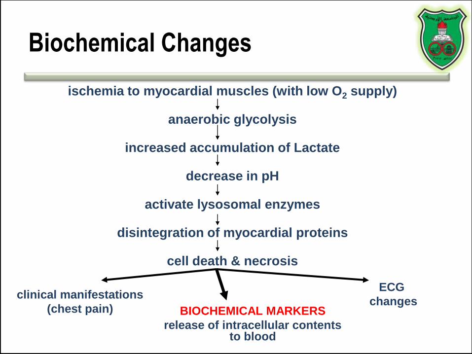
Biochemical Changes
BIOCHEMICAL MARKERS
release of intracellular contents to blood
clinical manifestations
(chest pain)
ECG
changes
ischemia to myocardial muscles (with low O2 supply)
anaerobic glycolysis
increased accumulation of Lactate
decrease in pH
activate lysosomal enzymes
disintegration of myocardial proteins
cell death & necrosis
What is a molecular biomarker?
A molecular alteration that is objectively measured and evaluated as an indicator of
normal biological processes, pathogenic processes, or pharmacologic responses to a
therapeutic intervention

Criteria for ideal markers for MI
• Specific: no false positive (present in the myocardium absent in nonmyocardial tissues)
• Sensitive: no false negative (produced at high concentrations that can be measurable)
• Prognostic: relation between plasma level & extent of damage
• Persists longer: can diagnose delayed admission
• Reproducible
• Simple, inexpensive
• Quick
• Acceptable (by patient and clinician)

What are biomarkers good?
• Diagnostic (yes or no; infarct vs. reinfarct)
• Differentiating (AMI, skeletal muscle damage, other cardiac conditions, renal disease, etc. )
• Risk-stratifying (low- vs. high-risk)
• Prognostic (degree of severity; infarct size)

MI biomarkers
• Inflammation (C-reactive proteins)
• Oxidative stress (myeloperoxidase)
• Extracellular matrix remodeling (proteases)
• Neurohormones
• Myocyte injury (troponins, creatine kinase, heart-type fatty acid binding protein, myoglobin)
• Myocyte stress (Brain-natriuretic peptide)
• New biomarkers

Alterations of molecular profile


TROPONINS
THE GOLD STANDARD

Structure
• Associated with tropomyosin, which forms a continuous chain along each actin thin filament
• A complex of the three subunits:
– TN-T: tropomyosin binding subunit
– TN-I: myosin ATPase inhibiting subunit
– TN-C: calcium binding subunit

Troponin isoforms
• Troponin I and T are highly specific for myocardial injury
– Levels in a healthy person are negligible
• cTNI indicates only cardiac troponin
• cTNT may cross-react with troponin found in other muscles
– non-cardiac injury such as skeletal myopathies and with renal failure
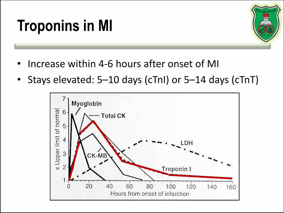
Troponins in MI
• Increase within 4-6 hours after onset of MI
• Stays elevated: 5–10 days (cTnI) or 5–14 days (cTnT)

Why is release of troponin prolonged?
• Most is bound to the contractile apparatus of the cardiomyocyte
• 3% of cTnI and 6% of cTnT exist free in the cytoplasm
• The initial elevation of cTnI or cTnT is thought to be a function of the free cytolsolic form
• The prolonged elevation is caused by degradation of the contractile pool

Troponin Influence on Prognosis
Since normal people have virtually nil levels of troponin in serum
Positive results: MI or chronic disease

Advantages
• Higher sensitivity than CK-MB
• Fewer false-positive results in the setting of trauma, surgery, and renal failure as compared to CK-MB
• Prognostic of death from acute coronary syndrome

Troponin and death

Disadvantages
• It lacks sensitivity in the early hours of AMI
• Pulmonary embolism, congestive heart failure, and myocarditis can all lead to cardiac troponin elevation
Serial Sampling
• When initial results are negative
• Serial sampling at presentation, 6–9 h later, and after 12 h is recommended if the earlier results are negative and clinical suspicion remains high
What is troponin test is not present,
then use that of creatine kinase

Sources of energy

Creatine phosphate

CK in MI
• Indicator of myocardial infarction

Total CK can be elevated
• AMI
• False positive (for MI) CK elevation can be seen in:
– Significant skeletal muscle injury
– Significant CNS damage (Stroke/Trauma)
– Occasionally from GI, renal, urologic disease
– Others: i.m. injection, hypothermia, exercise, intoxication and drug abuse
• Dose-related side effect in statin treatment
– Statin-related increases in CK mainly affect MM isozyme

CK isozymes
Serum Skeletal Muscle
Cardiac Muscle Brain
0 trace BB<6% MB
>94% MM
0 trace BB1% MB
99% MM
0% BB20% MB80% MM
97% BB3% MB0%MM

CK-MB
• Increase: 3 and 12 h after the onset of MI
• Peaks at 24 hr
• Reverts to normal values within 48–72 h
Useful for
diagnosis of
reinfarction

Cardiac relative index
• improves specificity, but may limit sensitivity
RI = (CK-MB mass / Total CK) x 100
More than 5 % is indicative for MI

Limitations of CK-MB
• Skeletal Muscle Involvement
• Duchenne Muscular Dystrophy
• Polymyositis
• Alcohol Myopathy
• Thermal or Electrical Burn Patients
• Carcinomas (Colon, Lung, Prostate, Endometrial..)
• Athletes (e.g. Marathon runners)

CK-MB isoforms and MI
• Two isoforms called 1 (plasma) and 2 (cellular)
• 2 to 1 ratio of > 1.5 can useful for early MI detection
– Requires a skilled technician
– False positive results with congestive heart failure and other conditions can occur

MYOGLOBIN (Mb)
• Rapidly released to the circulation after muscle injury
– An early marker that can be detected 1–2 hours after symptom onset, and remains elevated for up to 24 hours
• Sensitive in the absence of concomitant skeletal muscle trauma or renal failure
• Specimens collected serially every 1-2 hours during the first 2-10 hours after infarction
• Levels that double within 1-2 hours are highly suggestive of AMI
• Suited to excluding AMI at the earliest phase

But…
• low-specificity for MI – in patients with renal failure or skeletal muscle trauma
• Rises and falls rapidly in the setting of MI
• The level may normalize in patients that present >24 hours after symptom onset– indicated for the diagnosis of re-infarction
• Therefore, – potentially useful for ruling out but not for confirming the
diagnosis of AMI
– Is used in combination with CK-MB or cTn

SPECIFICITY OF CARDIAC
MARKERS
50
60
70
80
90
100
TROPONIN-I
70%
87%
92%
99%
CK-MB TOTAL CK MYOGLOBIN

SUMMARY

MARKER TISSUE
SOURCE
PHYSIOLOGIC
FUNCTION
“DIAGNOSTIC
WINDOW”
CLINICAL
UTILITY
Creatine
Kinase (CK)
Total Activity
Skeletal
muscle
Cardiac
muscle
Skeletal
muscle
Rephosphorylation
of ADP, forming
ATP in muscle
contraction
Rise: 6-8 hr
Peak: 24-36 hr
Normal: 3-4
days
Limited
diagnostic
value since it is
increased in
various disease
states.
CK isoenzyme
analysis is
more useful for
diagnosis

MARKER TISSUE
SOURCE
PHYSIOLOGIC
FUNCTION
“DIAGNOSTIC
WINDOW”
CLINICAL
UTILITY
CK-MB
Isoenzyme,
Mass (amount,
not activity)
Cardiac
muscle
Skeletal
muscle to a
much
lesser extent
Same as above Rise: 4-6 hr
Peak: 12-24 hr
Normal: >48 hr
Mass assay of
CK-MB
isoenzyme,
the current
“gold
standard” for
early
diagnosis of
AMI

MARKER TISSUE
SOURCE
PHYSIOLOGIC
FUNCTION
“DIAGNOSTIC
WINDOW”
CLINICAL
UTILITY
CK-MB
Isoforms and
Isoforms ratio
Same as
above
Same as above Rise: 2-6 hr
Peak: 6-12 hr
Normal: 24-36
hr
Early
marker of
AMI, more
specific than
myoglobin
Myoglobin Cardiac
muscle
Skeletal
muscle
Oxygen binding
protein
Rise: 2-3 hr
Peak: 6-9 hr
Normal: 24-36
hr
Non-specific
early marker
to rule
in/rule out
AMI

MARKER TISSUE
SOURCE
PHYSIOLOGIC
FUNCTION
“DIAGNOSTIC
WINDOW”
CLINICAL
UTILITY
Cardiac
Troponin I
Cardiac
muscle
Muscle contraction
regulatory protein;
bound to
tropomyosin and
actin
Rise 4-8 hr
Peak: 14- 18 hr
Normal: 5-9 days
Highly specific
for myocardial
injury
Useful for
patients with
atypical
symptoms or
those who delay
seeking medical
attention
Potential to
diagnose AMI in
patients who also
have
concomitant
skeletal muscle
trauma/disease
Potential usage
to risk stratify
angina pectoris

MARKER TISSUE
SOURCE
PHYSIOLOGIC
FUNCTION
“DIAGNOSTIC
WINDOW”
CLINICAL
UTILITY
Cardiac
Troponin T
(cTnT)
Cardiac
muscle;
regenerating
skeletal
muscle
Same as above Rise: 4-8 hr
Peak: 14-18 hr
Normal: >14
days
As above for
cTnI

OLD BIOMARKERS
Aspartate aminotransferase
Lactate dehydrogenase

Aspartate aminotransferase

Lactate dehydrogenase in MI
• Rises in 12 to 24 hours following MI,
• Peaks in 2 to 3 days
• Gradually disappearing in 5 to 14 days

Tissue distribution of LDH
Isoenzyme Composition Present in
LDH1 ( H4) Myocardium, RBC, kidney
LDH2(H3M1)
Myocardium, RBC,
serum, kidney
LDH3 (H2M2) Kidney, Skeletal muscle
LDH4 (H1M3) Kidney, Skeletal muscle
LDH5 (M4) Skeletal muscle, Liver

Normal vs. MI
NormalLD1:LD2 = 0.5-0.75
MILD1:LD2 > 1

Conditions causing flipped LD1/LD2
without AMI
• Hemolysis
• Megoblastic & Pernicious Anemia
• Renal Cortex Infarction
• Testicular Germ Cell Tumors
• Small Cell Lung Carcinoma
• Adenocarcinoma of the Ovary
• Acute Coronary Insufficiency (Unstable Angina)
• Exercise Induced Myocardial Ischemia
• Polymyositis
• Muscular Dystrophies
• Well Trained Athletes
• Rhabdomyolysis

Example
3. Control
8. Normal
1. MI
2. MI (hrs post)
5. MI (2d post)
6. MI (1d post)
4. Liver disease
7. Liver + HF

Interpretation
• Sample #3 represent results for a control
• Sample #8 results are from a normal specimen.
• Sample# 1 MI patient. The specimen was collected at a time when the activity of both LDH and CK were elevated. Note the LDH flip and the high relative activity of the MB isoenzyme.
• Sample# 2 MI patient who experienced chest pain only several hours previously. Total CK is significantly elevated with a high relative MB isoenzyme activity.
• Sample# 6 MI patient (the 1st day post MI); CK activity is definitely elevated with a high relative MB isoenzyme activity and the LDH flip is evident.
• Sample# 5 MI patient (2 days post MI) so that CK has almost returned to normal activity and the LDH flip is definite.
• Sample# 7 MI patient with complications of heart failure and passive liver congestion or the patient was involved in an accident as a consequence of the MI, and suffered a crushing muscle injury.
• Sample# 4 a patient with liver disease. Although the LDH isoenzyme pattern is indistinguishable from muscle disease or injury, the absence of at least a trace of CK-MB isoenzyme is inconsistent with the muscle CPK isoenzyme distribution as is the apparently normal total activity.

FUTURE MARKERS

Natriuretic peptides
• Atrial natriuretic peptide (ANP)
• B type natriuretic peptide (BNP)
• C type natriuretic peptide
• D type natriuretic peptide
• All hormones function in the homeostasis of sodium and water retention


Physiological actions of NPs

B-type Natriuretic Peptide (BNP)
• Secreted by the ventricles in response to tension
• BNP binds and activates receptors causing reduction in systemic vascular resistance, central venous pressure and natriuresis

Synthesis and secretion of BNP

Clinical utilization
• A prognostic indicator of death, heart failure, risk prediction of AMI recurrence (Higher BNP suggests
higher chance of AMI recurrence)
• May also guide treatment
• Not useful relative to other biomarkers
• However, it is useful in risk stratification

Illustrations of risk stratification

BNP and Mortality

Limitations
• The proper cutoff values has not been determined
• BNP and NT-proBNP levels are higher in women and increase with age
• Biologic variability
• Assays lack precision

Glycogen phosphorylase BB (GPBB)
• Heart and brain tissue
– Because of the blood–brain barrier, GP-BB can be heart muscle specific
• A rapid rise in blood levels can be seen in myocardial infarction and unstable angina.
• GP-BB elevated 1–3 hours after process of ischemia.
– Early diagnosis in acute coronary syndrome.
• High specificity and sensitivity

Heart-type fatty acid binding protein
(H-FABP)
Not heart-specific, but can identify patients at high-risk

Profile of GPBB and H-FABP release
after MI

C-Reactive Protein
• Pentameric structure consisting of five 23-kDa identical subunits
• Produced primarily in hepatocytes
• Plasma levels can increase rapidly to 1000x baseline levels in response to acute inflammation
• “Positive acute phase reactant”

CRP and CV Risk
• Elevated levels predictive of:
– Long-term risk of first MI
– Ischemic stroke
– All-cause mortality

Limitations to CRP in Screening
• Low specificity
• Gender and racial differences exist
• Affected by physiologic conditions , lifestyle behaviors (smoking, obesity, exercise, and alcohol use), and drugs
• Clinical value??
– No evidence that lowering CRP levels decreases CV risk

Ischemia-Modified Albumin (IMA)

Ischemia-Modified Albumin (IMA)
• Under conditions of ischemia, albumin undergoes a conformational change, so that it can no longer bind to transitional metals such as copper or cobalt
– Albumin cobalt binding (ACB) test
• Using the albumin cobalt binding test, the proportion of albumin modified by ischemia can be estimated
– But, low specificity and sensitivity
• A predictor of long-term outcome in patients with acute myocardial infarction
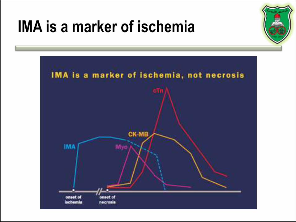
IMA is a marker of ischemia
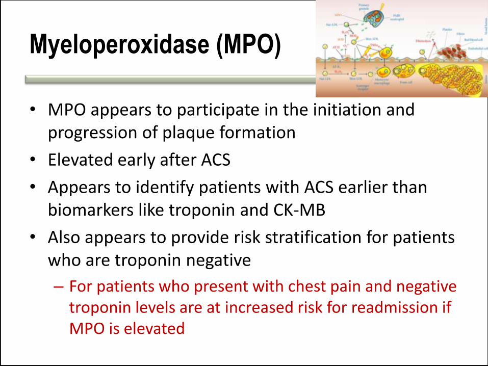
Myeloperoxidase (MPO)
• MPO appears to participate in the initiation and progression of plaque formation
• Elevated early after ACS
• Appears to identify patients with ACS earlier than biomarkers like troponin and CK-MB
• Also appears to provide risk stratification for patients who are troponin negative
– For patients who present with chest pain and negative troponin levels are at increased risk for readmission if MPO is elevated

Note
“Despite the multitude of cardiac biomarkers in production and under investigation, none have convincingly demonstrated their incremental utility beyond that of cTn.”
SJ Aldous
International Journal of Cardiology 164 (2013) 282–294

The Future of Cardiac Biomarkers
• Many experts are advocating the move towards a multimarker strategy for the purposes of diagnosis, prognosis, and treatment design

Why do we need multiple Markers?
• No single ideal marker exists for ACS
• Complicated diseases are not likely to be associated with single markers
• Multiple markers define disease categories
• Multi-marker panels can aid in differential diagnosis
It all goes back to $
“Conversely, multi-marker assessment has been shown to be associated with higher Emergency Department, coronary care and cardiac intervention costs but…[it] has not been shown to reduce overall costs despite reducing admissions.”
SJ Aldous
International Journal of Cardiology 164 (2013) 282–294